genetic control of biosynthesis and transport of riboflavin ... · achievements in 1937 and 1938,...
TRANSCRIPT

MICROBIOLOGY AND MOLECULAR BIOLOGY REVIEWS, June 2011, p. 321–360 Vol. 75, No. 21092-2172/11/$12.00 doi:10.1128/MMBR.00030-10Copyright © 2011, American Society for Microbiology. All Rights Reserved.
Genetic Control of Biosynthesis and Transport of Riboflavin andFlavin Nucleotides and Construction of Robust
Biotechnological Producers†Charles A. Abbas1 and Andriy A. Sibirny2,3*
Archer Daniels Midland Company, Decatur, Illinois 625261; Institute of Cell Biology, NAS of Ukraine, Lviv 79005, Ukraine2; andUniversity of Rzeszow, Rzeszow 35-601, Poland3
INTRODUCTION .......................................................................................................................................................322RIBOFLAVIN AND FLAVIN NUCLEOTIDES (FMN AND FAD) AND THEIR ROLE IN
METABOLISM ...................................................................................................................................................322Discovery and Occurrence .....................................................................................................................................322Chemical Structure and Properties......................................................................................................................323Analytical Methods .................................................................................................................................................323Other Natural Flavins............................................................................................................................................323Biological Role of Flavins ......................................................................................................................................325
Redox reactions ...................................................................................................................................................325Reactions with no net redox change ................................................................................................................325Light emission and other processes .................................................................................................................326Lumazine proteins ..............................................................................................................................................326FMN as the precursor of coenzyme B12 ..........................................................................................................326Free flavins ..........................................................................................................................................................326
BIOCHEMICAL PATHWAYS OF RIBOFLAVIN SYNTHESIS IN BACTERIA, FUNGI, AND PLANTS ....326GTP Cyclohydrolases II and III ...........................................................................................................................327Reductase and Deaminase.....................................................................................................................................327Dephosphorylation of 5-Amino-6-Ribitylamino-2,4(1H,3H)-Pyrimidinedione 5�-Phosphate........................3293,4-Dihydroxy-2-Butanone 4-Phosphate Synthase..............................................................................................329Lumazine Synthase.................................................................................................................................................330Riboflavin Synthase ................................................................................................................................................330Riboflavin Synthesis in Different Organisms......................................................................................................331
BIOSYNTHESIS AND DEGRADATION OF FLAVIN NUCLEOTIDES FMN AND FAD..............................331Riboflavin Kinase....................................................................................................................................................332FAD Synthetase.......................................................................................................................................................332Degradation of Flavin Nucleotides .......................................................................................................................332
BIOSYNTHESIS OF NATURAL FLAVIN ANALOGS..........................................................................................3335-Deazaflavins..........................................................................................................................................................333Roseoflavin...............................................................................................................................................................333
RIBOFLAVIN TRANSPORT INTO AND OUT OF THE CELL .........................................................................334Bacteria ....................................................................................................................................................................334Yeasts and Filamentous Fungi..............................................................................................................................335
Saccharomyces cerevisiae .....................................................................................................................................335Pichia guilliermondii ............................................................................................................................................335
(i) RF permease I ...........................................................................................................................................335(ii) RF permease II.........................................................................................................................................336(iii) RF excretase ............................................................................................................................................336
Ashbya gossypii (Eremothecium gossypii) ...........................................................................................................337Animals.....................................................................................................................................................................337
REGULATION OF RIBOFLAVIN SYNTHESIS....................................................................................................338Feedback Inhibition of GTP Cyclohydrolase II..................................................................................................338Transcriptional Regulation in Bacteria Using the Riboswitch Mechanism ...................................................338
Riboswitch (RFN element) in Bacillus..............................................................................................................338Regulation in E. coli ...........................................................................................................................................340
* Corresponding author. Mailing address: Institute of Cell Biology,NAS of Ukraine, Lviv 79005, Ukraine. Phone: 380 32 261 2108. Fax:380 32 261 2148. E-mail: [email protected].
† This review is dedicated to the memory Prof. Georgiy M. Shavlo-vsky, one of the pioneers in studies of riboflavin biosynthesis by yeasts,the mentor of one of the authors (A.A.S.).
321
on August 11, 2019 by guest
http://mm
br.asm.org/
Dow
nloaded from

Other bacteria .....................................................................................................................................................340Transcriptional Regulation in Mycelial Fungi ...................................................................................................340Transcriptional Regulation in Flavinogenic Yeasts by Iron.............................................................................341
THE RIBOFLAVIN BIOSYNTHESIS PATHWAY AS A TARGET FOR ANTIMICROBIAL DRUGS..........343FLAVIN SYNTHESIS BY FLAVINOGENIC MICROORGANISMS ..................................................................343INDUSTRIAL PRODUCTION OF RIBOFLAVIN AND FLAVIN NUCLEOTIDES AND THEIR
PRACTICAL APPLICATIONS .........................................................................................................................344CONSTRUCTION OF INDUSTRIAL RIBOFLAVIN PRODUCERS BY CLASSIC MUTAGENESIS AND
SELECTION AND BY METHODS OF METABOLIC ENGINEERING ...................................................344B. subtilis ..................................................................................................................................................................344Other Bacteria.........................................................................................................................................................345
Corynebacterium ammoniagenes ..........................................................................................................................345Lactic acid bacteria and propionibacteria............................................................................................345
A. gossypii .................................................................................................................................................................346E. ashbyii ..................................................................................................................................................................346C. famata (C. flareri) ...............................................................................................................................................346P. guilliermondii .......................................................................................................................................................348Methylotrophic Yeasts............................................................................................................................................348
CONSTRUCTION OF PRODUCERS OF FLAVIN NUCLEOTIDES ................................................................349UNRESOLVED ISSUES AND FUTURE PROSPECTS........................................................................................349ACKNOWLEDGMENTS ...........................................................................................................................................350REFERENCES ............................................................................................................................................................350
INTRODUCTION
Riboflavin [7,8-dimethyl-10-(1�-D-ribityl)isoalloxazine, vita-min B2] (RF) is an obligatory component of human and animaldiets, as it serves as a precursor of the flavin coenzymes flavinmononucleotide (FMN) and flavin adenine dinucleotide(FAD), which are involved in oxidative metabolism and otherprocesses. Commercially produced RF is used for animalfeed, as a dietary supplement, and as an additive by the foodindustry.
The biosynthetic pathway of RF synthesis in microorgan-isms and plants has been elucidated. It starts from GTP andribulose-5-phosphate and proceeds through pyrimidine andpteridine intermediates. Flavin nucleotides are synthesizedin two consecutive reactions from riboflavin in prokaryotesand eukaryotes. The pathway for the synthesis of the naturalRF analog 5-deazariboflavin shows some similarities to thatfor RF. The antibiotic roseoflavin, which is a natural RFanalog [7-methyl-8-dimethylamino-10(1�-D-ribityl)isoalloxa-zine], is probably synthesized from RF. Some microorgan-isms and all animal cells are capable of RF uptake, andmany microorganisms, especially RF overproducers, havedistinct systems for RF excretion (efflux) to the medium.
Regulation of RF synthesis occurs at the level of enzymeactivity and synthesis. In yeasts, the first enzyme of RF synthe-sis, GTP cyclohydrolase II, is regulated by allosteric inhibitionexerted by FAD and other nucleotides containing an adenylicmoiety. The physiological role of this regulation is not known.Regulation of RF synthesis at the gene level differs in bacteria,yeasts, and fungi. In bacteria, production of RF is repressed atthe transcriptional level by FMN, which binds to nascent non-coding mRNA and blocks further transcription (the so-calledriboswitch). In flavinogenic molds, RF overproduction starts atthe stationary phase due to derepression of enzymes involvedin RF synthesis and is accompanied by sporulation and myce-lial lysis. In flavinogenic yeasts, transcriptional repression ofRF synthesis is caused by iron ions. The putative transcriptionfactor encoded by SEF1 is somehow involved in this regulation.
Most commercial RF is currently produced by microbialsynthesis. For this, special selected strains of the bacteriumBacillus subtilis, the mold Ashbya gossypii, and the yeast Can-dida famata (Candida flareri) are used. Whereas earlier RFoverproducers were isolated by classical selection, current pro-ducers of RF and flavin nucleotides have been developed usingmodern approaches of metabolic engineering that involveoverexpression of structural and regulatory genes of the RFbiosynthetic pathway as well as genes involved in the overpro-duction of the purine precursor of riboflavin, GTP.
There are numerous reviews and one monograph on thetopic of the synthesis of riboflavin and other flavins. Most ofthem appeared several years ago and are cited in appropriateplaces in the current review. Readers are also referred to thetwo most recent reviews (115, 178), which cover biochemicalpathways and the biotechnology of RF synthesis, respectively.Here we have tried to provide balanced narratives on differentaspects of the biosynthesis and transport of RF, flavin nucle-otides, and some other natural flavins, including the biochem-istry, enzymology, and metabolic and genetic regulation ofthese processes, construction of flavin overproducers usingclassic selection and modern approaches of metabolic engi-neering, and data on industrial production of RF. This reviewis apparently the first in English to summarize the data oniron-dependent regulation of RF synthesis in flavinogenicyeasts and construction of yeast overproducers of RF andflavin nucleotides.
RIBOFLAVIN AND FLAVIN NUCLEOTIDES (FMN ANDFAD) AND THEIR ROLE IN METABOLISM
Discovery and Occurrence
Riboflavin (RF) (vitamin B2) was discovered in 1879 as ayellow pigment from milk and called lactoflavin. Its chemicalstructure was deciphered by Paul Karrer (Zurich, Switzerland)and Richard Kuhn (Heidelberg, Germany) in the 1930s (210,244). They were awarded the Nobel Prize for this and other
322 ABBAS AND SIBIRNY MICROBIOL. MOL. BIOL. REV.
on August 11, 2019 by guest
http://mm
br.asm.org/
Dow
nloaded from

achievements in 1937 and 1938, respectively. Kuhn first provedthat RF is an essential growth factor, viz., vitamin B2.
The main sources of RF in diets are milk, dairy, and meatproducts. In the United Kingdom, for example, milk and dairyproducts contribute 51% of RF intake in preschool children,35% in schoolchildren, 27% in adults, and 36% in the elderly(356). Cereals, meats, and fatty fish are also good sources ofRF, and certain fruits and vegetables, especially dark greenvegetables, contain reasonably high RF concentrations. Therecommended daily intake of RF is 1.3 mg/day for men and 1.1mg/day for women (122). RF deficiency is endemic in popula-tions that lack dairy and meat products. RF deficiency maycontribute to increased concentrations of plasma homocys-teine, with an associated increased risk of cardiovascular dis-ease. Deficiency may also cause impairment in iron metabo-lism and night blindness (356).
Chemical Structure and Properties
RF [7,8-dimethyl-10-(1�-D-ribityl)isoalloxazine] is a hetero-cyclic compound produced by all plants and most microorgan-isms. Animals and rare prokaryotic and eukaryotic micro-organisms (e.g., Corynebacterium pyogenes, Streptococcuspyogenes, Listeria monocytogenes, some lactic acid bacteria, my-coplasmas, spirochetes, rickettsiae, and protists) cannot syn-thesize RF and need to obtain it from their diets (or from themedium), so RF must be considered a vitamin for them (85,132, 208, 241, 356, 455, 494, 504). The growth response ofLactobacillus casei to RF has been used for developing a mi-crobiological assay for this vitamin (456). Proof of RF auxot-rophy differs between organisms, e.g., based on the absence orpresence of RF biosynthetic genes or direct determination ofgrowth dependence on exogenous RF.
In animals, RF deficiency results in retarded growth, failureto thrive, and eventual death (76). Experimental RF deficiencyresults in growth failure, weakness, ataxia, and inability tostand. Animals collapse, become comatose, and die. Deficiencyleads to dermatitis, hair loss, corneal opacity, cataracts, hem-orrhagic adrenals, fatty degeneration of the kidney and liver,and inflammation of the mucous membrane of the gastroin-testinal tract (180). RF deficiency also leads to developmentalabnormalities (321, 523). Postmortem studies of animals fed anRF-deficient diet showed that they had only a third of thenormal amount of RF in the liver (129), the main storage organfor RF. RF deficiency is rarely observed in developed coun-tries, though groups with a risk of low intake of RF are com-mon (pregnant and lactating women, children, athletes, andsome categories of patients on certain medicines (125, 356).
The currently used name RF recognizes the presence of thesugar alcohol ribitol in the molecule of this vitamin and theyellow color of the substance. RF (Fig. 1A) usually does nothave direct metabolic functions in the living cell but serves asa precursor for the synthesis of derivatives known as flavinnucleotides or flavin coenzymes, i.e., riboflavin-5�-phosphate(flavin mononucleotide [FMN]) (Fig. 1B) and flavin adeninedinucleotide (FAD) (Fig. 1C). RF, FMN, and FAD are themain representatives of the group of substances known as“flavins.” In general, flavins are designated as derivatives ofthe dimethylisoalloxazine {7,8-dimethylbenzo[g] pteridine-2,4(3H,10H)-dione} skeleton, with a substitution in the 10 po-
sition. The properties of natural flavins have been well studied(30, 33, 525a). RF and most other flavins are yellow com-pounds with a characteristic yellow-green fluorescence in UVlight. Peak absorbances by aqueous solutions of RF occur at223, 266, 373, and 445 nm. The maximum fluorescence emis-sion of neutral aqueous RF solution is at 535 nm. Light absor-bance and intense fluorescence are used in the analytical de-termination of flavins. Different forms can be easily separatedby liquid chromatography (503). RF is slightly soluble in waterand ethanol; its solubility is 100 to 130 mg/liter and 45 mg/literat room temperature in water and absolute ethanol, respec-tively (30). It is poorly soluble in allyl and benzyl alcohols, amylacetate, and phenol and practically insoluble in ether, chloro-form, acetone, and benzene. RF is soluble in alkaline solutionbut becomes unstable under these conditions. FMN is muchmore soluble in water (30 to 50 g/liter) (30, 279).
In an acid medium, flavin nucleotides are hydrolyzed to freeRF. Flavin molecules possess amphoteric properties. They flu-oresce only in the oxidized state. Optimal fluorescence occursat pH 3 to 8. The molecular coefficients of RF and FMNfluorescence are very close, whereas that of FAD is signifi-cantly lower (�20% of the molar fluorescence of RF andFMN) due to fluorescence quenching by the adenylic moiety(204). Irradiation of flavins leads to their decomposition, pro-ducing derivatives with totally or partially degraded ribitylchains; lumiflavin (7,8,10-trimethylisoalloxazine) is accumu-lated in alkaline solutions, whereas lumichrom (7,8-dimethyli-soalloxazine) is produced at neutral and acid pHs. RF andother flavins produced water-insoluble complexes with saltsof metal ions, e.g., Mg2�, Hg2�, Cu2�, Fe2�, Co2�, and Ni2�
(33, 374).
Analytical Methods
As mentioned above, flavins intensely absorb light and flu-oresce, making these properties important in most assays (30,503, 549). More complicated is assaying individual flavinswithin mixtures. Thus, different approaches have been pro-posed, e.g., paper (30, 56), ion-exchange (536), and thin-layer(147) chromatography, paper (533) and capillary (186) elec-trophoresis, specific RF extraction by 2-phenylethanol (478),or separation on silica gel and other resins (93). Currently,flavins are separated by high-performance liquid chromatog-raphy (379, 503, 549) with subsequent fluorescence detection,approaches that are approved by AOAC International (240).
Other Natural Flavins
In addition to RF, FMN, FAD, and products of their pho-tolysis (lumiflavin and lumichrome), there are other naturalflavins. Most well known are 5-deazaflavins, which are deriva-tives of flavins in which nitrogen in the 5 position of theisoalloxazine heterocyclic structure is replaced by carbon. Onedeazaflavin, 7,8-didemethyl-8-hydroxy-5-deazaflavin, has aribitylated derivative known as coenzyme F0, and an oligoglu-tamylated derivative of F0 (coenzyme F420) (Fig. 1D) is in-volved in hydride transfer during reductive transformation ofcarbon dioxide and acetate into methane in methanogenicarchaea (103, 151, 258, 299, 514). Coenzyme F420 has also beenfound in certain streptomycetes, in which it serves as a cofactor
VOL. 75, 2011 RF AND FLAVIN NUCLEOTIDE BIOSYNTHESIS AND TRANSPORT 323
on August 11, 2019 by guest
http://mm
br.asm.org/
Dow
nloaded from

in the biosynthesis of tetracycline and lincomycin (74, 189, 347,369). Mycobacterium and Nocardia spp. use coenzyme F420 as acofactor of glucose-6-phosphate dehydrogenase (359, 360).Cofactor F420 is required for the activation of experimentalantituberculosis drugs by Mycobacterium tuberculosis and My-cobacterium bovis strain BCG (43, 476). Although F420 con-
tains a 5-deazariboflavin moiety, its biochemistry is more sim-ilar to that of NAD(P) than to that of FMN/FAD (42). In itsdeprotonated 8-hydroxy form, coenzyme F0 is known to act asa cofactor of DNA photolyases from the cyanobacteria Syn-echocystis spp. as well as the eukaryotes Scenedesmus spp.,
FIG. 1. Chemical structures of flavins.
324 ABBAS AND SIBIRNY MICROBIOL. MOL. BIOL. REV.
on August 11, 2019 by guest
http://mm
br.asm.org/
Dow
nloaded from
Ostreococcus tauri, and Drosophila melanogaster (104, 105, 146,385).
Some natural flavins have a reddish-orange color, e.g.,the antibiotic roseoflavin [7-methyl-8-dimethylamino-(1�-D-ribityl)isoalloxazine] (Fig. 1E), which is produced by Strepto-myces davawensis and is active against Gram-positive bacteria(333, 334). The basidiomycete Schizophillum commune pro-duces two RF derivatives, known as schizoflavins: 7,8-dimethyl-l0-(2,3,4-trihydroxy-4-carboxybutyl)isoalloxazine (RF acid orriboflavinoic acid) and 7,8-dimethyl-l0-(2,3,4-trihydroxy-4-formylbutyl)isoalloxazine (RF-aldehyde or riboflavinal) (488).Their exact metabolic functions are unknown. Other closelyrelated compounds are molybdopterins (279), which consist ofa pyranopterin, a complex heterocycle featuring a pyran fusedto a pterin ring. In addition, the pyran ring has two thiolatesthat serve as ligands in molybdo- and tungstoenzymes (198).Natural flavins found as prosthetic groups of several enzymesin the strict anaerobe bacterium Peptostreptococcus elsdenii are6-hydroxy-7,8-dimethyl-isoalloxazine and 7-methyl-8-hydroxy-isoalloxazine (142). Nekoflavin, identified as 8�-hydroxyribo-flavin, was isolated from the choroid of cat eyes (297). Thisflavin, together with another hydroxyl derivative, 7�-hydroxy-riboflavin, was also found in human urine (326). Glycosidederivatives of RF and other isoalloxazines are quite common,i.e., RF glucosides, RF galactosides, and RF oligosaccharides,produced by some species of bacteria, yeasts, and mycelialfungi (485). Lampteroflavin, the riboflavinyl �-ribofuranoside,proved to be a light emitter in the mushroom Lampteromycesjaponicus (499). Plants frequently secrete RF and its deriva-tives RF-5�-sulfate and RF-3�-sulfate under conditions of ironstarvation (484).
Chemical syntheses resulted in a large collection of analogsof RF (30, 33, 249), and their biological activities have beenstudied in bacteria and animal models (152, 250, 279, 515).Some of them possessed significant antibacterial or antiprotistactivities. Strong antibacterial activity also was found for 8-N-alkyl analogs of roseoflavin (212, 213).
Biological Role of Flavins
The chemical entity responsible for the diverse biologicalactivity of flavin is the isoalloxazine moiety. It exists in threeredox states: (i) the oxidized or quinone state, (ii) the one-electron reduced or semiquinone (radical) state, and (iii) thetwo-electron reduced (fully reduced) or hydroquinone state.Flavin is an amphoteric molecule existing as neutral, anionic,and cationic species in all three redox states (290). The redoxpotential for the two-electron reduction of the flavin is about�200 mV. However, this value can greatly vary in flavoproteinsdue to the crucial role of the protein environment in theproperties of flavins, spanning a range from approximately�400 mV to �60 mV. In general, the proximity of a positivecharge is believed to increase the redox potential, and a neg-ative charge or a hydrophobic environment is expected tolower it (130).
Flavins are essential to the nutrition of all prokaryotic andeukaryotic cells. Their significant biological role in most casesis connected with the coenzyme functions of FMN and FAD.These nucleotides bind to proteins, producing flavoproteins(flavoenzymes). There is one exception: free RF is the active
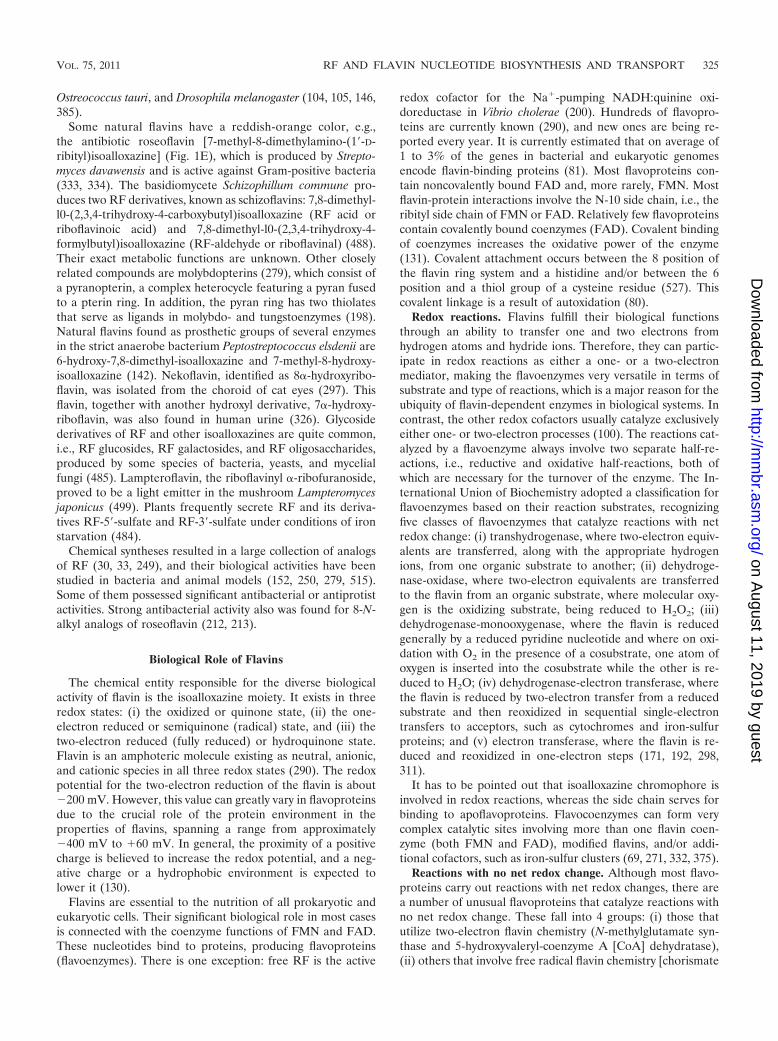
redox cofactor for the Na�-pumping NADH:quinine oxi-doreductase in Vibrio cholerae (200). Hundreds of flavopro-teins are currently known (290), and new ones are being re-ported every year. It is currently estimated that on average of1 to 3% of the genes in bacterial and eukaryotic genomesencode flavin-binding proteins (81). Most flavoproteins con-tain noncovalently bound FAD and, more rarely, FMN. Mostflavin-protein interactions involve the N-10 side chain, i.e., theribityl side chain of FMN or FAD. Relatively few flavoproteinscontain covalently bound coenzymes (FAD). Covalent bindingof coenzymes increases the oxidative power of the enzyme(131). Covalent attachment occurs between the 8 position ofthe flavin ring system and a histidine and/or between the 6position and a thiol group of a cysteine residue (527). Thiscovalent linkage is a result of autoxidation (80).
Redox reactions. Flavins fulfill their biological functionsthrough an ability to transfer one and two electrons fromhydrogen atoms and hydride ions. Therefore, they can partic-ipate in redox reactions as either a one- or a two-electronmediator, making the flavoenzymes very versatile in terms ofsubstrate and type of reactions, which is a major reason for theubiquity of flavin-dependent enzymes in biological systems. Incontrast, the other redox cofactors usually catalyze exclusivelyeither one- or two-electron processes (100). The reactions cat-alyzed by a flavoenzyme always involve two separate half-re-actions, i.e., reductive and oxidative half-reactions, both ofwhich are necessary for the turnover of the enzyme. The In-ternational Union of Biochemistry adopted a classification forflavoenzymes based on their reaction substrates, recognizingfive classes of flavoenzymes that catalyze reactions with netredox change: (i) transhydrogenase, where two-electron equiv-alents are transferred, along with the appropriate hydrogenions, from one organic substrate to another; (ii) dehydroge-nase-oxidase, where two-electron equivalents are transferredto the flavin from an organic substrate, where molecular oxy-gen is the oxidizing substrate, being reduced to H2O2; (iii)dehydrogenase-monooxygenase, where the flavin is reducedgenerally by a reduced pyridine nucleotide and where on oxi-dation with O2 in the presence of a cosubstrate, one atom ofoxygen is inserted into the cosubstrate while the other is re-duced to H2O; (iv) dehydrogenase-electron transferase, wherethe flavin is reduced by two-electron transfer from a reducedsubstrate and then reoxidized in sequential single-electrontransfers to acceptors, such as cytochromes and iron-sulfurproteins; and (v) electron transferase, where the flavin is re-duced and reoxidized in one-electron steps (171, 192, 298,311).
It has to be pointed out that isoalloxazine chromophore isinvolved in redox reactions, whereas the side chain serves forbinding to apoflavoproteins. Flavocoenzymes can form verycomplex catalytic sites involving more than one flavin coen-zyme (both FMN and FAD), modified flavins, and/or addi-tional cofactors, such as iron-sulfur clusters (69, 271, 332, 375).
Reactions with no net redox change. Although most flavo-proteins carry out reactions with net redox changes, there area number of unusual flavoproteins that catalyze reactions withno net redox change. These fall into 4 groups: (i) those thatutilize two-electron flavin chemistry (N-methylglutamate syn-thase and 5-hydroxyvaleryl-coenzyme A [CoA] dehydratase),(ii) others that involve free radical flavin chemistry [chorismate
VOL. 75, 2011 RF AND FLAVIN NUCLEOTIDE BIOSYNTHESIS AND TRANSPORT 325
on August 11, 2019 by guest
http://mm
br.asm.org/
Dow
nloaded from

synthase, DNA photolyase, (6-4)photolyase, and 4-hydroxybu-tyryl-CoA dehydratase], (iii) a number in which the role offlavin remain unclear [(R)-2-hydroxyacyl-CoA dehydratases,isopentenyl diphosphate isomerase, and UDPgalactopyranosemutase], and (iv) those that apparently do not involve theflavin directly in catalysis (acetohydroxyacid synthases and hy-droxynitrile lyase) (reviewed in reference 42).
Light emission and other processes. Flavins can also beinvolved in nonrelated processes, one of them being phototro-pism. FMN is the cofactor in phototropins of plants (53), whichare the blue light-sensitive photoreceptors responsible for pho-totropism (bending responses of plants toward or away fromlight sources) (188), chloroplast movement (166), and manyother functions. FMN is noncovalently bound in phototropin.The cofactors of cryptochromes are FAD and methenyl tetra-hydrofolate (269). FAD is also the redox- and light-sensitivenoncovalently bound chromophore in BLUF proteins (148).They are also involved in a variety of nonredox processes, suchas blue-light sensing in plants (70, 262, 386). FMN-containingfluorescent proteins have been engineered from the blue-lightphotoreceptors of Bacillus subtilis and Pseudomonas putida;after codon optimization, they have been heterologously ex-pressed in bacteria and yeasts (95, 489, 496). In contrast to thegreen fluorescent protein (GFP), the FMN-containing fluores-cent proteins fluoresce in both the presence and absence ofoxygen, which is important for studying proteins under anaer-obic conditions.
Flavins are also involved in circadian rhythm (135, 191, 384).RF induces disease resistance in plants by activating a signaltransduction pathway (5, 94, 550) and is involved as the apop-tosis-inducing factor in a mitochondrial flavoprotein (306).
Lumazine proteins. Some fluorescent bacteria of the generaPhotobacterium and Vibrio produce fluorescent proteins thatare also known as lumazine proteins. These proteins use 6,7-dimethyl-8-ribityllumazine, an RF immediate biosynthetic pre-cursor, as the noncovalently bound prosthetic group (236, 328,349). In addition to 6,7-dimethyl-8-ribityllumazine, RF, FMN,and 6-methyl-7-oxo-8-ribitylllumazine (the product of 6,7-di-methyl-8-ribityllumazine oxidation) can be used as prostheticgroups. Lumazine proteins act as optical transponders in theabove-mentioned fluorescent bacteria.
FMN as the precursor of coenzyme B12. FMN (but notFAD) also possesses an important biological function in beingthe biosynthetic precursor of the dimethylbenzimidazol part ofcoenzyme B12 (63, 366, 367). Conversion of FMN to dimeth-ylbenzimidazol involves a unique transformation reaction withno precedent in chemistry, which includes retro-aldol conden-sation sandwiched between two 2-electron oxidations (154,519). The corresponding oxidoreductase BluB from Sinorhizo-bium meliloti triggers the unprecedented fragmentation andcontraction of the bound FMNH2 and cleavage of the ribityltail to form dimethylbenzimidazol and d-erythrose 4-phos-phate (488a).
Free flavins. Some biological functions can be fulfilled byfree flavins, including RF, as a rule when secreted from thecells. In Helicobacter pylori, the excreted RF is thought to havea role in Fe3� reduction and hence in iron acquisition (529),and a similar role in Campylobacter jejuni has been suggested(78). Secreted FMN and RF mediate extracellular electrontransfer involved in Fe3� and other cation reduction in che-
motrophic bacteria of the genus Shewanella (77, 287, 509).Thus, free flavins participate as electron shuttles in the so-called Mtr respiration pathway (important for a chemotrophicmode of nutrition), in insoluble ion solubilization, and usuallyin some geochemical cycles. Secretion of RF by microorgan-isms, the physiological role of this process, and its regulationare described below. Avian eggs contain RF, FMN, FAD, andRF-binding protein (RBP), which are required for the activetransport of RF into the egg and storage of the RF neededlater in development (523, 535). Archaea also contain RF-binding proteins (dodecins), also known as lumichrome-bind-ing proteins, that are involved in the regulation of flavin ho-meostasis (157, 158).
BIOCHEMICAL PATHWAYS OF RIBOFLAVINSYNTHESIS IN BACTERIA, FUNGI,
AND PLANTS
Animals and a few prokaryotes (e.g., some lactic acid bac-teria) cannot synthesize RF de novo. All plants and fungi andmost bacteria are capable of RF production and are a sourceof vitamin B2 for animals, including humans. At the same time,all organisms, including animals, convert RF to the flavin co-enzymes FMN and FAD.
The biochemical pathway of RF synthesis was mostly estab-lished before 2000, based on research conducted in the UnitedStates, Japan, Ukraine, Russia, and Germany. The crucialbreakthroughs in deciphering the pathway of RF synthesiswere made by Adelbert Bacher and his colleagues in Munich,Germany. Biochemical reactions leading to synthesis of flavincoenzymes FMN and FAD were established many years ago(216, 218, 230, 403). At about the same time, the RF synthasereaction leading to synthesis of RF from two molecules of itsimmediate precursor 6,7-dimethyl-8-ribityllumazine was de-scribed (165, 355). The role of purine compounds as precursorsof RF was known from the works of Goodwin in the middle ofthe 20th century (150). The carbon atom of the purine precur-sor and all carbon atoms of the pyrimidine ring were incorpo-rated into the RF molecule (7). Later it was established thatsome guanylic compound at the nucleoside or nucleotide levelacts as precursor of RF and that this ribose moiety of thepurine precursor is transferred to the ribityl side chain of RF(18, 27, 272, 282). Finally, the first reaction of the pathwayleading to RF, that with GTP cyclohydrolase II, was described(123, 416). After this finding, it became clear that RF synthesisstarts from GTP, which in the GTP cyclohydrolase reaction isconverted to a phosphorylated pyrimidine derivative. Suchphosphorylated ribosylated pyrimidine has to be converted insome way to nonphosphorylated ribitylated pteridine (theabove-mentioned 6,7-dimethyl-8-ribityllumazine) and thenagain to the nonphosphorylated ribitylated modified isoallox-azine (RF). Further work on the pathway was hampered by theinstability of phosphorylated ribosylated pyrimidine interme-diates of RF synthesis. The most intricate was the identifica-tion of reactions involved in the conversion of the pyrimidineprecursor of RF to the pteridine precursor. In these reactionsan intermediate of the pentose phosphate pathway, ribose-5-phosphate or its derivative (273, 275a, 276), later identified asribulose-5-phosphate (506, 507), is involved.
Various approaches have been used to decipher the RF
326 ABBAS AND SIBIRNY MICROBIOL. MOL. BIOL. REV.
on August 11, 2019 by guest
http://mm
br.asm.org/
Dow
nloaded from

biosynthesis pathway. To identify the nature of what werethought to be the purine precursors of RF, cell feeding withradioactively labeled purines, inhibitors of purine interconver-sion, and mutants defective in specific steps of purine metab-olism were used (reviewed in references 10, 14, and 414).Identification of the 4-carbon compound involved in convert-ing the pyrimidine precursor of RF to the pteridine precursorwas based on synthesis of the pteridine precursor (6,7-dimethyl-8-ribityllumazine) by cell extracts of wild-type cells and RF-defective mutants after addition of the putative source of the4-carbon compound (275a, 276, 506). The intermediates in RFsynthesis were investigated after their accumulation in the cul-ture media of RF-deficient mutants of the yeasts Saccharomy-ces cerevisiae and Pichia (Candida) guilliermondii and ofBacillus subtilis; they were subsequently identified by physico-chemical methods (16, 17, 272, 331, 420). The tentative path-way was confirmed after isolation of the corresponding en-zymes, followed by cloning and mutation of the structuralgenes involved in the pathway (14, 115, 414). Additional modelorganisms used for studying RF biosynthesis are the flavino-genic yeast Candida famata (Candida flareri) and the moldsAshbya gossypii and Eremothecium ashbyii.
The biochemical pathways of RF synthesis appeared to besimilar, but not identical, in bacteria, fungi, and plants (seebelow). Surprisingly, the pathways of RF synthesis are identicalin eubacteria and plants but different in fungi and archaea(112–115). One important step in RF synthesis remains un-known, namely, the conversion of the phosphorylated pyrimi-dine derivative of RF to its nonphosphorylated derivative (thedephosphorylation step). The biochemical pathways leading toproduction of other natural flavins, such as deazaflavins androseoflavin, remain to be elucidated and await future study.
Comprehensive reviews of the biochemistry of RF synthesishave been published by Bacher and his colleagues (13, 14,112–115). The following brief overview of the RF biosynthesispathway is based on these reviews and recent publications.
The pathway of RF synthesis (Fig. 2) starts from two pre-cursors, GTP (one molecule) and ribulose-5-phosphate (twomolecules).
GTP Cyclohydrolases II and III
The first reaction of RF biosynthesis is catalyzed by GTPcyclohydrolase II (EC 3.5.4.25); this term is used to distinguishit from GTP cyclohydrolase I (EC 3.5.4.16), which is involvedin biosynthesis of folic acid and biopterin. GTP cyclohydrolaseII removes C-8 from GTP, producing formate; the enzyme alsoremoves pyrophosphate. It was first isolated from cell extractsof Escherichia coli (123); however, the role of the enzyme inRF biosynthesis was first established using RF auxotrophs ofthe flavinogenic yeast P. guilliermondii (416, 425). To someextent, the mechanisms of the GTP cyclohydrolase I and IIreactions are similar, although the final products of the reac-tions are different (46, 112). The product of the GTP cyclohy-drolase II reaction is 2,5-diamino-6-ribosylamino-4(3H)-pyrimidinedione phosphate (123, 124). Alternatively, GMP isformed as the reaction product with a rate of �10% of that forthe major pyrimidine product (373). GTP cyclohydrolase IIfrom E. coli is the homodimer and contains Zn2� ions as acofactor per subunit, being activated by Mg2� (206, 257, 371).
The enzyme properties were studied in the bacteria E. coli (35,123, 206), B. subtilis (185), Helicobacter pylori (32), and Strep-tomyces coelicolor (461), the flavinogenic yeast P. guilliermondii(412, 418, 425), and the model plant Arabidopsis thaliana (174).Yeasts and some bacteria (e.g., E. coli) contain separate genescoding for GTP cyclohydrolase II (in yeasts designated RIB1)(266, 330, 331, 420, 546), whereas plants and other bacteria(e.g., B. subtilis) contain the fused gene coding for a proteinwith two domains, one with GTP cyclohydrolase II activity andanother with the activity of 3,4-dihydroxy-2-butanone 4-phos-phate synthase (see below) (174, 185, 308). The three-dimen-sional structure of GTP cyclohydrolase II is known (365). Theactive center is formed by 3 cysteine residues, Cys54, Cys65, andCys67, which bind Zn2� as well as Arg128 and Tyr105 (206, 365).The reaction starts by pyrophosphate release, which appearedto be the rate-limiting step of the overall reaction, after whichimidazole ring opening and elimination of formate occur (206,373).
In H. pylori, GTP cyclohydrolase II is involved in determin-ing its hemolytic phenotype; heterologous expression of thecorresponding gene ribAB in E. coli induces hemolytic activityof the recipient strain (32, 108).
Archaea and some eubacteria also contain another GTPcyclohydrolase, GTP cyclohydrolase III, which catalyzes theconversion of GTP to 2-amino-5-formylamino-6-ribosylamino-4(3H)-pyrimidinone 5-phosphate, i.e., the formylated deriva-tive of the product of GTP cyclohydrolase II (151, 181, 461). Inother words, GTP cyclohydrolase III, in contrast to GTP cyc-lohydrolase II, hydrolyzes the imidazole ring of GTP but doesnot remove the resulting formyl group from the formamide. Itis noteworthy that the product of GTP cyclohydrolase III is theintermediate of the GTP cyclohydrolase II reaction (151, 402).In the archaeon Methanocaldococcus jannaschii, GTP cyclohy-drolase III is apparently involved in biosynthesis of RF (anddeazaflavin), as the product of this reaction undergoes formatecleavage by the specific formamide hydrolase (160). Thus,the 2,5-diamino-6-ribosylamino-4(3H)-pyrimidinedione phos-phate, the product of GTP cyclohydrolase II, is produced inarchaea by the consecutive action of GTP cyclohydrolase IIIand formamide hydrolase. It is also interesting that the genecoding for GTP cyclohydrolase III from archaea has no ho-mology with genes coding for GTP cyclohydrolase II, whereasthe genes display high homology in S. coelicolor (461).
Reductase and Deaminase
In the next two reactions, deamination of the amino group atposition 2 and reduction of the ribosyl side chain to ribityl takeplace (Fig. 2). The sequence of deamination and reduction isdistinct in fungi and archaea on the one hand and bacteria andplants on the other (14, 112, 113). In yeasts and fungi, theenzyme catalyzing the second reaction of RF biosynthesis, 2,5-diamino-6-ribosylamino-4(3H)-pyrimidinone 5-phosphate re-ductase [another name is 2,5-diamino-6-ribitylamino-4(3H)-pyrimidinone 5-phosphate synthase] (EC 1.1.1.193), usesNADPH for reduction of the product of the GTP cyclohydro-lase II reaction (21, 179, 305, 330, 377, 417). In S. cerevisiae thecorresponding gene is RIB7 (330), whereas in P. guilliermondiiit is RIB2 (420). In archaea and rare eubacteria, 2,5-diamino-6-ribosylamino-4(3H)-pyrimidinone 5-phosphate reductase,
VOL. 75, 2011 RF AND FLAVIN NUCLEOTIDE BIOSYNTHESIS AND TRANSPORT 327
on August 11, 2019 by guest
http://mm
br.asm.org/
Dow
nloaded from

like enzymes from fungi and yeasts, also acts as the distinctenzyme in the second step of RF biosynthesis (67, 153, 377). Inarchaea, it uses both NADPH and NADH as reductants. Thegenes coding for reductases from the pathogenic yeast Candidaglabrata, the extremophilic eubacterium Aquifex aeolicus, andthe archaeon M. jannaschii were cloned and expressed in E.coli, and the corresponding proteins from these organisms
were isolated in a purified state. All three enzymes catalyze anidentical reaction (67, 377). The 3-dimensional structure of theenzyme from M. jannaschii has been determined by X-raycrystallography (67).
In the third step, hydrolytic deamination of the 2,5-diamino-6-ribitylamino-4(3H)-pyrimidinone 5�-phosphate to 5-amino-6-ribitylamino-2,4(1H,3H)-pyrimidinedione 5�-phosphate oc-
FIG. 2. Biosynthesis of riboflavin and flavocoenzymes. (Reproduced from reference 112 with permission of the Royal Society of Chemistry.)
328 ABBAS AND SIBIRNY MICROBIOL. MOL. BIOL. REV.
on August 11, 2019 by guest
http://mm
br.asm.org/
Dow
nloaded from

curs in A. gossypii and S. cerevisiae (21, 179, 323). Thecorresponding enzyme (EC 3.5.4.26) is encoded by genes RIB2in S. cerevisiae and RIB3 in P. guilliermondii (331, 420). Theenzymes have been partially purified; however, the propertiesof the fungal deaminase involved in RF biosynthesis have notbeen studied in detail. No enzyme classification numbers havebeen assigned for reductases and deaminases from fungi andarchaea. In archaea, the deaminase gene has not yet beenidentified.
In eubacteria, e.g., E. coli and B. subtilis, the hydrolyticdeamination of 2,5-diamino-6-ribosylamino-4(3H)-pyrimidi-none 5-phosphate occurs before reduction of the ribosyl sidechain (14, 60, 119, 372). These bacteria possess bifunctional(2-domain-containing) deaminase-reductase enzymes encodedby one gene, called ribD in E. coli and ribG in B. subtilis (372).The deamination domain, diaminohydroxyphosphoribosyl-aminopyrimidine deaminase (EC 3.5.4.26), is located in theC-terminal part of the protein, whereas the reductase domain(EC 1.1.1.193) is located in the N-terminal region. Expres-sion of truncated genes encoding only deaminase or reduc-tase domains gave active enzymes, but these were unstable.Bifunctional deaminases-reductases, the products of the E.coli ribD and B subtilis ribG genes, were isolated in theirhomogenous state, and X-ray crystallography was used forstudying their structure (68, 473). The rate of the E. colideaminase reaction exceeds that of the reductase reaction,suggesting that there is no channeling between the two ac-tive sites (281).
Apparently plants, like eubacteria, contain a gene coding fordeaminase and reductase. The gene from A. thaliana has beencloned and has homology with the ribG gene of B. subtilis,coding for the bifunctional deaminase-reductase. A syntheticgene with optimized codon sequences encoding the N-terminalpart of the A. thaliana gene product was expressed in E. coli.The resulting protein was obtained in a homogenous state andcatalyzed the diaminohydroxyphosphoribosylaminopyrimidinedeaminase reaction of the RF biosynthetic pathway (119). Themechanism of the reductase reaction in plants has not beenstudied to date. The corresponding plant enzyme could not beidentified by genome comparison, apparently having a veryspecific amino acid sequence (113).
Dephosphorylation of 5-Amino-6-Ribitylamino-2,4(1H,3H)-Pyrimidinedione 5�-Phosphate
5-Amino-6-ribitylamino-2,4(1H,3H)-pyrimidinedione 5�-phos-phate, which is produced after the first three reactions of RFbiosynthesis, undergoes further dephosphorylation giving 5-amino-6-ribitylamino-2,4(1H,3H)-pyrimidinedione, and only this lastcompound is used in the lumazine synthase reaction, the next stepin RF synthesis (112, 113, 115). However, the mechanism ofdephosphorylation is not known. The corresponding mutantshave not been isolated after screening of hundreds of RF auxo-trophs of B. subtilis, E. coli, S. cerevisiae, P. guilliermondii, and C.famata (23, 331, 420, 511). The involvement of nonspecific phos-phatases in the biosynthesis of RF is unlikely, as this enzymewould not discriminate between the products of GTP cyclohydro-lase II, reductase and deaminase, which are all phosphorylated.One may assume that an unknown phosphatase is involved simul-taneously in the biosynthesis of RF and some other compound;
therefore, the corresponding mutants cannot be detected amongRF monoauxotrophs. The activity of this hypothetic phosphatasehas to be very high, as industrial B. subtilis recombinant RFproducers were isolated by overexpression of the RF operon,which apparently does not contain the phosphatase gene (185,343).
3,4-Dihydroxy-2-Butanone 4-Phosphate Synthase
The pyrimidine precursor of RF [5-amino-6-ribitylamino-2,4(1H,3H)-pyrimidinedione] is then converted to the pteri-dine compound, 6,7-dimethyl-8-ribityllumazine. Conversion ofone ring of the pyrimidine compound to two condensed-ringpteridines requires the joining of a 4-carbon compound. Theorigin of these 4 carbons was the subject of long-lasting dis-cussions. It has been suggested that the donor could be diac-etyl, acetoin, intermediates of pentose phosphate pathway,hexoses, trioses, and the ribityl residue of 5-amino-6-ribityl-amino-2,4(1H,3H)-pyrimidinedione (20, 112, 414). The struc-ture of the 4-carbon precursor of RF was elucidated using acell-free system of RF synthesis in the flavinogenic yeast P.guilliermondii (276). The pyrimidine precursor of RF, 5-amino-6-ribitylamino-2,4(1H,3H)-pyrimidinedione, was converted toRF in cell extracts of the wild-type strain and to 6,7-dimethyl-8-ribityllumazin in extracts of rib7 mutants defective in RFsynthase. Production of 6,7-dimethyl-8-ribityllumazin was stim-ulated by addition of ribose-5-phosphate but did not occur inthe P. guilliermondii rib6 RF auxotroph (273). Later it wasshown that the 4-carbon RF precursor was 3,4-dihydroxy-2-butanone 4-phosphate, which is produced in a single enzymaticstep from ribulose-5-phosphate. The corresponding enzymewas isolated from P. guilliermondii and then characterized(506–508). The enzyme (EC 4.1.99.12) catalyzes the elimina-tion of carbon 4 of the substrate as formate. Work with thepurified enzyme confirmed the skeletal rearrangement postu-lated on the basis of in vivo studies. This reaction is character-ized by extraordinary complexity. The 3,4-dihydroxy-2-bu-tanone 4-phosphate synthase has now been isolated from manyorganisms (112). The enzyme from E. coli is a homodimer witha molecular mass of 47 kDa (220, 370). Similarly, in B. subtilisand plants 3,4-dihydroxy-2-butanone 4-phosphate synthase isthe part of the fused protein also containing GTP cyclohydro-lase II (113, 174). GTP cyclohydrolase is located at the C-ter-minal end of the fused protein, whereas 3,4-dihydroxy-2-bu-tanone 4-phosphate synthase occupies the N-terminal part ofthe protein. The structure of this synthase was studied by X-raycrystallography and nuclear magnetic resonance (NMR) spec-troscopy (99, 220, 263, 264, 465, 466). In addition to its knownfunction in RF synthesis, the synthase also functions somehowin the regulation of mitochondrial respiration, as the corre-sponding S. cerevisiae rib3 knockout mutant grew with RF in aglucose medium but not in a glycerol or ethanol medium (197).
The active site of 3,4-dihydroxy-2-butanone 4-phosphatesynthase was localized by crystallographic analysis of the en-zymes from the archaeon M. jannaschii and the pathogenicyeast Candida albicans in a complex with ribulose-5-phosphate(99, 465, 466). A highly conserved loop comprised of severalacidic amino acid residues is essential for catalysis, as shown bystudies with a variety of mutant proteins (120).
VOL. 75, 2011 RF AND FLAVIN NUCLEOTIDE BIOSYNTHESIS AND TRANSPORT 329
on August 11, 2019 by guest
http://mm
br.asm.org/
Dow
nloaded from

Lumazine Synthase
6,7-Dimethyl-8-ribityllumazine synthase, or lumazine syn-thase (EC 2.5.1.B6), catalyzes the condensation of 5-amino-6-ribitylamino-2,4(1H,3H)-pyrimidinedione with 3,4-dihydroxy-2-butanone 4-phosphate. The enzyme was first isolated from B.subtilis as a complex with RF synthase, known as “heavy RFsynthase” (12). This designation led to some confusion. Study-ing lumazine synthase became possible after identificationof the aliphatic 4-carbon precursor of RF, 3,4-dihydroxy-2-butanone 4-phosphate (506, 507). Currently, lumazine syn-thases have been purified from many prokaryotic and eukary-otic organisms, and the corresponding genes have been clonedand sequenced (47, 137, 226, 248, 315, 316, 318, 346).
The lumazine synthase proteins can be divided into 2groups; enzymes from fungi, yeasts, and certain eubacteriaform c5-symmetric homopentamers, whereas those fromplants, archaea, and many eubacteria are represented by cap-sids of 60 identical subunits that are characterized by icosahe-dral 532 symmetry and a mass of �1 MDa. The icosahedrallumazine synthases can be best described as dodecamers ofpentamers. The subunit folds are all very similar. The topolog-ically equivalent active sites (5 in the case of the pentamericenzymes and 60 in the case of the icosahedral enzymes) are alllocated at the interfaces between adjacent subunits in the pen-tamer motif. The structural complexity of some of these pro-teins is in surprising contrast with the absence of any aminoacid residues that can individually be of major importance forthe enzyme-catalyzed reaction (112, 118). In Bacillaceae, luma-zine synthase and RF synthase form a complex comprising anicosahedral capsid of 60 lumazine synthase subunits and a coreof 3 RF synthase subunits (see below for more details) (12, 19).In spite of the difference in protein organization, alignment ofamino acid sequences shows high homology among all luma-zine synthases for which the crystal structure is known (139).The pentameric enzymes of S. cerevisiae and Brucella abortuscontain inserts of 4 amino acids between helices a4 and a5,which is hypothesized as being responsible for their inability toform an icosahedral capsid as a consequence of steric hin-drance (116, 231). Pathogenic bacteria belonging to the genusBrucella contain 2 types of lumazine synthases; one type hasvery low enzymatic activity and an unusual decametric struc-ture that appeared to be an immunodominant antigen (556).
Lumazine synthase from the fission yeast Schizosaccharomy-ces pombe is characterized by a bright yellow color, unlikethe case for all other lumazine synthases, which are color-less. The yellow color is due to noncovalent binding of RFtogether with small amounts of 6,7-dimethyl-8-ribitylluma-zine (116). The molecular structure of lumazine synthaseshas been analyzed in considerable detail by X-ray structureand electron microscopy analyses. This was done for thepentameric enzymes of S. cerevisiae and S. pombe and theicosahedral enzyme of the hyperthermophilic eubacteriumAquifex aeolicus, in complex with various structural analogsof substrate and product and of putative intermediates (139,231, 303, 551, 552).
Remarkably, the lumazine synthase reaction can proceedwithout enzyme catalysis in dilute neutral-pH solutions atroom temperature, and thus the acceleration caused by theenzyme is rather limited. Moreover, the activation energy of
the noncatalyzed reaction is lower than that of the enzyme-catalyzed reaction, at least in the case of the enzymes fromeubacteria and spinach (118, 225).
Riboflavin Synthase
The final step in RF biosynthesis catalyzed by RF synthase(EC 2.5.1.9) involves dismutation of 2 molecules of 6,7-dimethyl-8-ribityllumazine, in which an exchange of a 4-carbon unitoccurs, thus transforming one of them into RF and the otherone into 5-amino-6-ribitylamino-2,4(1H,3H)-pyrimidinedione,the substrate of the lumazine synthase reaction (Fig. 2). Thisenzyme was identified many years ago as the first enzyme ofthe pathway of RF biosynthesis in flavinogenic fungi (246, 284,291). Later, RF synthases from the flavinogenic fungus A.gossypii and baker’s yeast (S. cerevisiae) were isolated in apurified form, and the mechanism of the reaction was studiedin detail by Plaut and his colleagues (165, 352, 353, 355, 513).Currently, genes coding for RF synthases have been clonedfrom S. cerevisiae and other organisms, including the yeast S.pombe, the eubacteria B. subtilis and E. coli, and the plant A.thaliana. The properties of the corresponding enzymes havealso been studied in detail (117, 140, 265, 389). All the enzymesmentioned are homotrimers. However, RF synthases from ar-chaea are homopentamers, with unrelated amino acid se-quences (121, 362).
Whereas the RF synthases of the archaea M. jannaschii andMethanobacterium thermoautotrophicum share no similaritywith those of eubacteria and eukaryotes, they have significantsequence similarity with 6,7-dimethyl-8-ribityllumazine syn-thases (121), thus suggesting that RF synthases of archaea areparalogs of lumazine synthase.
The reaction catalyzed by RF synthase can be formally de-scribed as a dismutation involving the transfer of a 4-carbonmoiety between 2 identical substrate molecules of 6,7-dimethyl-8-ribityllumazine. One molecule serves as the donor of the4-carbon moiety used for conversion of the pteridine ring ofthe substrate to the isoalloxazine ring of RF (340, 354). Thesecond product of that dismutation, 5-amino-6-ribitylamino-2,4(1H,3H)-pyrimidinedione, serves as the substrate for thepreceding step of RF biosynthesis catalyzed by lumazine syn-thase and is recycled by this enzyme. By their joint action,lumazine synthase and RF synthase generate 1 mol of RF from1 mol of GTP and 2 mol of ribulose 5-phosphate.
It is interesting that a very complex RF synthase reaction,similar to the lumazine synthase reaction, goes spontaneouslywithout a catalyst. Appropriate conditions for the nonenzy-matic conversion of 2 molecules of 6,7-dimethyl-8-ribitylluma-zine to RF and 5-amino-6-ribitylamino-2,4(1H,3H)-pyrimidin-edione are boiling water solutions at acidic or neutral pH (28,29, 380).
A lumazine synthase/RF synthase complex with an unusualquaternary structure has been found in Bacillaceae and hasbeen studied in more detail in B. subtilis (15, 247, 248). Thecomplex, referred to before as “heavy RF synthase,” consists ofan RF synthase homotrimer enclosed in the central core spaceof the icosahedral lumazine synthase capsid (11, 114) (Fig. 3).The structural details of this complex remain unknown. Theenzyme complex catalyzes the formation of one equivalent ofRF from 1 mol of 5-amino-6-ribitylamino-2,4(1H,3H)-pyrimi-
330 ABBAS AND SIBIRNY MICROBIOL. MOL. BIOL. REV.
on August 11, 2019 by guest
http://mm
br.asm.org/
Dow
nloaded from

dinedione and 2 mol of 3,4-dihydroxy-2-butanone 4-phosphate.Kinetic analysis under steady-state conditions showed that RFis formed more rapidly from the above-mentioned lumazinesynthase substrates than from 6,7-dimethyl-8-ribityllumazine.This anomalous kinetic behavior has been attributed to sub-strate channeling due to the confinement of the RF synthasemodule inside the icosahedral capsids (114, 224). It is note-worthy that the cavity of recombinant icosahedral lumazinesynthase capsids can be used in vitro for the containment ofnanocrystalline iron oxide (428).
The 3-dimensional structures of RF synthases from E. coli,S. pombe, and plants have been determined by X-ray crystal-lography, and their intramolecular sequence structures arevery similar. Structural studies show a folding pattern of 2domains with close topologic similarity (140, 265, 401). This2-domain architecture has important implications for the dis-mutation mechanism. Recently, RF synthases without appar-ent sequence similarity to the enzymes from eubacteria, fungi,and plants have been cloned and characterized from the ar-chaea Methanobacterium thermoautotrophicum and M. jann-aschii (98, 121, 190). Sequence comparison indicates that thepentameric archaeal RF synthases must have separated fromthe lumazine synthase branch at a very early time in evolution.In archaea, there is a fundamental difference in the stereo-chemistry of the pentacyclic intermediate involved in the dis-mutation of 6,7-dimethyl-8-ribityllumazine into RF and 5-amino-6-ribitylamino-2,4(1H,3H)-pyrimidinedione in the laststep of RF biosynthesis (190). Notably, the active sites of theRF synthase from archaea have the same basic topology as thatof lumazine synthase (362).
Fluorescent lumazine proteins, as already mentioned, pos-
sess sequence homology to RF synthase, although they aredevoid of enzymatic activity and are monomeric, in contrast totrimeric RF synthases (236, 302, 349). The fluorescent proteinsmay play a role in light emission from the respective hostorganism. More specifically, energy is presumably transmittedfrom the excited state of bacterial luciferase to a luminescentprotein, and thus the fluorescent proteins can serve as opticaltransponders that modulate both the wavelength distributionand the quantum efficiency in this process (254, 255). Thecrystal structure of the lumazine protein from Photobacteriumkishitanii has been studied (393).
In general, the catalytic efficiencies and reaction rates ofthe enzymes involved in RF biosynthesis are very low, fre-quently around 1 catalytic cycle per min per enzyme subunit;only reductases from yeasts and eubacteria possess muchhigher catalytic rates (112). Low reaction rates of lumazineand RF synthases are especially surprising, as these reac-tions occur spontaneously without a catalyst. The above-mentioned low efficiency of the catalysis of RF biosynthesisenzymes apparently indicates the cell demand for a verysmall amount of RF and flavin coenzymes and can be thelimiting factor in the construction of more efficient indus-trial RF overproducers.
Riboflavin Synthesis in Different Organisms
Unexpectedly, the order of the reduction/deamination reac-tions of the RF pyrimidine precursor is identical in fungi andarchaea (reduction followed by deamination) and different ineubacteria and plants (112). Both eubacteria and plantspossess bifunctional GTP cyclohydrolase II/3,4-dihydroxy-2-butanone 4-phosphate synthase, whereas fungi have separateenzymes for each reaction (174). In bifunctional enzymes, theGTP cyclohydrolase II domain is located at the C-terminal endof the fusion protein. Presumably, there was either a singleevolutionary event in the gene or an unknown selection pres-sure that led to the fusion of the corresponding coding se-quences (174). Interestingly, the N-terminal sequences of plantRF biosynthesis enzymes contain putative signals for chloro-plast targeting (199). Thus, RF biosynthesis apparently occursinside chloroplasts, which may have evolved from ancient cy-anobacteria.
Also unexpectedly, archaeal RF biosynthesis is similar tothat in fungi but unlike that in eubacteria. Archaea and fungifirst reduce and subsequently deaminate the pyrimidine pre-cursor of RF. The archaeal RF biosynthesis pathway has sev-eral unique features in that it uses GTP cyclohydrolase III forcatalysis of the first reaction and an additional specific hydro-lase (see above). The RF synthases of archaea are dissimilar tothose from other organisms, although they have high homologyto lumazine synthases (153).
BIOSYNTHESIS AND DEGRADATION OF FLAVINNUCLEOTIDES FMN AND FAD
Organisms generally do not need free RF, which almostalways serves only as a precursor of flavin nucleotides. Incontrast to RF biosynthesis, which occurs only in plants, fungi,and most prokaryotes, all organisms, including animals, cansynthesize the flavin nucleotides FMN and FAD (for further
FIG. 3. Computer-generated model of a heavy riboflavin synthasecomplex. The capsid was generated using the coordinates from thelumazine synthase 60-mer of B. subtilis (Protein Data Bank [PDB]entry 1RVV) and from the riboflavin synthase trimer of E. coli (PDBentry 1I8D). (Reproduced from reference 114 with permission ofElsevier.)
VOL. 75, 2011 RF AND FLAVIN NUCLEOTIDE BIOSYNTHESIS AND TRANSPORT 331
on August 11, 2019 by guest
http://mm
br.asm.org/
Dow
nloaded from

details, see a recent review on microbial synthesis of flavinnucleotides [540]).
Riboflavin Kinase
FMN (RF-5�-phosphate) is produced by specific phosphor-ylation of RF at the 5� position of the ribityl chain in a reactioncatalyzed by RF kinase (EC 2.7.1.26). The corresponding ac-tivity in plants had been described many years ago (145, 310);however, a detailed study of the enzyme properties was madepossible after the corresponding gene had been cloned andoverexpressed. Two groups of RF kinases are recognized. Onegroup is represented in fungi, plants, animals, archaea, and(rarely) eubacteria by monofunctional RF kinase proteins (10,26, 73, 211, 214, 289, 387, 390, 458, 537). The structures of S.pombe and human RF kinases showed a novel family of phos-phoryl-transferring enzymes (26, 211). The role of monofunc-tional B. subtilis RF kinase in FMN synthesis in vivo has yet tobe clarified (177, 457, 458). In addition to monofunctional RFkinases, bifunctional RF kinase/FAD synthetase is the mainenzyme involved in eubacterial flavin nucleotide biosynthesis(101, 280, 286, 320). Regarding the bifunctional enzymes, theN-terminal and C-terminal domains are related to nucleotidyl-transferases and RF kinases, respectively (133). In plants, an-other type of bifunctional RF kinase has been discovered,which contains an FMN hydrolase domain (387). Located inthe N-terminal domain of the bifunctional protein, the FMNhydrolase belongs to the haloacid dehalogenase superfamily ofenzymes. Eukaryotic RF kinases share sequence similarity tothe RF kinase parts of bifunctional enzymes from eubacteria.
RF kinases use oxidized RF and ATP as substrates, althoughthere is a B. subtilis RF kinase that uses reduced RF (219), andthe enzyme from Brevibacterium ammoniagenes uses met-aphosphate as phosphate donor (320). The RF kinase reactionis irreversible. In the case of bifunctional RF kinase/FAD syn-thetase, phosphorylation of RF to FMN is essentially irrevers-ible, while adenylylation of FMN to FAD is readily reversible(101). In addition to providing cells with FMN, RF kinase canfulfill other functions. For example, in Streptococcus agalactiae,a gene (mreA) coding for RF kinase is responsible for resis-tance to macrolide antibiotics (73).
The archaebacterium M. jannaschii contains different RFkinases with no homology to RF kinases from other organisms(4, 289). Archaeal enzymes represent a unique class of kinasesthat use CTP instead of ATP as the phosphate donor. AnotherEC number (EC 2.7.1.161) has been assigned to this RF ki-nase.
Localization of RF kinases has been studied in several eu-karyotes. For example, in S. cerevisiae, RF kinase is found inboth microsomes and inner mitochondrial membranes (22,390). A mitochondrial location of RF kinases has been re-ported in rat liver (24) and plants (143). However, a bifunc-tional RF kinase/FMN hydrolase from A. thaliana is apparentlycytosolic (387). In S. cerevisiae, the RF kinase is a vital enzyme,since deleting FMN1 is lethal (390). Interestingly, RF kinasedirectly interacts with receptors of the tumor necrosis factor(TNF), activates NADPH oxidase, and consequently stimu-lates production of reactive oxygen species in mouse and hu-man cells. Exogenous FMN and FAD could substitute for RFkinase in this stimulation (543). RF kinase is rate limiting in
the synthesis of FAD, an essential prosthetic group of NADPHoxidase.
FAD Synthetase
The enzyme of FAD synthesis, FAD synthetase or FMNadenylyltransferase (EC 2.7.7.2), catalyzes transfer of adenylylmoieties from ATP to FMN. In eukaryotic organisms, onlymonofunctional FAD synthetases are known, whereas in bac-teria, FAD synthetases that act as part of bifunctional RFkinase/FAD synthetase were found (133, 134). Genome se-quence analysis of nearly 800 prokaryotes revealed a bifunc-tional RF kinase/FAD synthetase with conservation of severalconsensus regions and highly conserved residues. The N-ter-minal and C-terminal domains of the products of analyzedgenes are related to nucleotidyltransferases and RF kinases,respectively. The structure of the bifunctional enzyme in thethermophilic bacterium Thermotoga maritima has been re-ported. This structure shows that the enzyme is folded in 2domains and comprises one ATP-binding site in each of thedomains, with a single flavin-binding site (517, 518). A struc-tural model of the bifunctional enzyme from Corynebacteriumammoniagenes has been proposed (133, 173). In B. subtilis, abifunctional RF kinase/FAD synthetase, specific for reducedflavins, has been reported (219).
Amino acid sequencing shows that bacterial and eukaryoticFAD synthetases belong to 2 different protein superfamilies,which apparently utilize different sets of active-site residues toaccomplish the same reaction (184). Monofunctional eukary-otic FAD synthetases have been cloned and overexpressedfrom yeasts (403, 531), mammals (54, 300, 327), and plants(388). There is only one FAD synthetase in yeasts, whereashuman and plant cells contain 2 isoforms and correspondinggenes (54, 388, 531). There are reports of cytosolic (531) andmitochondrial (22) localization of FAD synthetase in S. cerevi-siae, whereas mitochondrially located FAD synthetases havebeen reported in plant cells (144, 388). In human cells, oneisoform is cytosolic whereas the second one is mitochondrial(497). FAD synthetase, similarly to RF kinase, is an essentialgene, with knockout of FAD1 in S. cerevisiae being lethal (531).Apparently, exogenous FMN and FAD cannot penetrate yeastcells to recover growth of yeast mutants defective in RF kinaseand FAD synthetase. The crystal structure of yeast FAD syn-thetases has been studied (184, 259).
Degradation of Flavin Nucleotides
FMN and FAD can be degraded to RF and FMN, respec-tively. FMN degradation is catalyzed by the nonspecific phos-phohydrolase FMN hydrolase. As discussed above, plants con-tain bifunctional enzymes that have RF kinase and FMNhydrolase domains (387), but the physiological function of thehydrolase domain is unknown. No specific FMN hydrolase hasbeen described so far, and the corresponding enzymes appearto be nonspecific toward many monophosphoric esters (2, 24,437, 443, 479). No EC number has been assigned to enzymeshydrolyzing FMN. FAD can be hydrolyzed to FMN and AMPby FAD pyrophosphatases (EC 3.6.1.18), which are also non-specific enzymes since they also hydrolyze NAD, NADH, andCoA (24, 222, 256, 363, 408). The physiological roles of en-
332 ABBAS AND SIBIRNY MICROBIOL. MOL. BIOL. REV.
on August 11, 2019 by guest
http://mm
br.asm.org/
Dow
nloaded from

zymes hydrolyzing FMN and FAD in maintaining intracellularand intraorganellar levels of free RF, FMN, and FAD have yetto be elucidated.
BIOSYNTHESIS OF NATURAL FLAVIN ANALOGS
As discussed above (“Other Natural Flavins”), there arenumerous natural flavins. However, their metabolic functionsand biosynthetic pathways remain unknown. In this section, webriefly summarize the available data on the biosynthesis of 2important natural flavin analogs, 5-deazariboflavins (flavinswith coenzyme functions) and the antibiotic roseoflavin.
5-Deazaflavins
Biosynthetic pathways for coenzymes F0 and F420 have beenstudied in methanogens. 7,8-Didemethyl-8-hydroxy-5-deazaf-lavin (and its N-10-ribitylated derivative known as coenzymeF0) is produced by the interaction of the RF precursor 5-amino-6-ribitylamino-2,4(1H,3H)-pyrimidinedione with 4-hydroxy-phenylpyruvate (151, 368). Thus, 5-amino-6-ribitylamino-2,4(1H,3H)-pyrimidinedione is the final common intermediatein the synthesis of RF and deazaflavines. Specific, detailedsteps in deazaflavin synthesis are not known. Oxalate is some-how involved in F0 biosynthesis in Methanobacterium ther-moautotrophicum (524). In M. jannaschii and Methanosarcinamazei, the synthesized 7,8-didemethyl-8-hydroxy-5-deazaflavin(and coenzyme F0) are further converted to coenzyme F420 bythe coupling of 2-phospho-L-lactate (126, 159). The enzyme ofM. jannaschii MJ0768 catalyzes the GTP-dependent additionof 2 glutamates to the L-lactyl phosphodiester of 7,8-dide-methyl-8-hydroxy-5-deazariboflavin (F0) to form F420-glu-tamyl-glutamate (261). This enzyme has no sequence similarityto any previously characterized protein. A hypothetical path-way of F0 cofactor synthesis is shown in Fig. 4. Nothing isknown about deazaflavin synthesis in eukaryotic cells, whichcontain F0 as a cofactor of DNA photolyase (104, 105, 146).There are homologs of archaeal enzymes involved in F0 syn-thesis from 5-amino-6-ribitylamino-2,4(1H,3H)-pyrimidinedi-one and 4-hydroxyphenylpyruvate in eukaryotic green algae(146). For other eukaryotes that use deazaflavins as cofactorsof DNA photolyase, these compounds can be considered vita-mins provided by some prokaryotes.
Roseoflavin
The antibiotic roseoflavin [7-methyl-8-dimethylamino-10-(1�-D-ribityl)-isoalloxazine], produced by Streptomyces davaw-ensis, is apparently synthesized from RF. Data on radioactiveguanine incorporation into roseoflavin and radioactivity dilu-tion in roseoflavin by nonradioactive RF support this hypoth-esis (296). Possible intermediates in the pathway from RF toroseoflavin are 8-amino- and 8-methylamino derivatives of RF(201). The mechanism of substitution of a methyl group atposition 7 of RF with an amino group remains unknown, but itmay include some additional intermediate(s). The correspond-ing enzymes involved in the conversion of RF to roseoflavinare also unknown. Intracellular roseoflavin is converted by RFkinase to roseoflavin-5�-phosphate (an analog of FMN) andfurther by FAD synthetase to roseoflavin adenine dinucleotide
(515). Roseoflavin derivatives of FMN and FAD are inactiveas coenzymes, apparently due to loss of their oxidizing abilityfollowing an intramolecular charge transfer from the 8-di-methylamino group to the pteridine moiety (434). Roseoflavin
FIG. 4. Biosynthesis of the F0 cofactor. (Reproduced from refer-ence 151 with permission of the Royal Society of Chemistry.)
VOL. 75, 2011 RF AND FLAVIN NUCLEOTIDE BIOSYNTHESIS AND TRANSPORT 333
on August 11, 2019 by guest
http://mm
br.asm.org/
Dow
nloaded from

producers normally convert roseoflavin to analogs of FMN andFAD, so presumably defects in these conversions cannot ex-plain S. davawensis resistance to roseoflavin (155). In roseo-flavin-sensitive B. subtilis strains, roseoflavin binds to the FMNriboswitch and thus inhibits transcription of the RF operon(253, 335). Riboswitch mutations in roseoflavin-resistant mu-tants disrupt ligand binding to FMN-binding aptamers. Per-haps the reason for the insensitivity of S. davawensis to roseo-flavin is the inability of the antibiotic produced to bind with theFMN riboswitch. RF biosynthesis genes have been cloned fromS. davawensis and expressed in E. coli (156). However, genesinvolved in RF conversion to roseoflavin remain unidentified.
RIBOFLAVIN TRANSPORT INTO ANDOUT OF THE CELL
RF must be capable of readily penetrating cells of animalsand lactic acid bacteria that are RF auxotrophs, since theirgrowth depends on the uptake of exogenous RF (356). Theability of RF prototrophic cells to take up exogenous RF is notobligatory and also is not that obvious. As discussed above, theflavin antibiotic roseoflavin is active only against Gram-positivebacteria. Many Gram-negative bacteria are resistant to roseo-flavin, probably due to the inability of this RF structural analogto enter cells (23, 156, 333, 334). Many microorganisms arecapable of RF oversynthesis and accumulation in a medium(see below), suggesting that these organisms can efficientlysecrete RF. Lactating mammary gland cells actively secrete RFinto milk (187, 501). In eukaryotic organisms, there probablyexist specific systems which provide transport of RF and/orflavin coenzymes to organelles (mitochondria, vacuoles, etc.).The data suggest the existence of specific cellular transportsystems for RF in many micro- and macroorganisms.
Below, the peculiarities of RF transport in different groupsof organisms are considered. No direct data on RF transport inarchaea and plants appear to be available in the literature.
Bacteria
RF transport has been studied experimentally in B. subtilisand Lactococcus lactis. E. coli neither transports exogenous RFnor possesses genes homologous to RF transport genes fromother bacteria (505). Hence, RF auxotrophic mutants of E. colineed very high concentrations of RF to grow (23, 422). Anal-ysis of RF genes coding for RF transporters (which are subjectto RF feedback regulation with the so-called RFN element[see below]) predicts the existence of 3 homolog classes: (i)homologs of ypaA (ribU) of B. subtilis, (ii) homologs of ribM ofL. lactis, and (iii) homologs of impX of Fusobacterium nuclea-tum (504).
The last class of genes has not yet been functionally charac-terized in bacteria. According to other classifications, ypaAfrom B. subtilis belongs to the bile/arsenite/RF transporter(BART) superfamily, which involves the family of RF trans-porters (285). The RF transporter family comprises 58 proteinsfrom eubacteria and archaea; all those with 5 transmembranedomains have their N termini outside and their C terminiinside the cells. Members of this family can be subdivided into4 clusters, with the ypaA protein being a member of the thirdcluster (285).
Studies with B. subtilis have shown that RF transport is aspecific carrier-mediated process with an extremely high affin-ity (apparent Km between 5 and 20 nM). RF transport was lowin RF prototrophic cells and was greatly stimulated in RFauxotrophs, apparently due to transporter protein repressionby RF (65). RF transport occurred against a concentrationgradient (50), and RF accumulated inside the cells in its co-enzyme forms, FMN and FAD (65). Membrane vesicles iso-lated from B. subtilis cells bound RF with high affinity, and thesolubilized RF-binding activity was characterized, like RFtransport, by high substrate specificity and affinity. Inhibitors ofenergy metabolism blocked RF transport (65, 505). However,it was not determined whether inhibitors suppress an RF trans-port process per se or subsequent ATP-dependent RF conver-sion to flavin nucleotides. The gene ypaA proved to code forthe RF transport protein in B. subtilis (243). Its knockoutabolished RF transport and drastically increased demand ofRF auxotrophs in exogenous RF, whereas overexpression ac-tivated RF transport (243, 505). FMN and FAD inhibiteduptake of radioactive RF, but it is unclear if these nucleotidesact in their intact forms or after conversion to RF as inhibitorsof RF transport. The corresponding gene codes for a proteinwith 5 transmembrane domains with a cytoplasmic C terminus(505). Exogenous RF repressed synthesis of ypaA protein(504).
RF transport has also been studied in detail in the lactic acidbacterium Lactobacillus lactis (57). The gene ribU is responsi-ble for RF transport in this organism. Exogenous roseoflavinand FMN inhibited RF transport, and deletions in ribU geneabolished the process. Exogenous RF and FMN repressedsynthesis of a transport protein. RF transport occurred in theabsence of an energy source; it is probably driven by equilibra-tion of internal and external RF pools via an exchange (coun-terflow) mechanism. This possibility is supported by the factthat accumulated radiolabeled RF in energized cells could bechased out of the cell with excess unlabeled exogenous RF(57). The ribU gene was overexpressed and the correspondingmembrane protein solubilized from membranes before purifi-cation. RF, FMN, and roseoflavin bound this RibU proteinwith extremely high affinity (Kd for RF, 0.6 nM), and Trp68 wasinvolved in binding RF (97). In L. lactis, the RF transporter isclassified as an energy-coupling factor (ECF) transporter(376), but the reported absence of energy dependency for RFtransport (57) confuses the overall picture of the energy needsfor this ECF transport system. A similar situation was found inCorynebacterium glutamicum. The corresponding structuralgene, pnuX (otherwise designated ribM), was isolated and ex-pressed in E. coli, resulting in transformants that acquired theability to take up RF from the medium (57). This processshowed saturation kinetics with an apparent Km of 11 �M.Only roseoflavin inhibited RF uptake; FMN was ineffective.Active transport inhibitors had no effect on RF uptake. RFauxotrophs of E. coli that expressed the heterologous ribM-encoded transporter may be involved in the synthesis of flavo-proteins with modified cofactors, e.g., with roseoflavin deriva-tives (293). A putative RF transporter-encoding gene (ribM)has also been identified in the roseoflavin producer, S. dava-wensis. Expression of this gene in E. coli resulted in RF-trans-porting transformants that were highly sensitive to roseoflavin(156).
334 ABBAS AND SIBIRNY MICROBIOL. MOL. BIOL. REV.
on August 11, 2019 by guest
http://mm
br.asm.org/
Dow
nloaded from

Recently, many bacterial RF transport proteins have beenclassified as members of a novel class of modular transportersthat mostly involve vitamin transporters (376). According totheir proposed modular structure, vitamin transporters consistof substrate-specific integral membrane proteins and are re-sponsible for substrate recognition and translocation, togetherwith additional energy-coupling modules (167, 172, 376): mod-ule A, containing ATPase (the same as in ABC superfamilytransporters [79]); module T, a characteristic transmembraneprotein with unknown function; and module S, which is specificto its own substrate. The A and T modules can be shared bydifferent transport systems. This new group of modular trans-porters are known as ECF (energy-coupling factor) transport-ers (376). The RF transporter from B. subtilis belongs to anECF transport system, since deletion of the ecfT gene, whichencodes module T, completely abolished RF transport, simi-larly to mutants defective in the ypaA gene. In contrast to ABCtransporters, ECF transporters never use soluble periplasmicsubstrate-binding proteins and share A and T modules be-tween many quite different S components (79, 376). Althoughthe L. lactis ribU gene has also been included in this classifi-cation (376), it apparently acts as facilitator, so the precise roleof A and T modules in RF transport remains unexplained.
Thus, numerous RF transport genes have been identified inprokaryotes. They form separate families of membrane pro-teins of the bile/arsenite/RF transporter (BART) superfamilyand apparently consist of several modules. The affinities ofinvestigated bacterial RF transporters for their substrates arevery high. Synthesis of RF transporters is repressed by RF. Assubstrate specificity of RF transporters from pathogenic bac-teria can differ from that of RF transporters from animals,identification of specific inhibitors of bacterial RF transportwill be of interest for development of novel antimicrobialagents. Studies of bacterial RF transport deal only with RFuptake from the medium, and while some bacteria (e.g., B.subtilis) overproduce and excrete RF as an industrial process,little is known about the mechanisms of RF excretion in bac-teria. A simple exchange (counterflow) of intra- and extracel-lular RF has been postulated for L. lactis (57), but this fails toaccount for unidirectional excretion of RF during its oversyn-thesis. RF cellular transport mechanisms await further eluci-dation.
Yeasts and Filamentous Fungi
The characteristics of RF transport (both uptake and excre-tion) have been studied in some detail in baker’s yeast (S.cerevisiae), the flavinogenic yeast P. guilliermondii, and to alesser extent the flavinogenic fungus A. gossypii.
Saccharomyces cerevisiae. RF auxotrophs of S. cerevisiae re-quire significant amounts of RF for growth (1 to 10 �g/ml),which significantly exceed the levels for the growth of RFauxotrophs of B. subtilis (50, 331, 364). RF prototrophic cells ofS. cerevisiae were unable to transport RF from the medium.However, anaerobically grown RF auxotrophic mutants cantake up radioactive RF (345). RF transport has saturationkinetics (Km, 15 �M; pH optimum, 7.5), and RF analogs inhibituptake. Uptake by S. cerevisiae RF auxotrophs could not bereproduced in other studies (364). Nevertheless, the gene cod-ing for the RF transporter has been cloned by selection for
multicopy suppressors that allow RF auxotrophs to grow ingreatly reduced concentrations of exogenous RF. This trans-porter gene showed strong homology to genes coding formonocarboxylate transporters and was designated MCH5(364). The corresponding protein was localized to the plasmamembrane. Uptake of radioactive RF was measurable only inRF auxotrophic �rib4 and �rib5 transformants overexpressingMcf5p. Exogenous RF repressed synthesis of Mch5 protein inS. cerevisiae, but the mechanism is unclear. Heterologous ex-pression of MCH5 in S. pombe produced transformants with anability to take up RF from the medium, seemingly by facilitateddiffusion, as inhibitors of energy metabolism did not affect theprocess. The data raise the question of the physiological rolesof the transport system, since S. cerevisiae and other yeastspecies are invariably RF prototrophs, implying that the trans-port systems are inoperative in wild-type cells.
RF is taken up by cells of RF-deficient mutants and releasedfrom S. cerevisiae cells during incubation in a vitamin-freemedium (345). This efflux, similar to uptake, shows saturationkinetics (Km, 48 �M) and a pH optimum of 5.0. Exogenoussugars stimulated RF excretion but did not influence RF up-take. Although RF excretion occurs in RF prototrophs, RFuptake was observed only in anaerobically grown RF auxo-trophs, which suggests that RF uptake and efflux are catalyzedby separate transporters (345). Nevertheless, other work hasdiscussed the role that the RF transporter Mcf5p plays infacilitating RF flux into and out of the cell (364). Activation ofthe transcription factor encoded by PUT3 is involved in adapt-ing S. cerevisiae RF auxotrophs to low concentrations of exog-enous RF (460).
Flavins have to be transported inside the cells to differentorganelles. In S. cerevisiae, RF transport from the cytosol tomitochondria is catalyzed by at least two separate transportsystems (22). FAD produced in mitochondria is exported intothe cytosol using a specific transport system distinct from thatfor RF mitochondrial uptake. The FLX1 gene, coding for themitochondrial FAD exporter, has been isolated and function-ally characterized. Flx1p is somehow involved in the regulationof synthesis or processing of mitochondrial flavoproteins. Themitochondrial FAD exporter also seems to exist in plants(144). Some indirect data on a specific S. cerevisiae FAD trans-porter into the lumen of the cytoplasmic reticulum have beenreported (358).
Pichia guilliermondii. P. guilliermondii is the only yeast spe-cies that possesses a system for active RF transport (involvingan RF permease). This enzyme is cryptic in wild-type strains,but it can be manifested in RF auxotrophs adapted to growthin a medium with very low concentrations of RF.
(i) RF permease I. The wild-type cells of the flavinogenicyeast P. guilliermondii, like those of S. cerevisiae, cannot take upRF from the medium, and RF auxotrophs of P. guilliermondiigrow only at very high concentrations of exogenous RF, muchhigher than those for S. cerevisiae. Optimal growth of P. guil-liermondii RF auxotrophs occurred only at very high RF con-centrations of �200 �g/ml (420, 449). These initial RF auxo-trophs were used for the isolation of auxotrophs with muchlower exogenous RF requirements of �2 �g/ml. The resultantstrains still did not take up exogenous RF from the mediumbut were characterized by multiple sensitivity to structurallyand functionally unrelated antibiotics and antimetabolites that
VOL. 75, 2011 RF AND FLAVIN NUCLEOTIDE BIOSYNTHESIS AND TRANSPORT 335
on August 11, 2019 by guest
http://mm
br.asm.org/
Dow
nloaded from

normally do not inhibit yeast growth (actinomycin D, structuralanalogs of RF, etc.) (447). One of these RF auxotrophs (rib2mutant) with decreased requirements for exogenous RF wasused to isolate RF auxotrophs growing at RF concentrations 1order of magnitude lower (0.1 to 0.3 �g/ml). One of the strains,MS1-3, has been studied in detail. It formed bright yellowcolonies in a medium with a high RF concentration (200 �g/ml). Washed cells grown in a sucrose medium with a low RFconcentration (0.5 �g/ml) and incubated in carbon-free orglycerol-containing media with a high RF concentration (100to 200 �g/ml) took up RF against a concentration gradient,accumulating it in vacuoles as crystals (427, 449, 450). The RFconcentration in cells could reach 20 mg/g (dry weight), char-acterized by saturation kinetics (Km of 0.17 mM, i.e., 1 order ofmagnitude higher than that in S. cerevisiae) (345), at a pHoptimum of 5.8. The corresponding transport system was des-ignated an RF permease (449, 450). Its activity was stronglyinhibited by energy poisons and showed strict substrate speci-ficity. It is noteworthy that RF permease was totally devoid ofthe ability to transport FMN and FAD (449). The ability totransport RF in strains displaying RF permease unexpectedlyappeared to be a recessive trait. RF prototrophs isolated frominitial auxotrophic RF-transporting strains took up this vitaminwith the same velocity; thus, RF permease was not regulated byRF. RF permease was strongly and competitively inhibited byglucose (Ki of 5.7 mM). However, RF permease did not trans-port glucose, since it inhibited RF permease with unchangedefficiency in a mutant that was totally defective in glucoseutilization and transport (442). In addition to glucose, RFpermease was inhibited by sucrose, maltose, trehalose, and�-methylglucoside and slightly by 2-deoxyglucose, whereasfructose and mannose had no effect. Glucose also stronglyinhibited uptake of the RF analog 8-piperidyl-10-(1�-D-ri-bityl)isoalloxazine, which competed with RF for uptake intothe cell; however, in contrast to RF, it could not have beenexcreted by the cells into the medium. Thus, glucose inhibitionof RF transport cannot be explained by activation of RF efflux.
RF permease was synthesized only in media with �-gluco-sides, including sucrose, maltose, �-methylglucoside, and mel-izitose, whereas cells grown on other substrates (glucose, fruc-tose, etc.) did not take up RF from the medium at all, and anRF auxotroph in a glucose medium required high concentra-tions of exogenous RF (100 �g/ml) for growth (452). Coordi-nated induction of RF permease and �-glucosidase has beenfound. Genetic control of RF permease synthesis showed tworegulatory genes of negative action, designated RPF80 andRPF81, and one gene of positive action, RPF82 (440). Reces-sive mutations rfp80 and rfp81 and dominant mutation RFP82c
led to the constitutive synthesis of RF permease (and �-glu-cosidase) in a glucose medium without �-glucosides, whereasrecessive mutation rfp82 rendered strains unable to synthesizeRF permease in media containing �-glucosides. However, themolecular nature of the these regulatory genes, as well as thatof the structural gene of RF permease, was not identified, as atthat time the molecular genetics of P. guilliermondii had notbeen developed.
Selection for growth of the RF auxotroph at very low con-centrations of RF in the presence of the structural nonmetabo-lizable analog 8-piperidyl-10-(1�-D-ribityl)isoalloxazine (whichcompetes with RF for transport) made it possible to isolate P.
guilliermondii mutants with increased RF permease affinity toRF (Km of 31 �M, which is close to the value for the RFtransport system of S. cerevisiae). Additional, unidentified mu-tations introduced in this strain allowed constitutive synthesisof RF permease in glucose media and resistance to glucoseinhibition of RF uptake. The corresponding RF auxotroph,strain 9i, grew in media containing 0.005 �g RF/ml and ap-peared to be very effective for microbiological RF assay, as wellas for removal of RF from solutions and its overaccumulationinside cells (A. A. Sibirny, unpublished data).
(ii) RF permease II. The isolation of independent strains ofP. guilliermondii capable of RF uptake was attempted. To doso, another RF auxotroph, an rib3 mutant, was selected for itsability to grow with very low RF concentrations. The mutantRA68-2 was selected, which produced yellow colonies onplates with RF. Washed cells of this strain took up RF from themedium against a concentration gradient and accumulated it invacuoles as crystals. However, the properties of the RF trans-port systems in strains MS1-3 and RA68-2 appeared to bequite different. The transport velocity of RA68-2 cells was 1order of magnitude lower than that of strain MS1-3; it wasidentical in cells grown in sucrose or glucose media, and thesesugars did not inhibit RF uptake. Due to such differences, theRF transport system in strain MS1-3 and its derivatives wasdesignated RF permease I, and that in strain RA68-2 wasdesignated RF permease II (426). Activity of RF permease II,like that of RF permease I, did not depend on RF auxotrophy.The genes coding for RF permeases I and II seem not to belinked.
Wild-type P. guilliermondii seems to contain a cryptic genefor RF permease that is not expressed for several reasons, oneof which could be the absence of an efficient promoter. Duringpositive selection for RF auxotrophs growing at very low con-centrations of exogenous RF, this gene is transferred under thecontrol of an efficient promoter or fused to a functional genethat is being expressed. Differences in the properties of RFpermeases I and II are related to sites of insertion of thiscryptic gene. Gene transfer could hypothetically take placeusing mobile genetic elements or some other recombinationevent. Cloning of the gene(s) coding RF permease(s) is aprerequisite for understanding the cryptic nature of this trans-port system in wild-type strains and its visualization duringselection for RF auxotrophs growing with very low RF concen-trations. It could also shed light on the nature of the vacuolarRF transport responsible for accumulation of RF crystals invacuoles. The development of P. guilliermondii molecular ge-netics (see below) opened up good prospects for these studies,which are now in progress. Some data show that the cells of thedistinct wild-type strain of P. guilliermondii can take up exog-enous RF and accumulate it (267, 477). This finding could notbe reproduced with other strains of this species; however, thoseauthors suggest that there does exist in many cases a cryptic RFpermease that expresses its activity under certain conditions.
(iii) RF excretase. P. guilliermondii cells of the RF auxotrophMS1-3 that were loaded with RF excreted this RF on incuba-tion in a vitamin-free medium. Sugars strongly activated thisprocess (by 10 to 13 times), whereas inhibitors of energy me-tabolism severely suppressed it (427, 451). The RF excretionsystem was characterized by substrate specificity that differedfrom that of RF permease; e.g., the RF structural analog 8-
336 ABBAS AND SIBIRNY MICROBIOL. MOL. BIOL. REV.
on August 11, 2019 by guest
http://mm
br.asm.org/
Dow
nloaded from
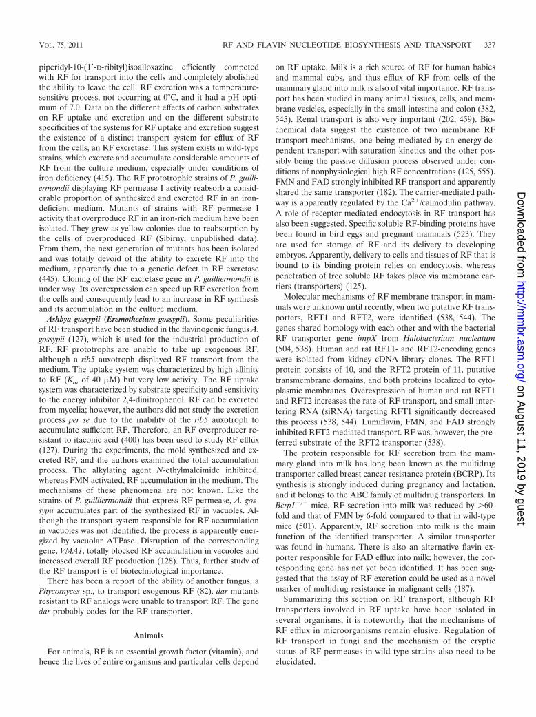
piperidyl-10-(1�-D-ribityl)isoalloxazine efficiently competedwith RF for transport into the cells and completely abolishedthe ability to leave the cell. RF excretion was a temperature-sensitive process, not occurring at 0°C, and it had a pH opti-mum of 7.0. Data on the different effects of carbon substrateson RF uptake and excretion and on the different substratespecificities of the systems for RF uptake and excretion suggestthe existence of a distinct transport system for efflux of RFfrom the cells, an RF excretase. This system exists in wild-typestrains, which excrete and accumulate considerable amounts ofRF from the culture medium, especially under conditions ofiron deficiency (415). The RF prototrophic strains of P. guilli-ermondii displaying RF permease I activity reabsorb a consid-erable proportion of synthesized and excreted RF in an iron-deficient medium. Mutants of strains with RF permease Iactivity that overproduce RF in an iron-rich medium have beenisolated. They grew as yellow colonies due to reabsorption bythe cells of overproduced RF (Sibirny, unpublished data).From them, the next generation of mutants has been isolatedand was totally devoid of the ability to excrete RF into themedium, apparently due to a genetic defect in RF excretase(445). Cloning of the RF excretase gene in P. guilliermondii isunder way. Its overexpression can speed up RF excretion fromthe cells and consequently lead to an increase in RF synthesisand its accumulation in the culture medium.
Ashbya gossypii (Eremothecium gossypii). Some peculiaritiesof RF transport have been studied in the flavinogenic fungus A.gossypii (127), which is used for the industrial production ofRF. RF prototrophs are unable to take up exogenous RF,although a rib5 auxotroph displayed RF transport from themedium. The uptake system was characterized by high affinityto RF (Km of 40 �M) but very low activity. The RF uptakesystem was characterized by substrate specificity and sensitivityto the energy inhibitor 2,4-dinitrophenol. RF can be excretedfrom mycelia; however, the authors did not study the excretionprocess per se due to the inability of the rib5 auxotroph toaccumulate sufficient RF. Therefore, an RF overproducer re-sistant to itaconic acid (400) has been used to study RF efflux(127). During the experiments, the mold synthesized and ex-creted RF, and the authors examined the total accumulationprocess. The alkylating agent N-ethylmaleimide inhibited,whereas FMN activated, RF accumulation in the medium. Themechanisms of these phenomena are not known. Like thestrains of P. guilliermondii that express RF permease, A. gos-sypii accumulates part of the synthesized RF in vacuoles. Al-though the transport system responsible for RF accumulationin vacuoles was not identified, the process is apparently ener-gized by vacuolar ATPase. Disruption of the correspondinggene, VMA1, totally blocked RF accumulation in vacuoles andincreased overall RF production (128). Thus, further study ofthe RF transport is of biotechnological importance.
There has been a report of the ability of another fungus, aPhycomyces sp., to transport exogenous RF (82). dar mutantsresistant to RF analogs were unable to transport RF. The genedar probably codes for the RF transporter.
Animals
For animals, RF is an essential growth factor (vitamin), andhence the lives of entire organisms and particular cells depend
on RF uptake. Milk is a rich source of RF for human babiesand mammal cubs, and thus efflux of RF from cells of themammary gland into milk is also of vital importance. RF trans-port has been studied in many animal tissues, cells, and mem-brane vesicles, especially in the small intestine and colon (382,545). Renal transport is also very important (202, 459). Bio-chemical data suggest the existence of two membrane RFtransport mechanisms, one being mediated by an energy-de-pendent transport with saturation kinetics and the other pos-sibly being the passive diffusion process observed under con-ditions of nonphysiological high RF concentrations (125, 555).FMN and FAD strongly inhibited RF transport and apparentlyshared the same transporter (182). The carrier-mediated path-way is apparently regulated by the Ca2�/calmodulin pathway.A role of receptor-mediated endocytosis in RF transport hasalso been suggested. Specific soluble RF-binding proteins havebeen found in bird eggs and pregnant mammals (523). Theyare used for storage of RF and its delivery to developingembryos. Apparently, delivery to cells and tissues of RF that isbound to its binding protein relies on endocytosis, whereaspenetration of free soluble RF takes place via membrane car-riers (transporters) (125).
Molecular mechanisms of RF membrane transport in mam-mals were unknown until recently, when two putative RF trans-porters, RFT1 and RFT2, were identified (538, 544). Thegenes shared homology with each other and with the bacterialRF transporter gene impX from Halobacterium nucleatum(504, 538). Human and rat RFT1- and RFT2-encoding geneswere isolated from kidney cDNA library clones. The RFT1protein consists of 10, and the RFT2 protein of 11, putativetransmembrane domains, and both proteins localized to cyto-plasmic membranes. Overexpression of human and rat RFT1and RFT2 increases the rate of RF transport, and small inter-fering RNA (siRNA) targeting RFT1 significantly decreasedthis process (538, 544). Lumiflavin, FMN, and FAD stronglyinhibited RFT2-mediated transport. RF was, however, the pre-ferred substrate of the RFT2 transporter (538).
The protein responsible for RF secretion from the mam-mary gland into milk has long been known as the multidrugtransporter called breast cancer resistance protein (BCRP). Itssynthesis is strongly induced during pregnancy and lactation,and it belongs to the ABC family of multidrug transporters. InBcrp1�/� mice, RF secretion into milk was reduced by �60-fold and that of FMN by 6-fold compared to that in wild-typemice (501). Apparently, RF secretion into milk is the mainfunction of the identified transporter. A similar transporterwas found in humans. There is also an alternative flavin ex-porter responsible for FAD efflux into milk; however, the cor-responding gene has not yet been identified. It has been sug-gested that the assay of RF excretion could be used as a novelmarker of multidrug resistance in malignant cells (187).
Summarizing this section on RF transport, although RFtransporters involved in RF uptake have been isolated inseveral organisms, it is noteworthy that the mechanisms ofRF efflux in microorganisms remain elusive. Regulation ofRF transport in fungi and the mechanism of the crypticstatus of RF permeases in wild-type strains also need to beelucidated.
VOL. 75, 2011 RF AND FLAVIN NUCLEOTIDE BIOSYNTHESIS AND TRANSPORT 337
on August 11, 2019 by guest
http://mm
br.asm.org/
Dow
nloaded from

REGULATION OF RIBOFLAVIN SYNTHESIS
Regulation of RF synthesis occurs mostly at the level ofthe synthesis of its biosynthetic enzymes. Effectors of thisregulation are flavins and, unexpectedly, iron ions and, to alesser extent, other metals (cobalt, chromium, zinc in flavi-nogenic yeasts, and magnesium in some in fungi) in manyorganisms.
Feedback Inhibition of GTP Cyclohydrolase II
Very little is known about the regulation of RF synthesis atthe level of modulation of enzyme activity. Apparently, GTPcyclohydrolase II is the rate-limiting reaction in RF synthesis,at least in B. subtilis (185). As a rule, activities of key enzymescatalyzing the first limiting step of the anabolic pathways areregulated by allosteric feedback inhibition exerted by the prod-uct of the reaction or entire pathway. However, very little isknown about the possible feedback control of RF biosynthesisat the level of GTP cyclohydrolase II, although feedback con-trol is well documented for GTP cyclohydrolase I, which isinvolved in the biosynthesis of folates and tetrahydrobiopterin(44, 237, 283). While the structures of bacterial GTP cyclohy-drolases from E. coli and B. subtilis have been studied in detail(206, 257, 365, 373), no information on feedback inhibition ofthis enzyme by RF, flavin nucleotides, or other metabolites isavailable. GTP cyclohydrolase II from the flavinogenic yeast P.guilliermondii was allosterically inhibited by FAD (412, 425).This inhibition was retained in P. guilliermondii GTP cyclohy-drolase II overexpressed in E. coli, whereas the native bacterialenzyme showed no such inhibition (546). RF and FMN had noeffect on enzyme activity. The physiological role of this inhibi-tion remains unclear, since in addition to FAD, other nucleo-tides containing an adenylic group (5�-AMP, 3�,5�-AMP, ADP,ATP, NAD, and NADP) inhibited enzyme activity to a similarextent (412, 425). No data are available on the effect of FADand other adenylic nucleotides on the activity of GTP cyclo-hydrolases from other yeasts, including S. cerevisiae. No P.guilliermondii mutants have proved to be defective in this kindof inhibition.
There is apparently another possible feedback regulation ofRF biosynthetic enzymes, namely, through 6,7-dimethyl-8-ribi-tyllumazine synthase from P. guilliermondii, which is inhibitedby RF but not FAD, whereas neither RF nor FAD influencedthe activities of 3,4-dihydroxy-2-butanone 4-phosphate syn-thase and RF synthase (274, 275). There are no data on thephysiological significance of 6,7-dimethyl-8-ribityllumazinesynthase inhibition obtained in experiments on the isolatedenzyme.
Transcriptional Regulation in Bacteria Usingthe Riboswitch Mechanism
The discovery of the riboswitch mechanism involved in reg-ulation of the RF operon in B. subtilis has been among themajor achievements in modern molecular genetics.
Riboswitch (RFN element) in Bacillus. The regulation of RFsynthesis has been studied in detail in B. subtilis. This organismwas initially chosen for investigation, and the first principal resultson the RF operon were obtained by S. E. Bresler, D. A. Perumov,
and colleagues in Russia. Later, investigators from Germany, theUnited States, and China were involved in these studies. In 1969,Bresler and his colleagues described a mutant of B. subtilis thatoverproduced RF (52). Its isolation, as well as that of RF auxo-trophs, initiated a series of experiments on the biochemical andgenetic aspects of RF synthesis in B. subtilis. All known mutationsleading to RF auxotrophy are linked and are localized close togenes involved in lysine biosynthesis (49, 51).
The RF operon (rib operon) consists of 5 genes (ribGBAHT)that encode the catalytic enzymes for RF synthesis from GTPand ribulose-5-phosphate. The operon is transcribed as onepolycistronic RNA of nearly 4,300 bp (308). The untranslatedleader region, designated ribO, of �300 bp (originally thoughtto be the operator region), has been identified upstream of thefirst gene in the operon (309). Although mutations in ribO ledto RF overproduction (221), searches for the gene coding forthe repressor protein were unsuccessful. Mutations in twotrans-acting genes, ribC and ribR, which caused RF oversyn-thesis have been identified (163, 280, 458); however, theycoded for bifunctional RF kinase/FAD synthetase and mono-functional RF kinase, respectively. Genes ribC and ribR are notlinked to the RF operon. The available data suggest that RibCand RibR proteins do not interact with the ribO region. At thesame time, mutants defective in RF kinases still proved to besensitive to repression of RF synthesis after addition of exog-enous FMN (48, 280). Inspection of the leader region of the riboperon revealed a sequence that could fold into a character-istic and evolutionarily well-conserved RNA structure of �140bp called the RFN element. Analysis of 23 different RFN ele-ments showed that 10 of 20 bp of the inverted repeats werecompletely conserved and that of the 47 nucleotides from thesingle-strand RNA filament, 24 were conserved (138, 221, 504).The RFN element directly binds with FMN, and the formationof this complex is important in the regulation of the rib operon(307, 528). Direct binding of the small molecule FMN to nas-cent mRNA of the RFN element leads to a conformationalchange resulting in terminator hairpin formation, which leadsto attenuation of transcription. In the absence of FMN, theconformation of the RFN element forms an antiterminatorstructure that allows normal transcription of the entire operon(307). The RFN element serves as the receptor for a metabo-lite-dependent riboswitch that directly binds FMN in the ab-sence of proteins (528). The RFN element is a natural FMN-binding aptamer, the allosteric character of which is used fortight control in the expression of the RF operon (Fig. 5). RFNelements have been found upstream of RF biosynthesis andRF transporter genes in many bacteria (504).
Similar metabolite-binding riboswitches are involved in theregulation of other operons and separate genes in bacteria.Riboswitches are found in many classes of eubacteria but onlya few archaea and eukaryotes (25). Riboswitches involved invitamin and coenzyme synthesis constitute the most abundantclass of untranslated mRNA regions capable of direct sensingof cellular metabolites (75, 325, 378, 405). Riboswitches arethe nontranslated mRNAs that bind effectors (low-molecu-lar-weight metabolites) using selective binding domains(otherwise called ligand-binding pockets or aptamers) with-out a requirement for any proteins. Regulation of gene
338 ABBAS AND SIBIRNY MICROBIOL. MOL. BIOL. REV.
on August 11, 2019 by guest
http://mm
br.asm.org/
Dow
nloaded from

expression by the riboswitch mechanism without participa-tion of regulatory proteins is probably a molecular relic ofthe ancestral RNA world.
Riboswitches typically contain disordered regions in theirconserved aptamer cores that become structured upon metab-olite binding. These changes may trigger rearrangements inadditional expression platform structures located outside the
aptamer, giving two alternative conformations. Riboswitchesare able to interconvert between these ligand-bound and li-gand-free structures in the context of the full-length RNA (25,405, 406). The crystal structure of the FMN riboswitch con-trolling the putative RF transporter encoded by impX inFusobacterium nucleatum bound with FMN, RF, and roseofla-vin has been determined (404). Binding of the riboswitch to
FIG. 5. FMN causes transcription termination of the ribD RNA in vitro. (A) Sequence and secondary structure of the leader sequence ribDRNA. Shaded regions identify nucleotides that are complementary and might serve as an antiterminator structure. Nucleotides denoted with anasterisk have been altered from the wild-type sequence to generate a restriction site. The nucleotide sequence comprising the 62- and 30-nucleotideregions (not shown) is presented in references 16 and 17. (B) Model for the FMN-dependent riboswitch. Shaded regions identify the putativeantiterminator structure that is disrupted after binding of FMN and formation of the P1 structure. (Reproduced from reference 528 withpermission of the publisher. Copyright 2002 National Academy of Sciences, U.S.A.)
VOL. 75, 2011 RF AND FLAVIN NUCLEOTIDE BIOSYNTHESIS AND TRANSPORT 339
on August 11, 2019 by guest
http://mm
br.asm.org/
Dow
nloaded from

FMN depends on Mg2� ions and is further enhanced by K�
ions. FAD, RF, and lumiflavin have much lower affinity for theriboswitch than FMN. The roseoflavin-bound structure adopts aconformation similar to that of the RF-bound structure, withsome additional conformational adjustments. The data explainthe effect of regulatory mutations in the FMN riboswitch on geneexpression and can be used for developing strategies to search forspecific inhibitors of RF synthesis in pathogenic bacteria by actingon the FMN riboswitch. The structure of the FMN-bound ribo-switch is shown in Fig. 6.
The carboxy terminus of the B. subtilis ribR monofunctionalRF kinase binds to the FMN riboswitch, but this binding ispartially lost in cells bearing mutations in the RFN element(177). The data suggest that riboswitch regulation using FMNas an effector can be supplemented with the action of mono-functional RF kinase, thus acting as a regulatory protein.
Regulation in E. coli. It is noteworthy that in many groups ofbacteria, e.g., in Gammaproteobacteria, including Enterobacte-riaceae, the FMN riboswitch attenuates translation, or bothtranscription and translation, mechanisms (25, 504). It usesFMN-induced structure changes of the riboswitch structure toblock the initiation of translation. Unlike riboswitches withtranscription control mechanisms that require very specific ter-minator structures in their expression platforms, the RNAstructures that prevent translation initiation can be morevaried.
In physiological terms, RF synthesis in E. coli occurs consti-tutively (422, 526). Genes of RF synthesis are scattered on theE. coli chromosome (23, 495). 5� noncoding regions of the RFstructural genes contain RFN elements apparently acting at thetranslation level (25, 504); however, the physiological role ofthese riboswitches in the regulation of RF synthesis in E. coliremains unknown. The E. coli ribA gene, coding for GTPcyclohydrolase II, belongs to the soxRS regulon, which is in-
duced by superoxide-generating agents. The localization of thesuperoxide-responsive element in the promoter has been de-termined (232, 233). The exact role of this regulation is un-known; however, it may indicate that GTP cyclohydrolase II orthe final product of the pathway, RF or flavin nucleotides, issomehow involved in defense against oxidative stress. Reactiveoxygen forms convert GTP to 8-hydroxy-dGTP, which acts as amutagen. GTP cyclohydrolase II fulfills a supplementary rolein the detoxification of 8-hydroxy-dGTP to mutT protein (229).
Other bacteria. The ribBA gene of H. pylori, coding forbifunctional GTP cyclohydrolase II//3,4-dihydroxybutanonephosphate synthase, is derepressed under conditions of irondeficiency, which leads to increased flavin synthesis (529). Het-erologous expression of this gene in E. coli doubled RF syn-thesis and ferrireductase activity.
As mentioned above, flavins participate in bacterial lumines-cence. In Photobacterium phosphoreum and Photobacteriumleiognathi, RF biosynthesis genes are linked to the luciferase(lux) operon and are transcribed as one polycistronic mRNA(251, 252, 270). In other luminescent species, e.g., Vibrio fisch-eri, an unlinked ribB gene homologous to the E. coli genecoding for 3,4-dihydroxy-2-butanone 4-phosphate synthaseforms a joint regulon with the lux operon (61).
It is well known that Clostridium acetobutylicum is capable ofoverproducing RF under conditions of iron deficiency (175),but the mechanism of regulation remains obscure.
Transcriptional Regulation in Mycelial Fungi
Of the mycelial fungi that naturally overproduce RF, two(the phytopathogenic [cotton pathogens] fungi Eremotheciumashbyii [162] and Eremothecium [Ashbya] gossypii [525]) belongto the group of the most flavinogenic natural organisms known(83). RF accumulates in the mycelia, giving them a brightyellow color (128). The ability to overproduce RF is an unsta-ble feature in E. ashbyii, and highly flavinogenic clones easilylose their potential during lyophilization or storage at roomtemperature (313). Therefore, most work has been conductedon E. gossypii (its generally used name remains A. gossypii,which is used throughout this review). However, E. ashbyiioverproduces not only RF but also FAD, whereas A. gossypiidoes not overproduce flavin nucleotides (337, 534). A. gossypiiis characterized by a high level of gene order conservation(synteny) between its genome and that of the yeast S. cerevisiaeand thus became a popular model for the biology of fungaldevelopment (350, 522). Methods of molecular genetics for A.gossypii, including insertional mutagenesis, have been devel-oped (391, 467, 468, 530). The genome of A. gossypii has beensequenced and has strong similarities to that of S. cerevisiae.With a size of only 9.2 Mb, carrying 4,718 protein-codinggenes, the A. gossypii genome is the smallest of those of free-living eukaryotes yet characterized (45, 86).
RF oversynthesis by both of the above-mentioned fungi oc-curs after vegetative growth has ceased, during mycelium lysisand spore formation (209, 463). As a rule, similar kinetics ofproduction have been seen during microbial synthesis of sec-ondary metabolites, e.g., antibiotics, but not primary metabo-lites. Therefore, RF overproduction by the mycelial fungi E.ashbyii and A. gossypii has been called “pseudosecondary bio-synthesis” (66). The physiological reasons for RF overproduc-
FIG. 6. Overall riboswitch structure in a ribbon representation. P1to P6, domains; L1 to L6, loops. The J1-2 segment participates inantiterminator formation in the absence of FMN, whereas in theFMN-bound state, it is locked up in the junction. (Reproduced fromreference 404 with permission of Macmillan Publishers Ltd., copyright2009.)
340 ABBAS AND SIBIRNY MICROBIOL. MOL. BIOL. REV.
on August 11, 2019 by guest
http://mm
br.asm.org/
Dow
nloaded from
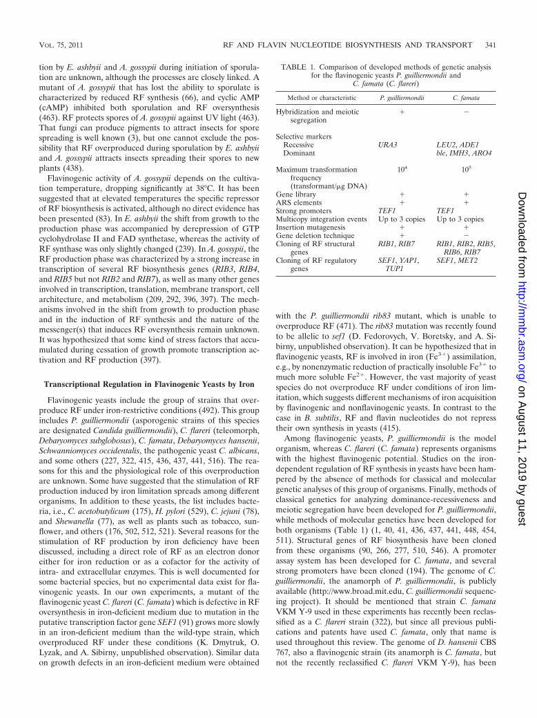
tion by E. ashbyii and A. gossypii during initiation of sporula-tion are unknown, although the processes are closely linked. Amutant of A. gossypii that has lost the ability to sporulate ischaracterized by reduced RF synthesis (66), and cyclic AMP(cAMP) inhibited both sporulation and RF oversynthesis(463). RF protects spores of A. gossypii against UV light (463).That fungi can produce pigments to attract insects for sporespreading is well known (3), but one cannot exclude the pos-sibility that RF overproduced during sporulation by E. ashbyiiand A. gossypii attracts insects spreading their spores to newplants (438).
Flavinogenic activity of A. gossypii depends on the cultiva-tion temperature, dropping significantly at 38°C. It has beensuggested that at elevated temperatures the specific repressorof RF biosynthesis is activated, although no direct evidence hasbeen presented (83). In E. ashbyii the shift from growth to theproduction phase was accompanied by derepression of GTPcyclohydrolase II and FAD synthetase, whereas the activity ofRF synthase was only slightly changed (239). In A. gossypii, theRF production phase was characterized by a strong increase intranscription of several RF biosynthesis genes (RIB3, RIB4,and RIB5 but not RIB2 and RIB7), as well as many other genesinvolved in transcription, translation, membrane transport, cellarchitecture, and metabolism (209, 292, 396, 397). The mech-anisms involved in the shift from growth to production phaseand in the induction of RF synthesis and the nature of themessenger(s) that induces RF oversynthesis remain unknown.It was hypothesized that some kind of stress factors that accu-mulated during cessation of growth promote transcription ac-tivation and RF production (397).
Transcriptional Regulation in Flavinogenic Yeasts by Iron
Flavinogenic yeasts include the group of strains that over-produce RF under iron-restrictive conditions (492). This groupincludes P. guilliermondii (asporogenic strains of this speciesare designated Candida guilliermondii), C. flareri (teleomorph,Debaryomyces subglobosus), C. famata, Debaryomyces hansenii,Schwanniomyces occidentalis, the pathogenic yeast C. albicans,and some others (227, 322, 415, 436, 437, 441, 516). The rea-sons for this and the physiological role of this overproductionare unknown. Some have suggested that the stimulation of RFproduction induced by iron limitation spreads among differentorganisms. In addition to these yeasts, the list includes bacte-ria, i.e., C. acetobutylicum (175), H. pylori (529), C. jejuni (78),and Shewanella (77), as well as plants such as tobacco, sun-flower, and others (176, 502, 512, 521). Several reasons for thestimulation of RF production by iron deficiency have beendiscussed, including a direct role of RF as an electron donoreither for iron reduction or as a cofactor for the activity ofintra- and extracellular enzymes. This is well documented forsome bacterial species, but no experimental data exist for fla-vinogenic yeasts. In our own experiments, a mutant of theflavinogenic yeast C. flareri (C. famata) which is defective in RFoversynthesis in iron-deficient medium due to mutation in theputative transcription factor gene SEF1 (91) grows more slowlyin an iron-deficient medium than the wild-type strain, whichoverproduced RF under these conditions (K. Dmytruk, O.Lyzak, and A. Sibirny, unpublished observation). Similar dataon growth defects in an iron-deficient medium were obtained
with the P. guilliermondii rib83 mutant, which is unable tooverproduce RF (471). The rib83 mutation was recently foundto be allelic to sef1 (D. Fedorovych, V. Boretsky, and A. Si-birny, unpublished observation). It can be hypothesized that inflavinogenic yeasts, RF is involved in iron (Fe3�) assimilation,e.g., by nonenzymatic reduction of practically insoluble Fe3� tomuch more soluble Fe2�. However, the vast majority of yeastspecies do not overproduce RF under conditions of iron lim-itation, which suggests different mechanisms of iron acquisitionby flavinogenic and nonflavinogenic yeasts. In contrast to thecase in B. subtilis, RF and flavin nucleotides do not represstheir own synthesis in yeasts (415).
Among flavinogenic yeasts, P. guilliermondii is the modelorganism, whereas C. flareri (C. famata) represents organismswith the highest flavinogenic potential. Studies on the iron-dependent regulation of RF synthesis in yeasts have been ham-pered by the absence of methods for classical and moleculargenetic analyses of this group of organisms. Finally, methods ofclassical genetics for analyzing dominance-recessiveness andmeiotic segregation have been developed for P. guilliermondii,while methods of molecular genetics have been developed forboth organisms (Table 1) (1, 40, 41, 436, 437, 441, 448, 454,511). Structural genes of RF biosynthesis have been clonedfrom these organisms (90, 266, 277, 510, 546). A promoterassay system has been developed for C. famata, and severalstrong promoters have been cloned (194). The genome of C.guilliermondii, the anamorph of P. guilliermondii, is publiclyavailable (http://www.broad.mit.edu, C. guilliermondii sequenc-ing project). It should be mentioned that strain C. famataVKM Y-9 used in these experiments has recently been reclas-sified as a C. flareri strain (322), but since all previous publi-cations and patents have used C. famata, only that name isused throughout this review. The genome of D. hansenii CBS767, also a flavinogenic strain (its anamorph is C. famata, butnot the recently reclassified C. flareri VKM Y-9), has been
TABLE 1. Comparison of developed methods of genetic analysisfor the flavinogenic yeasts P. guilliermondii and
C. famata (C. flareri)
Method or characteristic P. guilliermondii C. famata
Hybridization and meioticsegregation
� �
Selective markersRecessive URA3 LEU2, ADE1Dominant ble, IMH3, ARO4
Maximum transformationfrequency(transformant/�g DNA)
104 105
Gene library � �ARS elements � �Strong promoters TEF1 TEF1Multicopy integration events Up to 3 copies Up to 3 copiesInsertion mutagenesis � �Gene deletion technique � �Cloning of RF structural
genesRIB1, RIB7 RIB1, RIB2, RIB5,
RIB6, RIB7Cloning of RF regulatory
genesSEF1, YAP1,
TUP1SEF1, MET2
VOL. 75, 2011 RF AND FLAVIN NUCLEOTIDE BIOSYNTHESIS AND TRANSPORT 341
on August 11, 2019 by guest
http://mm
br.asm.org/
Dow
nloaded from

sequenced in the framework of the French National ProgramGenolevures (http://cbi.labri.fr/Genolevures/elt/DEHA)).
Iron deficiency leads to derepression of all of the P. guilli-ermondii enzymes involved in RF synthesis, with the exceptionof the reductase, an enzyme in the second step of the pathwaythat is constitutively synthesized (417). Iron content did notinfluence synthesis of enzymes involved in RF conversion toflavin coenzymes (408, 424). Derepression occurred at thetranscriptional level, as iron starvation caused increased RIB1(GTP cyclohydrolase II) and RIB7 (RF synthase) mRNA ac-cumulation (38). Regulation by iron at the transcriptional levelwas also seen for the RIB1 and RIB3 (3,4-dihydroxy-2-butanone-4-phosphate synthase) genes of a strain of anotherflavinogenic species, Candida membranifaciens subsp. flavinog-enie W14-3 (516). The activity of the RIB2 product, reductase,is so high that when it dropped below the detection level in theleaky rib2 mutant of P. guilliermondii, this did not cause RFauxotrophy (417).
Numerous mutants of P. guilliermondii that are defective inthe regulation of RF synthesis by iron have been isolated byseveral innovative selection procedures. One of them seems tobe straightforward, since it has been based on the isolation ofmutants resistant to RF analogs. The assumption was thatexogenous RF must alleviate the inhibitory action of RF ana-logs on yeast growth, and thus RF overproducers must be moreresistant to these compounds. However, wild-type strains of P.guilliermondii were resistant to very high concentrations of RFanalogs, apparently due to their inability to penetrate the yeastcell. This obstacle was overcome either by using mutants withmultiple sensitivity to antibiotics and antimetabolites (447) orby addition of high concentrations of phosphates or sulfatesthat make wild-type cells susceptible to the inhibitory action ofRF analogs (444). In the mutants resistant to RF analogs thathave been isolated, overproduction of RF in the medium wasseen, as expected, when a sufficient amount of iron was present(421, 407). The second method for the selection of mutantsoverproducing RF in an iron-rich medium was with leaky rib2mutants partially defective in activity of reductase (the secondenzyme in biosynthesis). Activity was so low that the leakystrains required exogenous RF in the medium and a high ironcontent, although they grew without RF in an iron-deficientmedium. The leaky rib2 suppressor mutants that acquired theability to grow in a medium with a high iron content withoutRF were characterized by derepression of GTP cyclohydrolaseII, and after substitution of the leaky rib2 with the wild-typeRIB2 allele, RF was overproduced in an iron-rich medium(411, 419). Another method was similar in using a mutant witha thermosensitive GTP cyclohydrolase II that grew like theparental strain without exogenous RF only in an iron-deficientmedium. Mutants with impaired regulation of RF synthesiswere isolated as revertants growing at the minimal restrictivetemperature in an iron-sufficient medium without exogenousRF. In meiotic segregants that contained the wild-type allele ofthe RIB1 gene, mutants overproduced RF in an iron-rich me-dium (9). An additional approach was based on isolation ofrib1 mutants with decreased requirements for exogenous RF(472) or on the leaky rib1 mutant (with a missense mutationleading to substitution G620A) growing without exogenous RFin an iron-rich medium (38, 469, 470). Mutants with dere-pressed synthesis of RF in an iron-rich medium were also
isolated from the yeast C. famata by selection of mutants re-sistant to the RF structural analog 7-methyl-8-trifluoromethyl-10(1�-D-ribityl)isoalloxazine in a medium with a high sulfateconcentration (439; Sibirny, unpublished data).
In P. guilliermondii, all isolated mutations were analyzedusing hybridization and meiotic segregation analyses. All led toRF overproduction and derepression of RF biosynthetic en-zymes in iron-rich media and were recessive. They were di-vided into 9 complementation classes: rib80, rib81, hit1, andred1 to red6 (407, 411, 470, 472). Mutations rib81 and red6showed constitutive derepression of RIB1 gene transcription inan iron-rich medium (38). Most RF-overproducing mutantsalso had derepressed iron transport and an elevated content ofnonhemin iron (109, 110, 409, 410, 415). These data suggest acoordinated regulation of RF synthesis and iron acquisition inP. guilliermondii. RF overproduction in P. guilliermondii causesCo2� ion stress (106) and oxidative stress (357). Later, it wasshown that Co2� ions, iron deficiency, and the mutations rib80,rib81, and hit1 all lead to oxidative stress (39). These observa-tions can be partially explained by hypothesizing that RF actsas an antioxidant.
Elucidation of the identified gene action required cloning ofthe genes as the first step. However, mutants overexpressingRF do not differ significantly from wild-type cells in theirgrowth characteristics, which hampered transformant selectionwith the restored wild-type phenotype. One additional RF-overproducing mutant of P. guilliermondii was obtained usingthe molecular approach of knockout of the yeast frataxin ho-molog gene YFH1, encoding a mitochondrial protein involvedin iron trafficking and storage (8, 64, 392). The resulting �yfh1mutant, like mutants isolated by conventional mutagenesis,overproduced RF in an iron-rich medium, possessed an ele-vated intracellular iron content, and was hypersensitive to ox-idative stress. However, the �yfh1 mutation was nonallelic tomutations rib80, rib81, hit1, and red6 (361). It seems that mu-tations in numerous genes involved in iron transport, assimi-lation, and storage lead to RF overproduction in P. guillier-mondii. Knowledge of the precise role of the identified genes inregulation of RF synthesis awaits further studies. Recently,insertion mutagenesis was used in the isolation of P. guillier-mondii mutants with derepressed RF synthesis in iron-suffi-cient medium. Among them, the gene VMA1, coding for vac-uolar ATPase, was tagged. Insertion and knockout vma1mutants overproduced RF in an iron-rich medium (41a). Therole of vacuolar ATPase in the regulation of RF synthesisremains obscure.
Mutants with the opposite phenotype, i.e., an inability tooverproduce RF in iron-deficient media, were also isolatedfrom flavinogenic yeasts. In P. guilliermondii, such mutantswere isolated as mutants lacking the ability to excrete RF iniron-deficient medium. The corresponding recessive mutationwas designated rib83 (413). Mutation rib83 is epistatic overmutations leading to RF oversynthesis, i.e., rib80, rib81, andhit1 (471; D. Fedorovych, unpublished data). The iron contentin rib83 cells was similar to that in the wild-type strain (39). InC. famata, insertion mutagenesis was used to isolate RF-non-overproducing mutants and tag the corresponding genes.Three mutants unable to overproduce RF contained an inser-tional cassette in the promoter regions of the structural genecoding for GTP cyclohydrolase II (RIB1) and in homologs of
342 ABBAS AND SIBIRNY MICROBIOL. MOL. BIOL. REV.
on August 11, 2019 by guest
http://mm
br.asm.org/
Dow
nloaded from

the gene MET2, involved in methionine biosynthesis, and thegene SEF1, coding for a putative transcription factor (91).Intact orthologs of these genes from D. hansenii restored nor-mal regulation of RF synthesis. Data on Sef1p functions inother yeasts are poor. Kluyveromyces lactis SEF1 and its S.cerevisiae homolog can suppress a mutation in Rpm2p, which isa protein subunit of yeast mitochondrial RNase P, an enzymeresponsible for the 5� maturation of mitochondrial tRNAs.DNA sequence analysis of the K. lactis SEF1 gene showed thatit contained the Zn(2)-Cys(6) binuclear cluster motif found ina growing number of yeast transcription factors (161). Disrup-tion of the SEF1 homolog in P. guilliermondii led to isolation ofmutants unable to overproduce RF. Complementation analysisshowed that �sef1 P. guilliermondii is allelic to rib83, which wasisolated earlier by classical selection (D. Fedorovych, V. Bo-retsky, and A. Sibirny, unpublished data).
Expression of C. famata SEF1 is derepressed under condi-tions of iron starvation, which suggests autoregulation of geneexpression. In the nonflavinogenic yeasts S. cerevisiae andPichia stipitis, expression of this gene is not regulated by iron.The industrial strain C. famata dep8 possesses constitutivelyderepressed SEF1 which does not depend on iron content (K.Dmytruk and A. Sibirny, unpublished data). Introduction ofthe D. hansenii SEF1 gene into �sef1 C. famata restored theability to overproduce RF, whereas introduction of SEF1 fromthe nonflavinogenic species P. stipitis in the same �sef1 C.famata strain did not. The coding sequences of SEF1 are verysimilar in flavinogenic and nonflavinogenic yeast species; wetherefore suggest that the inability of P. stipitis SEF1 to restoreRF overproduction in �sef1 C. famata depends on its lowconstitutive expression. SEF1 may play a central role in regu-lating RF synthesis in flavinogenic yeasts, but the mechanismof its action remains to be elucidated.
THE RIBOFLAVIN BIOSYNTHESIS PATHWAY AS ATARGET FOR ANTIMICROBIAL DRUGS
Most microorganisms, including pathogens, synthesize RFde novo, and many of them (especially Gram-negative bacteriaand pathogenic fungi) cannot take up RF from the growthmedium. Therefore, for microorganisms that cannot transportRF, specific inhibitors of RF synthesis enzymes should be po-tent antimicrobial drugs where they can penetrate the cell.Mammals do not possess RF biosynthetic enzymes and thushave no target for potential antimicrobial drugs; they are ca-pable of very efficient RF transport. In the light of the rapiddevelopment of resistance to virtually all current antibiotics,exploration of new targets and development of novel anti-infective drugs are urgent medical needs (37, 112, 278, 365).
Inhibitors of metabolic reactions have rarely been used asantimicrobial agents, although sulfonamides, the first antibac-terial agents discovered before antibiotics, act against a broadspectrum of bacterial pathogens by competing with the naturalsubstrate of dihydropteroate synthase, p-aminobenzoate,thereby inhibiting this enzyme, which is involved in folic acidbiosynthesis (301). In this case, sulfonamides, but not folicacid, can be transported by pathogens, whereas human cellseffectively absorb folic acid and do not have an intrinsic path-way for biosynthesis of folic acid. Thus, the search for inhibi-tors of the enzymes involved in RF synthesis as potential an-
timicrobial agents is based on the premise that they will havethe same mode of action on folic acid biosynthesis as sulfon-amides.
All RF biosynthetic enzymes are potential targets for inhi-bition. However, GTP cyclohydrolase II, reductase, deaminase,and 3,4-dihydroxy-2-butanone 4-phosphate synthase act onphosphorylated substrates and products; most probably, there-fore, structural inhibitors of these enzymes would not pene-trate the microbial cells in their native form. Lumazine syn-thase and RF synthase are more promising targets forinhibition because they act with nonphosphorylated com-pounds as substrates or products [5-amino-6-ribitylamino-2,4(1H,3H)-pyrimidinedione and 6,7-dimethyl-8-ribitylluma-zine, respectively]. It would probably be difficult for the lattercompound (being a pteridine) to penetrate the cell envelope.Rybitiled pyrimidine derivatives are more suitable as inhibitorsof lumazine and RF synthases, although many pyrimidine an-alogs could have an influence on other processes, such asnucleotide metabolism and nucleic acid biosynthesis.
Throughput screening methods that are based on thecompetitive binding of tested inhibitors with lumazine syn-thase or RF synthase were developed (68, 205). Almost100,000 compounds have been tested for inhibition of RFbiosynthetic enzymes (279, 314, 316, 491, 553). Some com-pounds were very efficient inhibitors in vitro, and a few ofthem displayed antibiotic activity against M. tuberculosis.Therefore, this work is promising.
Alternative use of the RF biosynthetic pathway as a targetfor novel antimicrobial drug can be explored for pathogenicGram-positive bacteria that can effectively transport RF. Asdescribed above, the natural RF analog roseoflavin binds withthe FMN riboswitch, thereby repressing RF operon transcrip-tion (253, 335, 404). If roseoflavin binding to the FMN ribo-switch is the major target for its antimicrobial action, thesearch for structural analogs of this antibiotic with more potentor specific binding to the riboswitch could be another use ofthe RF biosynthetic pathway for the identification of new an-timicrobial drugs (36, 278). Specificity of the action of a selec-tive corepressor of RF biosynthesis could be achieved due tothe absence of this pathway in the host organism (humans oranimals). These RF or roseoflavin analogs, however, shouldnot be metabolized to analogs of flavin nucleotides, whichmight be toxic for host organisms.
FLAVIN SYNTHESIS BY FLAVINOGENICMICROORGANISMS
Flavins are present in each cell of living organisms. Cellularflavins are present preferentially in nucleotide forms, withFAD being the most abundant. The content of free intracel-lular RF does not exceed 7% of the total flavins (438). Thepool of free flavins (not bound to proteins) in microorganismsvaries between 20 and 200 �g/g biomass. Some microorganismshave much larger intracellular pools of free flavins, e.g., theflavinogenic fungi E. ashbyii and A. gossypii (66). Many micro-organisms excrete some portion of synthesized flavins to themedium, preferentially as free RF (83). This feature allows RFbiosynthesis to be considered a poorly controlled process. Forexample, exponentially growing wild-type cells of E. coli, B.subtilis, and Pseudomonas fluorescens secrete into the medium
VOL. 75, 2011 RF AND FLAVIN NUCLEOTIDE BIOSYNTHESIS AND TRANSPORT 343
on August 11, 2019 by guest
http://mm
br.asm.org/
Dow
nloaded from

several times more flavins than they contain inside the cells incoenzyme forms (526). The role of the excreted RF remainsunknown, but it could be hypothesized that it participates inthe mobilization of practically insoluble Fe3� by its reductionto the more soluble Fe2� form (287).
According to Demain (83), microorganisms that accumulate�10 mg RF/liter are overproducers. They can be divided into3 groups: weak (producing around 10 mg RF/liter), moderate(producing up to 600 mg RF/liter), and highly active or flavi-nogenic (producing �10 g RF/liter). The overproducers in-clude the bacteria C. acetobutylicum, Mycobacterium smegma-tis, and Corynebacterium diphtheriae, the yeasts Candida(Pichia) guilliermondii, Candida ghoshii, Candida parapsilosis,C. flareri, C. famata, D. subglobosus, D. hansenii, S. occidentalis,and Torulopsis candida, and the mycelial fungi Aspergillus niger,A. gossypii, E. ashbyii, and others. The first observation of RFoverproduction by bacteria was by Japanese authors who de-scribed the accumulation of RF in C. acetobutylicum (539). RFoverproduction in yeasts was reported at approximately thesame time (59, 348). The maximal amount of RF produced byyeast under conditions of iron deficiency was 560 mg RF/literin C. flareri (260). In addition to the concentration of iron ions,some other factors act on RF synthesis. Strong stimulation ofRF synthesis in flavinogenic yeasts was caused by Co2� (87,106), and some stimulation was seen with Cr6�, Mn2�, andZn2� (111, 228, 487). In the fungus A. niger, RF synthesis wasstimulated by a deficiency of Mg2� ions (319, 435). Somespecies of flavinogenic yeasts overproduce RF in iron-sufficientmedia containing n-alkanes as the sole carbon source (88, 329,423), but the mechanisms of these stimulatory effects remainunknown.
INDUSTRIAL PRODUCTION OF RIBOFLAVINAND FLAVIN NUCLEOTIDES AND THEIR
PRACTICAL APPLICATIONS
The vitamin market was $2.3 billion in 2003, with mostvitamins being produced microbiologically. In 2003, the annualproduction of RF was 4,600 tons, with a total value of $134million (84). In 2008, the total world riboflavin productioncapacity was around 10,000 tons, whereas annual demand isaround 6,000 tons estimated to be worth between $150 millionand $500 million depending on the price per kilogram (http://www.articlepros.com/business/Chinese-Marketing/article-660667.html and http://www.articlesbase.com/management-articles/riboflavin-market-development-situation-and-future-trends-2077296.html). Most RF is currently produced biotechnologi-cally using advanced microbial producers constructed from B.subtilis and A. gossypii (178). Production with the yeast C.famata was shut down several years ago by ADM Co. due tothe instability (at that time) of the available producer, dep8.The largest producer of RF currently is China (Hubei GuangjiPharmaceuticals, Shanghai Desano Vitamins Co.), althoughthis vitamin is also produced in other countries in Europe andAsia (Aventis, BASF, Daicel, DSM, Kyowa, Mitsui, Roche,and Takeda).
Over 80% of RF is used in agriculture as an additive topremixes for animal feeding (poultry, pigs, etc.) (462). RF alsois used in the food industry as the yellow colorant E-101 forbeverages, as well as in medicine as a component of vitamin
mixtures and for treatment of cataracts, migraine, malaria,methemoglobinemia, some organic acidurias, and other dis-eases of the skin, eyes, and nervous system (31, 483). RF in thepresence of long-wavelength UV irradiation effectively inacti-vates different viruses, which can be important for vaccinedevelopment (62).
Flavin nucleotides are used by the pharmaceutical and foodindustries. FMN is 200 times more soluble in water, so it isoften offered by pharmaceutical companies. However, itshould be pointed out that FMN offered as medicine is syn-thesized chemically and contains �30% of impurities of aflavin nature (324), which can act as antivitamins and/or RFantagonists. Reduced FMN has an interesting potential appli-cation in the detoxification of chlorinated xenobiotics (72).FAD is used in medicine and is produced biotechnologically(430). It was recently found that FAD could be an efficienttreatment of some inheritable diseases, e.g., chronic granulo-matous diseases caused by mutations in leukocyte NADPHoxidase genes (183) and Friedreich ataxia caused by the lack ofthe mitochondrial protein frataxin (149).
CONSTRUCTION OF INDUSTRIAL RIBOFLAVINPRODUCERS BY CLASSIC MUTAGENESISAND SELECTION AND BY METHODS OF
METABOLIC ENGINEERING
The history of industrial RF production has been describedin detail in previous reviews (83, 178, 438, 462). After the firstperiod of microbial production of RF based on natural micro-bial isolates and a subsequent period of chemical production,most RF is currently produced using genetically engineeredmicroorganisms. Both methods of classic mutagenesis and se-lection and modern methods of metabolic engineering havebeen used. Naturally, these approaches differed depending onthe organism used. In the case of B. subtilis, most effort wasdirected at impairment of regulation of the RF operon andamplification of the copy number of the structural gene. In A.gossypii, most attention has been paid to increasing the RFpathway supply with purine precursors and activation of theglyoxylic acid cycle, which improves catabolism of oil, the pre-ferred carbon substrate for RF synthesis. In C. famata, im-provement of RF productivity has been achieved by introduc-ing several unidentified mutations leading to resistance againstdifferent toxic agents and, recently, by increasing the copynumber of transcription factor SEF1 along with the genesinvolved in purine nucleotide interconversion and the RF bio-synthetic pathway. As some plants overproduce RF under con-ditions of iron deficiency, they can be used for selection ofRF-enriched plants as the source of vitamin B2 in food sup-plements (176).
Below, a short description of the main approaches used inthe isolation of efficient RF producers from bacteria, mycelialfungi, and yeasts is given.
B. subtilis
Wild-type strains of B. subtilis do not overproduce RF. RF-overproducing mutants are easy to isolate but produce verysmall amounts (52). Industrial RF producers of B. subtilis havebeen isolated using several selection steps for resistance to
344 ABBAS AND SIBIRNY MICROBIOL. MOL. BIOL. REV.
on August 11, 2019 by guest
http://mm
br.asm.org/
Dow
nloaded from

different antimetabolites and subsequently introducing regula-tory mutations alleviating repression by FMN and increasingthe copy numbers of the RF operon and of the ribA gene,coding for the first enzyme of RF biosynthesis (178, 341–344).Additionally, RF production in B. subtilis can be increased byderepression of the ZWF1 gene, encoding glucose-6-phosphatedehydrogenase (96), and by protein engineering of GTP cyc-lohydrolase II to improve its kinetic characteristics (257).
Work on B. subtilis RF industrial producers started afterisolation of mutants showing, successively, resistance to 8-aza-guanine, decoyinine, and methionine sulfoxide (343). Mutantsresistant to the purine analog 8-azaguanine often arose due toderepression of purine nucleotide synthesis (395). Decoyinineinhibits GMP synthetase, and decoyinine-resistant mutants canpossess derepressed levels of this enzyme (295). Mutants re-sistant to methionine sulfoxide seem to possess elevated activ-ity of IMP dehydrogenase (294). An isolated triple mutant(producing small amounts of RF [�7 mg/liter]) was used toselect roseoflavin-resistant mutants. About 1 to 2% of themgrew as yellow colonies and possessed mutations in the ribCgene, coding for bifunctional RF kinase/FAD synthetase (163,280). The yellow roseoflavin-resistant mutant RB50 accumu-lated elevated titers of RF (up to 40 mg/liter) due to derepres-sion of RF operon transcription (343).
Several copies of the DNA fragment containing the RFoperon were introduced into the genome of the RB50 strain,which led to a 10-fold increase in RF accumulation by thetransformants (up to 700 mg/liter). Subsequently, their own RFoperon promoters were replaced with the P15 promoter of B.subtilis phage SPO1. After optimization of the cultivation con-ditions in the fermentor for the constructed strain, 14 g RF/liter was obtained in 48 h and 15 g/liter was accumulated in56 h (344). Finally, introduction in this strain of one additionalcopy of the ribA gene under the control of the P15 promoter,coding for bifunctional GTP cyclohydrolase II/3,4-dihydroxy-2-butanone 4-phosphate synthase, led to strain VB2XL1,which was characterized by a further 25% increase in RFaccumulation (185). Overexpression of other individual genesof the RF operon did not increase RF synthesis (178). Asimilar protocol for the isolation of RF-overproducing B. sub-tilis strains has been used by Chinese authors (429).
However, the achieved RF yield in B. subtilis is quite lowcompared to the theoretical maximal yield (394). Recent workshows that B. subtilis possesses the potential for further in-creases in RF synthesis. A series of mutated GTP cyclohydro-lases has been obtained using error-prone mutagenesis; severalof them have shown improved kinetic characteristics (specificactivity and Vmax). Apparently the introduction of this kineti-cally improved enzyme (which is known in B. subtilis to catalyzethe rate-limiting reaction of the overall pathway) into indus-trial RF producers can further increase RF yield and produc-tivity (257). RF synthesis in the RF-overproducing strain hasbeen improved by a genetic defect in transketolase (141) or byredirecting carbon flow through phosphoenolpyruvate carboxi-nase (547). Overexpression of the ZWF1 gene, coding for glu-cose-6-phosphate dehydrogenase, in the RF-overproducingstrain also increased the RF yield by 25% (96). Reduction ofmaintenance metabolism by knockout of the major cyto-chrome oxidase that catalyzes the less efficient branch of therespiratory chain significantly increased the yield in an RF-
overproducing strain (548). Transposon mutagenesis of B. sub-tilis showed novel and unexpected genes involved in the regu-lation of RF synthesis: disruption of the fliP, yjaU, and yloNgenes led to a 25 to 35% increase in RF yield, whereas trans-poson insertion in numerous genes (nearly 30) significantlydecreased RF synthesis (493). The effect of many such inser-tions cannot yet be rationally explained. Overexpression of oneof the identified genes, gapB (coding for gluconeogenic glyc-eraldehyde-3-phosphate dehydrogenase), due to knockout ofits regulator ccpN, improved yield in the industrial RF pro-ducer. These data clearly show that insertion mutagenesis is apromising approach for the identification of novel genes in-volved in RF synthesis and their subsequent manipulation(493). Transcriptome analysis of a B. subtilis wild-type strainand the RF-overproducing mutant RH33 suggested that thepotential bottleneck in RF overproduction is the low pool ofphosphoribosyl pyrophosphate. Simultaneous overexpressionof two genes, prs and ywlF, involved in the biosynthetic path-way of phosphoribosyl pyrophosphate from ribulose-5-phos-phate in the RF overproducer RH33 increased RF productionby 25% (429). Synthesis of RF by some of the constructedstrains has been optimized using model-predictive control,based on artificial neural networks (242) and statistical design(532). Flux response studies of RF-overproducing B. subtilissuggested a role of the transhydrogenase-like mechanism inmaintaining the balance of reducing equivalents during RFoversynthesis (381).
Genetic manipulation of central carbon metabolism couldalso have a positive effect on RF production in B. subtilis. Forexample, disruption of the pta gene (encoding phosphotrans-acetylase) and simultaneous overexpression of the als gene(encoding acetolactate synthase) increased RF production(554). The authors explained this phenomenon as due to anelevated intracellular level of ATP.
Other Bacteria
Corynebacterium ammoniagenes. Corynebacterium ammoni-agenes is used for the industrial production of purine andpyrimidine nucleotides (136, 317) and therefore was chosen fordeveloping an alternative bacterial RF producer. In the firststep, the structural genes of RF, located in a cluster, werecloned (234). To isolate the RF overproducer, a strong pro-moter of C. ammoniagenes was cloned and substituted for thenatural promoter that controls the expression of the RF bio-synthesis genes in the wild-type strain. This led to a 3-foldincrease in RF yield (235). Additionally, the ribosome-bindingsequence of the bifunctional gene coding for GTP cyclohydro-lase II/3,4-dihydroxy-2-butanone 4-phosphate synthase was op-timized, which strongly increased RF synthesis. Under opti-mized conditions, the engineered strain accumulated 15.3 gRF/liter in 72 h, which is comparable to the yield from B.subtilis. No selection for resistance to RF analogs was used inthis work.
Lactic acid bacteria and propionibacteria. Selection ofroseoflavin-resistant mutants seemed an efficient approach forthe isolation of RF-overproducing strains of several species oflactic acid bacteria (L. lactis, Lactobacillus plantarum, and Leu-conostoc mesenteroides) as well as Propionibacterium freuden-reichii (58, 486). All the analyzed mutations were localized in
VOL. 75, 2011 RF AND FLAVIN NUCLEOTIDE BIOSYNTHESIS AND TRANSPORT 345
on August 11, 2019 by guest
http://mm
br.asm.org/
Dow
nloaded from

the riboswitch regions of RF gene clusters and not in the ribCgene, coding for RF kinase/FAD synthetase. The resultingmutants showed rather modest RF production, several thou-sandfold lower than that of industrial RF producers. For ex-ample, roseoflavin-resistant mutants of L. lactis accumulatedto only near 1.2 mg RF/liter (486), mutants of L. mesenteroidesand L. plantarum produced maximally 0.5 to 0.6 mg RF/liter,and P. freudenreichii accumulated up to 3 mg RF/liter (58).However, yogurts obtained with RF-overproducing mutants oflactic acid bacteria and propionibacteria as starter culturescould be used to increase the RF content in milk products.
A. gossypii
With A. gossypii, much attention was paid to optimization offermentation conditions favoring RF oversynthesis (83, 268).RF overproduction occurs on different residues, including oilywaste-activated bleaching earth (490). Selection for resistanceto RF analogs was not reported, apparently due to the resis-tance of A. gossypii to these compounds (462). No informationis available on A. gossypii mutants resistant to analogs of pu-rines, although exogenous purines and the purine precursorglycine activate RF production (268). As oil stimulates RFproduction in A. ashbyii, activation of the glyoxylic acid cycleshould be important for strain improvement. Itaconate inhibitsthe key enzyme of the glyoxylate shunt, isocitrate lyase, andmutants resistant to itaconate produced enhanced levels of RF(399, 400). Improvement of RF synthesis was also obtained inoxalate- and itaconate-resistant mutants (339, 481). The exactnature of these mutations was not elucidated.
The flavinogenic potential of A. gossypii was substantiallyimproved using modern approaches of metabolic engineering.These were directed at activation of the glyoxylate cycle andsupply with purine precursors of RF. At the same time, noinformation is available on improvement of RF production byoverexpression of RF structural genes. Introduction of an ad-ditional copy of the ICL1 gene, encoding isocitrate lyase, en-hanced RF production in a medium containing soybean oil(203). Overexpression of the second enzyme of the pathway,malate synthase, also improved oil consumption and RFproduction (480). Disruption of VMA1, encoding vacuolarATPase, which energized active RF transport from the cyto-plasm to the vacuole, resulted in complete excretion of synthe-sized RF into the medium and increased the total productionof RF (128). However, disruption of VMA1 in the flavinogenicyeast P. guilliermondii also resulted in RF oversynthesis, al-though normally this organism does not accumulate the RFproduced in vacuoles (41a).
More attention has been paid to activation of RF synthesisin A. gossypii with the purine precursor. Overexpression ofGLY1, encoding the glycine biosynthetic enzyme threoninealdolase, strongly enhanced RF production, apparently due toan improved supply of glycine, the purine precursor (312). Anincrease in RF production was also achieved by disruption ofthe SHM2 gene, encoding one of two isoenzymes of serinehydroxymethyltransferase, due to a defect in the conversion ofglycine to serine, which again improved the glycine supply(398). Conversion of glyoxylate into glycine was improved byheterologous expression of the alanine:glyoxylate aminotrans-ferase gene from S. cerevisiae (217). To enhance the metabolic
flux through the purine nucleotide biosynthesis pathway, thekey regulatory gene ADE4, encoding phosphoribosyl pyro-phosphate amidotransferase, was cloned and overexpressedunder the control of the strong constitutive promoter ofglyceraldehyde phosphate dehydrogenase to abolish repressioncaused by exogenous adenine (195). To alleviate feedback in-hibition of this enzyme by ATP and GTP, site-specific mu-tagenesis of the ADE4 gene by replacement of three aminoacid residues was carried out. Constitutive overexpression ofthis engineered enzyme resulted in a 10-fold increase in RFsynthesis (195). A. gossypii contains two phosphoribosyl pyro-phosphate synthetases, encoded by genes PRS2,4 and PRS3.Overexpression of each of them resulted in an increase of RFproduction. The activity of these enzymes is feedback inhibitedby GDP. Two conserved amino acid residues in the products ofthe PRS2,4 and PRS3 genes were changed to release this inhi-bition. Overexpression of the engineered PRS2,4 and PRS3genes produced strains with further elevated RF production,almost twice that of the wild-type strain (196). A protocol forinsertion mutagenesis of A. gossypii has been developed (391),and a disruption mutant defective in gene BAS1 (encoding amember of the Myb family of transcription factors) has beentagged. Corresponding disruption and knockout strains over-expressed RF (292). BAS1 proved to be involved in adenine-mediated repression of the de novo biosynthesis of purinenucleotides. Furthermore, it participates in the regulation ofthe glycine pathway, spore germination, and the duration ofthe trophic phase. Deletion of the C-terminal part of BAS1resulted in constitutive activation of the adenine and glycinepathways, thereby enhancing RF overproduction (292).
Data indicating that A. gossypii industrial RF producers ac-cumulate �15 g RF/liter (462) did not take into account morerecent publications, and so current producers could accumu-late even higher titers of RF.
E. ashbyii
E. ashbyii, which is closely related to A. gossypii (304, 336),had been used for industrial RF production in the UnitedStates (462) and in Pinsk (Belarus) in the former Soviet Union.However, these businesses were shut down, mainly due to therelatively low productivity of E. ashbyii (well below 4 g RF/liter) (207) and its high genetic instability (475). However, E.ashbyii, in contrast to A. gossypii, overproduces, in addition toRF, small amounts of FAD (291, 534). As FAD has pharma-ceutical and neutraceutical applications and is produced com-mercially (430), E. ashbyii still possesses its own biotechnolog-ical potential.
Much attention has been paid to optimization of media andcultivation conditions for E. ashbyii (34, 207, 238, 336, 337).Mutants resistant to 8-azaguanine with RF production en-hanced by 2 to 4 times have been isolated (474). No tools formolecular research on E. ashbyii have been reported.
C. famata (C. flareri)
The yeast industrial RF producer C. famata dep8 (ATCC20849) was isolated by conventional mutagenesis from strain C.famata VKM Y-9 without application of molecular tools. Itneeds to be pointed out that strain VKM Y-9 has been recently
346 ABBAS AND SIBIRNY MICROBIOL. MOL. BIOL. REV.
on August 11, 2019 by guest
http://mm
br.asm.org/
Dow
nloaded from

reidentified as C. flareri (322). However, as scientific and pat-ent literature designates the yeast RF producer C. famata, thisname is used throughout this review.
Selection consisted of 10 steps of mutagenesis and one stepof protoplast fusion (168–170). At the first stage of selection, amutant with deregulated purine synthesis growing as a pinkcolony was isolated. It accumulated 3-fold more RF than thewild-type strain. Independently, the wild-type strain was mu-tagenized and a mutant overproducing RF in the presence of5�-AMP was picked up with a 6-fold increased productivity.Both these mutants were used for protoplast fusion, of whichone hybrid colony accumulated RF crystals inside the cells,produced 14-fold more RF, and was picked up for furtherselection. After mutagenesis and plating on a poor medium,one mutant growing as a yellow colony was selected. Its RFproductivity had increased 27-fold relative to the wild-typestrain. Further, mutants resistant to 2-deoxyglucose were iso-lated, as it had been suggested that resistant strains shouldpossess enhanced glucose conversion to RF. The most flavino-genic of them was used at the next step, where the adenineanalog 4-aminopyrazolo-(3,4-d)pyrimidine (a known inhibitorof purine biosynthesis) (351) was used as the selection agent.The best selected strain accumulated up to 7.5 g RF/liter after6 days of cultivation in shake flasks and 1.1 g RF/liter in testtubes. This strain was mutagenized and selected for resistanceto 2-deoxyglucose. The most flavinogenic mutant, designatedstrain D, accumulated 1.58 mg RF/liter in test tubes. Mu-tagenized cells of this strain D were plated onto a medium witha high concentration of FeCl3, and yellow colonies were pickedup. The most flavinogenic strain was mutagenized and platedonto an exhausted medium obtained by filtering the culturemedium after prolonged cultivation of this strain supple-mented with glycine and sucrose. Several yellow colonies werepicked up, of which the most flavinogenic mutant accumulated2.5 g RF/liter and was designated strain A (also dep8, depos-ited as ATCC 20849). Only this strain was used for industrialproduction of RF.
Several attempts were made to isolate even more productivestrains. For this, strain dep8 was mutagenized and plated ontoa diluted exhausted medium obtained after its cultivation for10 days with supplements of sucrose and glycine. Among themutants growing in this medium, strain F accumulated 3.4 gRF/liter of medium. At the next stage, cells of strain F weremutagenized and plated onto a medium with the antibioticadenosine analog tubercidin (7-deaza-adenosine). The mostflavinogenic strains, B and G, accumulated up to 3.8 mg RF/liter. As their high flavinogenic activity was an unstable feature,strain B was used for isolation of a UV-sensitive mutant. Thecorresponding strain, designated H, accumulated 3.1 g RF/liter(168, 170). The molecular nature of the introduced mutationsis unknown. Instability remains the most serious drawback inthe industrial strain dep8.
Cultivation conditions in fermentors that favored maximalRF accumulation were developed for strain A (dep8). In a14-liter fermentor, 16 g RF/liter was accumulated after 240 h ofcultivation, whereas in a 450-liter fermentor, the concentrationreached 21 g/liter after 200 h (169, 170).
Strain C. famata dep8 appeared to be rather unstable, re-verting to totally nonflavinogenic variants, although these dif-fered from the wild-type strains of C. famata because iron
deficiency did not stimulate RF production by revertants (K.Dmytruk, O. Lyzak, and A. Sibirny, unpublished data). It wasalso observed that strain dep8 failed to use ethanol as the solecarbon source, whereas the wild-type strain and nonflavino-genic revertants normally grow on ethanol. The reasons for thedefects in ethanol utilization, and correlations between theinability to utilize ethanol and RF overproduction in straindep8, remain unknown. Reversions occurred invariably as aresult of frameshifts and creation of nonsense codons in theearlier-identified gene SEF1, coding for a putative transcrip-tion factor (91; Dmytruk et al., unpublished data). Introduc-tion of an additional copy of the SEF1 gene ortholog isolatedfrom the sequenced strain D. hansenii CBS 767 into strain dep8greatly improved the stability of the resultant transformants;moreover, it resulted in enhancement of RF production(Dmytruk et al., unpublished data).
An independent C. famata RF overproducer has recentlybeen isolated using a combination of random mutagenesis andrational approaches of metabolic engineering. First, the RF-overproducing strain AF-4 was isolated from the wild-typestrain C. famata VKM Y-9 in six consecutive steps of conven-tional mutagenesis (439). The selection scheme is given inTable 2. In the first step, mutants resistant to the RF structuralanalog 7-methyl-8-trifluoromethyl-10-(1�-D-ribityl)isoalloxa-zine were selected. Isolation of the resistant mutants was car-ried out on plates with the RF analog (200 mg/liter) and 0.6 MK2SO4. The most flavinogenic strain was used for isolation ofmutants resistant to 8-azaguanine (200 mg/liter). The mostflavinogenic strain was used in the third step of selection with6-azauracil (200 mg/liter). 6-Azauracil inhibits yeast IMP de-hydrogenase (107), and exogenous guanine alleviates the in-hibitory action of 6-azauracil on C. famata growth. The mostflavinogenic strain was selected for resistance to 6-diazo-5-oxo-L-norleucine (5 mg/liter), which inhibits purine biosynthesis(71), and the inhibition of C. famata growth by 6-diazo-5-oxo-L-norleucine was restored with exogenous guanine. It was un-expectedly found that C. famata growth was inhibited by thenatural nucleoside guanosine (100 mg/liter), and this inhibitioncould not be restored by guanine. It was decided to isolateguanosine-resistant mutants and check them for RF oversyn-thesis. The best RF producer obtained in the previous step wasmutagenized and plated with guanosine (300 mg/liter). In thelast step of the conventional selection procedure, the originalobservation that cultivation of C. famata RF-overproducingmutants on plates with a buffered medium at pH 6.8 to 7.0
TABLE 2. Steps for selection of C. famata RF-overproducingstrain AF-4
Step Strain Selective factor Riboflavin,mg/litera
I AA 7-Methyl-8-trifluoromethyl-10-(1�-D-ribityl)isoalloxazine
35
II AB 8-Azaguanine 78III AC 6-Azauracil 176IV AD 2-Diazo-5-oxo-L-norleucine 380V AE Guanosine 450VI AF-4 Search for yellow colonies at high pH 688
a Riboflavin synthesis was analyzed on a semisynthetic medium supplementedwith yeast extract on the fifth day of incubation on a shaker (200 rpm).
VOL. 75, 2011 RF AND FLAVIN NUCLEOTIDE BIOSYNTHESIS AND TRANSPORT 347
on August 11, 2019 by guest
http://mm
br.asm.org/
Dow
nloaded from

strongly inhibited their RF production but did not suppressgrowth and the colonies appear white, was used. After mu-tagenesis and plating, the best RF producer was isolated inmedium with guanosine on plates containing 0.1 M phosphatebuffer (pH 6.8), and following an analysis of �3,000 colonies,brightly yellow ones were picked up. By testing them in liquidmedium, the most flavinogenic mutant, designated AF-4, wasselected, which accumulated about 680 mg RF/liter, whereasthe wild-type strain under these conditions accumulated only 2to 3 mg RF/liter (92, 439) (Table 2).
This strain, AF-4, was relatively stable and, in contrast toindustrial strain C. famata dep8, did not revert to RF-nonover-producing revertants during several (�10) years of storage onagar slants and cultivation. Its flavinogenic activity was, how-ever, �30% lower than that of the industrial RF producer C.famata dep8 (Fig. 7). Molecular events that evolved duringeach step of selection of strain AF-4 have not been studied.
Metabolic engineering approaches for the selection of theadvanced RF producer involved introduction of additionalcopies of transcription factor gene SEF1 and IMH3 (encodingIMP dehydrogenase) orthologs from D. hansenii and the ho-mologous genes RIB1 (encoding GTP cyclohydrolase II) andRIB7 (encoding RF synthase), which encoded the first and lastenzymes of the RF biosynthesis pathway. Overexpression of allthese genes in the RF overproducer AF-4, obtained by classicalselection, resulted in a 4.1-fold increase of RF production inshake flask experiments, although separate introduction of anadditional one or two copies of SEF1and overexpression ofIMH3 also had considerable positive effects on RF production(92) (Fig. 7). The constructed strain is also much more pro-ductive than the industrial strain C. famata dep8. These au-thors believe that the constructed strain might well be em-ployed in biotechnological production of vitamin B2 because itis very productive and nonreverting.
P. guilliermondii
The flavinogenic potential of P. guilliermondii is weaker thanthat of C. famata (C. flareri), and thus the known P. guillier-mondii RF overproducers cannot compete with those isolatedfrom other organisms (245). Nevertheless, some P. guilliermon-dii mutants, especially those capable of RF uptake and accu-
mulation, can also be applied in biotechnology. RF permeaseentirely lacks the ability to transport FMN, though it efficientlytakes up and accumulates RF in cells (449). It was proposedthat this feature could be used to concentrate RF inside cellsfrom diluted solutions of this vitamin to thereby enrich yeastwith RF (453) and to separate RF from FMN, which is impor-tant for assaying RF kinase (215). RF-overproducing mutantswith genetic defects in RF excretase accumulate all synthesizedRF inside the cells. The RF content of these cells exceeds 500to 1,000 times the normal RF content in yeast cells (445).Dried RF-enriched biomass can be directly used as an inex-pensive source of RF for animal feeding. RF prototrophicstrains of P. guilliermondii overproducing the RF biosyntheticprecursor 6,7-dimethyl-8-ribityllumazine have been isolated(446).
Methylotrophic Yeasts
Methylotrophic yeasts grown on methanol synthesize hugeamounts of the FAD-containing enzyme alcohol oxidase,which catalyzes the first reaction of methanol catabolism (338).Under certain cultivation conditions, alcohol oxidase can com-prise nearly 30% of soluble protein in the cells. Correspond-ingly, during methylotrophic growth, yeast cells contain anelevated content of FAD (up to 1.5 mg FAD/g dry biomass),with most of it being bound to flavoproteins (6, 89, 500).Methanol induces synthesis of alcohol oxidase as well as RFsynthase, RF kinase, and FAD synthetase in methylotrophicyeasts (55, 102, 431, 432). The mechanisms of this inductionare not known. A mutant of Hansenula polymorpha unable togrow on methanol was characterized by a 10-fold decrease inboth the amount of alcohol oxidase protein and the level oftotal intracellular FAD (498). The content of free intracellularFAD, however, was identical in methanol-induced cells of thewild-type strain and also in the mutant of the glucose-growncells of both strains. The content of FAD in another FAD-containing enzyme, D-amino acid oxidase, remained un-changed in the mutant cells. Apparently, the absence of theapoprotein in some way prevents induction of FAD-synthesiz-ing enzymes. It is noteworthy that single mutations in the H.polymorpha gene GCR1, coding for a glucose transporter ho-molog, allow constitutive synthesis of FAD-containing alcohol
FIG. 7. Evaluation of the improved C. famata riboflavin overproducers isolated by metabolic engineering. RF synthesis was assayed after 5 daysof cultivation in yeast extract-peptone-dextrose (YPD) medium. (Reproduced from reference 92 with permission of Elsevier.)
348 ABBAS AND SIBIRNY MICROBIOL. MOL. BIOL. REV.
on August 11, 2019 by guest
http://mm
br.asm.org/
Dow
nloaded from

oxidase in a glucose medium without methanol (464). Theavailable data suggest an involvement of alcohol oxidase apo-protein in the regulation of FAD synthesis. High-cell-densitycultures of methylotrophic yeasts grown on methanol accumu-late flavins, including free RF (482). However, the amounts ofaccumulated flavins in culture liquids of wild-type cells ofmethylotrophic yeasts are very small.
Overexpression of the first gene of RF synthesis (P. pastorisRIB1 or a heterologous gene) coding for GTP cyclohydrolaseII, under the control of a strong constitutive promoter ofglyceraldehyde-3-phosphate dehydrogenase substantially in-creased RF production in Pichia pastoris. Overexpression of all6 structural genes of this pathway led to accumulation of 20 mgRF/liter in a glucose medium during shake flask experimentsand of 175 mg RF/liter during high-density cultivation in afermentor (288). The data show that RF producers can beengineered in nonflavinogenic yeasts; however, the RF yieldachieved is very low relative to those found in industrial pro-ducers. Apparently the RF pathway in the constructed straincould not be sufficiently supplied with the purine precursorGTP. It could be interesting to incubate cells of the con-structed strain with methanol, which, being the natural inducerof flavin synthesis, could provide cells with abundant amountsof some signals or substrates which limit RF oversynthesis andare produced in insufficient amounts during glucose cultiva-tion.
CONSTRUCTION OF PRODUCERS OFFLAVIN NUCLEOTIDES
Readers are referred to a recent review on construction offlavin nucleotide producers (540). Flavin nucleotides are usedin the pharmaceutical and food industries because they aremore efficient than RF in treating some diseases and havesignificantly higher solubility in water. FMN (RF-5�-phos-phate) is currently produced by chemical phosphorylation ofRF; however, even the most purified preparations of chemi-cally synthesized FMN contain �25% of impurities of a flavinnature, i.e., isomeric RF phosphates, RF cyclophosphates, andRF bisphosphates (33, 324). These compounds can act as an-timetabolites and thus be toxic. FAD is produced biotechno-logically, with an annual production of 10 tons (430); however,this compound is much more expensive than RF. FAD (95%purity) is �1,000 times more expensive than RF, and syntheticFMN containing 30% impurities is even more expensive (seehttp://www.sigmaaldrich.com/united-states.html for prices).
FAD could be isolated from the mycelium of E. ashbyii (291,534) or produced by biotransformation of exogenous FMN orRF and ATP using bacterial cells. The first process in FADproduction used a mutant of Sarcina lutea that is defective inadenosine deaminase in a medium supplemented with FMN.D-Cycloserine stimulated this process, and the maximal yield ofFAD reached 0.7 g/liter after 5 days of cultivation (383, 520).Enzymatic conversion of FMN and ATP to FAD using FADsynthetase from Arthrobacter globiformis is known but is veryexpensive (433). More recently, efficient transformation of ex-ogenous FMN and ATP to FAD was achieved using cells of arecombinant strain of Brevibacterium ammoniagenes with a 20-fold derepression of bifunctional RF kinase/FAD synthetase(164). Under optimal conditions, 1.2 g FAD/liter was produced
from exogenously added FMN and ATP. When metaphos-phate was used as the phosphate donor instead of ATP, therecombinant strain accumulated FMN from RF and ATPwithout the production of FAD. FMN production reached10 g/liter (320). Heterologous overexpression of bifunc-tional RF kinase/FAD synthetase in E. coli, Enterobacter,and Pseudomonas resulted in increased maximal concentra-tions of FAD in a medium with FMN and ATP of up to 18g/liter. A strong disadvantage of these processes is thatexpensive additives (FMN and especially ATP) are used inhigh concentrations. Apparently there is an intrinsic draw-back in the development of bacterial organisms for produc-tion of flavin nucleotides because FMN is a corepressor ofRFN riboswitches, which prevents accumulation of largeamounts of FMN and FAD during de novo biosynthesis.
Flavinogenic yeasts possess a quite different regulatory sys-tem where normally iron, and not flavin nucleotides, repressesthe enzymes involved in RF biosynthesis. Therefore, it is pos-sible to construct yeast strains that are capable of FMN andFAD oversynthesis de novo. To construct an effective producerof FMN, FMN1, encoding RF kinase, has been cloned from thesequenced flavinogenic strain D. hansenii CBS 767 and over-expressed in C. famata under the control of its own strongregulatory promoter RIB1 and its own strong constitutive pro-moter TEF1. C. famata transformants with several (6 to 8)integrated copies of the native D. hansenii FMN1 gene underthe control of the TEF1 promoter had a 200-fold-enhancedactivity of RF kinase compared to the wild-type strain andaccumulated much larger amounts of FMN in an iron-deficientmedium (in which no FMN accumulation occurred) (193). Toconstruct FMN producers that can overproduce this nucleotidein an iron-rich medium, the D. hansenii FMN1 gene was inte-grated in several (5 or 6) copies in the C. famata RF-overpro-ducing strain AF-4. The resulting transformants had high RFkinase activity and accumulated 130 mg FMN/liter in the cul-ture medium after 24 h of cultivation (542). Optimization ofthe medium content and cultivation conditions raised this tonearly 300 mg FMN/liter in 30 h (Fig. 8) (541).
To construct an FAD overproducer, the gene FAD1 of D.hansenii, encoding FAD synthetase under the control of TEF1promoter, was integrated in 2 copies into a strain capable ofFMN overproduction. The resulting transformants had a 15-fold-increased activity of FAD synthetase compared to theparental strain, and in optimized medium they accumulated450 mg FAD/liter after 40 h of cultivation (V. Yatsyshyn, D.Fedorovych, and A. Sibirny, unpublished data). Constructedproducers of FMN and FAD still accumulate a large portion ofRF in the culture medium, which could be explained by thehigh activity of FMN- and FAD-degrading enzymes in thesecases. To construct more efficient overproducers of FMN andFAD, multicopy integration of FMN1 and FAD1 genes has tobe achieved in the more efficient RF producers that are avail-able (92), and genes responsible for hydrolysis of FMN andFAD have to be inactivated.
UNRESOLVED ISSUES AND FUTURE PROSPECTS
There are unresolved questions in both basic and appliedresearch on flavin synthesis. In the biochemistry of RF synthe-sis, one stage—namely, dephosphorylation of 5-amino-6-ribi-
VOL. 75, 2011 RF AND FLAVIN NUCLEOTIDE BIOSYNTHESIS AND TRANSPORT 349
on August 11, 2019 by guest
http://mm
br.asm.org/
Dow
nloaded from

tylamino-2,4(1H,3H)-pyrimidinedione 5�-phosphate—remainsto be deciphered. In the physiology of RF synthesis, the sig-nificance of the very frequent secretion of part of the synthe-sized RF into the medium remains a mystery. Such secretoryactivity could be the result of imprecise regulation of thisminor biosynthetic pathway, which was suggested many yearsago (83). It cannot be excluded, however, that secreted RFplays some role, such as being involved in iron mobilization oroxidative stress defense. Especially important are investiga-tions into the reasons for and mechanisms of RF overproduc-tion in some bacteria, flavinogenic yeasts, and plants underconditions of iron deficiency. There are apparently strong rea-sons for the wide distribution of iron-dependent repression ofRF synthesis among prokaryotes, fungi, and plants.
In applied studies of flavinogenesis, the search for potentand specific inhibitors of RF synthesis as effective anti-infec-tives remains a promising and important task. For Gram-neg-ative bacteria and pathogenic fungi, which are incapable of RFtransport into the cell, investigations need to be concentratedon the search for specific and effective inhibitors of the inter-mediate reactions of RF synthesis. For Gram-positive bacteria,which efficiently transport exogenous RF, the search for RFand roseoflavin analogs that are not converted to analogs offlavin nucleotides, while they potently block RF biosynthesisthrough riboswitch binding, will continue. Much needs to bedone to improve current RF biotechnological producers sincethey are not all free of serious drawbacks. Low productivity isapparently the most important problem, as usually not morethan 4% of the carbon source is converted to RF in the avail-able strains. For yeast and bacterial industrial producers, ge-netic stability is an additional concern. For many purposes, RFhas to be replaced by flavin coenzymes, especially in medicineand the food industry. However, current biotechnological pro-ducers of FMN and FAD give low productivities and yields.These problems nevertheless provide a good perspective forsuccessful developments in the nearest future.
ACKNOWLEDGMENTS
A.A.S. is grateful to colleagues from the Institute of Cell Biology,NAS of Ukraine, Lviv, namely, Y. R. Boretsky, K. V. Dmytruk, D. V.Fedorovych, V. M. Ubiyvovk, and V. Y. Yatsyshyn, for critical readingof the manuscript and stimulating discussions. We are grateful toDenys N. Wheatley for helpful comments on the manuscript and to
BioMedES Limited (Aberdeen, United Kingdom) for help in present-ing this review in the best possible English.
REFERENCES
1. Abbas, C., et al. March 2007. Transformation systems for flavinogenic yeast.U.S. patent 7,009,045.
2. Akiyama, T., J. Selhub, and I. H. Rosenberg. 1982. FMN phosphatase andFAD pyrophosphatase in rat intestinal brush borders: role in intestinalabsorption of dietary riboflavin. J. Nutr. 112:263–268.
3. Alexopoulos, C. J., C. W. Mims, and M. Blackwell. 1996. Introductorymycology, 4th ed. John Wiley & Sons, Inc., New York, NY.
4. Ammelburg, M., et al. 2007. A CTP-dependent archaeal riboflavin kinaseforms a bridge in the evolution of cradle-loop barrels. Structure 15:1577–1590.
5. Asai, S., K. Mase, and H. Yoshioka. 2010. A key enzyme for flavin synthesisis required for nitric oxide and reactive oxygen species production in diseaseresistance. Plant J. 62:911–924.
6. Ashin, V. V., and Y. A. Trotsenko. 1998. Formation and distribution ofmodified FAD between isozymes of alcohol oxidase in the methylotrophicyeast Pichia methanolica. Biochemistry (Moscow) 63:1407–1413.
7. Audley, B. G., and T. W. Goodwin. 1962. Studies on the biosynthesis ofriboflavin. 7. The incorporation of adenine and guanine into riboflavin andinto nucleic acid purines in Eremothecium ashbyii and Candida flareri.Biochem. J. 84:587–592.
8. Babcock, M., et al. 1997. Regulation of mitochondrial iron accumulation byYfh1p, a putative homolog of frataxin. Science 276:1709–1712.
9. Babyak, L. Y., A. A. Sibirnyi, and G. M. Shavlovskii. 1993. Selection andsome properties of the mutants rib81 with impaired regulation of riboflavinbiosynthesis. Tsitol. Genet. 27:28–32. (In Russian.)
10. Bacher, A. 1991. Biosynthesis of flavins, p. 215–249. In F. Muller (ed.),Chemistry and biochemistry of flavoenzymes, vol. 1. CRC Press, BocaRaton, FL.
11. Bacher, A., R. Bauer, U. Eggers, H. Harders, and H. Schnepple. 1976.Riboflavin synthases of Bacillus subtilis, p. 729–732. In T. P. Singer (ed.),Flavins and flavoproteins. Elsevier, Amsterdam, Netherlands.
12. Bacher, A., et al. 1980. Riboflavin synthases of Bacillus subtilis. Purificationand properties. J. Biol. Chem. 255:632–637.
13. Bacher, A., et al. 2001. Biosynthesis of riboflavin. Vitam. Horm. 61:1–49.14. Bacher, A., D. Eberhardt, M. Fischer, K. Kis, and G. Richter. 2000. Bio-
synthesis of vitamin B2 (riboflavin). Annu. Rev. Nutr. 20:153–167.15. Bacher, A., et al. 1997. Biosynthesis of riboflavin: lumazine synthase and
riboflavin synthase. Methods Enzymol. 280:389–399.16. Bacher, A., and F. Lingens. 1970. Biosynthesis of riboflavin. Formation of
2,5-diamino-6-hydroxy-4(1�-D-ribitylamino)pyrimidine in riboflavin auxo-troph. J. Biol. Chem. 245:4647–4652.
17. Bacher, A., and F. Lingens. 1971. Biosynthesis of riboflavin. Formation of6-hydroxy-2,4,5-triaminopyrimidine in RIB7 mutants of Saccharomycescerevisiae. J. Biol. Chem. 246:7018–7022.
18. Bacher, A., and B. Mailander. 1973. Biosynthesis of riboflavin. The struc-ture of the purine precursor. J. Biol. Chem. 248:6227–6231.
19. Bacher, A., and B. Mailander. 1978. Biosynthesis of riboflavin in Bacillussubtilis: function and genetic control of the riboflavin synthase complex. J.Bacteriol. 134:476–482.
20. Bacher, A., et al. 1993. Biosynthesis of flavins, p. 147–192. In H. Dugas andF. P. Schmidtchen (ed.), Bioorganic chemistry frontiers. Springer Verlag,Berlin, Germany.
21. Bacher, A., et al. 1997. Biosynthesis of riboflavin: GTP cyclohydrolase II,deaminase, and reductase. Methods Enzymol. 280:382–389.
FIG. 8. RF kinase activity (A) and FMN accumulation in the culture medium (B) by C. famata strains overexpressing the FMN1 gene. wt, wildtype. (Reproduced from reference 542 with permission of Elsevier.)
350 ABBAS AND SIBIRNY MICROBIOL. MOL. BIOL. REV.
on August 11, 2019 by guest
http://mm
br.asm.org/
Dow
nloaded from

22. Bafunno, V., et al. 2004. Riboflavin uptake and FAD synthesis in Saccha-romyces cerevisiae mitochondria: involvement of the Flx1p carrier in FADexport. J. Biol. Chem. 279:95–102.
23. Bandrin, S. V., M. I. Beburov, P. M. Rabinovich, and A. I. Stepanov. 1979.Riboflavin auxotrophs of Escherichia coli. Genetika 15:2063–2065. (In Rus-sian.)
24. Barile, M., et al. 1997. Flavin adenine dinucleotide and flavin mononucle-otide metabolism in rat liver—the occurrence of FAD pyrophosphatase andFMN phosphohydrolase in isolated mitochondria. Eur. J. Biochem. 249:777–785.
25. Barrick, J. E., and R. R. Breaker. 2007. The distributions, mechanisms, andstructures of metabolite-binding riboswitches. Genome Biol. 8:R239.
26. Bauer, S., et al. 2003. Crystal structure of Schizosaccharomyces pomberiboflavin kinase reveals a novel ATP and riboflavin-binding fold. J. Mol.Biol. 326:1463–1473.
27. Baugh, C. M., and C. L. Krumdieck. 1969. Biosynthesis of riboflavine inCorynebacterium species: the purine precursor. J. Bacteriol. 98:1114–1119.
28. Beach, R., and G. W. Plaut. 1969. The formation of riboflavin from 6,7-dimethyl-8-ribityllumazine in acid media. Tetrahedron Lett. 40:3489–3492.
29. Beach, R. L., and G. W. Plaut. 1970. Stereospecificity of the enzymaticsynthesis of the o-xylene ring of riboflavin. J. Am. Chem. Soc. 92:2913–2916.
30. Beinert, H. 1960. Flavin coenzymes, p. 339–416. In P. Boyer, H. Lardy, andK. Myrback (ed.), The enzymes, vol. 2. Academic Pres, New York, NY.
31. Bender, D. A. 2003. Nutritional biochemistry of the vitamins, 2nd ed., p.172–199. Cambridge University Press, Cambridge, United Kingdom.
32. Bereswill, S., et al. 1998. Hemolytic properties and riboflavin synthesis ofHelicobacter pylori: cloning and functional characterization of the ribA geneencoding GTP-cyclohydrolase II that confers hemolytic activity to Esche-richia coli. Med. Microbiol. Immunol. 186:177–187.
33. Berezovskii, V. M. 1973. Chemistry of vitamins. Food Industry PublishingHouse, Moscow, USSR.
34. Bigelis, R. 1989. Industrial products of biotechnology: application of genetechnology, p. 243. In H. J. Rehm and G. Reed (ed.), Biotechnology, vol.7b. VCH, Weinheim, Germany.
35. Blau, N., and A. Niederwieser. 1985. GTP-cyclohydrolases: a review. J. Clin.Chem. Clin. Biochem. 23:169–176.
36. Blount, K. F., and R. R. Breaker. 2006. Riboswitches as antibacterial drugtargets. Nat. Biotechnol. 24:1558–1564.
37. Bonomi, H. R., et al. 2010. An atypical riboflavin pathway is essential forBrucella abortus virulence. PLoS One 5:e9435.
38. Boretsky, Y. R., et al. 2005. Positive selection of mutants defective intranscriptional repression of riboflavin synthesis by iron in the flavinogenicyeast Pichia guilliermondii. FEMS Yeast Res. 5:829–837.
39. Boretsky, Y. R., et al. 2007. Mutations and environmental factors affectingregulation of riboflavin synthesis and iron assimilation also cause oxidativestress in the yeast Pichia guilliermondii. J. Basic Microbiol. 47:371–377.
40. Boretsky, Y. R., et al. 2007. Development of a transformation system forgene knock-out in the flavinogenic yeast Pichia guilliermondii. J. Microbiol.Methods 70:13–19.
41. Boretsky, Y., et al. 1999. Identification of an ARS element and developmentof a high efficiency transformation system for Pichia guilliermondii. Curr.Genet. 36:215–221.
41a.Boretsky, Y. R., et al. 2011. Identification of the genes affecting regulationof riboflavin synthesis in the flavinogenic yeast Pichia guilliermondii usinginsertion mutagenesis. FEMS Yeast Res. 11:307–314.
42. Bornemann, S. 2002. Flavoenzymes that catalyse reactions with no netredox change. Nat. Prod. Rep. 19:761–772.
43. Boshoff, H. I., and C. E. Barry III. 2006. Is the mycobacterial cell wall ahopeless drug target for latent tuberculosis? Drug Disc. Today Dis. Mech.3:237–245.
44. Bowling, K. M., et al. 2008. Direct binding of GTP cyclohydrolase andtyrosine hydroxylase: regulatory interactions between key enzymes in do-pamine biosynthesis. J. Biol. Chem. 283:31449–31459.
45. Brachat, S., et al. 2003. Reinvestigation of the Saccharomyces cerevisiaegenome annotation by comparison to the genome of a related fungus:Ashbya gossypii. Genome Biol. 4:R45.
46. Bracher, A., et al. 1998. Biosynthesis of pteridines. NMR studies on thereaction mechanisms of GTP cyclohydrolase I, pyruvoyltetrahydropterinsynthase, and sepiapterin reductase. J. Biol. Chem. 273:28132–28141.
47. Braden, B. C., C. A. Velikovsky, A. A. Cauerhff, I. Polikarpov, and F. A.Goldbaum. 2000. Divergence in macromolecular assembly: X-ray crystallo-graphic structure analysis of lumazine synthase from Brucella abortus. J.Mol. Biol. 297:1031–1036.
48. Bresler, S. E., E. A. Glazunov, T. P. Chernik, and D. A. Perumov. 1973.Study of the operon of riboflavin biosynthesis in Bacillus subtilis. V. Flavinmononucleotide and flavin adenine dinucleotide as effectors in the operonof riboflavin biosynthesis. Genetika 9:84–91. (In Russian.)
49. Bresler, S. E., E. A. Glazunov, G. F. Gorinchuk, T. P. Chernik, and D. A.Perumov. 1978. Riboflavin biosynthesis operon of Bacillus subtilis. XIV.Operator-constitutive mutants. Genetika 14:1530–1538.
50. Bresler, S. E., E. A. Glazunov, and D. A. Perumov. 1972. Study of the
operon of riboflavin biosynthesis in Bacillus subtilis. IV. Regulation of thesynthesis of riboflavin synthetase. Study of riboflavin transport through cellenvelope. Genetika 8:109–117. (In Russian.)
51. Bresler, S. E., E. A. Glazunov, D. A. Perumov, and T. P. Chernik. 1977.Riboflavin biosynthesis operon of Bacillus subtilis. XIII. Genetic and bio-chemical study of mutants with regard to intermediate stages of biosynthe-sis. Genetika 13:2006–2016. (In Russian.)
52. Bresler, S. E., V. L. Kalinin, A. S. Kriviskiy, and D. A. Perumov. 1969. Themutant of Bacillus subtilis producing large amounts of riboflavin. Genetika5:133–138. (In Russian.)
53. Briggs, W. R., and J. M. Christie. 2002. Phototropins 1 and 2: versatile plantblue-light receptors. Trends Plant Sci. 7:204–210.
54. Brizio, C., et al. 2006. Over-expression in Escherichia coli and character-ization of two recombinant isoforms of human FAD synthetase. Biochem.Biophys. Res. Commun. 344:1008–1016.
55. Brooke, A. G., L. Dijkhuizen, and W. Harder. 1986. Regulation of flavinbiosynthesis in the methylotrophie yeast Hansenula polymorpha. Arch. Mi-crobiol. 145:62–70.
56. Brown, J. A., and M. M. Marsh. 1952. Paper chromatographic separationand determination of some water-soluble vitamins. Anal. Chem. 24:1952–1956.
57. Burgess, C. M., et al. 2006. The riboflavin transporter RibU in Lactococcuslactis: molecular characterization of gene expression and the transportmechanism. J. Bacteriol. 188:2752–2760.
58. Burgess, C. M., E. J. Smid, G. Rutten, and D. van Sinderen. 2006. A generalmethod for selection of riboflavin-overproducing food grade micro-organ-isms. Microb. Cell Fact. 5:24.
59. Burkholder, P. R. 1943. Synthesis of riboflavin by yeast. Proc. Natl. Acad.Sci. U. S. A. 29:166–172.
60. Burrows, R. B., and G. M. Brown. 1978. Presence of Escherichia coli of adeaminase and a reductase involved in biosynthesis of riboflavin. J. Bacte-riol. 136:657–867.
61. Callahan, S. M., and P. V. Dunlap. 2000. LuxR- and acyl-homoserine-lactone-controlled non-lux genes define a quorum-sensing regulon in Vibriofischeri. J. Bacteriol. 182:2811–2822.
62. Callahan, S. M., et al. 2008. Controlled inactivation of recombinant viruseswith vitamin B2. J. Virol. Methods 148:132–145.
63. Campbell, G. R. O., et al. 2006. Sinorhizobium meliloti bluB is necessary forproduction of 5,6-dimethylbenzimidazole, the lower ligand of B12. Proc.Natl. Acad. Sci. U. S. A. 103:4634–4639.
64. Campuzano, V., et al. 1996. Friedreich’s ataxia: autosomal recessive diseasecaused by an intronic GAA triplet repeat expansion. Science 271:1423–1427.
65. Cecchini, G., M. Perl, J. Lipsick, T. P. Singer, and E. B. Kearney. 1979.Transport and binding of riboflavin by Bacillus subtilis. J. Biol. Chem.254:7295–7301.
66. Cerletti, P., R. Strom, M. G. Giordano, D. Barra, and S. Giovenco. 1965.Flavin coenzymes, flavinogenesis and reproduction in Ashbya gossypii.J. Biochem. 57:773–786.
67. Chatwell, L., et al. 2006. Biosynthesis of riboflavin: structure and propertiesof 2,5-diamino-6-ribosylamino-4(3H)-pyrimidinone 5�-phosphate reductaseof Methanocaldococcus jannaschii. J. Mol. Biol. 359:1334–1351.
68. Chen, S. C., Y. C. Chang, C. H. Lin, and S. H. Liaw. 2006. Crystal structureof a bifunctional deaminase and reductase from Bacillus subtilis involved inriboflavin biosynthesis. J. Biol. Chem. 281:7605–7613.
69. Cheng, V. W., E. Ma, Z. Zhao, R. A. Rothery, and J. H. Weiner. 2006. Theiron-sulfur clusters in Escherichia coli succinate dehydrogenase direct elec-tron flow. J. Biol. Chem. 281:27662–27668.
70. Christie, J. M., and W. R. Briggs. 2001. Blue light sensing in higher plants.J. Biol. Chem. 276:11457–11460.
71. Chu, S. Y., and J. F. Henderson. 1972. Inhibition of the phosphoribosyl-formylglycineamidine synthetase of Ehrlich ascites tumor cells by glutamineanalogues. Biochem. Pharmacol. 21:401–406.
72. Ciptadjaya, C. G., W. Guo, J. M. Angeli, and S. O. Obare. 2009. Controllingthe reactivity of chlorinated ethylenes with flavin mononucleotide hydro-quinone. Environ. Sci. Technol. 43:1591–1597.
73. Clarebout, G., C. Villers, and R. Leclercq. 2001. Macrolide resistance genemreA of Streptococcus agalactiae encodes a flavokinase. Antimicrob. AgentsChemother. 45:2280–2286.
74. Coats, J. H., G. P. Li, M. S. Kuo, and D. A. Yurek. 1989. Discovery,production, and biological assay of an unusual flavenoid cofactor involvedin lincomycin biosynthesis. J. Antibiot. (Tokyo). 42:472–474.
75. Cochrane, J. C., and S. A. Strobel. 2008. Riboswitch effectors as proteinenzyme cofactors. RNA 14:993–1002.
76. Cooperman, J. M., and R. Lopez. 1991. Riboflavin, p. 283–310. In J. Mack-lin (ed.), Handbook of vitamins. Marcel Dekker, New York, NY.
77. Coursolle, D. D. B. Baron, D. R. Bond, and J. A. Gralnick. 2010. The Mtrrespiratory pathway is essential for reducing flavins and electrodes in She-wanella oneidensis. J. Bacteriol. 192:467–474.
78. Crossley, R. A., et al. 2007. Riboflavin biosynthesis is associated with as-similatory ferric reduction and iron acquisition by Campylobacter jejuni.Appl. Environ. Microbiol. 73:7819–7825.
VOL. 75, 2011 RF AND FLAVIN NUCLEOTIDE BIOSYNTHESIS AND TRANSPORT 351
on August 11, 2019 by guest
http://mm
br.asm.org/
Dow
nloaded from

79. Davidson, A. L., E. Dassa, C. Orelle, and J. Chen. 2008. Structure, function,and evolution of bacterial ATP-binding cassette systems. Microbiol. Mol.Biol. Rev. 72:317–364.
80. Decker, K., and R. Brandsch. 1997. Determining covalent flavinylation.Methods Enzymol. 280:413–423.
81. De Colibus, L., and A. Mattevi. 2006. New frontiers in structural flavoen-zymology. Curr. Opin. Struct. Biol. 16:722–728.
82. Delbruck, M., and T. Ootaki. 1979. An unstable nuclear gene in Phycomy-ces. Genetics 92:27–48.
83. Demain, A. L. 1972. Riboflavin oversynthesis. Annu. Rev. Microbiol. 26:369–388.
84. Demain, A. L. 2007. The business of biotechnology. Ind. Biotechnol. 3:269–283.
85. Diamond, L. S., and C. C. Cunnick. 2007. A serum-free, partly definedmedium, PDM-805, for axenic cultivation of Entamoeba histolyticaSchaudinn, 1903 and other Entamoeba. J. Eukaryot. Microbiol. 38:211–216.
86. Dietrich, F. S., et al. 2004. The Ashbya gossypii genome as a tool formapping the ancient Saccharomyces cerevisiae genome. Science 304:304–307.
87. Dikanskaia, E. M. 1971. Yeast variants producing riboflavin and resistant tocobalt. Mikrobiologiia 40:1077–1083. (In Russian.)
88. Dikanskaia, E. M., and T. A. Gorobtsova. 1975. Increased flavin synthesis inyeasts utilizing hydrocarbons. Mikrobiologiia 44:784–790. (In Russian.)
89. Dikanskaia, E. M., T. A. Gorobtsova, and V. P. Kulikova. 1976. Flavino-genesis in methylotrophic yeasts. Mikrobiologiia 45:955–959. (In Russian.)
90. Dmytruk, K. V., et al. 2004. Cloning of structural genes involved in ribo-flavin synthesis of the yeast Candida famata. Ukr. Biokhim. Zhurn. 76:78–87.
91. Dmytruk, K. V., A. Y. Voronovsky, and A. A. Sibirny. 2006. Insertionmutagenesis of the yeast Candida famata (Debaryomyces hansenii) by ran-dom integration of linear DNA fragments. Curr. Genet. 50:183–191.
92. Dmytruk, K. V., V. Y. Yatsyshyn, D. V. Fedorovych, and A. A. Sibirny. 2011.Metabolic engineering and classic selection of the yeast Candida famata(Candida flareri) for construction of the strains with enhanced riboflavinproduction. Metab. Eng. 13:82–88.
93. Dobler, W., M. Eggersdorfer, and J. Paust. January 1991. Purification ofsalts of riboflavin 5�-phosphate, in particular of monosodium riboflavin5�-phosphate. U.S. patent 4,987,229.
94. Dong, H., and S. V. Beer. 2000. Riboflavin induces disease resistance inplants by activating a novel signal transduction pathway. Phytopathology90:801–811.
95. Drepper, T., et al. 2007. Reporter proteins for in vivo fluorescence withoutoxygen. Nat. Biotechnol. 25:443–445.
96. Duan, Y. X., T. Chen, X. Chen, and X. M. Zhao. 2010. Overexpression ofglucose-6-phosphate dehydrogenase enhances riboflavin production in Ba-cillus subtilis. Appl. Microbiol. Biotechnol. 85:1907–1914.
97. Duurkens, R. H., M. B. Tol, E. R. Geertsma, H. P. Permentier, and D. J.Slotboom. 2007. Flavin binding to the high affinity riboflavin transporterRibU. J. Biol. Chem. 282:10380–10386.
98. Eberhardt, S., S. Korn, F. Lottspeich, and A. Bacher. 1997. Biosynthesis ofriboflavin: an unusual riboflavin synthase of Methanobacterium thermoau-totrophicum. J. Bacteriol. 179:2938–2943.
99. Echt, S., et al. 2004. Potential anti-infective targets in pathogenic yeasts:structure and properties of 3,4-dihydroxy-2-butanone 4-phosphate synthaseof Candida albicans. J. Mol. Biol. 341:1085–1096.
100. Edwards, A. M. 2006. General properties of flavins, p. 1–11. In E. Silva andA. M. Edwards (ed.), Comprehensive series in photochemical and photo-biological sciences, vol. 6. Flavins: photochemistry and photobiology. RSC,Cambridge, United Kingdom.
101. Efimov, I., V. Kuusk, X. Zhang, and W. S. McIntire. 1998. Proposed steady-state kinetic mechanism for Corynebacterium ammoniagenes FAD synthe-tase produced by Escherichia coli. Biochemistry 37:9716–9723.
102. Eggeling, L., H. Sahm, and F. Wagner. 1977. Induction of FMN adenylyl-transferase in the methanol utilizing yeast Candida boidinii. FEMS Micro-biol. Lett. 1:205–211.
103. Eker, A. P., J. K. Hessels, and R. H. Dekker. 1986. Photoreactivatingenzyme from Streptomyces griseus. VI. Action spectrum and kinetics ofphotoreactivation. Photochem. Photobiol. 44:197–205.
104. Eker, A. P. M., P. Kooiman, J. K. Hessels, and A. Yasui. 1990. DNAphotoreactivating enzyme from the cyanobacterium Anacystis nidulans.J. Biol. Chem. 265:8009–8015.
105. Eker, A. P., C. Quayle, I. Chaves, and G. T. van der Horst. 2009. DNArepair in mammalian cells: direct DNA damage reversal: elegant solutionsfor nasty problems. Cell Mol. Life Sci. 66:968–980.
106. Enary, T. M. 1955. Effect of cobalt and iron on riboflavin by Candidaguilliermondii. Acta Chem. Scand. 9:1726–1729.
107. Exinger, F., and F. Lacroute. 1992. 6-Azauracil inhibition of GTP biosyn-thesis in Saccharomyces cerevisiae. Curr. Genet. 22:9–11.
108. Fassbinder, F., M. Kist, and S. Bereswill. 2000. Structural and functionalanalysis of the riboflavin synthesis genes encoding GTP cyclohydrolase II(ribA), DHBP synthase (ribBA), riboflavin synthase (ribC), and riboflavin
deaminase/reductase (ribD) from Helicobacter pylori strain P1. FEMS Mi-crobiol. Lett. 191:191–197.
109. Fedorovich, D. V., I. V. Kityk, V. I. Dzhala, O. V. Protchenko, and G. M.Shavlovskii. 1997. Accumulation and redox transformations of iron on theyeast Pichia guilliermondii and its flavinogenic mutants. Mikrobiologiia 66:60–64. (In Rusian.)
110. Fedorovich, D., O. Protchenko, and E. Lesuisse. 1999. Iron uptake by theyeast Pichia guilliermondii. Flavinogenesis and reductive iron assimilationare co-regulated processes. Biometals 12:295–300.
111. Fedorovych, D., H. Kszeminska, L. Babjak, P. Kaszycki, and H. Koloczek.2001. Hexavalent chromium stimulation of riboflavin synthesis in flavino-genic yeast. Biometals 14:23–31.
112. Fischer, M., and A. Bacher. 2005. Biosynthesis of flavocoenzymes. Nat.Prod. Rep. 22:324–350.
113. Fischer, M., and A. Bacher. 2006. Biosynthesis of vitamin B2 in plants.Physiol. Plant 126:304–318.
114. Fischer, M., and A. Bacher. 2008. Biosynthesis of vitamin B2: structure andmechanism of riboflavin synthase. Arch. Biochem. Biophys. 474:252–265.
115. Fischer, M., and A. Bacher. 2010. Riboflavin biosynthesis, p. 3–36. In L.Mander, and H. W. Liu (ed.), Comprehensive natural products. II. Chem-istry and biology, vol. 7. Cofactors. Elsevier, Philadelphia, PA.
116. Fischer, M., et al. 2002. Biosynthesis of riboflavin: 6,7-dimethyl-8-ribityl-lumazine synthase of Schizosaccharomyces pombe. Eur. J. Biochem. 269:519–526.
117. Fischer, M., et al. 2005. Evolution of vitamin B2 biosynthesis: riboflavinsynthase of Arabidopsis thaliana and its inhibition by riboflavin. Biol. Chem.386:417–428.
118. Fischer, M., et al. 2003. Enzyme catalysis via control of activation entropy:site-directed mutagenesis of 6,7-dimethyl-8-ribityllumazine synthase. J.Mol. Biol. 326:783–793.
119. Fischer, M., et al. 2004. Evolution of vitamin B2 biosynthesis: structural andfunctional similarity between pyrimidine deaminases of eubacterial andplant origin. J. Biol. Chem. 279:36299–36308.
120. Fischer, M., et al. 2002. Biosynthesis of riboflavin in archaea: studies on themechanism of 3,4-dihydroxy-2-butanone-4-phosphate synthase of Methano-coccus jannaschii. J. Biol. Chem. 277:41410–41416.
121. Fischer, M., et al. 2004. Evolution of vitamin B2 biosynthesis. A novel classof riboflavin synthase in Archaea. J. Mol. Biol. 343:267–278.
122. Food and Nutrition Board, Institute of Medicine, National Academy ofSciences. 1998. Riboflavi, n, p. 87–122. In Dietary reference intakes: thia-min, riboflavin, niacin, vitamin B6, vitamin B12, pantothenic acid, biotin,folate and choline. The National Academies Press, Washington, DC.
123. Foor, F., and G. M. Brown. 1975. Purification and properties of guanosinetriphosphate cyclohydrolase II from Escherichia coli. J. Biol. Chem. 250:3545–3551.
124. Foor, F., and G. M. Brown. 1980. GTP cyclohydrolase II from Escherichiacoli. Methods Enzymol. 66:303–307.
125. Foraker, A. B., C. M. Khantwal, and P. W. Swaan. 2003. Current perspec-tives in cellular uptake and trafficking of riboflavin. Adv. Drug Deliv. Rev.55:1467–1483.
126. Forouhar, F., et al. 2008. Molecular insights into the biosynthesis of the F420coenzyme. J. Biol. Chem. 283:11832–11840.
127. Forster, C., J. L. Revuelta, and R. Kramer. 2001. Carrier-mediated trans-port of riboflavin in Ashbya gossypii. Appl. Microbiol. Biotechnol. 55:85–89.
128. Forster, C., M. A. Santos, S. Ruffert, R. Kramer, and J. L. Revuelta. 1999.Physiological consequence of disruption of the VMA1 gene in the riboflavinoverproducer Ashbya gossypii. J. Biol. Chem. 274:9442–9448.
129. Foy, H., and V. Mbaya. 1977. Riboflavin, Prog. Food Nutr. Sci. 2:357–394.130. Fraaije, M. V., and A. Mattevi. 2000. Flavoenzymes: diverse catalysts with
recurrent features. Trends Biochem. Sci. 25:126–132.131. Fraaije, M. W., R. H. van den Heuvel, W. J. van Berkel, and A. Mattevi.
1999. Covalent flavinylation is essential for efficient redox catalysis invanillyl-alcohol oxidase. J. Biol. Chem. 274:35514–35520.
132. Fraga, A. A., and C. A. Reddy. 1982. Nutritional requirements of Coryne-bacterium pyogenes. J. Clin. Microbiol. 16:334–340.
133. Frago, S., M. Martínez-Julvez, A. Serrano, and M. Medina. 2008. Structuralanalysis of FAD synthetase from Corynebacterium ammoniagenes. BMCMicrobiol. 8:160.
134. Frago, S., A. Velazquez-Campoy, and M. Medina. 2009. The puzzle ofligand binding to Corynebacterium ammoniagenes FAD synthetase. J. Biol.Chem. 284:6610–6619.
135. Froehlich, A. C., Y. Liu, J. J. Loros, and J. C. Dunlap. 2002. White Collar-1,a circadian blue light photoreceptor, binding to the frequency promoter.Science 297:815–819.
136. Fujio, T., and A. Maruyama. 1997. Enzymatic production of pyrimidinenucleotides using Corynebacterium ammoniagenes cells and recombinantEscherichia coli cells: enzymatic production of CDP-choline from oroticacid and choline chloride (part I). Biosci. Biotechnol. Biochem. 61:956–959.
137. García-Ramírez, J. J., M. A. Santos, and J. L. Revuelta. 1995. The Saccha-romyces cerevisiae RIB4 gene codes for 6,7-dimethyl-8-ribityllumazine syn-thase involved in riboflavin biosynthesis. Molecular characterization of the
352 ABBAS AND SIBIRNY MICROBIOL. MOL. BIOL. REV.
on August 11, 2019 by guest
http://mm
br.asm.org/
Dow
nloaded from

gene and purification of the encoded protein. J. Biol. Chem. 270:23801–23807.
138. Gelfand, M. S., A. A. Mironov, J. Jomantas, Y. I. Kozlov, and D. A.Perumov. 1999. A conserved RNA structure element involved in the reg-ulation of bacterial riboflavin synthesis genes. Trends Genet. 15:439–442.
139. Gerhardt, S., et al. 2002. The structural basis of riboflavin binding toSchizosaccharomyces pombe 6,7-dimethyl-8-ribityllumazine synthase. J.Mol. Biol. 318:1317–1329.
140. Gerhardt, S., et al. 2002. Studies on the reaction mechanism of riboflavinsynthase: X-ray crystal structure of a complex with 6-carboxyethyl-7-oxo-8-ribityllumazine. Structure 10:1371–1381.
141. Gershanovich, V. N., A. I. Kukanova, Z. M. Galushkina, and A. I. Stepanov.2000. Transketolase mutation in riboflavin-synthesizing strains of Bacillussubtilis. Mol. Gen. Mikrobiol. Virusol. 3:3–7. (In Russian.)
142. Ghisla, S., and S. G. Mayhew. 1976. Identification and properties of 8-hy-droxyflavin-adenine dinucleotide in electron-transferring flavoprotein fromPeptostreptococcus elsdenii. Eur. J. Biochem. 63:373–390.
143. Giancaspero, T. A., V. Locato, M. C. de Pinto, L. De Gara, and M. Barile.2009. The occurrence of riboflavin kinase and FAD synthetase ensuresFAD synthesis in tobacco mitochondria and maintenance of cellular redoxstatus. FEBS J. 276:219–231.
144. Giancaspero, T. A., R. Wait, E. Boles, and M. Barile. 2008. Succinatedehydrogenase flavoprotein subunit expression in Saccharomyces cerevi-siae—involvement of the mitochondrial FAD transporter, Flx1p. FEBS J.275:1103–1117.
145. Giri, K. V., P. W. Krishnaswamy, and N. A. Rao. 1957. Occurrence offlavokinase activity in plants. Nature 179:1134–1135.
146. Glas, A. F., et al. 2009. The archaeal cofactor F0 is a light-harvestingantenna chromophore in eukaryotes. Proc. Natl. Acad. Sci. U. S. A. 106:11540–11545.
147. Gliszczynska, A., and A. Koziolowa. 1998. Chromatographic determinationof flavin derivatives in baker’s yeast. J. Chromatogr. A 822:59–66.
148. Gomelsky, M., and G. Klug. 2002. BLUF: a novel FAD-binding domaininvolved in sensory transduction in microorganisms. Trends Biochem. Sci.27:497–500.
149. Gonzalez-Cabo, P., S. Ros, and F. Palau. 2010. Flavin adenine dinucleotiderescues the phenotype of frataxin deficiency. PLoS One 5:e8872.
150. Goodwin, T. W., and D. McEvoy. 1959. Studies on the biosynthesis ofriboflavin. 5. General factors controlling flavinogenesis in the yeast Candidaflareri. Biochem. J. 71:742–748.
151. Graham, D. E., and R. H. White. 2002. Elucidation of methanogenic coen-zyme biosyntheses: from spectroscopy to genomics. Nat. Prod. Rep. 19:133–147.
152. Graham, D. W., J. E. Brown, W. T. Ashton, R. D. Brown, and E. F. Rogers.1977. Anticoccidial riboflavine antagonists. Experientia 33:1274–1276.
153. Graupner, M., H. Xu, and R. H. White. 2002. The pyrimidine nucleotidereductase step in riboflavin and F420 biosynthesis in Archaea proceeds bythe eukaryotic route to riboflavin. J. Bacteriol. 184:1952–1957.
154. Gray, M. J., and J. C. Escalante-Semerena. 2007. Single-enzyme conversionof FMNH2 to 5,6-dimethylbenzimidazole, the lower ligand of B12. Proc.Natl. Acad. Sci. U. S. A. 104:2921–2926.
155. Grill, S., S. Busenbender, M. Pfeiffer, U. Kohler, and M. Mack. 2008. Thebifunctional flavokinase/flavin adenine dinucleotide synthetase from Strep-tomyces davawensis produces inactive flavin cofactors and is not involved inresistance to the antibiotic roseoflavin. J. Bacteriol. 190:1546–1553.
156. Grill, S., et al. 2007. Identification and characterization of two Streptomycesdavawensis riboflavin biosynthesis gene clusters. Arch. Microbiol. 188:377–387.
157. Grininger, M., H. Staudt, P. Johansson, J. Wachtveitl, and D. Oesterhelt.2009. Dodecin is the key player in flavin homeostasis of archaea. J. Biol.Chem. 284:13068–13076.
158. Grininger, M., K. Zeith, and D. Oesterhelt. 2006. Dodecins: a family oflumichrome binding proteins. J. Mol. Biol. 357:842–857.
159. Grochowski, L. L., H. Xu, and R. H. White. 2008. Identification and char-acterization of the 2-phospho-L-lactate guanylyltransferase involved in co-enzyme F420 biosynthesis. Biochemistry 47:3033–3037.
160. Grochowski, L. L., H. Xu, and R. H. White. 2009. An iron(II) dependentformamide hydrolase catalyzes the second step in the archaeal biosyntheticpathway to riboflavin and 7,8-didemethyl-8-hydroxy-5-deazariboflavin. Bio-chemistry 48:4181–4188.
161. Groom, K. R., H. C. Heyman, M. C. Steffen, L. Hawkins, and N. C. Martin.1998. Kluyveromyces lactis SEF1 and its Saccharomyces cerevisiae homo-logue bypass the unknown essential function, but not the mitochondrialRNase P function, of the S. cerevisiae RPM2 gene. Yeast 14:77–87.
162. Guilliermond, A., M. Fontaine, and A. Raffy. 1935. Sur l’existence dansl’Eremothecium ashbyii d’un pigment jaune se rapportant au groupe desflavines. C. R. Hebd. Seances Acad. Sci. 201:1077–1080.
163. Gusarov, I. I., et al. 1997. Primary structure and functional activity of theBacillus subtilis ribC gene. Mol. Biol. (Moscow) 31:820–825.
164. Hagihara, T., T. Fujio, and K. Aisaka. 1995. Cloning of FAD synthetasegene from Corynebacterium ammoniagenes and its application to FAD andFMN production. Appl. Microbiol. Biotechnol. 42:724–729.
165. Harvey, R. A., and G. W. Plaut. 1966. Riboflavin synthetase from yeast.Properties of complexes of the enzyme with lumazine derivatives and ribo-flavin. J. Biol. Chem. 241:2120–2136.
166. Haupt, W. 1999. Chloroplast movement: from phenomenology to molecularbiology. Prog. Bot. 60:3–35.
167. Hebbeln, P., D. A. Rodionov, A. Alfandega, and T. Eitinger. 2007. Biotinuptake in prokaryotes by solute transporters with an optional ATP-bindingcassette-containing module. Proc. Natl. Acad. Sci. U. S. A. 104:2909–2914.
168. Heefner, D. L. A. Boyts, L. Burdzinski, and M. Yarus. July 1993. Efficientriboflavin production with yeast. U.S. patent 5,231,007.
169. Heefner, D., et al. December 1988. Riboflavin producing strains of micro-organisms, method for selecting, and method for fermentation. Patent WO88/09822.
170. Heefner, D. L., C. A. Weaver, M. J. Yarus, and L. A. Burdzinski. November1992. Method for producing riboflavin with Candida famata. U.S. patent5,164,303.
171. Hemmerich, P., V. Massey, and H. Fenner. 1977. Flavin and 5-deazaflavin:a chemical evaluation of ‘modified’ flavoproteins with respect to the mech-anisms of redox biocatalysis. FEBS Lett. 84:5–21.
172. Henderson, G. B., E. M. Zevely, and G. M. Huennekens. 1979. Coupling ofenergy to folate transport in Lactobacillus casei. J. Bacteriol. 139:552–559.
173. Herguedas, B., M. Martínez-Julvez, S. Frago, M. Medina, and J. A. Her-moso. 2010. Oligomeric state in the crystal structure of modular FADsynthetase provides insights into its sequential catalysis in prokaryotes. J.Mol. Biol. 400:218–230.
174. Herz, S., S. Eberhardt, and A. Bacher. 2000. Biosynthesis of riboflavin inplants. The ribA gene of Arabidopsis thaliana specifies a bifunctional GTPcyclohydrolase II/3,4-dihydroxy-2-butanone 4-phosphate synthase. Phyto-chemistry 53:723–731.
175. Hickey, R. J. 1945. The inactivation of iron by 2,2�-bipyrimidine and itseffect on riboflavin synthesis by Clostridium acetobutylicum. Arch. Biochem.8:439–447.
176. Higa, A., E. Miyamoto, L. ur Rahman, and Y. Kitamura. 2008. Roottip-dependent, active riboflavin secretion by Hyoscyamus albus hairy rootsunder iron deficiency. Plant Physiol. Biochem. 46:452–460.
177. Higashitsuji, Y., A. Angerer, S. Berghaus, B. Hobl, and M. Mack. 2007.RibR, a possible regulator of the Bacillus subtilis riboflavin biosyntheticoperon, in vivo interacts with the 5�-untranslated leader of rib mRNA.FEMS Microbiol. Lett. 274:48–54.
178. Hohmann, H. P., and K. P. Stahmann. 2010. Biotechnology of riboflavinproduction, p. 115–139. In L. Mander, and H. W. Liu (ed.), Comprehensivenatural products. II. Chemistry and biology, vol. 7. Cofactors. Elsevier,Philadelphia, PA.
179. Hollander, I., and G. M. Brown. 1979. Biosynthesis of riboflavin: reductaseand deaminase of Ashbya gossypii. Biochem. Biophys. Res. Commun. 89:759–763.
180. Horwitt, M. K. 1972. Riboflavine. XII. Requirements and factors influenc-ing them, p. 73–88. In W. H. Sebrell and R. S. Harris (ed.), The vitamins,vol. 5. Academic Press, New York, NY.
181. Howell, D. M., and R. H. White. 1997. D-Erythro-neopterin biosynthesis inthe methanogenic archaea Methanococcus thermophila and Methanobacte-rium thermoautotrophicum deltaH. J. Bacteriol. 179:5165–5170.
182. Huang, S. N., and P. W. Swaan. 2001. Riboflavin uptake in human tropho-blast-derived BeWo cell monolayers: cellular translocation and regulatorymechanisms. J. Pharmacol. Exp. Ther. 298:264–271.
183. Huang, Y. F., S. Y. Liu, C. L. Yen, P. W. Yang, and C. C. Shieh. 2009.Thapsigargin and flavin adenine dinucleotide ex vivo treatment rescuestrafficking-defective gp91phox in chronic granulomatous disease leuko-cytes. Free Radic. Biol. Med. 47:932–940.
184. Huerta, C., D. Borek, M. Machius, N. V. Grishin, and H. Zhang. 2009.Structure and mechanism of a eukaryotic FMN adenylyltransferase. J. Mol.Biol. 389:388–400.
185. Humbelin, M., et al. 1999. GTP cyclohydrolase II and 3,4-dihydroxy-2-butanone 4-phosphate synthase are rate-limiting enzymes in riboflavin syn-thesis of an industrial Bacillus subtilis strain used for riboflavin production.J. Ind. Microbiol. Biotechnol. 22:1–7.
186. Hustad, S., P. M. Ueland, and J. Schneede. 1999. Quantification of ribo-flavin, flavin mononucleotide, and flavin adenine dinucleotide in humanplasma by capillary electrophoresis and laser-induced fluorescence detec-tion. Clin. Chem. 45:862–868.
187. Ifergan, I., V. Goler-Baron, and Y. G. Assaraf. 2009. Riboflavin concentra-tion within ABCG2-rich extracellular vesicles is a novel marker for multi-drug resistance in malignant cells. Biochem. Biophys. Res. Commun. 380:5–10.
188. Iino, M. 2001. Phototropism in higher plants, p. 659–811. In D. P. Haderand M. Lebert (ed.), Photomovement. Elsevier, Amsterdam, Netherlands.
189. Ikeno, S., D. Aoki, M. Hamada, M. Hori, and K. S. Tsuchiya. 2006. DNAsequencing and transcriptional analysis of the kasugamycin biosyntheticgene cluster from Streptomyces kasugaensis M338-M1. J. Antibiot. (Tokyo)59:18–28.
190. Illarionov, B., W. Eisenreich, N. Schramek, A. Bacher, and M. Fischer.2005. Biosynthesis of vitamin B2: diastereomeric reaction intermediates of
VOL. 75, 2011 RF AND FLAVIN NUCLEOTIDE BIOSYNTHESIS AND TRANSPORT 353
on August 11, 2019 by guest
http://mm
br.asm.org/
Dow
nloaded from

archaeal and non-archaeal riboflavin synthases. J. Biol. Chem. 280:28541–28546.
191. Imaizumi, T., H. G. Tran, T. E. Swartz, W. R. Briggs, and S. A. Kay. 2003.FKF1 is essential for photoperiodic-specific light signalling in Arabidopsis.Nature 426:302–306.
192. International Union of Biochemistry. 1984. Nomenclature of electron-transfer proteins. In Enzyme nomenclature, recommendations. AcademicPress, Orlando, FL.
193. Ishchuk, O. P., et al. 2006. Construction of the flavinogenic yeast Candidafamata strains with high riboflavin kinase activity using gene engineering.Ukr. Biokhim. Zh. 78:63–69. (In Ukrainian.)
194. Ishchuk, O. P., et al. 2008. Development of a promoter assay system for theflavinogenic yeast Candida famata based on the Kluyveromyces lactis -ga-lactosidase LAC4 reporter gene. Enzyme Microb. Technol. 42:208–215.
195. Jimenez, A., M. A. Santos, M. Pompejus, and J. L. Revuelta. 2005. Meta-bolic engineering of the purine pathway for riboflavin production in Ashbyagossypii. Appl. Environ. Microbiol. 71:5743–5751.
196. Jimenez, A., M. A. Santos, and J. L. Revuelta. 2008. Phosphoribosyl pyro-phosphate synthetase activity affects growth and riboflavin production inAshbya gossypii. BMC Biotechnol. 8:67.
197. Jin, C., A. Barrientos, and A. Tsagoloff. 2003. Yeast dihydroxybutanonephosphate synthase, an enzyme of the riboflavin biosynthetic pathway, hasa second unrelated function in expression of mitochondrial respiration.J. Biol. Chem. 278:14698–14703.
198. Johnson, J. L., N. R. Bastian, and K. V. Rajagopalan. 1990. Molybdopteringuanine dinucleotide: a modified form of molybdopterin identified in themolybdenum cofactor of dimethyl sulfoxide reductase from Rhodobactersphaeroides forma specialis denitrificans. Proc. Natl. Acad. Sci. U. S. A.87:3190–3194.
199. Jordan, D. B., K. O. Bacot, T. J. Carlson, M. Kessel, and P. V. Viitanen.1999. Plant riboflavin biosynthesis. Cloning, chloroplast localization, ex-pression, purification, and partial characterization of spinach lumazine syn-thase. J. Biol. Chem. 274:22114–22121.
200. Juarez, O., M. J. Nilges, P. Gillespie, J. Cotton, and B. Barquera. 2008.Riboflavin is an active redox cofactor in the Na�-pumping NADH:quinoneoxidoreductase (Na�-NQR) from Vibrio cholerae. J. Biol. Chem. 283:33162–33167.
201. Juri, N., et al. 1987. Formation of roseoflavin from 8-amino- and 8-meth-ylamino-8-demethyl-D-riboflavin. J. Biochem. 101:705–711.
202. Jusko, W. J., and G. Levy. 1970. Pharmacokinetic evidence for saturablerenal tubular reabsorption of riboflavin. J. Pharm. Sci. 59:765–772.
203. Kaesler, B., et al. January 1997. Riboflavin production process by meansof microorganisms with modified isocitrate lyase activity. Patent WO9703208-A.
204. Kainosho, M., and Y. Kyogoku. 1972. High-resolution proton and phos-phorus nuclear magnetic resonance spectra of flavin-adenine dinucleotideand its conformation in aqueous solution. Biochemistry 11:741–752.
205. Kaiser, J., et al. 2007. A high-throughput screening platform for inhibitorsof the riboflavin biosynthesis pathway. Anal. Biochem. 365:52–61.
206. Kaiser, J., et al. 2002. Biosynthesis of vitamin B2. Eur. J. Biochem. 269:5264–5270.
207. Kalingan, A. E., and M. R. V. Krishnan. 1997. Application of agro-indus-trial by-products for riboflavin production by Eremothecium ashbyii NRRL1363. Appl. Microbiol. Biotechnol. 47:226–230.
208. Kanehisa, M., S. Goto, S. Kawashima, and A. Nakaya. 2002. The KEGGdatabases at GenomeNet. Nucleic Acids Res. 30:42–46.
209. Karos, M., C. Vilarino, C. Bollschweiler, and J. L. Revuelta. 2004. Agenome-wide transcription analysis of a fungal riboflavin overproducer.J. Biotechnol. 113:69–76.
210. Karrer, P., K. Schopp, and F. Benz. 1935. Synthesen von Flavinen. Helv.Chim. Acta 18:426–429.
211. Karthikeyan, S., Q. Zhou, A. L. Osterman, and H. Zhang. 2003. Ligandbinding-induced conformational changes in riboflavin kinase: structuralbasis for the ordered mechanism. Biochemistry 42:12532–12538.
212. Kasai, S., et al. 1978. Anti-riboflavin activity of 8N-alkyl analogues ofroseoflavin in some Gram-positive bacteria. J. Nutr. Sci. Vitaminol. (To-kyo) 24:339–350.
213. Kasai, S., S. Yamanaka, S. C. Wang, and K. Matsui. 1979. Anti-riboflavinactivity of 8-O-alkyl derivatives of riboflavin in some Gram-positive bacte-ria. J. Nutr. Sci. Vitaminol. (Tokyo) 25:289–298.
214. Kashchenko, V. E., and G. M. Shavlovskii. 1976. Purification and propertiesof the riboflavin kinase of the yeast Pichia guilliermondii. Biokhimiia 41:376–383. (In Russian.)
215. Kashchenko, V. E., E. N. Preobrazhenskaya, and A. A. Sibirny. November1991. The method for determination of riboflavin kinase activity. USSRauthor’s certificate no. 1631089.
216. Katagiri, H., H. Yamada, and K. Imai. 1959. Biosynthesis of flavin coen-zymes by microorganisms. II. Enzymatic synthesis of flavin adenine dinu-cleotide in Escherichia coli. J. Vitaminol. (Kyoto) 5:307–311.
217. Kato, T., and E. Y. Park. 2006. Expression of alanine:glyoxylate amino-transferase gene from Saccharomyces cerevisiae in Ashbya gossypii. Appl.Microbiol. Biotechnol. 71:46–52.
218. Kearney, E. B., and S. Englard. 1951. The enzymatic phosphorylation ofriboflavin. J. Biol. Chem. 193:821–834.
219. Kearney, E. B., J. Goldenberg, J. Lipsick, and M. Perl. 1979. Flavokinaseand FAD synthetase from Bacillus subtilis specific for reduced flavins.J. Biol. Chem. 254:9551–9557.
220. Kelly, M. J., et al. 2001. The NMR structure of the 47-kDa dimeric enzyme3,4-dihydroxy-2-butanone-4-phosphate synthase and ligand binding studiesreveal the location of the active site. Proc. Natl. Acad. Sci. U. S. A. 98:13025–13030.
221. Kil, Y. V., V. N. Mironov, I. Y. Gorishin, R. A. Kreneva, and D. A. Perumov.1992. Riboflavin operon of Bacillus subtilis: unusual symmetric arrangementof the regulatory region. Mol. Gen. Genet. 233:483–486.
222. Kim, J. K., J. Ezaki, M. Himeno, K. Kato, and S. Kim. 1993. Purificationand characterization of flavine-adenine dinucleotide phosphohydrolasefrom rat liver lysosomal membranes. J. Biochem. 114:126–131.
223. Reference deleted.224. Kis, K., and A. Bacher. 1995. Substrate channeling in the lumazine syn-
thase/riboflavin synthase complex of Bacillus subtilis. J. Biol. Chem. 270:16788–16795.
225. Kis, K., K. Kugelbrey, and A. Bacher. 2001. Biosynthesis of riboflavin. Thereaction catalyzed by 6,7-dimethyl-8-ribityllumazine synthase can proceedwithout enzymatic catalysis under physiological conditions. J. Org. Chem.66:2555–2559.
226. Klinke, S., et al. 2007. Structural and kinetic properties of lumazine syn-thase isoenzymes in the order Rhizobiales. J. Mol. Biol. 373:664–680.
227. Knight, S. A., E. Lesuisse, R. Stearman, R. D. Klausner, and A. Dancis.2002. Reductive iron uptake by Candida albicans: role of copper, iron andthe TUP1 regulator. Microbiology 148:29–40.
228. Knusel, F. 1957. Biosynthesis of riboflavin in Candida guilliermondii (A.Cast) Langeron and Guerra and some related species; influence of traceelements, especially iron and zinc. Arch. Mikrobiol. 27:219–259. (In Ger-man.)
229. Kobayashi, M., et al. 1998. Potential of Escherichia coli GTP cyclohydrolaseII for hydrolyzing 8-oxo-dGTP, a mutagenic substrate for DNA synthesis.J. Biol. Chem. 273:26394–26399.
230. Kobayashi, T., and T. Suzue. 1961. Flavin adenine dinucleotide-synthesiz-ing enzyme in Eremothecium ashbyii. J. Vitaminol. (Kyoto) 7:42–47.
231. Koch, M., et al. 2004. Structural basis of charge transfer complex formationby riboflavin bound to 6,7-dimethyl-8-ribityllumazine synthase. Eur.J. Biochem. 271:3208–3214.
232. Koh, Y. S., J. Choih, J. H. Lee, and J. H. Roe. 1996. Regulation of the ribAgene encoding GTP cyclohydrolase II by the soxRS locus in Escherichia coli.Mol. Gen. Genet. 251:591–598.
233. Koh, Y. S., W. H. Chung, J. H. Lee, and J. H. Roe. 1999. The reversedSoxS-binding site upstream of the ribA promoter in Escherichia coli. Mol.Gen. Genet. 261:374–380.
234. Koizumi, S., and S. Teshiba. 1998. Riboflavin biosynthetic genes of Coryne-bacterim ammoniagenes. J. Ferment. Bioeng. 86:130–133.
235. Koizumi, S., Y. Yonetani, A. Maruyama, and S. Teshiba. 2000. Productionof riboflavin by metabolically engineered Corynebacterium ammoniagenes.Appl. Microbiol. Biotechnol. 53:674–679.
236. Koka, P., and J. Lee. 1979. Separation and structure of the prosthetic groupof the blue fluorescence protein from the bioluminescent bacterium Pho-tobacterium phosphoreum. Proc. Natl. Acad. Sci. U. S. A. 76:3068–3072.
237. Kolinsky, M. A., and S. S. Gross. 2004. The mechanism of potent GTPcyclohydrolase I inhibition by 2,4-diamino-6-hydroxypyrimidine: require-ment of the GTP cyclohydrolase I feedback regulatory protein. J. Biol.Chem. 279:40677–40682.
238. Kolonne, S., R. J. Seviour, and B. M. McDougall. 1994. Effect of pH onexocellular riboflavin production by Eremothecium ashbyii. Biotechnol.Lett. 16:79–84.
239. Koltun, L. V., G. M. Shavlovskii, V. E. Kashchenko, and V. M. Trach. 1984.Changes in the enzyme activity of flavinogenesis in the process of culturingthe fungus Eremothecium ashbyii. Mikrobiologiia 53:43–47. (In Russian.)
240. Konings, E. J. M. 2006. Water-soluble vitamins. J. AOAC Intern. 89:285–288.
241. Koser, S. A. 1968. Vitamin requirements of bacteria and yeasts. Charles C.Thomas, Springfield, IL.
242. Kovarova-Kovar, K., et al. 2000. Application of model-predictive controlbased on artificial neural networks to optimize the fed-batch process forriboflavin production. J. Biotechnol. 79:39–52.
243. Kreneva, R. A., et al. 2000. Study of the phenotypic occurrence of ypaA geneinactivation in Bacillus subtilis. Genetika 36:1166–1168. (In Russian.)
244. Kuhn, R., and F. Weygand. 1934. Synthetic vitamin B2. Berichte 67B:2084–2085.
245. Kutsiaba, V. I., N. N. Stenchuk, and D. V. Fedorovich. 2002. Riboflavinoverproduction in 4-aminopyrazole[3,4-d]pyrimidine-treated yeast Pichiaguilliermondii. Prikl. Biokhim. Mikrobiol. 38:268–272. (In Russian.)
246. Kuwada, S., T. Masuda, T. Kishi, and M. Asai. 1958. On the biosynthesis ofriboflavin in the culture of Eremothecium ashbyii. J. Vitaminol. (Tokyo)4:217–225.
247. Ladenstein, R., K. Ritsert, R. Huber, G. Richter, and A. Bacher. 1994. The
354 ABBAS AND SIBIRNY MICROBIOL. MOL. BIOL. REV.
on August 11, 2019 by guest
http://mm
br.asm.org/
Dow
nloaded from

lumazine synthase/riboflavin synthase complex of Bacillus subtilis. X-raystructure analysis of hollow reconstituted beta-subunit capsids. Eur.J. Biochem. 223:1007–1017.
248. Ladenstein, R., et al. 1988. Heavy riboflavin synthase from Bacillus subtilis.Crystal structure analysis of the icosahedral beta 60 capsid at 3.3 A reso-lution. J. Mol. Biol. 203:1045–1070.
249. Lambooy, J. P. 1975. Biological activities of analogs of riboflavin. Plenum,New York, NY.
250. Lambooy, J. P., and C. S. Shaffner. 1977. Utilization of analogues ofriboflavin by the riboflavin-deficient chick embryo. J. Nutr. 107:245–250.
251. Lee, C. Y., and E. A. Meighen. 1992. The lux genes in Photobacteriumleiognathi are closely linked with genes corresponding in sequence to ribo-flavin synthesis genes. Biochem. Biophys. Res. Commun. 186:690–697.
252. Lee, C. Y., D. J. O’Kane, and E. A. Meighen. 1994. Riboflavin synthesisgenes are linked with the lux operon of Photobacterium phosphoreum. J.Bacteriol. 176:2100–2104.
253. Lee, E. R., K. F. Blount, and R. R. Breaker. 2009. Roseoflavin is a naturalantibacterial compound that binds to FMN riboswitches and regulates geneexpression. RNA Biol. 6:187–194.
254. Lee, J. 1993. Lumazine protein and the excitation mechanism in bacterialbioluminescence. Biophys. Chem. 48:149–158. Rev.
255. Lee, J., D. J. O’Kane, and A. J. Visser. 1985. Spectral properties andfunction of two lumazine proteins from Photobacterium. Biochemistry 24:1476–1483.
256. Lee, R. S.-F., and H. C. Ford. 1997. Purification and characterization of5�-nucleotidase/FAD pyrophosphatase from human placenta. Methods En-zymol. 280:424–436.
257. Lehmann, M., et al. 2009. Biosynthesis of riboflavin. Screening for animproved GTP cyclohydrolase II mutant. FEBS J. 276:4119–4129.
258. Lessner, D. J., et al. 2006. An unconventional pathway for reduction of CO2to methane in CO-grown Methanosarcina acetivorans revealed by proteom-ics. Proc. Natl. Acad. Sci. U. S. A. 103:17921–17926.
259. Leulliot, N., et al. 2010. Crystal structure of yeast FAD synthetase (Fad1) incomplex with FAD. J. Mol. Biol. 398:641–646.
260. Levine, H., J. E. Oyaas, L. Wasserman, J. C. Hoogerheide, and R. M. Stern.1949. Riboflavin production by Candida yeasts. Ind. Eng. Chem. 41:1665–1668.
261. Li, H., M. Graupner, H. Xu, and R. H. White. 2003. CofE catalyzes theaddition of two glutamates to F420-0 in F420 coenzyme biosynthesis inMethanococcus jannaschii. Biochemistry 42:9771–9778.
262. Li, Z., S. Wakao, B. B. Fischer, and K. K. Niyogi. 2009. Sensing andresponding to excess light. Annu. Rev. Plant Biol. 60:239–260.
263. Liao, D. I., J. C. Calabrese, Z. Wawrzak, P. V. Viitanen, and D. B. Jordan.2001. Crystal structure of 3,4-dihydroxy-2-butanone 4-phosphate synthaseof riboflavin biosynthesis. Structure 9:11–18.
264. Liao, D. I., P. V. Viitanen, and D. B. Jordan. 2000. Cloning, expression,purification and crystallization of dihydroxybutanone phosphate synthasefrom Magnaporthe grisea. Acta Crystallogr. D Biol. Crystallogr. 56:1495–1497.
265. Liao, D. I., Z. Wawrzak, J. C. Calabrese, P. V. Viitanen, and D. B. Jordan.2001. Crystal structure of riboflavin synthase. Structure 9:399–408.
266. Liauta-Teglivets, O., M. Hasslacher, I. R. Boretskii, S. D. Kohlwein, andG. M. Shavlovskii. 1995. Molecular cloning of the GTP-cyclohydrolasestructural gene RIB1 of Pichia guilliermondii involved in riboflavin biosyn-thesis. Yeast 11:945–952.
267. Liesegang, A., G. Straube, W. Fritsche, and H. Reinbothe. 1974. Correla-tion between growth and uptake of excreted riboflavin in Candida guillier-mondii. Z. Allg. Mikrobiol. 14:691–699. (In German.)
268. Lim, S. H., J. S. Choi, and E. Y. Park. 2001. Microbial production ofriboflavin uasing riboflavin overproducers, Ashbya gossypii, Bacillus subtilisand Candida famata: an overview. Biotechnol. Bioprocess Eng. 6:75–88.
269. Lin, C., et al. 1995. Association of flavin adenine dinucleotide with theArabidopsis blue light receptor CRY1. Science 269:968–970.
270. Lin, J. W., Y. F. Chao, and S. F. Weng. 2001. Riboflavin synthesis genesribE, ribB, ribH, ribA reside in the lux operon of Photobacterium leiognathi.Biochem. Biophys. Res. Commun. 284:587–595.
271. Liu, X. D., T. Mazumdar, Y. Xu, E. D. Getzoff, and N. T. Eissa. 2009.Identification of a flavin mononucleotide module residue critical for activityof inducible nitrite oxide synthase. J. Immunol. 183:5977–5982.
272. Logvinenko, E. M., G. M. Shavlovskiı, L. V. Koltun, and G. P. Kshemin-skaia. 1975. Nature of riboflavin precursors in Pichia guilliermondii yeast.Mikrobiologiia 44:48–54. (In Russian.)
273. Logvinenko, E. M., G. M. Shavlovskii, and N. Y. Kontorovskaya. 1987. Onbiochemical functions of the products of genes RIB5 and RIB6 involved inriboflavin biosynthesis in the yeast Pichia guilliermondii. Genetika 23:1699–1701. (In Russian.)
274. Logvinenko, E. M., G. M. Shavlovskii, V. M. Trach, and V. A. Sibirnyi.1973. Role of flavins in regulating riboflavin synthetase synthesis in Pichiaguilliermondii and Candida utilis. Mikrobiologiia 42:1008–1014. (In Rus-sian.)
275. Logvinenko, E. M., G. M. Shavlovskii, A. E. Zakalskii, and V. A. Samarskii.1989. Regulation of the activity and synthesis of enzymes participating in
the formation of 6,7-dimethyl-8-ribityllumazine, a riboflavin precursor inyeast. Ukr. Biokhim. Zhurn. 61:28–32. (In Russian.)
275a.Logvinenko, E. M., G. M. Shavlovskii, A. E. Zakalskii, and E. Z. Seniuta.1980. Detection of phosphorylated pyrimidine precursors of riboflavin inyeasts. Biokhimiia 45:1284–1292. (In Russian.)
276. Logvinenko, E. M., G. M. Shavlovskii, A. E. Zakalskii, and I. V. Zakhodylo.1982. Biosynthesis of 6,7-dimethyl-8-ribityllumazine in the extracts of theyeast Pichia guilliermondii. Biokhimiia 47:931–936. (In Russian.)
277. Logvinenko, E. M., et al. 1993. Cloning of the RIB7 gene encoding theriboflavin synthase of the yeast Pichia guilliermondii. Genetika 29:922–927.(In Russian.)
278. Long, Q., L. Ji, H. Wang, and J. Xie. 2010. Riboflavin biosynthetic andregulatory factors as potential novel anti-infective drug targets. Chem. Biol.Drug Des. 75:339–347.
279. Mack, M., and S. Grill. 2006. Riboflavin analogs and inhibitors of riboflavinbiosynthesis. Appl. Microbiol. Biotechnol. 71:265–275.
280. Mack, M., A. P. van Loon, and H. P. Hohmann. 1998. Regulation ofriboflavin biosynthesis in Bacillus subtilis is affected by the activity of theflavokinase/flavin adenine dinucleotide synthetase encoded by ribC. J. Bac-teriol. 180:950–955.
281. Magalhaes, M. L., A. Argyrou, S. M. Cahill, and J. S. Blanchard. 2008.Kinetic and mechanistic analysis of the Escherichia coli ribD-encoded bi-functional deaminase-reductase involved in riboflavin biosynthesis. Bio-chemistry 47:6499–6507.
282. Mailander, B., and A. Bacher. 1976. Biosynthesis of riboflavin. Structure ofthe purine precursor and origin of the ribityl side chain. J. Biol. Chem.251:3623–3628.
283. Maita, N., K. Hatakeyama, K. Okada, and T. Hakoshima. 2004. Structuralbasis of biopterin-induced inhibition of GTP cyclohydrolase I by GFRP, itsfeedback regulatory protein. J. Biol. Chem. 279:51534–51540.
284. Maley, G. F., and G. W. Plaut. 1959. The isolation, synthesis, and metabolicproperties of 6,7-dimethyl-8-ribityllumazine. J. Biol. Chem. 234:641–647.
285. Mansour, N. M., M. Sawhney, D. G. Tamang, C. Vogl, and M. H. Saier, Jr.2007. The bile/arsenite/riboflavin transporter (BART) superfamily. FEBS J.274:612–629.
286. Manstein, D. J., and E. F. Pai. 1986. Purification and characterization ofFAD synthetase from Brevibacterium ammoniagenes. J. Biol. Chem. 261:16169–16173.
287. Marsili, E., et al. 2008. Shewanella secretes flavins that mediate extracel-lular electron transfer. Proc. Natl. Acad. Sci. U. S. A. 105:3968–3973.
288. Marx, H., D. Mattanovich, and M. Sauer. 2008. Overexpression of theriboflavin biosynthetic pathway in Pichia pastoris. Microb. Cell Fact. 7:23.
289. Mashhadi, Z., H. Zhang, H. Xu, and R. H. White. 2008. Identification andcharacterization of an archaeon-specific riboflavin kinase. J. Bacteriol. 190:2615–2618.
290. Massey, V. 2000. The chemical and biological versality of riboflavin.Biochem. Soc. Trans. 28:283–296.
291. Masuda, T. 1955. Application of chromatography. XXVIII. On the forma-tion of FAD in the culture of Eremothecium ashbyii. Pharm. Bull. 3:434–440.
292. Mateos, L., A. Jimenez, J. L. Revuelta, and M. A. Santos. 2006. Purinebiosynthesis, riboflavin production, and trophic-phase span are controlledby a Myb-related transcription factor in the fungus Ashbya gossypii. Appl.Environ. Microbiol. 72:5052–5060.
293. Mathes, T., C. Vogl, J. Stolz, and P. Hegemann. 2009. In vivo generation offlavoproteins with modified cofactors. J. Mol. Biol. 385:1511–1518.
294. Matsui, H., K. Sato, H. Enei, and Y. Hirose. 1977. Mutation of an inosine-producing strain of Bacillus subtilis to DL-methionine sulfoxide resistancefor guanosine production. Appl. Environ. Microbiol. 34:337–341.
295. Matsui, H., K. Sato, H. Enei, and Y. Hirose. 1979. Production of guanosineby psicofuranine and decoyinine resistant mutants of Bacillus subtilis. Agric.Biol. Chem. 43:1739–1744.
296. Matsui, K., N. Juri, Y. Kubo, and S. Kasai. 1979. Formation of roseoflavinfrom guanine through riboflavin. J. Biochem. 86:167–175.
297. Matsui, K., and S. Kasai. 1996. Identification of nektoflavin as 7-alpha-hydroxyriboflavin. J. Biochem. 119:441–447.
298. Mattevi, A. 2006. To be or not to be an oxidase: challenging the oxygenreactivity of flavoenzymes. Trends Biochem. Sci. 31:276–283.
299. McCormick, J. R. D., and G. O. Morton. 1982. Identity of cosynthetic factor1 of Streptomyces aureofaciens and fragment F0 from coenzyme F420 ofMethanobacterium sp. J. Am. Chem. Soc. 104:4014–4015.
300. McCormick, D. B., M. Oka, D. M. Bowers-Komro, Y. Yamada, and H. A.Hartman. 1997. Purification and properties of FAD synthetase from liver.Methods Enzymol. 280:407–413.
301. McCullough, J. L., and T. H. Maren. 1973. Inhibition of dihydropteroatesynthetase from Escherichia coli by sulfones and sulfonamides. Antimicrob.Agents Chemother. 3:665–669.
302. Meighen, E. A. 1993. Bacterial bioluminescence: organization, regulation,and application of the lux genes. FASEB J. 7:1016–1022.
303. Meining, W., et al. 2000. The atomic structure of pentameric lumazinesynthase from Saccharomyces cerevisiae at 1.85 A resolution reveals the
VOL. 75, 2011 RF AND FLAVIN NUCLEOTIDE BIOSYNTHESIS AND TRANSPORT 355
on August 11, 2019 by guest
http://mm
br.asm.org/
Dow
nloaded from
binding mode of a phosphonate intermediate analogue. J. Mol. Biol. 299:181–197.
304. Messner, R., H. J. Prillinger, M. Ibl, and G. Himmler. 1995. Sequences ofribosomal genes and internal transcribed spacers move three plant parasiticfungi, Eremothecium ashbyi, Ashbya gossypii, and Nematospora coryli, to-wards Saccharomyces cerevisiae. J. Gen. Appl. Microbiol. 41:31–42.
305. Miersch, J., E. M. Logvinenko, A. E. Zakalsky, G. M. Shavlovsky, and H.Reinbothe. 1978. Origin of the ribityl side-chain of riboflavin from theribose moiety of guanosine triphosphate in Pichia guilliermondii yeast.Biochim. Biophys. Acta 543:305–312.
306. Miramar, M. D., et al. 2001. NADH oxidase activity of mitochondrialapoptosis-inducing factor. J. Biol. Chem. 276:16391–16398.
307. Mironov, A. S., et al. 2002. Sensing small molecules by nascent RNA: amechanism to control transcription in bacteria. Cell 111:747–756.
308. Mironov, V. N., et al. 1994. Functional organization of the riboflavin bio-synthesis operon from Bacillus subtilis SHgw. Mol. Gen. Genet. 242:201–208.
309. Mironov, V. N., D. A. Perumov, A. S. Kraev, A. I. Stepanov, and K. G.Skriabin. 1990. Unusual structure of the regulatory region of the riboflavinbiosynthesis operon in Bacillus subtilis. Mol. Biol. (Moscow) 24:256–261.
310. Mitsuda, H., Y. Tomozawa, and F. Kawai. 1963. Studies on plant flavoki-nase. II. The purification and some properties of bean flavokinase. J. Vi-taminol. (Tokyo) 66:142–148.
311. Miura, R. 2001. Versatility and specificity in flavoenzymes: control mech-anisms of flavin reactivity. Chem. Rec. 1:183–194.
312. Monschau, N., H. Sahm, and K. P. Stahmann. 1998. Threonine aldolaseoverexpression plus threonine supplementation enhanced riboflavin pro-duction in Ashbya gossypii. Appl. Environ. Microbiol. 64:4283–4290.
313. Moore, H. N., G. de Becze, and E. Schraffenberger. 1947. Studies onEremothecium ashbyii. J. Bacteriol. 53:502.
314. Morgunova, E., et al. 2006. Structural and thermodynamic insights into thebinding mode of five novel inhibitors of lumazine synthase from Mycobac-terium tuberculosis. FEBS J. 273:4790–4804.
315. Morgunova, E., et al. 2005. Crystal structure of lumazine synthase fromMycobacterium tuberculosis as a target for rational drug design: bindingmode of a new class of purinetrione inhibitors. Biochemistry 44:2746–2758.
316. Morgunova, E., et al. 2007. Lumazine synthase from Candida albicans as ananti-fungal target enzyme: structural and biochemical basis for drug design.J. Biol. Chem. 282:17231–17241.
317. Mori, H., A. Iida, T. Fujio, and S. Teshiba. 1997. A novel process of inosine5�-monophosphate production using overexpressed guanosine/inosine ki-nase. Appl. Microbiol. Biotechnol. 48:693–698.
318. Mortl, S., et al. 1996. Biosynthesis of riboflavin. Lumazine synthase ofEscherichia coli. J. Biol. Chem. 271:33201–33327.
319. Naik, M. S., and N. B. Das. 1964. Effect of copper and zinc deficiency on thesynthesis of protein and riboflavin by Aspergillus niger. Indian J. Exp. Biol.2:59–64.
320. Nakagawa, S., et al. 1995. Nucleotide sequence of the FAD synthetase genefrom Corynebacterium ammoniagenes and its expression in Escherichia coli.Biosci. Biotechnol. Biochem. 59:694–702.
321. Natraj, U., R. A. Kumar, and P. Kadam. 1987. Termination of pregnancy inmice with antiserum to chicken riboflavin-carrier protein. Biol. Reprod.36:677–685.
322. Nguyen, H. V., C. Gaillardin, and C. Neuveglise. 2009. Differentiation ofDebaryomyces hansenii and Candida famata by rRNA gene intergenicspacer fingerprinting and reassessment of phylogenetic relationships amongD. hansenii, C. famata, D. fabryi, C. flareri (D. subglobosus) and D. proso-pidis: description of D. vietnamensis sp. nov. closely related to D. nepalensis.FEMS Yeast Res. 9:641–662.
323. Nielsen, P., and A. Bacher. 1981. Biosynthesis of riboflavin. Characteriza-tion of the product of the deaminase. Biochim. Biophys. Acta 662:312–317.
324. Nielsen, P., P. Rauschenbach, and A. Bacher. 1983. Phosphates of ribofla-vin and riboflavin analogs: a reinvestigation by high-performance liquidchromatography. Anal. Biochem. 130:359–368.
325. Nudler, E., and A. S. Mironov. 2004. The riboswitch control of bacterialmetabolism. Trends Biochem. Sci. 29:11–17.
326. Ohkawa, H., N. Ohnishi, and K. Yagi. 1983. New metabolites of riboflavinappear in human purine. J. Biol. Chem. 258:5623–5628.
327. Oka, M., and D. B. McCormick. 1987. Complete purification and generalcharacterization of FAD synthetase from rat liver. J. Biol. Chem. 262:7418–7422.
328. O’Kane, D. J., B. Woodward, J. Lee, and D. C. Prasher. 1991. Borrowedproteins in bacterial bioluminescence. Proc. Natl. Acad. Sci. U. S. A. 88:1100–1104.
329. Olczyk, C. 1978. n-Alkanes as a substratum for riboflavin production. I.Investigations of the dynamics of the flavinogenesis in chosen yeasts of thegenus Candida. Pol. J. Pharmacol. Pharm. 30:83–88.
330. Oltmanns, O., and A. Bacher. 1972. Biosynthesis of riboflavine in Saccha-romyces cerevisiae: the role of genes rib1 and rib7. J. Bacteriol. 110:818–822.
331. Oltmanns, O., A. Bacher, F. Lingens, and F. K. Zimmermann. 1969. Bio-chemical and genetic classification of riboflavine deficient mutants of Sac-charomyces cerevisiae. Mol. Gen. Genet. 105:306–313.
332. O’Neill, H., S. G. Mayhew, and G. Butler. 1998. Cloning and analysis of thegenes for a novel electron-transferring flavoprotein from Megasphaera els-denii. Expression and characterization of the recombinant protein. J. Biol.Chem. 273:21015–21024.
333. Otani, S. 1976. Studies on roseoflavin: isolation, physical, chemical andbiological properties, p. 323–327. In T. P. Singer (ed.), Flavins and flavo-proteins. Elsevier Scientific Publishing Co., Amsterdam, Netherlands.
334. Otani, S., M. Takatsu, M. Nakano, S. Kasa, and R. Miura. 1974. Roseo-flavin, a new antimicrobial pigment from Streptomyces. J. Antibiot. (Tokyo)27:86–87.
335. Ott, E. J. Stolz, M. Lehmann, and M. Mack. 2009. The RFN riboswitch ofBacillus subtilis is a target for the antibiotic roseoflavin produced by Strep-tomyces davawensis. RNA Biol. 6:276–280.
336. Ozbas, T., and T. Kutsal. 1986. Riboflavin production by Eremotheciumashbyii in a batch stirred tank fermentor. Biotechnol. Lett. 8:441–444.
337. Ozbas, T., and T. Kutsal. 1986. Comparative study of riboflavin productionby two organisms Eremothecium ashbyii and Ashbya gossypii. Enzyme Mi-crob. Technol. 3:593–596.
338. Ozimek, P., M. Veenhuis, and I. J. van der Klei. 2005. Alcohol oxidase: acomplex peroxisomal, oligomeric flavoprotein. FEMS Yeast Res. 5:975–983.
339. Park, E. Y., J. H. Zhang, S. Tajima, and L. Dwiarti. 2007. Isolation ofAshbya gossypii mutant for an improved riboflavin production targeting forbiorefinery technology. J. Appl. Microbiol. 103:468–476.
340. Paterson, T., and H. C. Wood. 1972. The biosynthesis of pteridines. VI.Studies of the mechanism of riboflavin biosynthesis. J. Chem. Soc. Perkin 18:1051–1056.
341. Perkins, J. B., and J. Pero. 2002. Vitamin biosynthesis, p. 271–286. In A.Sonenshein, J. Hoch, and R. Losick (ed.), Bacillus subtilis and its closestrelatives: from genes to cells. ASM Press, Washington DC.
342. Perkins, J. B., J. G. Pero, and A. Sloma. January 1991. Riboflavin overpro-ducing strains of bacteria. European patent EP 0 405 370 B2.
343. Perkins, J. B., et al. 1999. Genetic engineering of Bacillus subtilis for thecommercial production of riboflavin. J. Ind. Microbiol. Biotechnol. 22:8–18.
344. Perkins, J. B., et al. July 1999. Bacterial strains which overproduce ribo-flavin. U.S. patent 5,925,538.
345. Perl, M., E. B. Kearney, and T. P. Singer. 1976. Transport of riboflavin intoyeast cells. J. Biol. Chem. 251:3221–3228.
346. Persson, K., G. Schneider, D. B. Jordan, P. V. Viitanen, and T. Sandalova.1999. Crystal structure analysis of a pentameric fungal and an icosahedralplant lumazine synthase reveals the structural basis for differences in as-sembly. Protein Sci. 8:2355–2365.
347. Peschke, U., H. Schmidt, H. Z. Zhang, and W. Piepersberg. 1995. Molecularcharacterization of the lincomycin-production gene cluster of Streptomyceslincolnensis 78-11. Mol. Microbiol. 16:1137–1156.
348. Pett, T. 1936. Lactoflavin in microorganisms. Biochem. J. 30:1438.349. Petushkov, V. N., and J. Lee. 1997. Purification and characterization of
flavoproteins and cytochromes from the yellow bioluminescence marinebacterium Vibrio fischeri strain Y1. Eur. J. Biochem. 245:790–796.
350. Philippsen, P., A. Kaufmann, and H. P. Schmitz. 2005. Homologues ofyeast polarity genes control the development of multinucleated hyphae inAshbya gossypii. Curr. Opin. Microbiol. 8:370–377.
351. Pickering, W. R., and R. A. Woods. 1973. Genetics of resistance to 4-amino-pyrazolo-(3,4-d)-pyrimidine in Saccharomyces cerevisiae. Mol. Gen. Genet.122:231–242.
352. Plaut, G. W. 1960. Studies on the stoichiometry of the enzymic conversionof 6,7-dimethyl-8-ribityllumazine to riboflavin. J. Biol. Chem. 235:PC41–PC42.
353. Plaut, G. W. 1963. Studies on the nature of the enzymic conversion of6,7-dimethyl-8-ribityllumazine to riboflavin. J. Biol. Chem. 238:2225–2243.
354. Plaut, G. W. E. 1971. The biosynthesis of riboflavin, p. 11–45. In M. Florkinand E. H. Stolz (ed.), Elsevier, Amsterdam, Netherlands.
355. Plaut, G. W., R. L. Beach, and T. Aogaichi. 1970. Studies on the mechanismof elimination of protons from the methyl groups of 6,7-dimethyl-8-ribityl-lumazine by riboflavin synthetase. Biochemistry 9:771–785.
356. Powers, H. J. 2003. Riboflavin (vitamin B-2) and health. Am. J. Clin. Nutr.77:1352–1360.
357. Protchenko, O. V., Y. R. Boretsky, T. M. Romanyuk, and D. V. Fedorovych.2000. Oversynthesis of riboflavin by yeast Pichia guilliermondii in responseto oxidative stress. Ukr. Biokhim. Zh. 72:19–23.
358. Protchenko, O., R. Rodriguez-Suarez, R. Androphy, H. Bussey, and C. C.Philpott. 2006. A screen for genes of heme uptake identifies the FLC familyrequired for import of FAD into the endoplasmic reticulum. J. Biol. Chem.281:21445–21457.
359. Purwantini, E., and L. Daniels. 1996. Purification of a novel coenzymeF420-dependent glucose-6-phosphate dehydrogenase from Mycobacteriumsmegmatis. J. Bacteriol. 178:2861–2866.
360. Purwantini, E., and L. Daniels. 1998. Molecular analysis of the gene en-coding F420-dependent glucose-6-phosphate dehydrogenase from Mycobac-terium smegmatis. J. Bacteriol. 180:2212–2219.
361. Pynyaha, Y. V., et al. 2009. Deficiency in frataxin homologue YFH1 in theyeast Pichia guilliermondii leads to missregulation of iron acquisition and
356 ABBAS AND SIBIRNY MICROBIOL. MOL. BIOL. REV.
on August 11, 2019 by guest
http://mm
br.asm.org/
Dow
nloaded from

riboflavin biosynthesis and affects sulfate assimilation. Biometals 22:1051–1061.
362. Ramsperger, A., et al. 2006. Crystal structure of an archaeal pentamericriboflavin synthase in complex with a substrate analog inhibitor: stereo-chemical implications. J. Biol. Chem. 281:1224–1232.
363. Ravindranath, S. D., and N. A. Rao. 1971. Nucleotidases in plants. V.Purification & properties of an enzyme hydrolysing flavin adenine dinucle-otide at acid pH value from Phaseolus radiatus. Indian J. Biochem. 8:219–226.
364. Reihl, P., and J. Stolz. 2005. The monocarboxylate transporter homologMch5p catalyzes riboflavin (vitamin B2) uptake in Saccharomyces cerevisiae.J. Biol. Chem. 280:39809–39817.
365. Ren, J., et al. 2005. GTP cyclohydrolase II structure and mechanism. J. Biol.Chem. 280:36912–36919.
366. Renz, P. 1999. Biosynthesis of the 5,6-dimethylbenzimidazole moiety ofcobalamin and of the other bases found in natural corrinoids, p. 557–575. In R. Banerjee (ed.), Chemistry and Biochemistry of B12. Wiley,New York, NY.
367. Renz, P. 2007. Investigations on the biosynthesis of the 5,6-dimethylbenz-imidazole moiety of vitamin B12, p. 119–130. In B. Krautler, D. Arigoni, andB. T. Golding (ed.), Vitamin B12 and B12 proteins. Wiley-VCH, Weinheim,Germany.
368. Reuke, B., S. Korn, W. Eisenreich, and A. Bacher. 1992. Biosyntheticprecursors of deazaflavins. J. Bacteriol. 174:4042–4049.
369. Rhodes, P. M., N. Winskill, E. J. Friend, and M. Warren. 1981. Biochemicaland genetic characterisation of Streptomyces rimosus mutants impaired inoxytetracycline biosynthesis. J. Gen. Microbiol. 124:329–338.
370. Richter, G., et al. 1992. Biosynthesis of riboflavin: cloning, sequencing, andexpression of the gene coding for 3,4-dihydroxy-2-butanone 4-phosphatesynthase of Escherichia coli. J. Bacteriol. 174:4050–4056.
371. Richter, G., et al. 1993. Biosynthesis of riboflavin: cloning, sequencing,mapping, and expression of the gene coding for GTP cyclohydrolase II inEscherichia coli. J. Bacteriol. 175:4045–4051.
372. Richter, G., et al. 1997. Biosynthesis of riboflavin: characterization of thebifunctional deaminase-reductase of Escherichia coli and Bacillus subtilis. J.Bacteriol. 179:2022–2028.
373. Ritz, H., et al. 2001. Biosynthesis of riboflavin: studies on the mechanism ofGTP cyclohydrolase II. J. Biol. Chem. 276:22273–22277.
374. Rivlin, R. S. (ed.). 1975. Riboflavin. Plenum Press, New York, NY.375. Roberts, G. A., et al. 2003. A self-sufficient cytochrome P450 with a primary
structural organization that includes a flavin domain and a [2Fe-2S] redoxcenter. J. Biol. Chem. 278:48914–48920.
376. Rodionov, D. A., et al. 2009. A novel class of modular transporters forvitamins in prokaryotes. J. Bacteriol. 191:42–51.
377. Romisch-Margl, W., W. Eisenreich, I. Haase, A. Bacher, and M. Fischer.2008. 2,5-Diamino-6-ribitylamino-4(3H)-pyrimidinone 5�-phosphate syn-thases of fungi and archaea. FEBS J. 275:4403–4414.
378. Roth, A., and R. R. Breaker. 2009. The structural and functional diversity ofmetabolite-binding riboswitches. Annu. Rev. Biochem. 78:305–334.
379. Roughead, Z. K., and D. B. McCormick. 1990. Qualitative and quantitativeassessment of flavins in cow’s milk. J. Nutr. 120:382–388.
380. Rowan, T., and H. C. Wood. 1968. The biosynthesis of pteridines. V. Thesynthesis of riboflavin from pteridine precursors. J. Chem. Soc. Perkin 14:452–458.
381. Ruhl, M., N. Zamboni, and U. Sauer. 2010. Dynamic flux responses inriboflavin overproducing Bacillus subtilis to increasing glucose limitation infed-batch culture. Biotechnol. Bioeng. 105:795–804.
382. Said, H. M., and P. Arianas. 1991. Transport of riboflavin in human intes-tinal brush border membrane vesicles. Gastroenterology 100:82–88.
383. Sakai, T., T. Watanabe, and I. Chibata. 1973. Selection of microorganismproducing flavin-adenine dinucleotide from FMN and adenine (AMP) andproduction of flavin-adenine dinucleotide by Sarcina lutea. Agric. Biol.Chem. 37:849–856.
384. Sancar, A. 2000. Cryptochrome: the second photoactive pigment in the eyeand its role in circadian photoreception. Annu. Rev. Biochem. 69:31–67.
385. Sancar, A. 2003. Structure and function of DNA photolyase and crypto-chrome blue-light photoreceptors. Chem. Rev. 103:2203–2237.
386. Sancar, A. 2004. Photolyase and cryptochrome blue-light photoreceptors.Adv. Protein Chem. 69:73–100.
387. Sandoval, F. J., and S. Roje. 2005. An FMN hydrolase is fused to a ribo-flavin kinase homolog in plants. J. Biol. Chem. 280:38337–38345.
388. Sandoval, F. Y., Y. Zhang, and S. Roje. 2008. Flavin nucleotide metabolismin plants: monofunctional enzymes synthesize FAD in plastids. J. Biol.Chem. 283:30890–30900.
389. Santos, M. A., J. J. García-Ramírez, and J. L. Revuelta. 1995. Riboflavinbiosynthesis in Saccharomyces cerevisiae. Cloning, characterization, and ex-pression of the RIB5 gene encoding riboflavin synthase. J. Biol. Chem.270:437–444.
390. Santos, M. A., A. Jimenez, and J. L. Revuelta. 2000. Molecular character-ization of FMN1, the structural gene for the monofunctional flavokinase ofSaccharomyces cerevisiae. J. Biol. Chem. 275:28618–28624.
391. Santos, M. A., L. Mateos, K. P. Stahmann, and J. L. Revuelta. 2006.
Insertional mutagenesis in the vitamin B2 producer fungus Ashbya gossypii,p. 283–300. In J. L. Barredo (ed.), Methods in biotechnology, vol. 18.Microbial processes and products. Humana Press Inc., Totowa, NJ.
392. Santos, R., et al. 2004. Candida albicans lacking the frataxin homologue: arelevant yeast model for studying the role of frataxin. Mol. Microbiol.54:507–519.
393. Sato, Y., et al. 2010. Crystal structures of the lumazine protein from Pho-tobacterium kishitanii in complexes with the authentic chromophore, 6,7-dimethyl-8-(1�-D-ribityl) lumazine, and its analogues, riboflavin and flavinmononucleotide, at high resolution. J. Bacteriol. 192:127–133.
394. Sauer, U., D. C. Cameron, and J. E. Bailey. 1998. Metabolic capacity ofBacillus subtilis for the production of purine nucleosides, riboflavin, andfolic acid. Biotechnol. Bioeng. 59:227–238.
395. Saxild, H. H., and P. Nygaard. 1987. Genetic and physiological character-ization of Bacillus subtilis mutants resistant to purine analogs. J. Bacteriol.169:2977�2983.
396. Schlosser, T., G. Schmidt, and K. P. Stahmann. 2001. Transcriptionalregulation of 3,4-dihydroxy-2-butanone 4-phosphate synthase. Microbiol-ogy 147:3377–3386.
397. Schlosser, T., et al. 2007. Growth stress triggers riboflavin overproductionin Ashbya gossypii. Appl. Microbiol. Biotechnol. 76:569–578.
398. Schlupen, C., et al. 2003. Disruption of the SHM2 gene, encoding one oftwo serine hydroxymethyltransferase isoenzymes, reduces the flux fromglycine to serine in Ashbya gossypii. Biochem. J. 369:263–273.
399. Schmidt, G., K. P. Stahmann, B. Kaesler, and H. Sahm. 1996. Correlationof isocitrate lyase activity and riboflavin formation in the riboflavin over-producer Ashbya gossypii. Microbiology 142:419–426.
400. Schmidt, G., K. P. Stahmann, and H. Sahm. 1996. Inhibition of purifiedisocitrate lyase identified itaconate and oxalate as potential antimetabolitesfor the riboflavin overproducer Ashbya gossypii. Microbiology 142:411–417.
401. Schott, K., J. Kellermann, F. Lottspeich, and A. Bacher. 1990. Riboflavinsynthases of Bacillus subtilis. Purification and amino acid sequence of thealpha subunit. J. Biol. Chem. 265:4204–4209.
402. Schramek, N., A. Bracher, and A. Bacher. 2001. Biosynthesis of riboflavin.Single turnover kinetic analysis of GTP cyclohydrolase II. J. Biol. Chem.276:44157–44162.
403. Schrecker, A. W., and A. Kornberg. 1950. Reversible enzymatic synthesis offlavin-adenine dinucleotide. J. Biol. Chem. 182:795–803.
404. Serganov, A., L. Huang, and D. J. Patel. 2009. Coenzyme recognition andgene regulation by a flavin mononucleotide riboswitch. Nature 458:233–237.
405. Serganov, A., and D. J. Patel. 2007. Ribozymes, riboswitches and beyond:regulation of gene expression without proteins. Nat. Rev. Genet. 8:776–790.
406. Serganov, A., and D. J. Patel. 2008. Towards deciphering the principlesunderlying an mRNA recognition code. Curr. Opin. Struct. Biol. 18:120–129.
407. Shavlovskii, G. M., L. I. Babiak, A. A. Sibirnyi, and E. M. Logvinenko. 1985.Genetic control of riboflavin biosynthesis in Pichia guilliermondii yeasts.The detection of a new regulator gene RIB81. Genetika 21:368–374.
408. Shavlovskii, G. M., and D. V. Fedorovich. 1977. The activity of enzymesinvolved in synthesis and hydrolysis of flavin adenine dinucleotide in Pichiaguilliermondii: studies at different levels of flavinogenesis. Mikrobiologiia46:904–911. (In Russian.)
409. Shavlovskii, G. M., Fedorovich, and L. Y. Babiak. 1993. Effects of rib81mutation on riboflavin biosynthesis and iron transport in the yeast Pichiaguilliermondii. Mikrobiologiia 62:897–904. (In Russian.)
410. Shavlovskii, G. M., D. V. Fedorovich, V. I. Kutsiaba, L. Y. Babiak, and N. N.Stenchuk. 1992. Involvement of gene RIB80 in regulation of riboflavinbiosynthesis and iron transport in Pichia guilliermondii. Genetika 28:25–32.(In Russian.)
411. Shavlovskii, G. M., D. V. Fedorovich, E. M. Logvinenko, and L. V. Koltun.1985. Isolation and characterization of the flavinogenic strains of Pichiaguilliermondii bearing regulatory mutation rib80 (ribR). Mikrobiologiia 54:919–925. (In Russian.)
412. Shavlovskii, G. M., L. V. Koltun, and V. E. Kashchenko. 1978. Regulationof the activity of GTP-cyclohydrolase, the enzyme of the first step of flavi-nogenesis in yeasts. Biokhimiia 43:2074–2081. (In Russian.)
413. Shavlovskii, G. M., L. V. Koltun, B. V. Kshanovskaya, E. M. Logvinenko,and N. N. Stenchuk. 1989. Regulation of biosynthesis of riboflavin byelements of positive control in Pichia guilliermondii yeast. Genetika 25:250–258. (In Russian.)
414. Shavlovskii, G. M., and E. M. Logvinenko. 1985. Flavin biogenesis in yeasts.Ukr. Biokhim. Zh. 57:98–112. (In Russian.)
415. Shavlovskii, G. M., and E. M. Logvinenko. 1988. Supersynthesis of flavinsin microorganisms and its molecular mechanism (review of the literature).Prikl. Biokhim Mikrobiol. 24:435–447. (In Russian.)
416. Shavlovskii, G. M., E. M. Logvinenko, V. E. Kashchenko, L. V. Koltun, andA. E. Zakalskii. 1976. Detection in Pichia guilliermondii of GTP-cyclohy-drolase, an enzyme involved in the 1st stage of flavinogenesis. Dokl. Akad.Nauk SSSR 230:1485–1487. (In Russian.)
417. Shavlovskii, G. M., E. M. Logvinenko, A. A. Sibirnyi, D. V. Fedorovich, andA. E. Zakalskii. 1981. Activity of the enzyme of the 2d step of flavinogen-
VOL. 75, 2011 RF AND FLAVIN NUCLEOTIDE BIOSYNTHESIS AND TRANSPORT 357
on August 11, 2019 by guest
http://mm
br.asm.org/
Dow
nloaded from

esis, 2,5-diamino-6-hydroxy-4-ribosylaminopyrimidine-5�-phosphate reduc-tase, in Pichia guilliermondii yeasts. Mikrobiologiia 50:1008–1011. (In Rus-sian.)
418. Shavlovskii, G. M., E. M. Logvinenko, and A. E. Zakalskii. 1983. Purifica-tion and some properties of GTP cyclohydrolase of the yeast Pichia guilli-ermondii. Biokhimia 48:837–843. (In Russian.)
419. Shavlovskii, G. M., A. A. Sibirnyi, D. V. Fedorovich, and E. Z. Seniuta.1982. Selection and mutant properties of Pichia guilliermondii yeasts withderepressed GTP cyclohydrolase, the enzyme of the 1st step in flavinogen-esis. Mikrobiologiia 51:96–101. (In Russian.)
420. Shavlovskii, G. M., A. A. Sibirnyi, B. V. Kshanovskaia, L. V. Koltun, andE. M. Logvinenko. 1979. Genetic classification of riboflavin-dependent mu-tants of Pichia guilliermondii yeasts. Genetika 15:1561–1568. (In Russian.)
421. Shavlovskii, G. M., A. A. Sibirnyi, G. P. Ksheminskaya, and G. E. Pinchuk.1980. Overproduction of riboflavin in mutants of Pichia guilliermondii yeastsresistant to 7-methyl-8-trifluoromethyl-10-(1�-D-ribityl)isoalloxazine. Mik-robiologiia 49:702–707. (In Russian.)
422. Shavlovskii, G. M., G. E. Tesliar, and L. P. Strugovshchikova. 1982. Fla-vinogenesis regulation in riboflavin-dependent Escherichia coli mutants.Mikrobiologiia 51:986–992. (In Russian.)
423. Shavlovskii, G. M., et al. 1978. Flavinogenic activity of natural strains of theyeast Pichia guilliermondii. Prikl. Biokhim. Mikrobiol. 14:184–189. (In Rus-sian.)
424. Shavlovsky, G. M., and V. E. Kashchenko. 1975. Determination of ribofla-vin kinase activity in yeast. Ukr. Biokhim. Zh. 47:536–541. (In Ukrainian.)
425. Schavlovsky, G. M., et al. 1980. First reaction of riboflavin biosynthesis:catalysis by a guanosine triphosphate cyclohydrolase from yeast. Arch.Microbiol. 124:255–259.
426. Shavlovsky, G. M., and A. A. Sibirny. 1985. Riboflavin transport in yeastsand its regulation, p. 385–392. In I. S. Kulaev, D. W. Tempest, and E. A.Dawes (ed.), Environmental regulation of microbial metabolism. AcademicPress, London, United Kingdom.
427. Shavlovsky, G. M., A. A. Sibirny, and G. P. Ksheminskaya. 1977. Permeaseand “excretase” for riboflavin in the mutants of Pichia guilliermondii yeast.Biochem. Physiol. Pflanzen. 171:139–145.
428. Shenton, W., S. Mann, H. Colfen, A. Bacher, and M. Fischer. 2001. Syn-thesis of nanophase irone oxide in lumazine synthase capsids. AngewChem. Intern. Edit. Engl. 40:442–445.
429. Shi, S., T. Chen, Z. Zhang, X. Chen, and X. Zhao. 2009. Transcriptomeanalysis guided metabolic engineering of Bacillus subtilis for riboflavin pro-duction. Metab. Eng. 11:243–252.
430. Shimizu, S. 2001. Vitamins and related compounds: microbial production,p. 322–326. In H. Rehm (ed)., Biotechnology, vol. 10. Wiley-VCH, Wein-heim, Germany.
431. Shimizu, S., M. Ishida, N. Kato, Y. Tani, and K. Ogata. 1977. Derepressionof FAD pyrophosphorylase and flavin changes during growth of Kloeckerasp. no. 2201 on methanol. Agric. Biol. Chem. 41:2215–2220.
432. Shimizu, S., M. Ishida, Y. Tani, and K. Ogata. 1977. Flavin changes ofKloeckera sp. no. 2201 during adaptation to methanol. Agric. Biol. Chem.41:423–424.
433. Shimizu, S., K. Yamane, Y. Tani, and H. Yamada. 1983. Enzymatic synthe-sis of flavin adenine dinucleotide. Appl. Biochem. Biotechnol. 8:237–247.
434. Shinkai, S., et al. 1986. Coenzyme models. 40. Spectral and reactivitystudies of roseoflavin analogs: correlation between reactivity and spectralparameters. Bioorg. Chem. 14:119–133.
435. Shukla, J. P., and K. A. Prabku. 1961. Studies on the production of ribo-flavin by Aspergillus niger. J. Sci. Ind. Res. 40:20–24.
436. Sibirny, A. A. 1996. Pichia guilliermondii, p. 255–272. In K. Wolf (ed.),Nonconventional yeasts in biotechnology. Springer Verlag, Berlin, Ger-many.
437. Sibirny, A. A., and Y. R. Boretsky. 2009. Pichia guilliermondii, p. 113–134. InT. Satyanarayana and G. Kunze (ed.), Yeast biotechnology: diversity andapplications. Springer Science, New York, NY.
438. Sibirny, A. A., D. V. Fedorovych, Y. R. Boretsky, and A. Y. Voronovsky.2006. Microbial synthesis of flavins. Naukova Dumka, Kiev, Ukraine. (InUkrainian.)
439. Sibirny, A. A., K. V. Dmytruk, and D. V. Fedorovych. May 2010. The yeaststrain Candida famata IMB Y-5034, the riboflavin producer. UA patent no.90754.
440. Sibirny, A. A., and G. M. Shavlovsky. 1984. Identification of regulatorygenes of riboflavin permease and �-glucosidase in the yeast Pichia guillier-mondii. Curr. Genet. 8:107–114.
441. Sibirny, A. A., and A. Y. Voronovsky. 2009. Candida famata (Debaryomyceshansenii), p. 85–111. In T. Satyanarayana and G. Kunze (ed.), Yeast bio-technology: diversity and applications. Springer Science, New York, NY.
442. Sibirnyi, A. A., G. P. Ksheminskaya, A. G. Orlovskaya, and G. M. Shav-lovskii. 1981. Riboflavin transport in the mutant of Pichia guilliermondiiyeast defective in glucose uptake. Biokhimiia 46:1761–1763. (In Russian.)
443. Sibirnyi, A. A., and G. M. Shavlovskii. 1978. On the inhibition of alkalinephosphatase I of the yeast Pichia guilliermondii in vitro and in vivo. Ukr.Biokhim. Zhurn. 50:226–231. (In Russian.)
444. Sibirnyi, A. A., and G. M. Shavlovskii. 1981. Increase in yeast and bacterial
sensitivity to inhibitors and riboflavin as affected by high sulfate and phos-phate concentrations. Mikrobiologiia 50:242–248. (In Russian.)
445. Sibirnyi, A. A., and G. M. Shavlovskii. August 1984. Mutants of the yeastPichia guilliermondii accumulating high amounts of riboflavin in the cells.USSR author’s certificate no. 207914.
446. Sibirnyi, A. A., and G. M. Shavlovskii. January 1984. The method of theproduction of 6,7-dimethyl-8-ribityllumazine. USSR author’s certificate no.1092952.
447. Sibirnyi, A. A., G. M. Shavlovskii, and G. V. Goloshchapova. 1977. Themutants of the yeast Pichia guilliermondii with multiple sensitivity to anti-biotics and antimetabolites. Selection and some properties of the mutants.Genetika 13:872–879. (In Russian.)
448. Sibirnyi, A. A., G. M. Shavlovskii, G. P. Ksheminskaya, and G. I. Naumov.1977. Hybridization and meiotic segregation in the paraffin-utilizing yeastPichia guilliermondii Wickerham. Genetika 13:314–321. (In Russian.)
449. Sibirnyi, A. A., G. M. Shavlovskii, G. P. Ksheminskaya, and A. G. Orlovs-kaya. 1977. Active riboflavin transport in the yeast Pichia guilliermondii.Finding and properties of the cryptic riboflavin permease. Biokhimiia 42:1841–1851. (In Russian.)
450. Sibirnyi, A. A., G. M. Shavlovskii, G. P. Ksheminskaya, and A. G. Orlovs-kaya. 1977. On riboflavin transport in the cells of riboflavinless yeast mu-tants. Mikrobiologiia 46:376–378. (In Russian.)
451. Sibirnyi, A. A., G. M. Shavlovskii, G. P. Ksheminskaya, and A. G. Orlovs-kaya. 1978. The influence of glucose and some its derivates on the systemsfor uptake and excretion of riboflavin in yeast Pichia guilliermondii. Biokh-imiia 43:1414–1422. (In Russian.)
452. Sibirnyi, A. A., G. M. Shavlovskii, G. P. Ksheminskaya, and A. G. Orlovs-kaya. 1979. Coordinate regulation of riboflavin permease and �-glucosidasesynthesis in the yeast Pichia guilliermondii. Biokhimiia 44:1558–1568. (InRussian.)
453. Sibirnyi, A. A., V. M. Trach, and G. M. Shavlovskii. September 1984. Themethod for riboflavin sorption from solutions. USSR author’s certificate no.209240.
454. Sibirnyi, A. A., V. P. Zharova, B. V. Kshanovskaia, and G. M. Shavlovskii.1977. Selection of a genetic strain of Pichia guilliermondii yeasts capable offorming a significant quantity of spores. Tsitol. Genet. 11:330–333. (InRussian.)
455. Siddiqi, R., and M. A. Khan. 1982. Vitamin and nitrogen base requirementsfor Listeria monocytogenes and haemolysin production. Zentralbl. Bakteriol.Mikrobiol. Hyg. A. 253:225–235.
456. Snell, E. E., and F. M. Strong. 1939. A microbiological assay for riboflavin.Ind. Eng. Chem. Anal. ed. 11:346–350.
457. Solovieva, I. M., R. A. Kreneva, L. Errais Lopez, and D. A. Perumov. 2005.The riboflavin kinase encoding gene ribR of Bacillus subtilis is a part of a 10kb operon, which is negatively regulated by the yrzC gene product. FEMSMicrobiol. Lett. 243:51–58.
458. Solovieva, I. M., R. A. Kreneva, D. J. Leak, and D. A. Perumov. 1999. TheribR gene encodes a monofunctional riboflavin kinase which is involved inregulation of Bacillus subtilis riboflavin operon. Mikrobiologiia 145:67–73.
459. Spector, R. 1982. Riboflavin transport by rabbit kidney slices: characteriza-tion and relation to cyclic organic acid transport. J. Pharmacol. Exp. Ther.221:394–398.
460. Spitzner, A., A. F. Perzlmaier, K. E. Geillinger, P. Reihl, and J. Stolz. 2008.The proline-dependent transcription factor Put3 regulates the expression ofthe riboflavin transporter MCH5 in Saccharomyces cerevisiae. Genetics 180:2007–2017.
461. Spoonamore, J. E., A. L. Dahlgran, N. E. Jacobsen, and V. Bandarian. 2006.Evolution of new function in the GTP cyclohydrolase II proteins of Strep-tomyces coelicolor. Biochemistry 45:12144–12155.
462. Stahmann, K. P., J. L. Revuelta, and H. Seulberger. 2000. Three biotech-nical processes using Ashbya gossypii, Candida famata, or Bacillus subtiliscompete with chemical riboflavin production. Appl. Microbiol. Biotechnol.53:509–516.
463. Stahmann, K. P., et al. 2001. Riboflavin, overproduced during sporulationof Ashbya gossypii, protects its hyaline spores against ultraviolet light. En-viron. Microbiol. 3:545–550.
464. Stasyk, O. V., et al. 2004. A hexose transporter homologue controls glucoserepression in the methylotrophic yeast Hansenula polymorpha. J. Biol.Chem. 279:8116–8125.
465. Steinbacher, S., S. Schiffmann, A. Bacher, and M. Fischer. 2004. Metal sitesin 3,4-dihydroxy-2-butanone 4-phosphate synthase from Methanococcusjannaschii in complex with the substrate ribulose 5-phosphate. Acta Crys-tallogr. D Biol. Crystallogr. 60:1338–1340.
466. Steinbacher, S., et al. 2003. Structure of 3,4-dihydroxy-2-butanone 4-phos-phate synthase from Methanococcus jannaschii in complex with divalentmetal ions and the substrate ribulose 5-phosphate: implications for thecatalytic mechanism. J. Biol. Chem. 278:42256–42265.
467. Steiner, S., and P. Philippsen. 1994. Sequence and promoter analysis of thehighly expressed TEF gene of the filamentous fungus Ashbya gossypii. Mol.Gen. Genet. 242:263–271.
468. Steiner, S., J. Wendland, M. C. Wright, and P. Philippsen. 1995. Homol-ogous recombination as the main mechanism for DNA integration and
358 ABBAS AND SIBIRNY MICROBIOL. MOL. BIOL. REV.
on August 11, 2019 by guest
http://mm
br.asm.org/
Dow
nloaded from

cause of rearrangements in the filamentous ascomycete Ashbya gossypii.Genetics 140:973–987.
469. Stenchuk, M. M., and K. E. Kapustyak. 1999. The new gene RED1 con-trolling biosynthesis of riboflavin and ferrireductase activity in the yeastPichia guilliermondii. Biopolymers Cell 15:522–528. (In Ukrainian.)
470. Stenchuk, N. N., and K. E. Kapustyak. 2003. The red mutations impairregulation of flavinogenesis and metal homeostasis in the yeast Pichiaguilliermondii. Genetika 39:1026–1032. (In Russian.)
471. Stenchuk, N. N., V. I. Kutsiaba, B. V. Kshanovskaia, and D. V. Fedorovich.2001. Effect of rib83 mutation on riboflavin biosynthesis and iron assimila-tion in Pichia guilliermondii. Mikrobiologiia 70:753–758. (In Russian.)
472. Stenchuk, N. N., O. V. Protchenko, D. V. Fedorovich, and G. M. Shavlov-skii. 1991. The mutants of Pichia guilliermondii with enhanced ability toreduce iron ions and riboflavin. Genetika 27:561–564. (In Russian.)
473. Stenmark, P., M. Moche, D. Gurmu, and P. Nordlund. 2007. The crystalstructure of the bifunctional deaminase/reductase RibD of the riboflavinbiosynthetic pathway in Escherichia coli: implications for the reductivemechanism. J. Mol. Biol. 373:48–64.
474. Stepanov, A. I., M. Y. Beburov, and V. G. Zhdanov. 1974. Mutants ofEremothecium ashbyii resistant to 8-azaguanine. Communication I. Isola-tion of mutants and study of the level of riboflavin biosynthesis. Sov. Genet.8:729–733.
475. Stepanov, A. I., and V. G. Zhdanov. 1974. The use of mutagenic factors inthe selection of the riboflavin producer Eremothecium ashbyii. Sov. Genet.8:745–749.
476. Stover, C. K., et al. 2000. A small-molecule nitroimidazopyran drug candi-date for the treatment of tuberculosis. Nature 405:962–966.
477. Straube, G. 1980. Riboflavin accumulation by cells of the yeast Pichia(Candida) guilliermondii. Z. Allg. Mikrobiol. 20:215–218.
478. Stripp, B. 1975. Intestinal absorption of riboflavin in man. Acta Pharmacol.Toxicol. 22:353–362.
479. Strugovshchikova, L. P., G. M. Shavlovskiı, I. P. Fedorovich, and R. V.Kucheras. 1973. Purification and some properties of the alkaline phospha-tase I of Pichia guilliermondii yeasts. Ukr. Biokhim. Zhurn. 45:312–317. (InUkrainian.)
480. Sugimoto, T., S. Kanamasa, T. Kato, and E. Y. Park. 2009. Importance ofmalate synthase in the glyoxylate cycle of Ashbya gossypii for the efficientproduction of riboflavin. Appl. Microbiol. Biotechnol. 83:529–539.
481. Sugimoto, T., A. Morimoto, M. Nariyama, T. Kato, and E. Y. Park. 2010.Isolation of an oxalate-resistant Ashbya gossypii strain and its improvedriboflavin production. J. Ind. Microbiol. Biotechnol. 37:57–64.
482. Surribas, A., D. Resina, P. Ferrer, and F. Valero. 2007. Riboflavin mayinterfere with on-line monitoring of secreted green fluorescence proteinfusion proteins in Pichia pastoris. Microb. Cell Fact. 6:15.
483. Survase, S. A., I. B. Bajaj, and R. S. Singhal. 2006. Biotechnological pro-duction of vitamins. Food Technol. Biotechnol. 44:381–396.
484. Susin, S., et al. 1993. Riboflavin 3�-sulfate and 5�-sulfate, two novel flavinsaccumulating in the roots of iron-deficient sugar beet (Beta vulgaris). J. Biol.Chem. 268:20958–20965.
485. Suzuki, Y., and H. Katagiri. 1963. Studies on dextransucrase. II. Factorsaffecting the formation of riboflavinylglucoside in growing cultures of Leu-conostoc mesenteroides. J. Vitaminol. (Kyoto) 10:293–298.
486. Sybesma, W., C. Burgess, M. Starrenburg, D. V. Sinderen, and J. Hugen-holtz. 2004. Multivitamin production in Lactococcus lactis using metabolicengineering. Metab. Eng. 6:109–115.
487. Sydorovych, I. B., and D. V. Fedorovych. 2002. Influence of manganese oniron accumulation and flavinogenesis in yeast Debaryomyces hansenii. Mik-robiol. Z. 64:47–52. (In Ukrainian.)
488. Tachibana, S., T. Murakami, and T. Ninomiya. 1975. Identification of thechemical structures of schizoflavins as 7,8-dimethyl-10-(2,3,4-trihydroxy-4-formylbutyl)isoalloxazine and 7,8-dimethyl-10-(2,3,4-trihydroxy-4-carboxy-butyl)isoalloxazine. J. Nutr. Sci. Vitaminol. 21:347–353.
488a.Taga, M. E., N. A. Larsen, A. R. Howard-Jones, C. T. Walsh, and G. C.Walker. 2007. BluB cannibalizes flavin to form the lower ligand of vitaminB12. Nature 446:449–453.
489. Taheri, P., and S. Tarighi. 2010. Riboflavin induces resistance in riceagainst Rhizoctonia solani via jasmonate-mediated priming of phenylpro-panoid pathway. J. Plant Physiol. 167:201–208.
490. Tajima, S., Y. Itoh, T. Sugimoto, T. Kato, and E. Y. Park. 2009. Increasedriboflavin production from activated bleaching earth by a mutant strain ofAshbya gossypii. J. Biosci. Biotechnol. 108:325–329.
491. Talukdar, A., et al. 2009. Discovery and development of a small moleculelibrary with lumazine synthase inhibitory activity. J. Org. Chem. 74:5123–5134.
492. Tanner, F. W., Jr., C. Voinovich, and J. M. Van Lanen. 1945. Riboflavinproduction by Candida species. Science 101:180–181.
493. Tannler, S., N. Zamboni, C. Kiraly, S. Aymerich, and U. Sauer. 2008.Screening of Bacillus subtilis transposon mutants with altered riboflavinproduction. Metab. Eng. 10:216–226.
494. Terrade, N., and R. Mira de Orduna. 2009. Determination of the essentialnutrient requirements of wine-related bacteria from the genera Oenococcusand Lactobacillus. Int. J. Food Microbiol. 133:8–13.
495. Tesliar, G. E., and G. M. Shavlovskii. 1983. Localization of the genescoding for GTP cyclohydrolase II and riboflavin synthase on the chromo-some of Escherichia coli K-12. Tsitol. Genet. 17:54–56. (In Russian.)
496. Tielker, D., I. Eichhof, K. E. Jaeger, and J. F. Ernst. 2009. Flavin mono-nucleotide-based fluorescent protein as an oxygen-independent reporter inCandida albicans and Saccharomyces cerevisiae. Eukaryot. Cell 8:913–915.
497. Torchetti, E. M., C. Brizio, M. Coletta, M. Galuccio, and T. A. Giancaspero.2010. Mitochondrial localization of human FAD synthetase isoform 1.Mitochondrion 10:263–273.
498. Ubiivovk, V. M., and A. A. Sibirnyi. 1991. Biochemical investigation of theproperties of a mutant of the methylotrophic yeast Hansenula polymorphawith reduced FAD and alcohol oxidase content. Biochemistry (Moscow)56:1570–1576.
499. Uyakul, D., M. Isobe, and T. Goto. 1990. Lampteroflavin, the first ribofla-vinyl alpha ribofuranoside as light emitter in the luminous mushroomLampteromyces japonicus. Tetrahedron 46:1367–1378.
500. van der Klei, I. J., W. Harder, and M. Veenhuis. 1991. Biosynthesis andassembly of alcohol oxidase, a peroxisomal matrix protein in methyl-otrophic yeasts: a review. Yeast 7:195–209.
501. van Herwaarden, A. E., et al. 2007. Multidrug transporter ABCG2/breastcancer resistance protein secretes riboflavin (vitamin B2) into milk. Mol.Cell. Biol. 27:1247–1253.
502. Vardja, T., R. Vardja, K. Pudersell, and A. Tohver. 2004. Riboflavin excre-tion from the excised roots of Hyoscyamus niger. Pharm. Biol. 42:353–359.
503. Vinas, P., N. Balsalobre, C. Lopez-Erroz, and M. Hernandez-Cordoba.2004. Liquid chromatographic analysis of riboflavin vitamers in foods usingfluorescence detection. J. Agric. Food Chem. 52:1789–1794.
504. Vitreschak, A. G., D. A. Rodionov, A. A. Mironov, and M. S. Gelfand. 2002.Regulation of riboflavin biosynthesis and transport genes in bacteria bytranscriptional and translational attenuation. Nucleic Acids Res. 30:3141–3151.
505. Vogl, C., et al. 2007. Characterization of riboflavin (vitamin B2) transportproteins from Bacillus subtilis and Corynebacterium glutamicum. J. Bacte-riol. 189:7367–7375.
506. Volk, R., and A. Bacher. 1988. Biosynthesis of riboflavin. The structure ofthe four-carbon precursor. J. Am. Chem. Soc. 110:3651–3653.
507. Volk, R., and A. Bacher. 1990. Studies on the 4-carbon precursor in thebiosynthesis of riboflavin. Purification and properties of L-3,4-dihydroxy-2-butanone-4-phosphate synthase. J. Biol. Chem. 265:19479–19485.
508. Volk, R., and A. Bacher. 1991. Biosynthesis of riboflavin. Studies on themechanism of L-3,4-dihydroxy-2-butanone 4-phosphate synthase. J. Biol.Chem. 266:20610–20618.
509. von Canstein, H., J. Ogawa, S. Shimizu, and J. R. Lloyd. 2008. Secretion offlavins by Shewanella species and their role in extracellular electron trans-fer. Appl. Environ. Microbiol. 74:615–623.
510. Voronovsky, A. Y., et al. 2004. Candida famata (Debaryomyces hansenii)DNA sequences containing genes involved in riboflavin synthesis. Yeast21:1307–1316.
511. Voronovsky, A. Y., et al. 2002. Development of a transformation system forthe flavinogenic yeast Candida famata. FEMS Yeast Res. 2:381–388.
512. Vorwieger, A., et al. 2007. Iron assimilation and transcription factor con-trolled synthesis of riboflavin in plants. Planta 226:147–158.
513. Wacker, H., R. A. Harvey, C. H. Winestock, and G. W. Plaut. 1964. 4-(1�-D-Ribitylamino)-5-amino-2,6-dihydroxypyrimidine, the second product ofthe riboflavin synthetase reaction. J. Biol. Chem. 239:3493–3497.
514. Walsh, C. 1986. Naturally occurring 5-deazaflavin coenzymes: biologicalredox roles. Acc. Chem. Res. 19:216–221.
515. Walsh, C., et al. 1978. Chemical and enzymatic properties of riboflavinanalogues. Biochemistry 17:1942–1951.
516. Wang, L., et al. 2008. Isolation and characterization of Candida membrani-faciens subsp. flavinogenie W14-3, a novel riboflavin-producing marineyeast. Microbiol. Res. 163:255–266.
517. Wang, W., R. Kim, J. Jancarik, H. Yokota, and S. H. Kim. 2003. Crystalstructure of a flavin-binding protein from Thermotoga maritima. Proteins52:633–635.
518. Wang, W., R. Kim, H. Yokota, and S. H. Kim. 2005. Crystal structure offlavin binding to FAD synthetase of Thermotoga maritima. Proteins 58:246–248.
519. Warren, M. J. 2006. Finding the final pieces of the vitamin B12 biosyntheticjigsaw. Proc. Natl. Acad. Sci. U. S. A. 103:4799–4800.
520. Watanabe, T., T. Uchida, J. Kato, and I. Chibata. 1974. Production offlavine-adenine dinucleotide from riboflavine by a mutant of Sarcina lutea.Arch. Microbiol. 27:531–536.
521. Weinstein, L. H., E. R. Purvis, A. N. Meiss, and R. L. Uhler. 1954. Chelates,absorption and translocation of ethylenediaminetetraacetic acid by sun-flower plants. J. Agric. Food Chem. 2:421–424.
522. Wendland, J., and A. Walther. 2005. Ashbya gossypii: a model for fungaldevelopmental biology. Nat. Rev. Microbiol. 3:421–429.
523. White, H. B., III, and A. H. Merrill, Jr. 1988. Riboflavin-binding proteins.Annu. Rev. Nutr. 8:279–299.
524. White, R. H. 2001. Biosynthesis of methanogenic cofactors. Vitamins Hor-mones 61:299–339.
VOL. 75, 2011 RF AND FLAVIN NUCLEOTIDE BIOSYNTHESIS AND TRANSPORT 359
on August 11, 2019 by guest
http://mm
br.asm.org/
Dow
nloaded from
525. Wickerham, L. J., M. H. Flickinger, and R. M. Johnston. 1946. The pro-duction of riboflavin by Ashbya gossypii. Arch. Biochem. 9:95–98.
525a.Wiley. 1984. Kirk-Othmer encyclopedia of chemical technology, p. 108–124.Wiley, New York, NY.
526. Wilson, A. C., and A. B. Pardee. 1962. Regulation of flavin synthesis byEscherichia coli. J. Gen. Microbiol. 28:283–303.
527. Winkler, A., F. Hartner, T. M. Kutchan, A. Glieder, and P. Macheroux.2006. Biochemical evidence that berberine bridge enzyme belongs to anovel family of flavoproteins containing a bi-covalently attached FAD co-factor. J. Biol. Chem. 281:21276–21285.
528. Winkler, W. C., S. Cohen-Chalamish, and R. R. Breaker. 2002. An mRNAstructure that controls gene expression by binding FMN. Proc. Natl. Acad.Sci. U. S. A. 99:15908–15913.
529. Worst, D. J., M. M. Gerrits, C. M. Vandenbrouke-Grauls, and J. G.Kusters. 1998. Helicobacter pylori ribBA-mediated riboflavin production isinvolved in iron acquisition. J. Bacteriol. 180:1473–1479.
530. Wright, M. C., and P. Philippsen. 1991. Replicative transformation of thefilamentous fungus Ashbya gossypii with plasmids containing Saccharomycescerevisiae ARS elements. Gene 109:99–105.
531. Wu, M., B. Repetto, D. M. Glerum, and A. Tzagoloff. 1995. Cloning andcharacterization of FAD1, the structural gene for flavin adenine dinucle-otide synthetase of Saccharomyces cerevisiae. Mol. Cell. Biol. 15:264–271.
532. Wu, Q. L., T. Chen, Y. Gan, X. Chen, and X. M. Zhao. 2007. Optimizationof riboflavin production by recombinant Bacillus subtilis RH44 using statis-tical designs. Appl. Microbiol. Biotechnol. 76:783–794.
533. Yagi, K., and Y. Matsuoka. 1955. Separating determination of riboflavinnucleotides by paper electrophoresis. J. Biochem. 42:757–762.
534. Yagi, K., Y. Matsuoka, S. Kuyama, and M. Tada. 1956. Preparation of flavinadenine dinucleotide from Eremothecium ashbyii. J. Biochem. 43:93–100.
535. Yagi, K., and F. Nagase. 1975. Flavins in chick embryo. J. Nutr. Sci. Vita-minol. 21:27–30.
536. Yagi, K., J. Okuda, and Y. Matsuoka. 1955. Separation of flavins by ion-exchange resins. Nature 175:555–556.
537. Yamada, Y., A. H. Merrill, Jr., and D. B. McCormick. 1990. Probablereaction mechanisms of flavokinase and FAD synthetase from rat liver.Arch. Biochem. Biophys. 278:125–130.
538. Yamamoto, S., et al. 2009. Identification and functional characterization ofrat riboflavin transporter 2. J. Biochem. 145:437–443.
539. Yamasaki, I., and W. Yositome. 1938. Uber die Flavingarung der Aceton-Butylalkoholbakterien. Biochem. Z. 297:398.
540. Yatsyshyn, V. Y., D. V. Fedorovych, and A. A. Sibirny. 2009. The microbialsynthesis of flavin nucleotides: a review. Appl. Biochem. Microbiol. 45:115–124.
541. Yatsyshyn, V. Y., D. V. Fedorovych, and A. A. Sibirny. 2010. Mediumoptimization for production of flavin mononucleotide by the recombinant
strain of the yeast Candida famata using statistical designs. Biochem. Eng.J. 49:52–60.
542. Yatsyshyn, V. Y., O. P. Ishchuk, A. Y. Voronovsky, D. V. Fedorovych, andA. A. Sibirny. 2009. Production of flavin mononucleotide by metabolicallyengineered yeast Candida famata. Metab. Eng. 11:163–167.
543. Yazdanpanah, B., et al. 2009. Riboflavin kinase couples TNF receptor 1 toNADPH oxidase. Nature 460:1159–1163.
544. Yonezawa, A., S. Masuda, T. Katsura, and K. Inui. 2008. Identification andfunctional characterization of a novel human and rat riboflavin transporter,RFT1. Am. J. Physiol. Cell Physiol. 295:C632–C641.
545. Yuasa, H. M. Hirobe, S. Tomei, and J. Watanabe. 2000. Carrier-mediatedtransport of riboflavin in the rat colon. Biopharm. Drug Dispos. 21:77–82.
546. Zakalskii, A. E., et al. 1990. Cloning of the RIB1 gene coding for the enzymeof the first stage of flavinogenesis in the yeast Pichia guilliermondi, GTP cyc-lohydrolase, in Escherichia coli cells. Genetika 26:614–620. (In Russian.)
547. Zamboni, N., H. Maaheimo, T. Szyperski, H. P. Hohmann, and U. Sauer.2004. The phosphoenolpyruvate carboxykinase also catalyzes C3 carboxy-lation at the interface of glycolysis and the TCA cycle of Bacillus subtilis.Metab. Eng. 6:277–284.
548. Zamboni, N., N. Mouncey, H. P. Hohmann, and U. Sauer. 2003. Reducingmaintenance metabolism by metabolic engineering of respiration improvesriboflavin production by Bacillus subtilis. Metab. Eng. 5:49–55.
549. Zandomeneghi, M., L. Carbonaro, and G. Zandomeneghi. 2007. Biochem-ical fluorometric method for the determination of riboflavin in milk. J.Agric. Food Chem. 55:5990–5994.
550. Zhang, S., et al. 2009. Riboflavin-induced priming for pathogen defense inArabidopsis thaliana. J. Integr. Plant Biol. 51:167–174.
551. Zhang, X., et al. 2003. A structure-based model of the reaction catalyzed bylumazine synthase from Aquifex aeolicus. J. Mol. Biol. 328:167–182.
552. Zhang, X., W. Meining, M. Fischer, A. Bacher, and R. Ladenstein. 2001.X-ray structure analysis and crystallographic refinement of lumazine syn-thase from the hyperthermophile Aquifex aeolicus at 1.6 A resolution:determinants of thermostability revealed from structural comparisons. J.Mol. Biol. 306:1099–1114.
553. Zhao, Y., et al. 2009. Discovery and development of the covalent hydrates oftrifluoromethylated pyrazoles as riboflavin synthase inhibitors with antibioticactivity against Mycobacterium tuberculosis. J. Org. Chem. 74:5297–5303.
554. Zhu, Y., X. Chen, T. Chen, and X. Zhao. 2007. Enhancement of riboflavinproduction by overexpression of acetolactate synthase in a pta mutant ofBacillus subtilis. FEMS Microbiol. Lett. 266:224–230.
555. Zielinska-Dawidziak, M., K. Grajek, A. Olejnik, K. Czaczyk, and W. Gra-jek. 2008. Transport of high concentration of thiamin, riboflavin and pyri-doxine across intestinal epithelial cells Caco-2. J. Nutr. Sci. Vitaminol.(Tokyo) 54:423–429.
556. Zylberman, V., et al. 2006. Evolution of vitamin B2 biosynthesis: 6,7-di-methyl-8-ribityllumazine synthases of Brucella. J. Bacteriol. 188:6135–6142.
Charles A. Abbas holds a B.S. in microbiol-ogy from the University of Minnesota, anM.S. in microbiology from the University ofMontana, and a Ph.D. in microbiology andcell science from the University of Florida.Currently he is the Director of Yeast andRenewable Research at Archer DanielsMidland Company. Areas of research in-clude bioprocessing of commodity crops tohigh-value-added products and develop-ment of yeast strains for the large-scale mi-crobial fermentation production of ethanol, polymers, amino acids,enzymes, vitamins, carotenoids, and organic acids. Dr. Abbas was therecipient of the Alltech Medal of Excellence for Alcohol Production in2001, was Chair of ASM Division O, has given numerous talks atscientific meetings, and has chaired sessions on topics including yeastbiotechnological applications, emerging biorefineries, food security,and system biology. He is the author of more than 30 research articles,review articles, and book chapters, more than 100 abstracts, and overa dozen patents and patent applications.
Andriy A. Sibirny graduated from Lviv Na-tional University (Ukraine); his Ph.D. andDr.Sc. studies on yeast biochemistry and ge-netics were performed at Lviv Division ofInstitute of Biochemistry, NAS of Ukraine.He had sabbaticals in Russia, Switzerland,the United States, and Italy. Currently, heworks as Director and Head of the Depart-ment of Molecular Genetics and Biotech-nology, Institute of Cell Biology, NAS ofUkraine (Lviv). He is also the Head of theDepartment of Biotechnology and Microbiology, University ofRzeszow (Poland). His scientific interests include yeast cell biology,namely, mechanisms of autophagic peroxisome degradation, cataboliterepression, and stress response, as well as yeast biotechnology, whichinvolve regulation of riboflavin synthesis, alcoholic fermentation, andproduction of heterologous proteins by nonconventional yeasts. He isthe Chair of the International Commission on Yeasts, the Correspond-ing Member of the NAS of Ukraine, and the President of the Ukrai-nian Society for Cell Biology.
360 ABBAS AND SIBIRNY MICROBIOL. MOL. BIOL. REV.
on August 11, 2019 by guest
http://mm
br.asm.org/
Dow
nloaded from