gene expression in ifn- -activated murine macrophages · relevant spot pairs using...
TRANSCRIPT
1795
Braz J Med Biol Res 37(12) 2004
Gene expression in murine macrophagesBrazilian Journal of Medical and Biological Research (2004) 37: 1795-1809ISSN 0100-879X
Gene expression in IFN-γγγγγ-activatedmurine macrophages
1Laboratório de Imunologia Viral, Instituto Butantan, São Paulo, SP, Brasil2Max Planck Institute of Immunobiology, Freiburg, Germany3Basel Institute for Immunology, and University Clinics, Basel, Switzerland
C.A. Pereira1, M. Modolell2,J.R. Frey3 and I. Lefkovits3
Abstract
Macrophages are critical for natural immunity and play a central rolein specific acquired immunity. The IFN-γ activation of macrophagesderived from A/J or BALB/c mice yielded two different patterns ofantiviral state in murine hepatitis virus 3 infection, which were relatedto a down-regulation of the main virus receptor. Using cDNA hybrid-ization to evaluate mRNA accumulation in the cells, we were able toidentify several genes that are differently up- or down-regulated byIFN-γ in A/J (267 and 266 genes, respectively, up- and down-regu-lated) or BALB/c (297 and 58 genes, respectively, up- and down-regulated) mouse macrophages. Macrophages from mice with differ-ent genetic backgrounds behave differently at the molecular level andcomparison of the patterns of non-activated and IFN-γ-activated A/Jor BALB/c mouse macrophages revealed, for instance, an up-regula-tion and a down-regulation of genes coding for biological functionssuch as enzymatic reactions, nucleic acid synthesis and transport,protein synthesis, transport and metabolism, cytoskeleton arrange-ment and extracellular matrix, phagocytosis, resistance and suscepti-bility to infection and tumors, inflammation, and cell differentiation oractivation. The present data are reported in order to facilitate futurecorrelation of proteomic/transcriptomic findings as well as of resultsobtained from a classical approach for the understanding of biologicalphenomena. The possible implication of the role of some of the geneproducts relevant to macrophage biology can now be further scruti-nized. In this respect, a down-regulation of the main murine hepatitisvirus 3 receptor gene was detected only in IFN-γ-activated macro-phages of resistant mice.
CorrespondenceC.A. Pereira
Laboratório de Imunologia Viral
Instituto Butantan
Av. Vital Brasil, 1500
05503-900 São Paulo, SP
Brasil
Fax: +55-11-3726-1505
E-mail: [email protected]
Research supported in part by
Fundação Butantan. C.A. Pereira is
a recipient of a CNPq-IA fellowship
and of the Swiss National
Foundation during part of this
research.
Publication supported by FAPESP.
The present address of J.R. Frey is
F. Hoffmann-La Roche Ltd.,
Preclinical CNS Research, PRBG-T,
Building 68/452A, 4070 Basel,
Switzerland, E-mail:
[email protected], and of
I. Lefkovits is Institute for
Physiology, University of Basel,
Vesalianum Vesalgasse 1,
4051 Basel, Switzerland, E-mail:
Received December 9, 2003
Accepted August 12, 2004
Key words• Gene array• mRNA• Mouse hepatitis virus 3
(MHV3)• IFN-γ• BALB/c mouse• A/J mouse• Macrophages
Introduction
The mononuclear phagocyte system con-stitutes the second major cell population ofthe immune system, with varied morpho-logic forms and functions in most of thetissues of the organism. Upon stimulation,these cells undergo striking physiologicalchanges playing important active roles.
Knowledge of macrophage functions de-pends to a large extent on the identificationof gene products exerting specific functionsunder given conditions and may constitute adecisive step towards the understanding ofboth innate and specific mechanisms of im-munity. Until recently most of our knowl-edge about the molecules expressed by vari-ous cell types was based on the study of their
1796
Braz J Med Biol Res 37(12) 2004
C.A. Pereira et al.
presence on the membranes of the cells un-der investigation. This was convenient, sincevarious techniques permitted raising anti-bodies against the surface molecules, andthe worldwide effort of identifying them ledto the powerful molecular definitions of the“clusters of differentiation”. Now that wepossess various cDNA libraries, as well asassays derived from them, we have on handa useful tool to probe the expression profileof transcribed molecules. By clusters of dif-ferentiation-molecular definition, importantstructural entities on the cell membraneshave been identified, and it is expected thatexpression profiling will permit us to focuson intracellular compartments and revealmeaningful regulatory polypeptides.
A/J mice have been described to be resis-tant to experimental infection with mousehepatitis virus 3 (MHV3), developing a milddisease that disappears after a few days. Onthe other hand, BALB/c mice develop acutehepatitis after infection and die some dayslater (1). Attempts to identify the mechan-isms involved in the resistance or suscepti-bility of mouse strains to MHV3 infectionhave led different groups to demonstrate theinvolvement of virus replication in macro-phages (2-4), the antiviral state induced byIFN-γ only in macrophages from resistantanimals (1), and the expression of a monokinewith procoagulant activity (5,6).
Studying the molecular basis of the virusresistance induced by IFN-γ in macrophages,we have recently shown that down-regula-tion of a viral receptor gene with conse-quences on the gene product synthesis maybe implicated in the resistance shown by A/J mice (7). In a previous study, by means ofproteomic analysis of proteins extracted fromA/J or BALB/c macrophages, we were ableto tag several gene products that were syn-thesized at elevated or diminished levels (8),indicating that macrophages from resistantand susceptible strains behave differently atthe molecular level upon IFN-γ activation.
The gene profiling technology adopted
for the present study (9-11) using an arrayconstructed from 1536 individual cDNAclones allowed us to study the mentioneddefined portion of expressed genes. Thechoice of the library is of high relevance tothe problem under study since in our librarythere are mainly immunologically relevantgene probes originating from an immuno-logically active organ, i.e., the fetal thymus.Not only can changes be detected at thequantitative and qualitative level of geneexpression, but it is also possible to identifythem by recognizing their protein productsince the expressed sequence tags have beenestablished for the individual entities of thecDNA library used for the evaluation of agiven mRNA sample by hybridization, andprotein products have been analyzed on 2D-SDS gels (10).
By using gene expression profiling toevaluate the mRNA content of IFN-γ-acti-vated macrophages we attempted to providedetailed information about up- or down-regu-lated gene products to be in turn linked to themodulation of biological macrophage func-tions. As an example using a classical ap-proach, we previously identified the regula-tion of the main MHV3 receptor gene ex-pression in IFN-γ-activated macrophages asa central feature of resistance (7). The geneexpression profiling shown here confirmedthese previous data. Several new regulatorymolecules can be considered for further scru-tiny.
Material and Methods
Macrophage cultures
Bone marrow-derived macrophages fromA/J and BALB/c mice were prepared aspreviously described (12) from bone mar-row cells collected from femurs of 6- to 8-week-old A/J and BALB/c mice from themouse colony of the Max-Planck Institutefor Immunobiology, Freiburg, Germany, ingas-permeable Teflon bags. Cells were de-
1797
Braz J Med Biol Res 37(12) 2004
Gene expression in murine macrophages
tached by repeated careful stretching of thebags, washed once with medium and used inthe experiments. They were cultivated inDulbecco’s modified Eagle’s medium con-taining 10% FCS at a concentration of 105
cells/well in 96-well plates.
MHV3 replication and receptor expression
Experiments were carried out in order toevaluate in the macrophages the induction ofanti-MHV3 state and the expression of virusreceptors mediated by exogenous IFN-γ.BALB/c or A/J mouse bone marrow-derivedmacrophage cultures were activated for 18 hwith 50 U/ml of IFN-γ (Genentech Inc., SouthSan Francisco, CA, USA) and for the virusreplication assay they were infected withMHV3 at multiplicity of infection of 0.1.Cell supernatants were then collected at dif-ferent times and virus titers determined by aplaque assay (13). The data, reported asplaque-forming units per milliliter, weremeasured in triplicate cultures. For virusreceptor expression, total cellular RNA wasextracted from IFN-γ-activated macrophagecultures and reverse transcribed and thecDNAs were submitted to the polymerasechain reaction containing specific primers.Samples were then submitted to agarose gelelectrophoresis for visualization (7).
RNA extraction from macrophages andreverse transcription
Total cellular RNA was extracted frommacrophage cultures (107 cells/5 ml) on 6-cm diameter Petriperm dishes (Sartorius,Goettingen, Germany) treated or not for 18 hwith 50 U/ml of IFN-γ by the isothiocyanatemethod (Trizol reagent; Invitrogen GmbH,Karlsruhe, Germany). Briefly, 20 ml Trizolwas added to the frozen pellets and solubili-zation was achieved by passing the lysatethrough the pipette. After addition of chloro-form, vigorous shaking, and standing on icefor 5 min, the suspension was centrifuged at
12,000 g for 15 min. The upper aqueousphase was transferred to a fresh tube and anequal volume of isopropanol was added. TheRNA precipitate was spun at 12,000 g for 15min and the pellet was washed with 75%alcohol and solubilized in water. Reversetranscription was performed using reversetranscriptase SuperScript II (InvitrogenGmbH) and applying non-radioactive dNTPcomponents.
Analysis of gene expression
A fetal thymus library was chosen forthis study because: a) the mRNA populationof the fetal thymus encompasses a muchwider spectrum of expressed entities thanany other source of immunocompetent cells,and, b) the availability of collections ofproteomic data base entries concerning the“translability” of the gene products into pro-tein molecules. So, a murine fetal thymuscDNA library was prepared from fetal thy-muses of BALB/c mice using cytoplasmicRNA samples, which were reversely tran-scribed into cDNAs. The resulting prepara-tions were amplified using cDNA packaginginto infectious phage particles and, uponsubsequent infection with Escherichia coli(LE 392/P2), plaques were collected. Theoriginal lambda ecc phage library was trans-formed into a plasmid-based library in orderto be manageable by a robot. The array wasconfigured as a stack of sixteen 96-well mi-croplates providing a total of 1536 clonalpositions. The geometry of the stack of six-teen microplates was carefully chosen sothat it would form a 3-D ordered library,which was sampled for proteomic analysison three coordinated axes. A clonal addressis provided as a six-digit number leading toidentification of the physical localization ofthe clone. Since this library was not normal-ized it contains several redundant clones.Nucleotide sequences from all the cloneswere established and are available to thescientific community upon request (9-11).
1798
Braz J Med Biol Res 37(12) 2004
C.A. Pereira et al.
In order to identify modified gene ex-pression in IFN-γ-activated macrophages thecDNA was hybridized to the arrayed DNA,and the relative hybridization intensity wasestablished by comparing the intensity ofrelevant spot pairs using chemoluminescenceas a readout. The procedure was carried outby labeling the probe with horseradish per-oxidase and cross-linking with glutaralde-hyde, followed by the light emitting reactionof luminol, which produces blue light uponoxidation. Relative hybridization intensitywas established by comparison to the inten-sity of control spots.
Nylon sheets with 1536 clones in dupli-cate were prepared as already described (10).Briefly, the robot first merged four 96-wellmicrotiter plates into a single 386-well plate,and each of the 386-well plates was spottedon the nylon sheet in duplicate. This wasdone with all original microtiter plates. Forgood visual orientation the duplicates werespaced with diagonal, horizontal and verti-cal orientation. The probe quantitation wasperformed via an image analysis system origi-nally implemented for analysis of proteinspots. The image analysis system is calledKepler (version 8.0; Large Scale BiologyCorporation, Rockville, MD, USA), devel-oped at Argonne National Laboratory, andthe spot intensity was modeled as a 3-DGaussian curve. No spot normalization wasperformed within one hybridization sheet.Differences in intensity within the dupli-cates were attributed either to experimentalvariations or to robotic deposition of thespots on the nylon sheet. The nylon sheetswere used to expose X-ray films (30 s to 6min) and those showing comparable cyto-chrome c spot intensity (cytochrome c con-trol spots served as a measure of chemilumi-nescence homogeneity) were subjected toimage analysis (Kepler). After signal quanti-fication, the data were processed for calcula-tion of arithmetic means. Only those expres-sion changes based on reliable modelingwere considered. A cut-off limit at 300 units
(since artifacts were present in low intensityspot modeling) and minimal ratios of 0.5 and2.0 for up- and down-regulation of genes,respectively, were applied. As a frame refer-ence we used the expression of the elonga-tion factor α gene, which is present in sixcopies in our ordered library. Each data setrefers to a clonal position for which in mostinstances the molecular identity of the cDNAis known. A/J and BALB/c samples werehybridized in parallel separately on two dif-ferent nylon filters, and the repetition wasperformed upon stripping on the other filter(cross-wise). Gene expression analysis wasrepeated several times, showing low experi-mental variation.
Some genes identified as being differen-tially expressed in IFN-γ-activated A/J andBALB/c mouse macrophages were submit-ted to database queries in order to investigatetheir possible involvement in macrophageregulatory processes. This topic is addressedin the discussion of the biological functionsof selected genes.
Results
The approaches used to elucidate the cel-lular and molecular basis of resistance againstMHV3 infection are indicated in Table 1. A/J mice were shown to be resistant and BALB/c mice were shown to be susceptible toexperimental infection with MHV3 (1). IFN-γ activation of macrophage cultures from A/J mice led to a partial restriction of MHV3growth, in contrast to the BALB/c macro-phage activation (1). Our control experi-ments showed at least 10 times lower virustiters in supernatants of IFN-γ-activated A/Jmacrophages than in the other cultures (datanot shown). Studies on the expression ofgenes coding for virus receptors and on virusbinding to membrane proteins from IFN-γ-activated macrophages showed down-regu-lation of virus receptor expression only inmacrophages from the resistant A/J mice (7).Thus, the in vitro ability of activated A/J
1799
Braz J Med Biol Res 37(12) 2004
Gene expression in murine macrophages
macrophages to restrict MHV3 growth cor-related with the diminished virus bindingand virus receptor gene expression, leadingus to suggest that these experimental obser-vations reflect the contribution to the in vivoresistance expressed by these mice follow-ing experimental virus infection (7).
Proteomic studies including computer-aided image comparison of gels obtainedfrom 2-D SDS-PAGE of extracted proteinsfrom IFN-γ-activated A/J and BALB/c mac-rophages revealed the up- and down-regula-tion of several gene products (Table 1). Byusing a similar approach, now focused onmRNA expression, in the present paper wereport gene expression profiling data show-ing that several up- and down-regulated geneswere detected in IFN-γ-activated macro-phages from A/J and BALB/c mice (Table1).
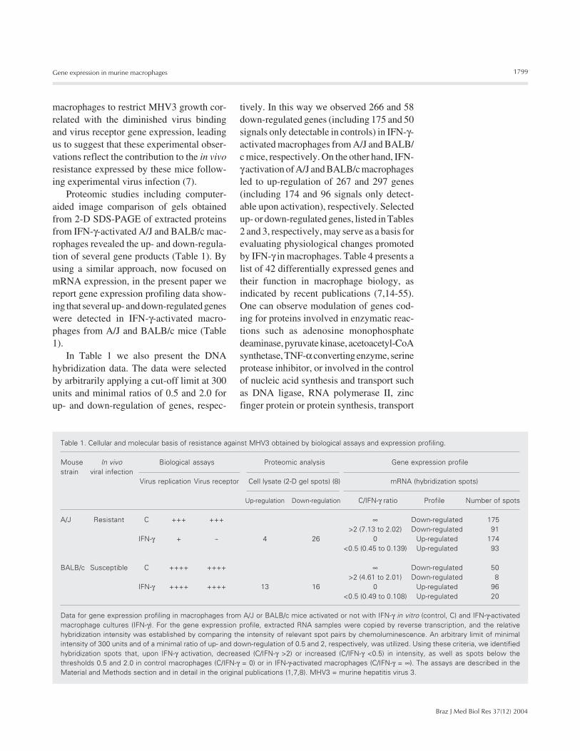
In Table 1 we also present the DNAhybridization data. The data were selectedby arbitrarily applying a cut-off limit at 300units and minimal ratios of 0.5 and 2.0 forup- and down-regulation of genes, respec-
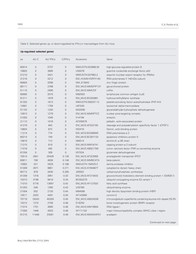
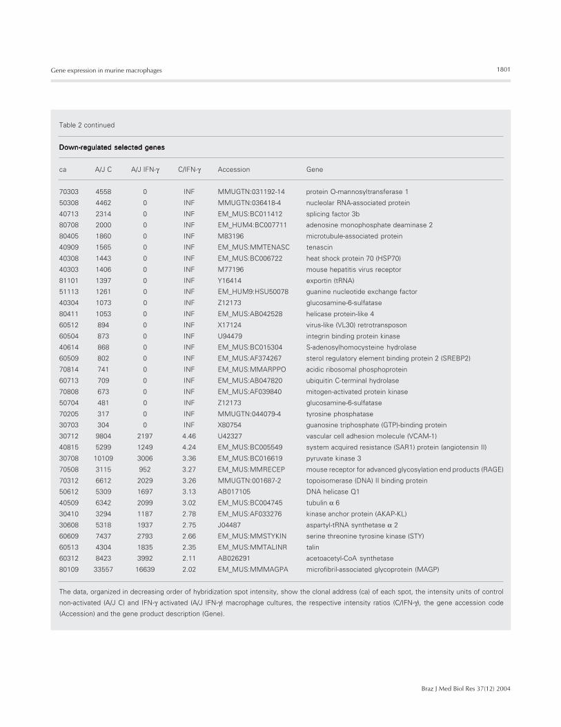
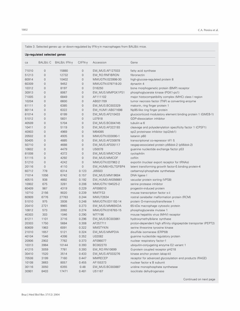
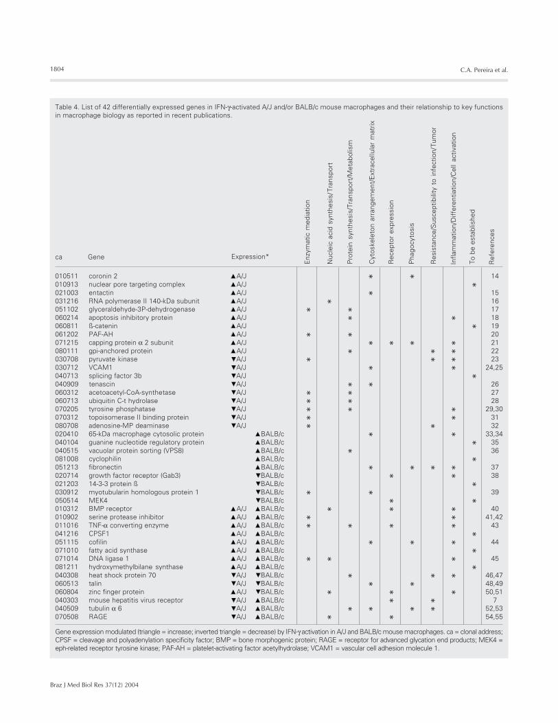
tively. In this way we observed 266 and 58down-regulated genes (including 175 and 50signals only detectable in controls) in IFN-γ-activated macrophages from A/J and BALB/c mice, respectively. On the other hand, IFN-γ activation of A/J and BALB/c macrophagesled to up-regulation of 267 and 297 genes(including 174 and 96 signals only detect-able upon activation), respectively. Selectedup- or down-regulated genes, listed in Tables2 and 3, respectively, may serve as a basis forevaluating physiological changes promotedby IFN-γ in macrophages. Table 4 presents alist of 42 differentially expressed genes andtheir function in macrophage biology, asindicated by recent publications (7,14-55).One can observe modulation of genes cod-ing for proteins involved in enzymatic reac-tions such as adenosine monophosphatedeaminase, pyruvate kinase, acetoacetyl-CoAsynthetase, TNF-α converting enzyme, serineprotease inhibitor, or involved in the controlof nucleic acid synthesis and transport suchas DNA ligase, RNA polymerase II, zincfinger protein or protein synthesis, transport
Table 1. Cellular and molecular basis of resistance against MHV3 obtained by biological assays and expression profiling.
Mouse In vivo Biological assays Proteomic analysis Gene expression profilestrain viral infection
Virus replication Virus receptor Cell lysate (2-D gel spots) (8) mRNA (hybridization spots)
Up-regulation Down-regulation C/IFN-γ ratio Profile Number of spots
A/J Resistant C +++ +++ ∞ Down-regulated 175>2 (7.13 to 2.02) Down-regulated 91
IFN-γ + - 4 26 0 Up-regulated 174<0.5 (0.45 to 0.139) Up-regulated 93
BALB/c Susceptible C ++++ ++++ ∞ Down-regulated 50>2 (4.61 to 2.01) Down-regulated 8
IFN-γ ++++ ++++ 13 16 0 Up-regulated 96<0.5 (0.49 to 0.108) Up-regulated 20
Data for gene expression profiling in macrophages from A/J or BALB/c mice activated or not with IFN-γ in vitro (control, C) and IFN-γ-activatedmacrophage cultures (IFN-γ). For the gene expression profile, extracted RNA samples were copied by reverse transcription, and the relativehybridization intensity was established by comparing the intensity of relevant spot pairs by chemoluminescence. An arbitrary limit of minimalintensity of 300 units and of a minimal ratio of up- and down-regulation of 0.5 and 2, respectively, was utilized. Using these criteria, we identifiedhybridization spots that, upon IFN-γ activation, decreased (C/IFN-γ >2) or increased (C/IFN-γ <0.5) in intensity, as well as spots below thethresholds 0.5 and 2.0 in control macrophages (C/IFN-γ = 0) or in IFN-γ-activated macrophages (C/IFN-γ = ∞). The assays are described in theMaterial and Methods section and in detail in the original publications (1,7,8). MHV3 = murine hepatitis virus 3.
1800
Braz J Med Biol Res 37(12) 2004
C.A. Pereira et al.
Table 2. Selected genes up- or down-regulated by IFN-γ in macrophages from A/J mice.
Up-regulated selected genesUp-regulated selected genesUp-regulated selected genesUp-regulated selected genesUp-regulated selected genes
ca A/J C A/J IFN-γ C/IFN-γ Accession Gene
60914 0 5737 0 MMUGTN:023998-30 high-glucose-regulated protein 8
10602 0 3685 0 U50078 guanine nucleotide exchange factor p53
51210 0 3421 0 MMUGTN:037962-2 exportin (nuclear export receptor for tRNAs)
31216 0 3212 0 EM_HUM9:HSRPII140 RNA polymerase II 140-kDa subunit
60804 0 2936 0 NM_013843 zinc finger protein
80111 0 2768 0 EM_MUS:MMGPIP137 gpi-anchored protein
51115 0 2490 0 EM_MUS:MMCOF cofilin
60905 0 2075 0 M92933 lymphocyte common antigen (Ly5)
81211 0 1879 0 EM_MUS:BC003861 hydroxymethylbilane synthase
61202 0 1813 0 MMUGTN:056337-12 platelet-activating factor acetylhydrolase (PAF-AH)
10901 0 1758 0 U87240 lysosomal alpha-mannosidase
51102 0 1293 0 M32599 glyceraldehyde-3-phosphate dehydrogenase
10913 0 1279 0 EM_MUS:MMNPTCC nuclear pore-targeting complex
21003 0 1040 0 X14194 entactin
21112 0 1016 0 AF205079 palladin, actin-associated protein
41216 0 943 0 EM_MUS:AF322193 cleavage and polyadenylation specificity factor 1 (CPSF1)
10604 0 873 0 X53416 filamin, actin-binding protein
11214 0 774 0 EM_MUS:BC006945 DNA polymerase α 2
60214 0 749 0 EM_MUS:BC007133 apoptosis inhibitory protein 5
10810 0 712 0 X84014 laminin-5, α 3B chain
71215 0 619 0 EM_MUS:MM16741 capping protein α 2 subunit
11016 0 492 0 EM_MUS:AB021709 tumor necrosis factor (TNF) α converting enzyme
61204 0 309 0 X57024 glutamate dehydrogenase
70514 2841 20439 0.139 EM_MUS:AF323958 prostaglandin transporter (PGT)
60811 706 4836 0.146 EM_MUS:MMBCATA beta-catenin
10902 531 2829 0.188 MMUGTN:194525-2 serine protease inhibitor
51009 2671 9851 0.271 EM_MUS:AY004877 cytoplasmic dynein heavy chain
60712 978 3430 0.285 J05503 carbamoyl-phosphate synthetase
81208 1316 3951 0.33 EM_MUS:AF210433 glucocorticoid modulatory element binding protein 1 (GMEB-1)
10313 3196 9419 0.34 BC002270 ubiquitin-conjugating enzyme E2 variant 1
71010 5778 13697 0.42 EM_MUS:AF127033 fatty acid synthase
51203 546 1260 0.43 U20780 ubiquitinating enzyme
21004 932 2130 0.44 M64098 high density lipoprotein binding protein (HBP)
10511 2033 4491 0.45 AF143956 coronin-2
70716 18243 40359 0.45 EM_MUS:AB024538 immunoglobulin superfamily containing leucine-rich repeat (ISLRI)
10312 1270 2756 0.46 D16250 bone morphogenetic protein (BMP) receptor
71014 1701 3690 0.46 EM_MUS:MM19604 DNA ligase I
71005 1446 3033 0.48 AF111102 major histocompatibility complex (MHC) class I region
61215 11446 23581 0.49 EM_MUS:MMADAPA1 α-adaptin
Continued on next page
1801
Braz J Med Biol Res 37(12) 2004
Gene expression in murine macrophages
Table 2 continued
Down-regulated selected genesDown-regulated selected genesDown-regulated selected genesDown-regulated selected genesDown-regulated selected genes
ca A/J C A/J IFN-γ C/IFN-γ Accession Gene
70303 4558 0 INF MMUGTN:031192-14 protein O-mannosyltransferase 1
50308 4462 0 INF MMUGTN:036418-4 nucleolar RNA-associated protein
40713 2314 0 INF EM_MUS:BC011412 splicing factor 3b
80708 2000 0 INF EM_HUM4:BC007711 adenosine monophosphate deaminase 2
80405 1860 0 INF M83196 microtubule-associated protein
40909 1565 0 INF EM_MUS:MMTENASC tenascin
40308 1443 0 INF EM_MUS:BC006722 heat shock protein 70 (HSP70)
40303 1406 0 INF M77196 mouse hepatitis virus receptor
81101 1397 0 INF Y16414 exportin (tRNA)
51113 1261 0 INF EM_HUM9:HSU50078 guanine nucleotide exchange factor
40304 1073 0 INF Z12173 glucosamine-6-sulfatase
80411 1053 0 INF EM_MUS:AB042528 helicase protein-like 4
60512 894 0 INF X17124 virus-like (VL30) retrotransposon
60504 873 0 INF U94479 integrin binding protein kinase
40614 868 0 INF EM_MUS:BC015304 S-adenosylhomocysteine hydrolase
60509 802 0 INF EM_MUS:AF374267 sterol regulatory element binding protein 2 (SREBP2)
70814 741 0 INF EM_MUS:MMARPPO acidic ribosomal phosphoprotein
60713 709 0 INF EM_MUS:AB047820 ubiquitin C-terminal hydrolase
70808 673 0 INF EM_MUS:AF039840 mitogen-activated protein kinase
50704 481 0 INF Z12173 glucosamine-6-sulfatase
70205 317 0 INF MMUGTN:044079-4 tyrosine phosphatase
30703 304 0 INF X80754 guanosine triphosphate (GTP)-binding protein
30712 9804 2197 4.46 U42327 vascular cell adhesion molecule (VCAM-1)
40815 5299 1249 4.24 EM_MUS:BC005549 system acquired resistance (SAR1) protein (angiotensin II)
30708 10109 3006 3.36 EM_MUS:BC016619 pyruvate kinase 3
70508 3115 952 3.27 EM_MUS:MMRECEP mouse receptor for advanced glycosylation end products (RAGE)
70312 6612 2029 3.26 MMUGTN:001687-2 topoisomerase (DNA) II binding protein
50612 5309 1697 3.13 AB017105 DNA helicase Q1
40509 6342 2099 3.02 EM_MUS:BC004745 tubulin α 6
30410 3294 1187 2.78 EM_MUS:AF033276 kinase anchor protein (AKAP-KL)
30608 5318 1937 2.75 J04487 aspartyl-tRNA synthetase α 2
60609 7437 2793 2.66 EM_MUS:MMSTYKIN serine threonine tyrosine kinase (STY)
60513 4304 1835 2.35 EM_MUS:MMTALINR talin
60312 8423 3992 2.11 AB026291 acetoacetyl-CoA synthetase
80109 33557 16639 2.02 EM_MUS:MMMAGPA microfibril-associated glycoprotein (MAGP)
The data, organized in decreasing order of hybridization spot intensity, show the clonal address (ca) of each spot, the intensity units of control
non-activated (A/J C) and IFN-γ activated (A/J IFN-γ) macrophage cultures, the respective intensity ratios (C/IFN-γ), the gene accession code
(Accession) and the gene product description (Gene).
1802
Braz J Med Biol Res 37(12) 2004
C.A. Pereira et al.
Table 3. Selected genes up- or down-regulated by IFN-γ in macrophages from BALB/c mice.
Up-regulated selected genesUp-regulated selected genesUp-regulated selected genesUp-regulated selected genesUp-regulated selected genes
ca BALB/c C BALB/c IFN-γ C/IFN-γ Accession Gene
71010 0 15880 0 EM_MUS:AF127033 fatty acid synthase
51213 0 12732 0 EM_RO:RNFIBRON fibronectin
60914 0 10402 0 MMUGTN:023998-30 high-glucose-regulated protein 8
60309 0 9452 0 MMUGTN:078718-20 dynactin 4
10312 0 8197 0 D16250 bone morphogenetic protein (BMP) receptor
30913 0 8067 0 EM_MUS:MMPGK1PS1 phosphoglycerate kinase (PGK1-ps1)
71005 0 6849 0 AF111102 major histocompatibility complex (MHC) class I region
10204 0 6600 0 AB021709 tumor necrosis factor (TNF) α converting enzyme
61111 0 6385 0 EM_MUS:BC003329 makorin, ring finger protein 1
80114 0 6322 0 EM_HUM1:AB071698 Np95-like ring finger protein
61014 0 6199 0 EM_MUS:AF210433 glucocorticoid modulatory element binding protein 1 (GMEB-1)
51012 0 5831 0 L07918 GDP-dissociation inhibitor
40509 0 5704 0 EM_MUS:BC004745 tubulin α 6
10411 0 5119 0 EM_MUS:AF322193 cleavage and polyadenylation specificity factor 1 (CPSF1)
40903 0 4969 0 M64085 spi2 proteinase inhibitor (spi2/eb1)
20502 0 4935 0 MMUGTN:033090-1 katanin p60
50405 0 4885 0 EM_MUS:AF230878 transcriptional co-repressor tif1 ß
50710 0 4688 0 EM_MUS:AF035117 rasgap-associated protein p56dok-2 (p56dok-2)
10602 0 4479 0 U50078 guanine nucleotide exchange factor p53
81008 0 4372 0 EM_MUS:MMCYCM cyclophilin
51115 0 4292 0 EM_MUS:MMCOF cofilin
51210 0 4242 0 MMUGTN:037962-2 exportin (nuclear export receptor for tRNAs)
20116 0 4110 0 EM_HUM8:HSLTGFBP4 latent transforming growth factor-ß binding protein-4
60712 778 6314 0.123 J05503 carbamoyl-phosphate synthetase
71014 1056 6742 0.157 EM_MUS:MM19604 DNA ligase I
40515 938 4892 0.192 EM_HUM3:AK056661 vacuolar protein sorting (VPS8)
10902 675 3281 0.206 MMUGTN:194525-2 serine protease inhibitor
60409 987 4319 0.229 AF006010 progestin-induced protein
10710 2156 9020 0.239 MMTFS3 mouse transcription factor s-ii
60909 6776 27783 0.244 MMU72634 rostral cerebellar malformation protein (RCM)
51010 975 3938 0.248 MMUGTN:031192-14 protein O-mannosyltransferase 1
20410 2721 9965 0.273 EM_MUS:MM65KDA 65-kDa macrophage cytosolic protein
10812 573 2092 0.274 MMUGTN:016783-15 phosphoglycerate mutase 1
40303 303 1046 0.290 M77196 mouse hepatitis virus (MHV) receptor
81211 1101 3716 0.296 EM_MUS:BC003861 hydroxymethylbilane synthase
20303 1750 5684 0.308 AF257711 proton-dependent high affinity oligopeptide transporter (PEPT2)
60609 1963 6091 0.322 MMSTYKIN serine threonine tyrosine kinase
21010 1657 5121 0.324 EM_MUS:MMPDIA disulfide isomerase (ERP59)
40104 1546 4398 0.352 U02082 guanine nucleotide regulatory protein
20906 2902 7782 0.373 AF098077 nuclear respiratory factor-1
10313 3984 10144 0.393 BC002270 ubiquitin-conjugating enzyme E2 variant 1
41215 3059 7781 0.393 EM_RO:RN10699 G-protein coupled receptor pH218
30410 1520 3514 0.433 EM_MUS:AF033276 kinase anchor protein (akap-kl)
70508 3199 7160 0.447 MMRECEP receptor for advanced glycosylation end products (RAGE)
10108 3663 8057 0.455 AF155373 nuclear factor κ B subunit
30116 3050 6355 0.48 EM_MUS:BC003887 uridine monophosphate synthetase
30901 8403 17471 0.481 U51167 isocitrate dehydrogenase
Continued on next page
1803
Braz J Med Biol Res 37(12) 2004
Gene expression in murine macrophages
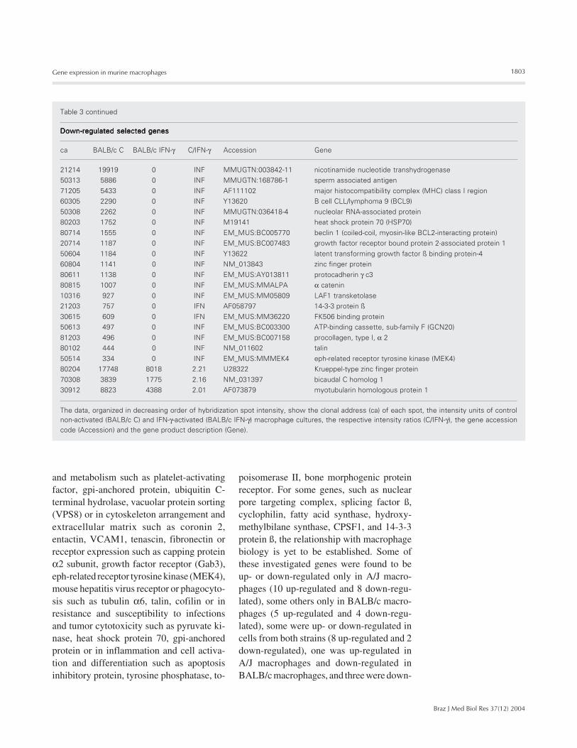
and metabolism such as platelet-activatingfactor, gpi-anchored protein, ubiquitin C-terminal hydrolase, vacuolar protein sorting(VPS8) or in cytoskeleton arrangement andextracellular matrix such as coronin 2,entactin, VCAM1, tenascin, fibronectin orreceptor expression such as capping proteinα2 subunit, growth factor receptor (Gab3),eph-related receptor tyrosine kinase (MEK4),mouse hepatitis virus receptor or phagocyto-sis such as tubulin α6, talin, cofilin or inresistance and susceptibility to infectionsand tumor cytotoxicity such as pyruvate ki-nase, heat shock protein 70, gpi-anchoredprotein or in inflammation and cell activa-tion and differentiation such as apoptosisinhibitory protein, tyrosine phosphatase, to-
poisomerase II, bone morphogenic proteinreceptor. For some genes, such as nuclearpore targeting complex, splicing factor ß,cyclophilin, fatty acid synthase, hydroxy-methylbilane synthase, CPSF1, and 14-3-3protein ß, the relationship with macrophagebiology is yet to be established. Some ofthese investigated genes were found to beup- or down-regulated only in A/J macro-phages (10 up-regulated and 8 down-regu-lated), some others only in BALB/c macro-phages (5 up-regulated and 4 down-regu-lated), some were up- or down-regulated incells from both strains (8 up-regulated and 2down-regulated), one was up-regulated inA/J macrophages and down-regulated inBALB/c macrophages, and three were down-
Table 3 continued
Down-regulated selected genesDown-regulated selected genesDown-regulated selected genesDown-regulated selected genesDown-regulated selected genes
ca BALB/c C BALB/c IFN-γ C/IFN-γ Accession Gene
21214 19919 0 INF MMUGTN:003842-11 nicotinamide nucleotide transhydrogenase
50313 5886 0 INF MMUGTN:168786-1 sperm associated antigen
71205 5433 0 INF AF111102 major histocompatibility complex (MHC) class I region
60305 2290 0 INF Y13620 B cell CLL/lymphoma 9 (BCL9)
50308 2262 0 INF MMUGTN:036418-4 nucleolar RNA-associated protein
80203 1752 0 INF M19141 heat shock protein 70 (HSP70)
80714 1555 0 INF EM_MUS:BC005770 beclin 1 (coiled-coil, myosin-like BCL2-interacting protein)
20714 1187 0 INF EM_MUS:BC007483 growth factor receptor bound protein 2-associated protein 1
50604 1184 0 INF Y13622 latent transforming growth factor ß binding protein-4
60804 1141 0 INF NM_013843 zinc finger protein
80611 1138 0 INF EM_MUS:AY013811 protocadherin γ c3
80815 1007 0 INF EM_MUS:MMALPA α catenin
10316 927 0 INF EM_MUS:MM05809 LAF1 transketolase
21203 757 0 IFN AF058797 14-3-3 protein ß
30615 609 0 IFN EM_MUS:MM36220 FK506 binding protein
50613 497 0 INF EM_MUS:BC003300 ATP-binding cassette, sub-family F (GCN20)
81203 496 0 INF EM_MUS:BC007158 procollagen, type I, α 2
80102 444 0 INF NM_011602 talin
50514 334 0 INF EM_MUS:MMMEK4 eph-related receptor tyrosine kinase (MEK4)
80204 17748 8018 2.21 U28322 Krueppel-type zinc finger protein
70308 3839 1775 2.16 NM_031397 bicaudal C homolog 1
30912 8823 4388 2.01 AF073879 myotubularin homologous protein 1
The data, organized in decreasing order of hybridization spot intensity, show the clonal address (ca) of each spot, the intensity units of controlnon-activated (BALB/c C) and IFN-γ-activated (BALB/c IFN-γ) macrophage cultures, the respective intensity ratios (C/IFN-γ), the gene accessioncode (Accession) and the gene product description (Gene).
1804
Braz J Med Biol Res 37(12) 2004
C.A. Pereira et al.
010511 coronin 2 A/J * * 14010913 nuclear pore targeting complex A/J *021003 entactin A/J * 15031216 RNA polymerase II 140-kDa subunit A/J * 16051102 glyceraldehyde-3P-dehydrogenase A/J * * 17060214 apoptosis inhibitory protein A/J * * 18060811 ß-catenin A/J * 19061202 PAF-AH A/J * * 20071215 capping protein α 2 subunit A/J * * * * 21080111 gpi-anchored protein A/J * * * 22030708 pyruvate kinase A/J * * * 23030712 VCAM1 A/J * * 24,25040713 splicing factor 3b A/J *040909 tenascin A/J * * 26060312 acetoacetyl-CoA-synthetase A/J * * 27060713 ubiquitin C-t hydrolase A/J * * 28070205 tyrosine phosphatase A/J * * * 29,30070312 topoisomerase II binding protein A/J * * 31080708 adenosine-MP deaminase A/J * * 32020410 65-kDa macrophage cytosolic protein BALB/c * * 33,34040104 guanine nucleotide regulatory protein BALB/c * 35040515 vacuolar protein sorting (VPS8) BALB/c * 36081008 cyclophilin BALB/c *051213 fibronectin BALB/c * * * * 37020714 growth factor receptor (Gab3) BALB/c * * 38021203 14-3-3 protein ß BALB/c *030912 myotubularin homologous protein 1 BALB/c * * 39050514 MEK4 BALB/c * *010312 BMP receptor A/J BALB/c * * * 40010902 serine protease inhibitor A/J BALB/c * * 41,42011016 TNF-α converting enzyme A/J BALB/c * * * * 43041216 CPSF1 A/J BALB/c *051115 cofilin A/J BALB/c * * * 44071010 fatty acid synthase A/J BALB/c *071014 DNA ligase 1 A/J BALB/c * * * 45081211 hydroxymethylbilane synthase A/J BALB/c *040308 heat shock protein 70 A/J BALB/c * * * 46,47060513 talin A/J BALB/c * * 48,49060804 zinc finger protein A/J BALB/c * * * 50,51040303 mouse hepatitis virus receptor A/J BALB/c * * 7040509 tubulin α 6 A/J BALB/c * * * * 52,53070508 RAGE A/J BALB/c * * 54,55
Infla
mm
atio
n/D
iffer
entia
tion/
Cel
l ac
tivat
ion
Ref
eren
ces
Expression*ca Gene
To b
e es
tabl
ishe
d
Pha
gocy
tosi
s
Enz
ymat
ic m
edia
tion
Nuc
leic
aci
d sy
nthe
sis/
Tran
spor
t
Pro
tein
syn
thes
is/T
rans
port
/Met
abol
ism
Cyt
oske
leto
n ar
rang
emen
t/E
xtra
cellu
lar
mat
rix
Rec
epto
r ex
pres
sion
Res
ista
nce/
Sus
cept
ibili
ty t
o in
fect
ion/
Tum
or
Table 4. List of 42 differentially expressed genes in IFN-γ-activated A/J and/or BALB/c mouse macrophages and their relationship to key functionsin macrophage biology as reported in recent publications.
Gene expression modulated (triangle = increase; inverted triangle = decrease) by IFN-γ activation in A/J and BALB/c mouse macrophages. ca = clonal address;CPSF = cleavage and polyadenylation specificity factor; BMP = bone morphogenic protein; RAGE = receptor for advanced glycation end products; MEK4 =eph-related receptor tyrosine kinase; PAF-AH = platelet-activating factor acetylhydrolase; VCAM1 = vascular cell adhesion molecule 1.
1805
Braz J Med Biol Res 37(12) 2004
Gene expression in murine macrophages
regulated in A/J macrophages and up-regu-lated in BALB/c macrophages.
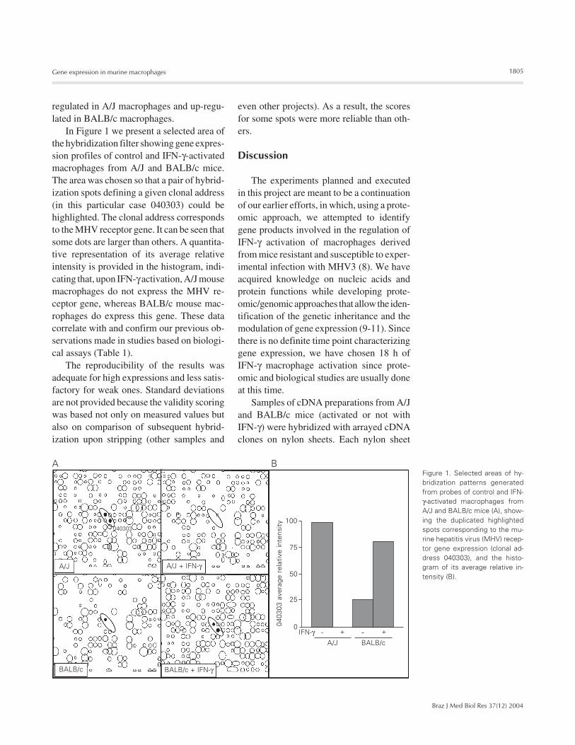
In Figure 1 we present a selected area ofthe hybridization filter showing gene expres-sion profiles of control and IFN-γ-activatedmacrophages from A/J and BALB/c mice.The area was chosen so that a pair of hybrid-ization spots defining a given clonal address(in this particular case 040303) could behighlighted. The clonal address correspondsto the MHV receptor gene. It can be seen thatsome dots are larger than others. A quantita-tive representation of its average relativeintensity is provided in the histogram, indi-cating that, upon IFN-γ activation, A/J mousemacrophages do not express the MHV re-ceptor gene, whereas BALB/c mouse mac-rophages do express this gene. These datacorrelate with and confirm our previous ob-servations made in studies based on biologi-cal assays (Table 1).
The reproducibility of the results wasadequate for high expressions and less satis-factory for weak ones. Standard deviationsare not provided because the validity scoringwas based not only on measured values butalso on comparison of subsequent hybrid-ization upon stripping (other samples and
even other projects). As a result, the scoresfor some spots were more reliable than oth-ers.
Discussion
The experiments planned and executedin this project are meant to be a continuationof our earlier efforts, in which, using a prote-omic approach, we attempted to identifygene products involved in the regulation ofIFN-γ activation of macrophages derivedfrom mice resistant and susceptible to exper-imental infection with MHV3 (8). We haveacquired knowledge on nucleic acids andprotein functions while developing prote-omic/genomic approaches that allow the iden-tification of the genetic inheritance and themodulation of gene expression (9-11). Sincethere is no definite time point characterizinggene expression, we have chosen 18 h ofIFN-γ macrophage activation since prote-omic and biological studies are usually doneat this time.
Samples of cDNA preparations from A/Jand BALB/c mice (activated or not withIFN-γ) were hybridized with arrayed cDNAclones on nylon sheets. Each nylon sheet
040303
A/J A/J + IFN-γ
BALB/c + IFN-γBALB/c
BALB/cA/J
IFN-γ
100
75
50
25
00403
03 a
vera
ge r
elat
ive
inte
nsity
++
A BFigure 1. Selected areas of hy-bridization patterns generatedfrom probes of control and IFN-γ-activated macrophages fromA/J and BALB/c mice (A), show-ing the duplicated highlightedspots corresponding to the mu-rine hepatitis virus (MHV) recep-tor gene expression (clonal ad-dress 040303), and the histo-gram of its average relative in-tensity (B).
1806
Braz J Med Biol Res 37(12) 2004
C.A. Pereira et al.
contained 1536 cDNA clones of fetal thy-mus origin in duplicate, as well as severalcontrol dots of cytochrome c origin. A chemi-luminescence readout followed by imageanalysis yielded data which indicated therelative intensity of hybridized entities.
As shown in Table 1, based on the obser-vation that A/J mice were resistant andBALB/c mice were susceptible to experi-mental infection with MHV3, classical bio-logical assays for the study of the cellularand molecular basis of resistance of mice toMHV3 led us to hypothesize and experimen-tally demonstrate that IFN-γ activation couldpartially restrict viral multiplication only inthe resistant A/J macrophages and that themolecular basis of this restriction relied onthe IFN-γ induced down-regulation of themain viral receptor (7). Taken together,proteomic experiments and gene expressionprofiling provided us not only with a confir-mation of these predictions but with thepossibility of a much deeper comprehensionof the biology of IFN-γ activation of macro-phages. We have demonstrated a panel ofup- and down-regulated genes as well asidentified their relationships to key func-tions in macrophages.
By expression profiling, we have evalu-ated the degree of up- or down-regulation ofseveral genes in macrophages from A/J andBALB/c mice upon activation by IFN-γ. Sincethis technology allows us to identify theexpression of genes that are modified uponactivation by IFN-γ, we can further examinethe biological role of the gene products andcategorize the possible modulation of bio-logical functions induced by IFN-γ activa-tion in macrophages from both mouse strains.We have on hand, in terms of hybridizationspots, several identified molecular entitiesof the cDNA library, and our semi-quantita-tive data show the overall influence of IFN-γ activation on macrophage gene expression.Contrary to the proteomic approach, in whichevery polypeptide species (if present in ad-equate amounts, if within the resolution lim-
its of the separation, and if it contains methi-onine in the amino acid sequence) present inthe 2-D SDS gel matrix shows up on theradiofluorogram, the hybridization readoutis constrained to the portion of cDNA mol-ecules present in the library. Among the1536 clonal entities, there are about onethousand different molecular clones, encom-passing about 470 low abundance ones. Sinceall of these clones are transcribable and trans-latable the number is satisfactory for ad-equate analysis. It represents about 20% ofthe messages present in a typical cell (thelymphocyte being the model) (56). Note thata cell has altogether about 40,000 mRNAmolecules, some present in a relatively largecopy number, others with only 3-5 copiesper cell. There are approximately 5,000 dif-ferent mRNA molecules in a cell (56). Ourexperiments revealed only those up- anddown-regulated which have members pres-ent among the 1536 entities tested.
In Figure 1 we show the hybridizationpatterns of a selected area of the gene ex-pression array obtained from control andIFN-γ-activated A/J and BALB/c macro-phages, with the duplicated gene spots cor-responding to the main MHV receptor (clonaladdress 040303) highlighted, as well as itshistograms of average relative intensity. Onecan observe that, as predicted by classicalassays performed in the past (7), there is adown-regulation of this receptor gene onlyin IFN-γ-activated macrophages from resis-tant A/J mice. Also, as previously shown (7),the receptor gene expression in IFN-γ-acti-vated BALB/c macrophages was high, al-though its basal expression in control macro-phages was found to be not always reproduc-ible, possibly due to variations in the physi-ological state of the cells.
Since most of the genes available in ourcDNA library have already been decodedand many of their protein products identi-fied, we now know the gene products likelyto be up- or down-regulated and have thepossibility of deducing modulation of bio-
1807
Braz J Med Biol Res 37(12) 2004
Gene expression in murine macrophages
logical functions induced by IFN-γ activa-tion in macrophages. As it is preliminarilyshown in Table 4, we could identify genescoding for proteins participating in processesof macrophage biology like enzymatic reac-tions, nucleic acid synthesis and transport,protein synthesis, transport and metabolism,cytoskeleton arrangement and extracellularmatrix, receptor expression, phagocytosis,resistance/susceptibility to infection or tu-mors and inflammation or cell differentia-tion. The data indicate that the overall generegulation by IFN-γ can be quite different inmacrophages originating from mice with dif-ferent genetic backgrounds and this panel ofIFN-γ-regulated genes may serve as a start-ing point for general and specific studies ofmacrophage biology.
This paper reveals a large assembly ofgenes differentially expressed in macro-phages of two murine genetic backgrounds(A/J and BALB/c) upon IFN-γ activation.These data will turn out to be very useful forgeneral studies of macrophage biology andcan be an alternative strategy to confirmhypothetical as well as already defined fea-tures of macrophages, such as that of MHVreceptor gene modulation upon IFN-γ acti-vation.
Acknowledgments
The Basel Institute of Immunology wasfounded and supported by F. Hoffmann-LaRoche and Co. Ltd., Basel, Switzerland.
References
1. Lucchiari MA, Martin JP, Modolell M & Pereira CA (1991). Acquiredimmunity of A/J mice to mouse hepatitis virus 3 infection. Depend-ence on interferon gamma synthesis and macrophage sensitivity tointerferon gamma. Journal of General Virology, 72: 1317-1322.
2. Arnheiter H, Baechi T & Haller O (1982). Adult mouse hepatocytesin primary monolayer culture express genetic resistance to mousehepatitis virus type 3. Journal of Immunology, 129: 1275-1281.
3. Pereira CA, Steffan AM & Kirn A (1984). Interaction between mousehepatitis viruses and primary cultures of Kupffer and endothelialliver cells from resistant and susceptible inbred mouse strains.Journal of General Virology, 65: 1617-1620.
4. Lamontagne L, Descoteaux JP & Jolicoeur P (1989). Mouse hepati-tis virus 3 replication in T and B lymphocytes correlate with viralpathogenicity. Journal of Immunology, 142: 4458-4465.
5. Dindzans VJ, Skamene E & Levy GA (1986). Susceptibility/resis-tance to mouse hepatitis virus strain 3 and macrophage procoagu-lant activity are genetically linked and controlled by two-non-H-2linked genes. Journal of Immunology, 137: 2355-2360.
6. Fingerote RJ, Abecassis M, Phillips MJ, Rao YS, Cole EH, LeibowitzJ & Levy GA (1996). Loss of resistance to MHV3 infection aftertreatment with corticosteroid is associated with induction of macro-phage PCA. Journal of Virology, 70: 4275-4282.
7. Vassão RC, De Franco MT, Hartz D, Modolell M, Sippel AE & PereiraCA (2000). Down-regulation of Bgp1a viral receptor by interferon γ isrelated to the antiviral state and resistance to mouse hepatitis virus3 infection. Virology, 274: 278-283.
8. Pereira CA, Lucchiari MA, Modolell M, Kuhn L & Lefkovits I (1993).An attempt to identify gene products related to the induction of anantiviral state in macrophages resistant and sensitive to IFN-gamma.Research in Virology, 144: 479-486.
9. Lefkovits I, Kettman JR & Frey JR (2001). Global analysis of geneexpression in cells of the immune system. I. Analytical limitations in
obtaining sequence information on polypeptides in two-dimensionalgel spots. Electrophoresis, 21: 2688-2693.
10. Frey JR, Nguyen C, Houlgatte R et al. (2001). Global analysis ofgene expression in cells of the immune system. II. Cell free transla-tion products and high-density filter hybridization data. Electropho-resis, 21: 2694-2702.
11. Lefkovits I, Kettman JR & Frey JR (2001). Proteomic analysis of raremolecular species of translated polypeptides from a mouse fetalthymus cDNA library. Proteomics, 1: 560-573.
12. Munder PG, Modolell M & Wallach DFH (1971). Cell propagation onfilms of polymeric fluorocarbon as a mean to regulate pericellularpH and pO2 in cultured monolayers. FEBS Letters, 15: 191-196.
13. Pereira CA, Mercier G, Oth D & Dupuy JM (1984). Induction ofnatural killer cells and interferon during mouse hepatitis virus infec-tion of resistant and susceptible inbred mouse strains. Immunobiol-ogy, 166: 35-42.
14. Schuller S, Neefjes J, Ottenhoff T, Thole J & Young D (2001).Coronin is involved in uptake of Mycobacterium bovis BCG in hu-man macrophages but not in phagosome maintenance. Cell Micro-biology, 3: 785-793.
15. Gronski Jr TJ, Martin RL, Kobayashi DK, Walsh BC, Holman MC,Huber M, Van Wart HE & Shapiro SD (1997). Hydrolysis of a broadspectrum of extracellular matrix proteins by human macrophageelastase. Journal of Biological Chemistry, 272: 12189-12194.
16. Ishida T, Matsuura K, Setoguchi M, Higuchi Y & Yamamoto S(1994). Enhancement of murine serum amyloid A3 mRNA expres-sion by glucocorticoids and its regulation by cytokines. Journal ofLeukocyte Biology, 56: 797-806.
17. Mateo RB, Reichner JS, Mastrofrancesco B, Kraft-Stolar D & AlbinaJE (1995). Impact of nitric oxide on macrophage glucose metabo-lism and glyceraldehyde-3-phosphate dehydrogenase activity.American Journal of Physiology, 268: 669-675.
1808
Braz J Med Biol Res 37(12) 2004
C.A. Pereira et al.
18. Horie T, Dobashi K, Iizuka K, Yoshii A, Shimizu Y, Nakazawa T &Mori M (1999). Interferon-gamma rescue TNF-alpha-induced apop-tosis mediated by up-regulation of TNFR2 on EoL-1 cells. Experi-mental Hematology, 27: 512-519.
19. Lapteva N, Ando Y, Nieda M, Hohjoh H, Okai M, Kikuchi A, DymshitsG, Ishikawa Y, Juji T & Tokunaga K (2001). Profiling of genesexpressed in human monocytes and monocyte-derived dendriticcells using cDNA expression array. British Journal of Haematology,114: 191-197.
20. Tselepis AD, Karabina SA, Stengel D, Piedagnel R, Chapman MJ &Ninio E (2001). N-linked glycosylation of macrophage-derived PAF-AH is a major determinant of enzyme association with plasma HDL.Journal of Lipid Research, 42: 1645-1654.
21. Witke W, Li W, Kwiatkowski DJ & Southwick FS (2001). Compari-sons of CapG and gelsolin-null macrophages: demonstration of aunique role for CapG in receptor-mediated ruffling, phagocytosis,and vesicle rocketing. Journal of Cell Biology, 154: 775-784.
22. Coelho PS, Klein A, Talvani A, Coutinho SF, Takeuchi O, Akira S,Silva JS, Canizzaro H, Gazzinelli RT & Teixeira MM (2002). Glycosyl-phosphatidyl inositol-anchored mucin like glycoproteins isolatedfrom Trypanosoma cruzi trypomastigotes induce in vivo leukocyterecruitment dependent on MCP-1 production by IFN-gamma-primedmacrophages. Journal of Leukocyte Biology, 71: 837-844.
23. Duncan JR, Potter CB, Cappellini MD, Kurtz JB, Anderson MJ &Weatherall DJ (1983). Aplastic crisis due to parvovirus infection inpyruvate kinase deficiency. Lancet, 2: 14-16.
24. Nansen A, Christensen JP, Ropke C, Marker O, Scheynius A &Thomsen AR (1998). Role of interferon-gamma in the pathogenesisof LCMV-induced meningitis: unimpaired leukocyte recruitment,but deficient macrophage activation in interferon-gamma knock-outmice. Journal of Neuroimmunology, 86: 202-212.
25. Peng HB, Spiedcker M & Liao JK (1998). Inducible nitric oxide: Anautoregulatory feedback inhibitor of vascular inflammation. Journalof Immunology, 161: 1970-1976.
26. Harkonen E, Virtanen I, Linnala A, Laitinen LL & Kinnula VL (1995).Modulation of fibronectin and tenascin production in human bron-chial epithelial cells by inflammatory cytokines in vitro. AmericanJournal of Respiratory Cell and Molecular Biology, 13: 109-115.
27. Bergstrom JD, Wong GA, Edwards PA & Edmond J (1984). Theregulation of acetoacetyl-CoA synthetase activity by modulators ofcholesterol synthesis in vivo and utilization of acetoacetate forcholesterogenesis. Journal of Biological Chemistry, 259: 14548-14553.
28. Glockzin S, von Knethen A, Scheffner M & Brune B (1999). Activa-tion of the cell death program by nitric oxide involves inhibition ofthe proteasome. Journal of Biological Chemistry, 274: 19581-19586.
29. Xaus J, Comalada M, Valledor AF, Cardo M, Herrero C, Soler C,Lloberas J & Celada A (2001). Molecular mechanisms involved inmacrophage survival, proliferation, activation and apoptosis. Immu-nobiology, 204: 543-550.
30. Simoncic PD, Lee-Loy A, Barber DL, Tremblay ML & McGlade CJ(2002). The T cell protein tyrosine phosphatase is a negative regula-tor of Janus family kinases 1 and 3. Current Biology, 12: 446-453.
31. Chiou WF, Chou CJ & Chenm CF (2001). Camptothecin suppressesnitric oxide biosynthesis in RAW 264.7 macrophages. Life Sci-ences, 69: 625-635.
32. Nikolajeva V, Eze D, Kamradze A, Indulena M & Muiznieks I (1996).Protective effect of adenylate deaminase (from Penicilliumlanosoviride) against acute infections in mice. Immunopharmacolo-gy, 35: 163-169.
33. Shinomiya H, Hagi A, Fukuzumi M, Mizobuchi M, Hirata H & Utsumi
S (1995). Complete primary structure and phosphorylation site ofthe 65-kDa macrophage protein phosphorylated by stimulation withbacterial lipopolysaccharide. Journal of Immunology, 154: 3471-3478.
34. Kikuchi H, Fujinawa T, Kuribayashi F, Nakanishi A, Imajoh-Ohmi S,Goto M & Kanegasaki S (1994). Induction of essential componentsof the superoxide generating system in human monoblastic leuke-mia U937 cells. Journal of Biochemistry, 116: 742-746.
35. Vestal DJ, Buss JE, McKercher SR, Jenkins NA, Copeland NG,Kelner GS, Asundi VK & Maki RA (1998). Murine GBP-2: a new IFN-gamma-induced member of the GBP family of GTPases isolatedfrom macrophages. Journal of Interferon and Cytokine Research,18: 977-985.
36. Luo W & Chang A (2000). An endosome-to-plasma membranepathway involved in trafficking of a mutant plasma membraneATPase in yeast. Molecular Biology of the Cell, 11: 579-592.
37. Jun CD, Yoon HJ, Kim HM & Chung HT (1995). Fibronectin activatesmurine peritoneal macrophages for tumor cell destruction in thepresence of IFN-gamma. Biochemical and Biophysical ResearchCommunications, 206: 969-974.
38. Wolf I, Jenkins BJ, Liu Y, Seiffert M, Custodio JM, Young P &Rohrschneider LR (2002). Gab3, a new DOS/Gab family member,facilitates macrophage differentiation. Molecular and Cellular Biol-ogy, 22: 231-244.
39. Nandurkar HH & Huysmans R (2002). The myotubularin family:novel phosphoinositide regulators. International Union of Biochem-istry and Molecular Biology Life, 53: 37-43.
40. Gould SE, Day M, Jones SS & Dorai H (2002). BMP-7 regulateschemokine, cytokine, and hemodynamic gene expression in proxi-mal tubule cells. Kidney International, 61: 51-60.
41. Hamerman JA, Hayashi F, Schroeder LA, Gygi SP, Haas AL,Hampson L, Coughlin P, Aebersold R & Aderem A (2002). Serpin 2ais induced in activated macrophages and conjugates to a ubiquitinhomolog. Journal of Immunology, 168: 2415-2423.
42. Kwak JY, Park SY, Han MK, Lee HS, Sohn MH, Kim UH, McGregorJR, Samlowski WE & Yim CY (1998). Receptor-mediated activationof murine peritoneal macrophages by antithrombin III acts as acostimulatory signal for nitric oxide synthesis. Cellular Immunology,188: 33-40.
43. Rovida E, Paccagnini A, Del Rosso M, Peschon J & Dello Sbarba P(2001). TNF-alpha-converting enzyme cleaves the macrophagecolony-stimulating factor receptor in macrophages undergoing acti-vation. Journal of Immunology, 166: 1583-1589.
44. Matsui S, Matsumoto S, Adachi R et al. (2002). LIM kinase 1modulates opsonized zymosan-triggered activation of macrophage-like U937 cells. Possible involvement of phosphorylation of cofilinand reorganization of actin cytoskeleton. Journal of Biological Chem-istry, 277: 544-549.
45. Khan Z & Francis GE (1987). Contrasting patterns of DNA strandbreakage and ADP-ribosylation-dependent DNA ligation duringgranulocyte and monocyte differentiation. Blood, 69: 1114-1119.
46. Breoler M, Dorner B, More SH, Roderian T, Fleischer B & von BoninA (2001). Heat shock proteins as “danger signals”: eukaryoticHsp60 enhances and accelerates antigen-specific IFN-gamma pro-duction in T cells. European Journal of Immunology, 31: 2051-2059.
47. Panjwani NN, Popova L & Srivastava PK (2002). Heath shock pro-teins gp96 and hsp70 activate the release of nitric oxide by APCs.Journal of Immunology, 168: 2997-3003.
48. Rossi AG, McCutcheon JC, Roy N, Chilvers ER, Haslett C &Dransfield I (1998). Regulation of macrophage phagocytosis of apop-totic cells by cAMP. Journal of Immunology, 160: 3562-3568.
1809
Braz J Med Biol Res 37(12) 2004
Gene expression in murine macrophages
49. Greenberg S, Burridge K & Silverstein SC (1990). Colocalization of F-actin and talin during Fc receptor-mediated phagocytosis in mousemacrophages. Journal of Experimental Medicine, 172: 1853-1856.
50. Heyninck K, De Valck D, Vanden Berghe W, Van Criekinge W,Contreras R, Fiers W, Haegeman G & Beyaert R (1999). The zincfinger protein A20 inhibits TNF-induced NF-kappa B-dependent geneexpression by interfering with an RIP- or TRAF2-mediated transacti-vation signal and directly binds to a novel NF-kappa B-inhibitingprotein ABIN. Journal of Cell Biology, 145: 1471-1482.
51. Shin JN, Kim I, Lee JS, Koh GY, Lee ZH & Kim HH (2002). A novelzinc finger protein that inhibits osteoclastogenesis and the functionof tumor necrosis factor-associated factor 6. Journal of BiologicalChemistry, 277: 8346-8353.
52. Siffert JC, Baldacini O, Kuhry JG, Wachsmann D, Benabdelmou-mene S, Faradji A, Monteil H & Poindron P (1993). Effects ofClostridium difficile toxin B on human monocytes and macrophages:possible relationship with cytoskeletal rearrangement. Infection and
Immunity, 61: 1082-1090.53. Rammes A, Roth J, Goebeler M, Klempt M, Hartmann M & Sorg C
(1997). Myeloid-related protein (MRP) 8 and MRP14, calcium-bind-ing proteins of the S100 family, are secreted by activated mono-cytes via a novel, tubulin-dependent pathway. Journal of BiologicalChemistry, 272: 9496-9502.
54. Schmidt AM, Hori O, Cao R, Yan SD, Brett J, Wautier JL, Ogawa S,Kuwabara K, Matsumoto M & Stern D (1996). RAGE: a novel cellularreceptor for advanced glycation end products. Diabetes, 45: S77-S80.
55. Ohgami N, Nagai R, Ikemoto M, Arai H, Kuniyasu A, Horiuchi S &Nakayama H (2001). CD36, a member of the class b scavengerreceptor family, as a receptor for advanced glycation end products.Journal of Biological Chemistry, 276: 3195-3202.
56. Lefkovits I (1995). ...and such are little lymphocytes made of. Re-search in Immunology, 146: 5-10.