gastroenterology referral guidance · persistent dyspepsia or inadequate response to treatment...
TRANSCRIPT
Gastroenterology Referral Guidance NHS Hounslow CCG
(May 2013)
Contents Page
Notes 2
Dyspepsia 3
Abnormal Liver Function Test (LFTs) 8
Rectal Bleeding 12
Inflammatory Bowel Disease 16
Irritable Bowel Syndrome 20
Family History of Bowel Cancer 23
Supplementary information
Referral Forms 26
Lower GI Endoscopy Referral criteria 29
Stoma Care Service at WMUH 30
Contact Information WMUH 31
Appendix: Abnormal LFTs 32
Version details
Version No. 4 Please email [email protected] for additional corrections, admissions or comments
Approved by Ian Perry (GP) Dr Joel Mawdsley (WMUH Gastroenterology Consultant)
Approval date May 2013
Review date
Page 2
Back to Contents Page
Gastroenterology Referral Guidance – NHS Hounslow v3
Gastroenterology Referral guidance for Hounslow Clinical Commissioning Group clinicians This is intended to be a guide only. It is not exhaustive and appropriate clinical judgement should be used for individual cases.
When referring to Gastroenterology, please provide information in accordance with the core required information fields of the Standard generic referral letter with particular attention to the following sections:
Past history: relevant family history, any co-morbidity, risk factors
Investigations: state whether the patient has had any relevant investigations (and attach results if available)
You may wish to consider certain investigations before referral (refer to relevant sections in the guidance).
Please note if you are concerned about your patient's condition and require urgent assessment it is not necessary to undertake routine tests unless this will significantly alter your referral decision.
It is important that you highlight what you – and your patient - expect from the referral. You should explicitly state whether you wish to continue management of the patient following investigations or if you would prefer the consultant to take over the case regardless of the outcome of initial assessment.
All new referrals for the attention of a Consultant Gastroenterologist should be sent via the Referral Facilitation Service (except for 999 Emergency admissions). RFS Information:
2nd Floor, Sovereign Court, 15-21 Staines Road,
Hounslow, Middlesex TW3 3HR.
Tel: 05511 434900
Fax: 0800 756 7754
There is also a free phone number: 0800 756 7751 and a landline number: 020 3416 3611 (local rate) for patients to call.
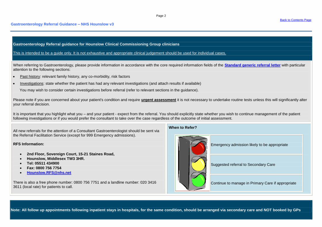
When to Refer?
Emergency admission likely to be appropriate
Suggested referral to Secondary Care
Continue to manage in Primary Care if appropriate
Note: All follow up appointments following inpatient stays in hospitals, for the same condition, should be arranged via secondary care and NOT booked by GPs
Page 3
Back to Contents Page
Dyspepsia Owner Ian Perry
Joel Mawdsley
External resources
Dyspepsia – Management of dyspepsia in adults in primary care (Quick reference guide) (NICE clinical guidelines 17, NICE, August 2002). LINK
Investigation and management of dyspepsia in primary care BMJ 2008. LINK
Patient.co.uk PIL: LINK
Version No 3
Approval
date
April 2013
Review date
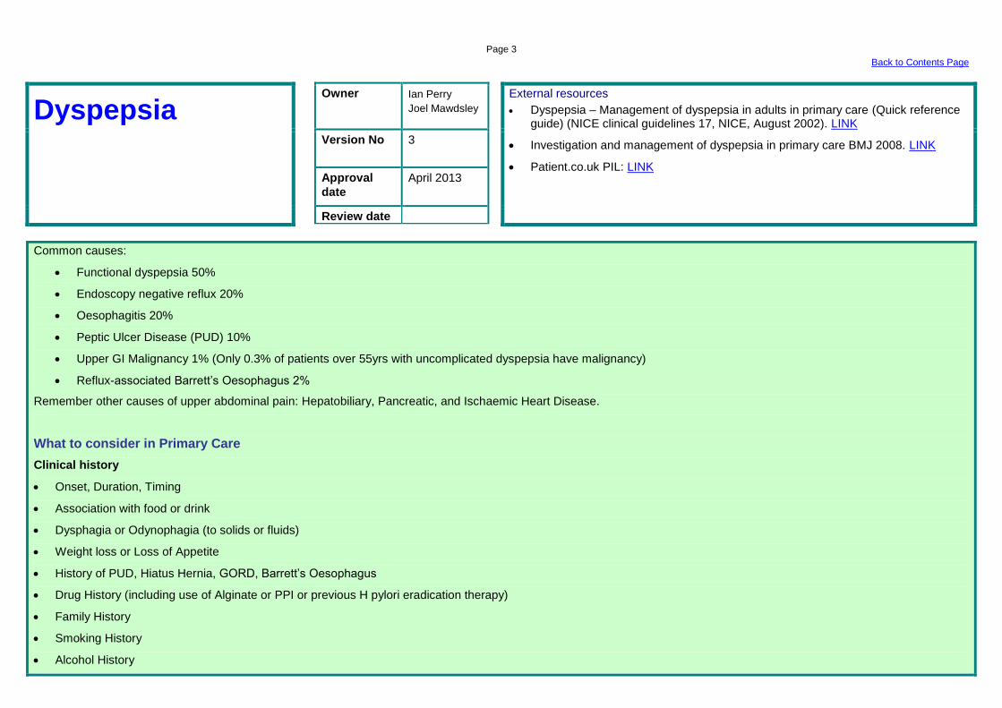
Common causes:
Functional dyspepsia 50%
Endoscopy negative reflux 20%
Oesophagitis 20%
Peptic Ulcer Disease (PUD) 10%
Upper GI Malignancy 1% (Only 0.3% of patients over 55yrs with uncomplicated dyspepsia have malignancy)
Reflux-associated Barrett’s Oesophagus 2%
Remember other causes of upper abdominal pain: Hepatobiliary, Pancreatic, and Ischaemic Heart Disease.
What to consider in Primary Care
Clinical history
Onset, Duration, Timing
Association with food or drink
Dysphagia or Odynophagia (to solids or fluids)
Weight loss or Loss of Appetite
History of PUD, Hiatus Hernia, GORD, Barrett’s Oesophagus
Drug History (including use of Alginate or PPI or previous H pylori eradication therapy)
Family History
Smoking History
Alcohol History
Page 4
Back to Contents Page
Physical examination
Abdominal exam
Weight and BMI
Investigations
Bloods
o FBC
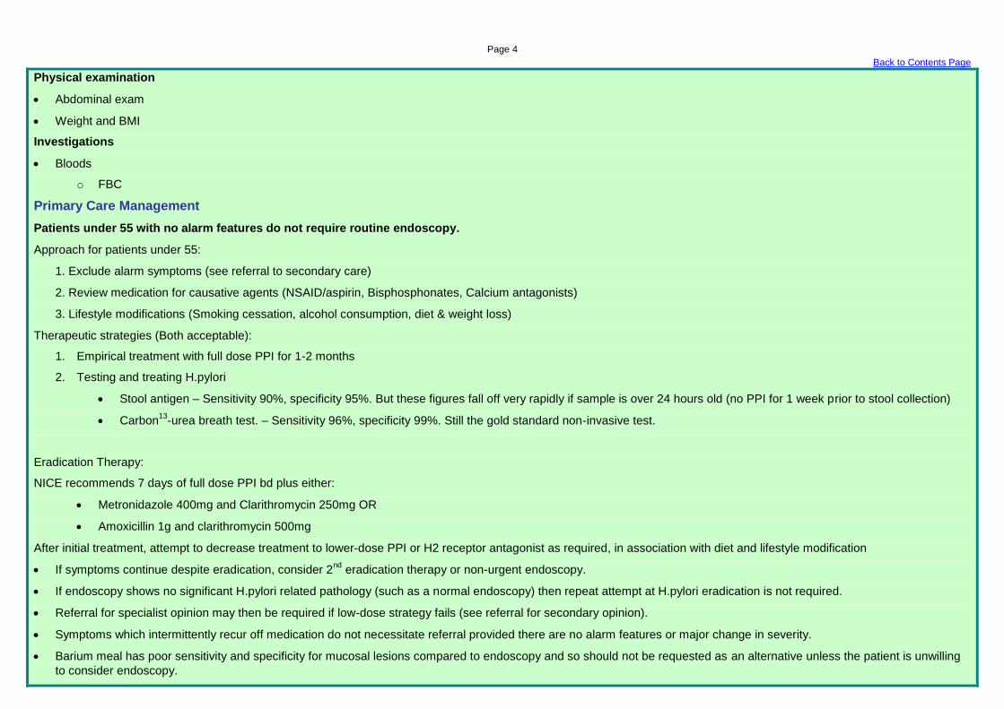
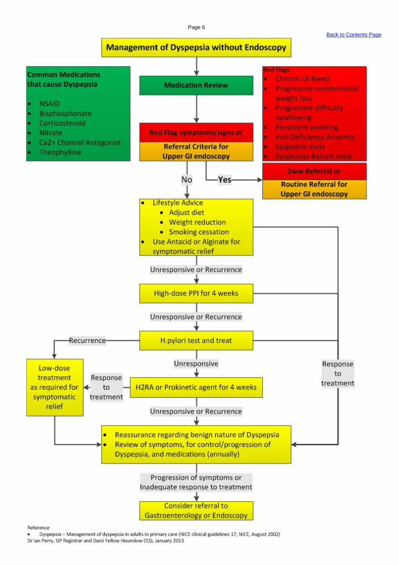
Primary Care Management
Patients under 55 with no alarm features do not require routine endoscopy.
Approach for patients under 55:
1. Exclude alarm symptoms (see referral to secondary care)
2. Review medication for causative agents (NSAID/aspirin, Bisphosphonates, Calcium antagonists)
3. Lifestyle modifications (Smoking cessation, alcohol consumption, diet & weight loss)
Therapeutic strategies (Both acceptable):
1. Empirical treatment with full dose PPI for 1-2 months
2. Testing and treating H.pylori
Stool antigen – Sensitivity 90%, specificity 95%. But these figures fall off very rapidly if sample is over 24 hours old (no PPI for 1 week prior to stool collection)
Carbon13
-urea breath test. – Sensitivity 96%, specificity 99%. Still the gold standard non-invasive test.
Eradication Therapy:
NICE recommends 7 days of full dose PPI bd plus either:
Metronidazole 400mg and Clarithromycin 250mg OR
Amoxicillin 1g and clarithromycin 500mg
After initial treatment, attempt to decrease treatment to lower-dose PPI or H2 receptor antagonist as required, in association with diet and lifestyle modification
If symptoms continue despite eradication, consider 2nd
eradication therapy or non-urgent endoscopy.
If endoscopy shows no significant H.pylori related pathology (such as a normal endoscopy) then repeat attempt at H.pylori eradication is not required.
Referral for specialist opinion may then be required if low-dose strategy fails (see referral for secondary opinion).
Symptoms which intermittently recur off medication do not necessitate referral provided there are no alarm features or major change in severity.
Barium meal has poor sensitivity and specificity for mucosal lesions compared to endoscopy and so should not be requested as an alternative unless the patient is unwilling
to consider endoscopy.
Page 5
Back to Contents Page
Page 6
Back to Contents Page

Page 7
Back to Contents Page
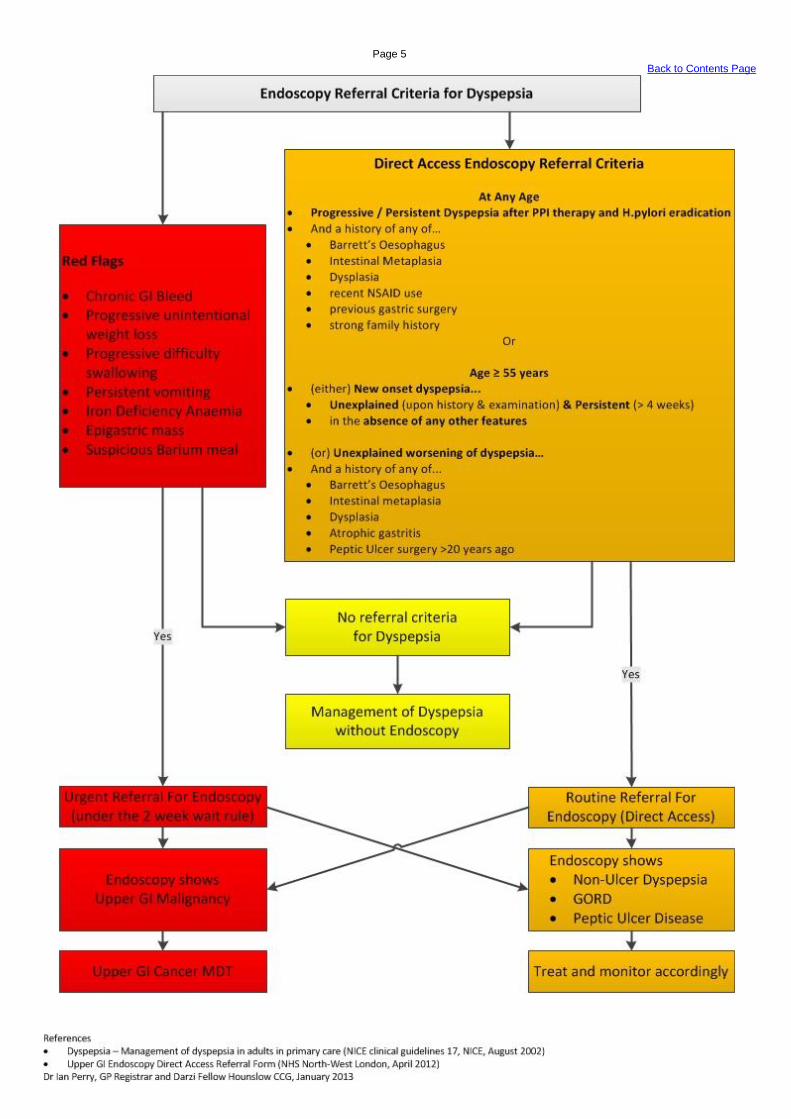
Referral Threshold
URGENT SECONDARY CARE REFERRAL
Urgently Refer for direct access endoscopy or for secondary care opinion if the history-taking, physical
examination or investigations show evidence of:
Anorexia
Progressive unintentional weight loss
Dysphagia
Persistent vomiting
High risk factors for cancer i.e.: previous gastric surgery or ulcer, known Barrett’s or atrophic gastritis
Epigastric mass
Iron deficient anaemia
Patients aged 55 years and older with unexplained and persistent recent onset dyspepsia (where
‘unexplained’ is defined as no clear cause identifiable such as NSAID use, and a sensible definition of
persistent would be 4-6 weeks).
ROUTINE SECONDARY CARE REFERRAL
Routine Referral for direct access endoscopy or secondary care opinion:
Persistent dyspepsia or inadequate response to treatment
Secondary care resource:
Refer to Gastroenterologist
Referral Threshold
EMERGENCY SECONDARY CARE REFERRAL
Active GI Bleeding
Marked abdominal tenderness on examination with rebound and guarding.
999 for emergency or urgent referral
Page 8
Back to Contents Page
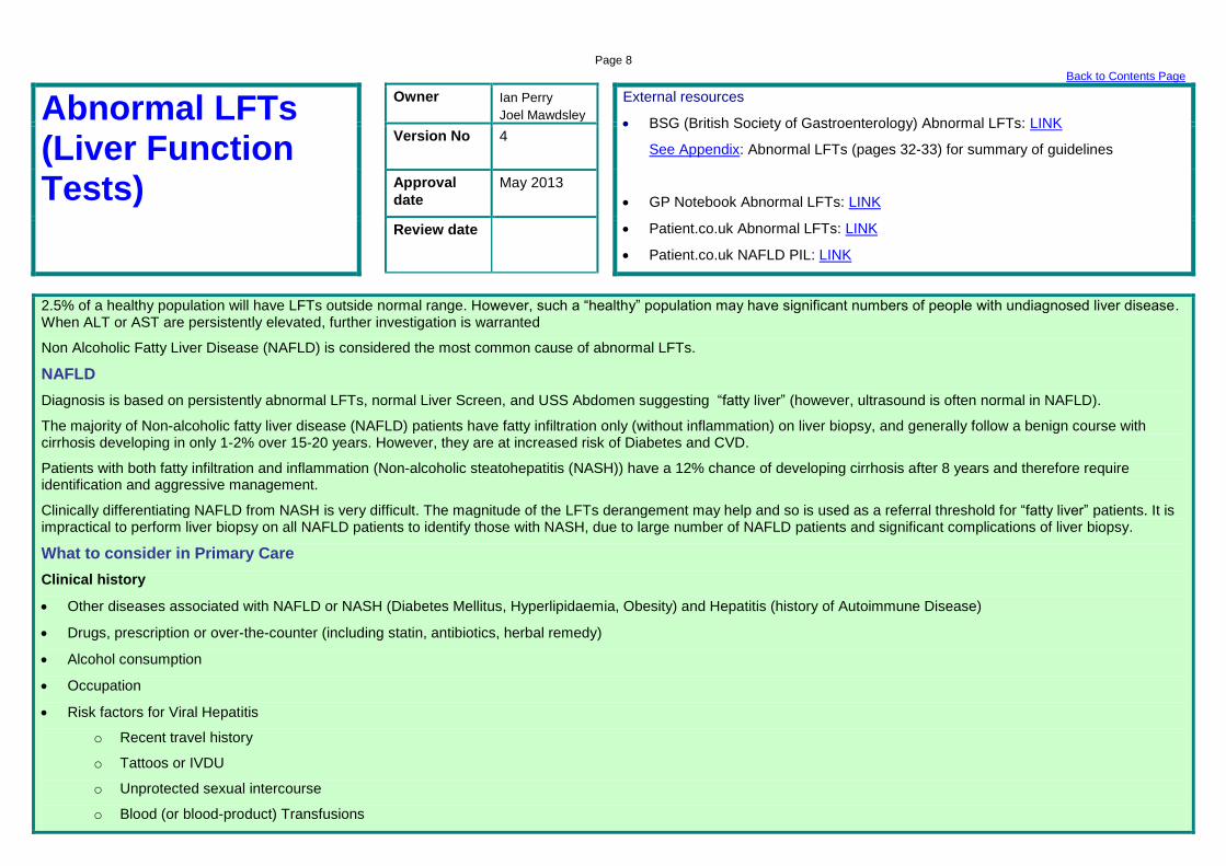
Abnormal LFTs (Liver Function Tests)
Owner Ian Perry
Joel Mawdsley
External resources
BSG (British Society of Gastroenterology) Abnormal LFTs: LINK
See Appendix: Abnormal LFTs (pages 32-33) for summary of guidelines
GP Notebook Abnormal LFTs: LINK
Patient.co.uk Abnormal LFTs: LINK
Patient.co.uk NAFLD PIL: LINK
Version No 4
Approval
date
May 2013
Review date
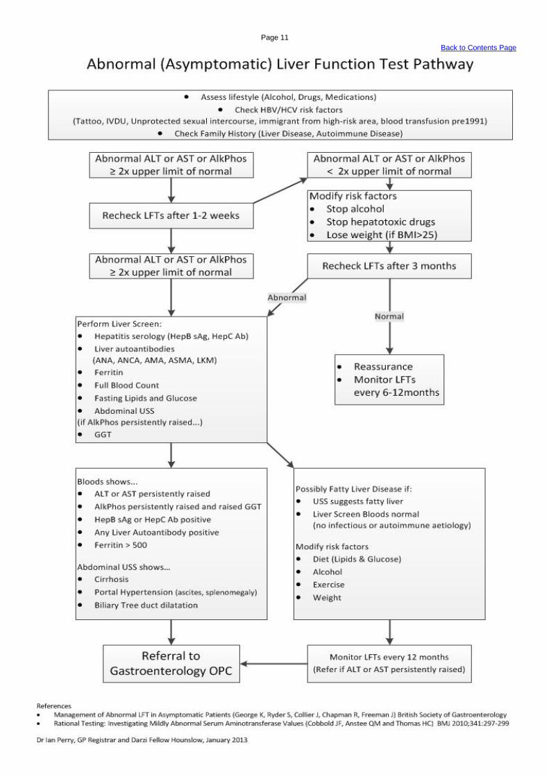
2.5% of a healthy population will have LFTs outside normal range. However, such a “healthy” population may have significant numbers of people with undiagnosed liver disease. When ALT or AST are persistently elevated, further investigation is warranted
Non Alcoholic Fatty Liver Disease (NAFLD) is considered the most common cause of abnormal LFTs.
NAFLD
Diagnosis is based on persistently abnormal LFTs, normal Liver Screen, and USS Abdomen suggesting “fatty liver” (however, ultrasound is often normal in NAFLD).
The majority of Non-alcoholic fatty liver disease (NAFLD) patients have fatty infiltration only (without inflammation) on liver biopsy, and generally follow a benign course with cirrhosis developing in only 1-2% over 15-20 years. However, they are at increased risk of Diabetes and CVD.
Patients with both fatty infiltration and inflammation (Non-alcoholic steatohepatitis (NASH)) have a 12% chance of developing cirrhosis after 8 years and therefore require identification and aggressive management.
Clinically differentiating NAFLD from NASH is very difficult. The magnitude of the LFTs derangement may help and so is used as a referral threshold for “fatty liver” patients. It is impractical to perform liver biopsy on all NAFLD patients to identify those with NASH, due to large number of NAFLD patients and significant complications of liver biopsy.
What to consider in Primary Care
Clinical history
Other diseases associated with NAFLD or NASH (Diabetes Mellitus, Hyperlipidaemia, Obesity) and Hepatitis (history of Autoimmune Disease)
Drugs, prescription or over-the-counter (including statin, antibiotics, herbal remedy)
Alcohol consumption
Occupation
Risk factors for Viral Hepatitis
o Recent travel history
o Tattoos or IVDU
o Unprotected sexual intercourse
o Blood (or blood-product) Transfusions
Page 9
Back to Contents Page
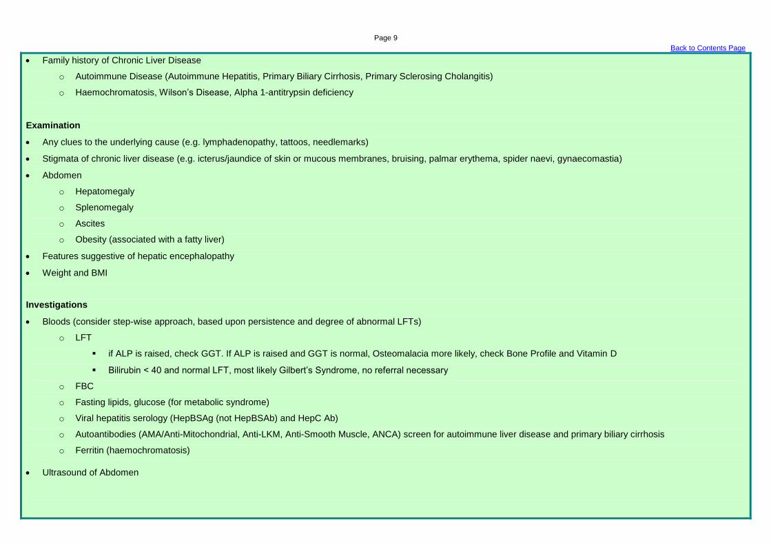
Family history of Chronic Liver Disease
o Autoimmune Disease (Autoimmune Hepatitis, Primary Biliary Cirrhosis, Primary Sclerosing Cholangitis)
o Haemochromatosis, Wilson’s Disease, Alpha 1-antitrypsin deficiency
Examination
Any clues to the underlying cause (e.g. lymphadenopathy, tattoos, needlemarks)
Stigmata of chronic liver disease (e.g. icterus/jaundice of skin or mucous membranes, bruising, palmar erythema, spider naevi, gynaecomastia)
Abdomen
o Hepatomegaly
o Splenomegaly
o Ascites
o Obesity (associated with a fatty liver)
Features suggestive of hepatic encephalopathy
Weight and BMI
Investigations
Bloods (consider step-wise approach, based upon persistence and degree of abnormal LFTs)
o LFT
if ALP is raised, check GGT. If ALP is raised and GGT is normal, Osteomalacia more likely, check Bone Profile and Vitamin D
Bilirubin < 40 and normal LFT, most likely Gilbert’s Syndrome, no referral necessary
o FBC
o Fasting lipids, glucose (for metabolic syndrome)
o Viral hepatitis serology (HepBSAg (not HepBSAb) and HepC Ab)
o Autoantibodies (AMA/Anti-Mitochondrial, Anti-LKM, Anti-Smooth Muscle, ANCA) screen for autoimmune liver disease and primary biliary cirrhosis
o Ferritin (haemochromatosis)
Ultrasound of Abdomen
Page 10
Back to Contents Page
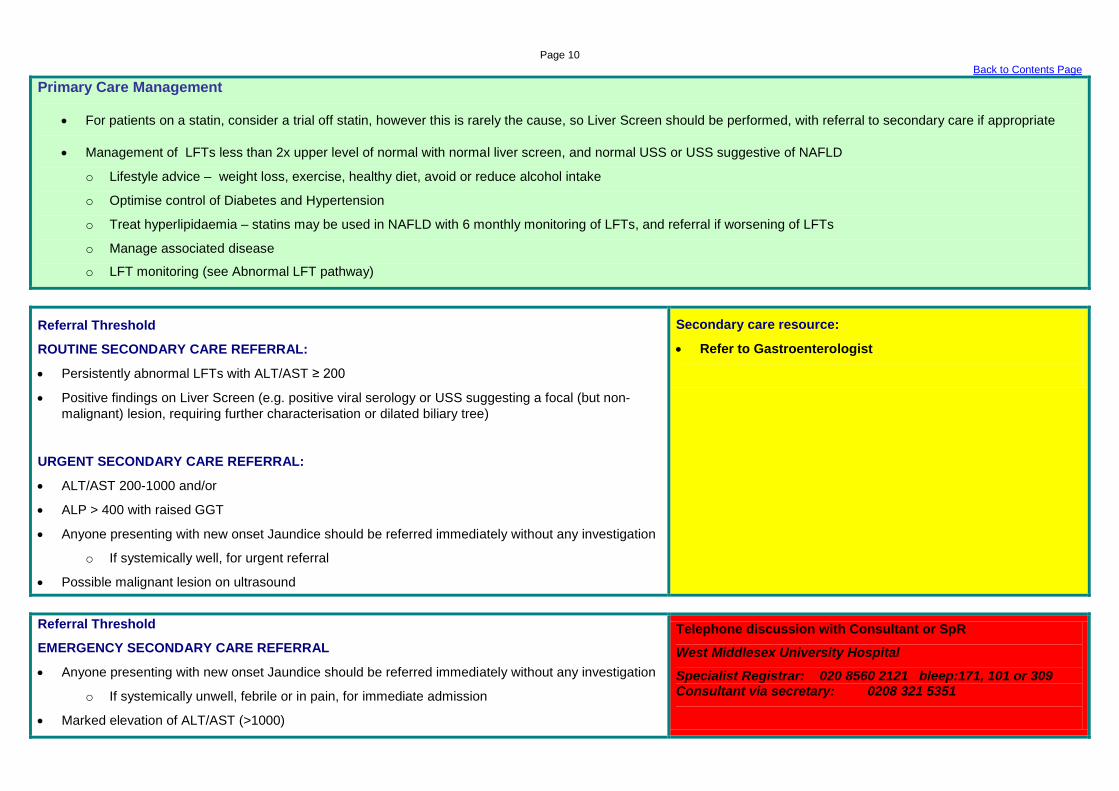
Primary Care Management
For patients on a statin, consider a trial off statin, however this is rarely the cause, so Liver Screen should be performed, with referral to secondary care if appropriate
Management of LFTs less than 2x upper level of normal with normal liver screen, and normal USS or USS suggestive of NAFLD
o Lifestyle advice – weight loss, exercise, healthy diet, avoid or reduce alcohol intake
o Optimise control of Diabetes and Hypertension
o Treat hyperlipidaemia – statins may be used in NAFLD with 6 monthly monitoring of LFTs, and referral if worsening of LFTs
o Manage associated disease
o LFT monitoring (see Abnormal LFT pathway)
Referral Threshold
ROUTINE SECONDARY CARE REFERRAL:
Persistently abnormal LFTs with ALT/AST ≥ 200
Positive findings on Liver Screen (e.g. positive viral serology or USS suggesting a focal (but non-
malignant) lesion, requiring further characterisation or dilated biliary tree)
URGENT SECONDARY CARE REFERRAL:
ALT/AST 200-1000 and/or
ALP > 400 with raised GGT
Anyone presenting with new onset Jaundice should be referred immediately without any investigation
o If systemically well, for urgent referral
Possible malignant lesion on ultrasound
Secondary care resource:
Refer to Gastroenterologist
Referral Threshold
EMERGENCY SECONDARY CARE REFERRAL
Anyone presenting with new onset Jaundice should be referred immediately without any investigation
o If systemically unwell, febrile or in pain, for immediate admission
Marked elevation of ALT/AST (>1000)
Telephone discussion with Consultant or SpR
West Middlesex University Hospital
Specialist Registrar: 020 8560 2121 bleep:171, 101 or 309 Consultant via secretary: 0208 321 5351
Page 11
Back to Contents Page
Page 12
Back to Contents Page
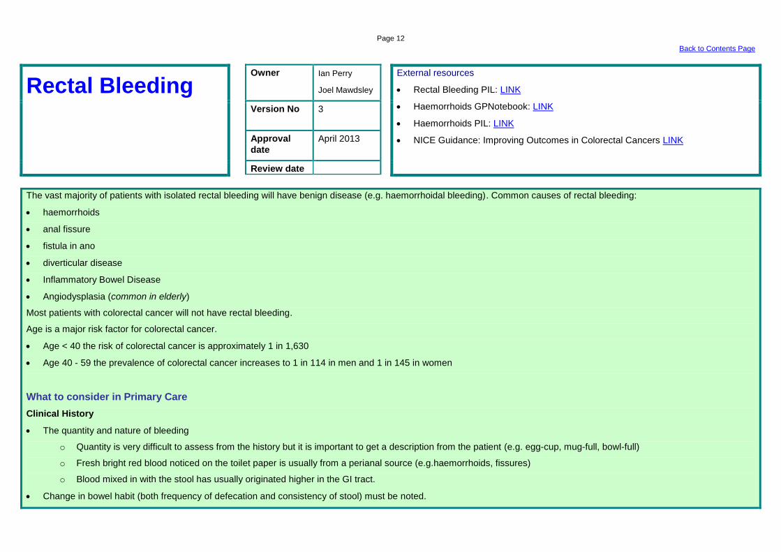
Rectal Bleeding Owner Ian Perry
Joel Mawdsley
External resources
Rectal Bleeding PIL: LINK
Haemorrhoids GPNotebook: LINK
Haemorrhoids PIL: LINK
NICE Guidance: Improving Outcomes in Colorectal Cancers LINK
Version No 3
Approval
date
April 2013
Review date
The vast majority of patients with isolated rectal bleeding will have benign disease (e.g. haemorrhoidal bleeding). Common causes of rectal bleeding:
haemorrhoids
anal fissure
fistula in ano
diverticular disease
Inflammatory Bowel Disease
Angiodysplasia (common in elderly)
Most patients with colorectal cancer will not have rectal bleeding.
Age is a major risk factor for colorectal cancer.
Age < 40 the risk of colorectal cancer is approximately 1 in 1,630
Age 40 - 59 the prevalence of colorectal cancer increases to 1 in 114 in men and 1 in 145 in women
What to consider in Primary Care
Clinical History
The quantity and nature of bleeding
o Quantity is very difficult to assess from the history but it is important to get a description from the patient (e.g. egg-cup, mug-full, bowl-full)
o Fresh bright red blood noticed on the toilet paper is usually from a perianal source (e.g.haemorrhoids, fissures)
o Blood mixed in with the stool has usually originated higher in the GI tract.
Change in bowel habit (both frequency of defecation and consistency of stool) must be noted.

Page 13
Back to Contents Page
Tenesmus
Anal symptoms (for example soreness, itching or prolapse, occur often with piles)
Progressive unintentional weight loss
Drug History (including warfarin and aspirin)
Family history of bowel cancer or polyposis
Examination
Abdominal exam
DRE (check for haemorrhoids, tags, fissures, rectal mass)
Investigations
Bloods
o FBC
o Ferritin
(Carcinoembryonic antigen (CEA) is not sufficiently sensitive or specific test for general population bowel cancer screening and should not be used)
PRIMARY CARE MANAGEMENT
If assessment suggests the source of bleeding is very likely to be local perianal disease such as simple haemorrhoids or anal fissure then secondary care referral may not be necessary. For example intermittent bright red blood noticed on the paper in a patient under 60 with no associated change in bowel habit and no alarm symptoms:
Conservative management for 6 weeks (GP Note Book and Patient.co.uk PIL)
o failure to respond should prompt a review of diagnosis and discussion with gastroenterologist
Page 14
Back to Contents Page
Page 15
Back to Contents Page
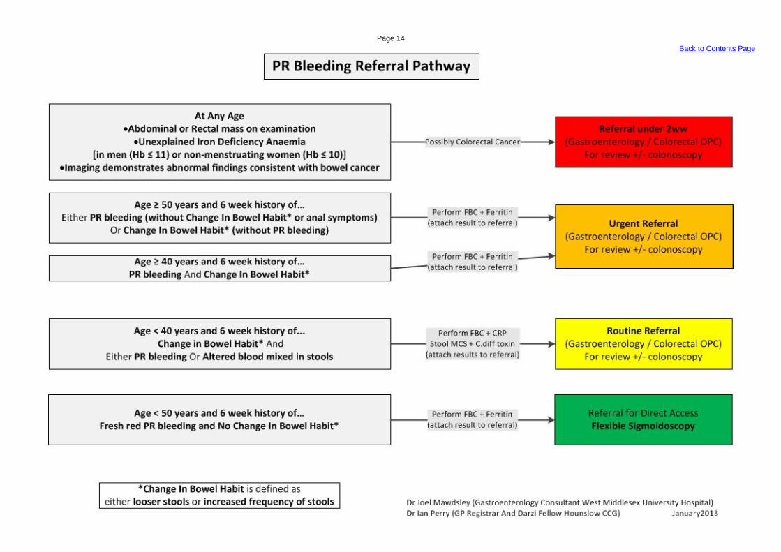
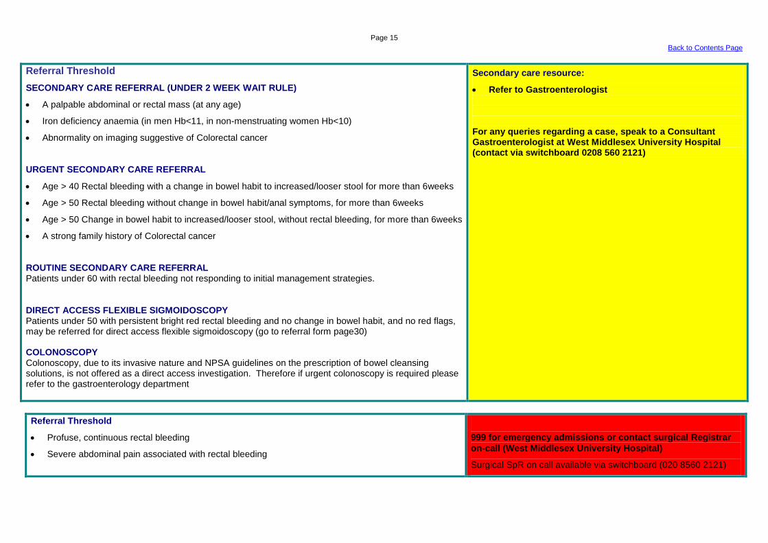
Referral Threshold
SECONDARY CARE REFERRAL (UNDER 2 WEEK WAIT RULE)
A palpable abdominal or rectal mass (at any age)
Iron deficiency anaemia (in men Hb<11, in non-menstruating women Hb<10)
Abnormality on imaging suggestive of Colorectal cancer
URGENT SECONDARY CARE REFERRAL
Age > 40 Rectal bleeding with a change in bowel habit to increased/looser stool for more than 6weeks
Age > 50 Rectal bleeding without change in bowel habit/anal symptoms, for more than 6weeks
Age > 50 Change in bowel habit to increased/looser stool, without rectal bleeding, for more than 6weeks
A strong family history of Colorectal cancer
ROUTINE SECONDARY CARE REFERRAL Patients under 60 with rectal bleeding not responding to initial management strategies.
DIRECT ACCESS FLEXIBLE SIGMOIDOSCOPY Patients under 50 with persistent bright red rectal bleeding and no change in bowel habit, and no red flags, may be referred for direct access flexible sigmoidoscopy (go to referral form page30) COLONOSCOPY Colonoscopy, due to its invasive nature and NPSA guidelines on the prescription of bowel cleansing solutions, is not offered as a direct access investigation. Therefore if urgent colonoscopy is required please refer to the gastroenterology department
Secondary care resource:
Refer to Gastroenterologist For any queries regarding a case, speak to a Consultant Gastroenterologist at West Middlesex University Hospital (contact via switchboard 0208 560 2121)
Referral Threshold
Profuse, continuous rectal bleeding
Severe abdominal pain associated with rectal bleeding
999 for emergency admissions or contact surgical Registrar
on-call (West Middlesex University Hospital)
Surgical SpR on call available via switchboard (020 8560 2121)
Page 16
Back to Contents Page
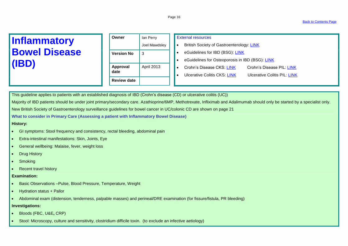
Inflammatory Bowel Disease (IBD)
Owner Ian Perry
Joel Mawdsley
External resources
British Society of Gastroenterology: LINK
eGuidelines for IBD (BSG): LINK
eGuidelines for Osteoporosis in IBD (BSG): LINK
Crohn’s Disease CKS: LINK Crohn’s Disease PIL: LINK
Ulcerative Colitis CKS: LINK Ulcerative Colitis PIL: LINK
Version No 3
Approval
date
April 2013
Review date
This guideline applies to patients with an established diagnosis of IBD (Crohn’s disease (CD) or ulcerative colitis (UC))
Majority of IBD patients should be under joint primary/secondary care. Azathioprine/6MP, Methotrexate, Infliximab and Adalimumab should only be started by a specialist only.
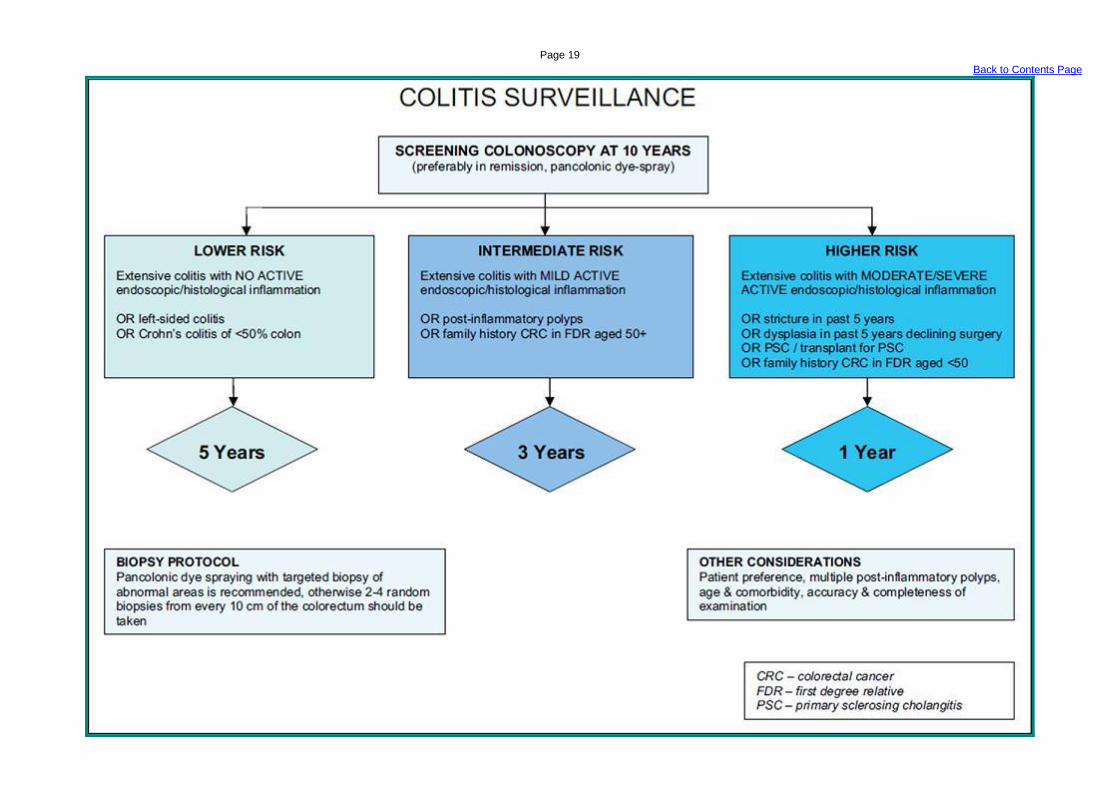
New British Society of Gastroenterology surveillance guidelines for bowel cancer in UC/colonic CD are shown on page 21
What to consider in Primary Care (Assessing a patient with Inflammatory Bowel Disease)
History:
GI symptoms: Stool frequency and consistency, rectal bleeding, abdominal pain
Extra-intestinal manifestations: Skin, Joints, Eye
General wellbeing: Malaise, fever, weight loss
Drug History
Smoking
Recent travel history
Examination:
Basic Observations –Pulse, Blood Pressure, Temperature, Weight
Hydration status + Pallor
Abdominal exam (distension, tenderness, palpable masses) and perineal/DRE examination (for fissure/fistula, PR bleeding)
Investigations:
Bloods (FBC, U&E, CRP)
Stool: Microscopy, culture and sensitivity, clostridium difficile toxin. (to exclude an infective aetiology)
Page 17
Back to Contents Page
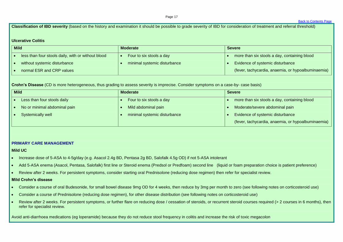
Classification of IBD severity (based on the history and examination it should be possible to grade severity of IBD for consideration of treatment and referral threshold)
Ulcerative Colitis
Mild Moderate Severe
less than four stools daily, with or without blood
without systemic disturbance
normal ESR and CRP values
Four to six stools a day
minimal systemic disturbance
more than six stools a day, containing blood
Evidence of systemic disturbance
(fever, tachycardia, anaemia, or hypoalbuminaemia)
Crohn’s Disease (CD is more heterogeneous, thus grading to assess severity is imprecise. Consider symptoms on a case-by- case basis)
Mild Moderate Severe
Less than four stools daily
No or minimal abdominal pain
Systemically well
Four to six stools a day
Mild abdominal pain
minimal systemic disturbance
more than six stools a day, containing blood
Moderate/severe abdominal pain
Evidence of systemic disturbance
(fever, tachycardia, anaemia, or hypoalbuminaemia)
PRIMARY CARE MANAGEMENT
Mild UC
Increase dose of 5-ASA to 4-5g/day (e.g. Asacol 2.4g BD, Pentasa 2g BD, Salofalk 4.5g OD) if not 5-ASA intolerant
Add 5-ASA enema (Asacol, Pentasa, Salofalk) first line or Steroid enema (Predsol or Predfoam) second line (liquid or foam preparation choice is patient preference)
Review after 2 weeks. For persistent symptoms, consider starting oral Prednisolone (reducing dose regimen) then refer for specialist review.
Mild Crohn’s disease
Consider a course of oral Budesonide, for small bowel disease 9mg OD for 4 weeks, then reduce by 3mg per month to zero (see following notes on corticosteroid use)
Consider a course of Prednisolone (reducing dose regimen), for other disease distribution (see following notes on corticosteroid use)
Review after 2 weeks. For persistent symptoms, or further flare on reducing dose / cessation of steroids, or recurrent steroid courses required (> 2 courses in 6 months), then refer for specialist review.
Avoid anti-diarrhoea medications (eg loperamide) because they do not reduce stool frequency in colitis and increase the risk of toxic megacolon
Page 18
Back to Contents Page
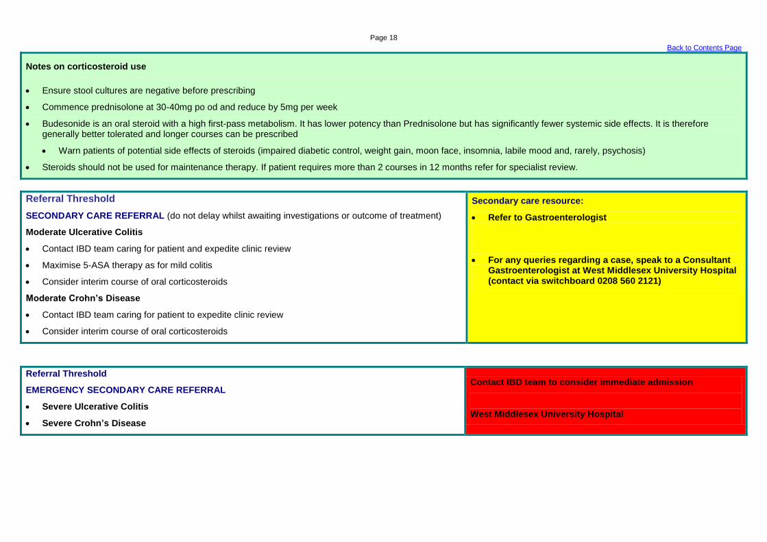
Notes on corticosteroid use
Ensure stool cultures are negative before prescribing
Commence prednisolone at 30-40mg po od and reduce by 5mg per week
Budesonide is an oral steroid with a high first-pass metabolism. It has lower potency than Prednisolone but has significantly fewer systemic side effects. It is therefore generally better tolerated and longer courses can be prescribed
Warn patients of potential side effects of steroids (impaired diabetic control, weight gain, moon face, insomnia, labile mood and, rarely, psychosis)
Steroids should not be used for maintenance therapy. If patient requires more than 2 courses in 12 months refer for specialist review.
Referral Threshold
SECONDARY CARE REFERRAL (do not delay whilst awaiting investigations or outcome of treatment)
Moderate Ulcerative Colitis
Contact IBD team caring for patient and expedite clinic review
Maximise 5-ASA therapy as for mild colitis
Consider interim course of oral corticosteroids
Moderate Crohn’s Disease
Contact IBD team caring for patient to expedite clinic review
Consider interim course of oral corticosteroids
Secondary care resource:
Refer to Gastroenterologist
For any queries regarding a case, speak to a Consultant Gastroenterologist at West Middlesex University Hospital (contact via switchboard 0208 560 2121)
Referral Threshold
EMERGENCY SECONDARY CARE REFERRAL
Severe Ulcerative Colitis
Severe Crohn’s Disease
Contact IBD team to consider immediate admission
West Middlesex University Hospital
Page 19
Back to Contents Page
Page 20
Back to Contents Page
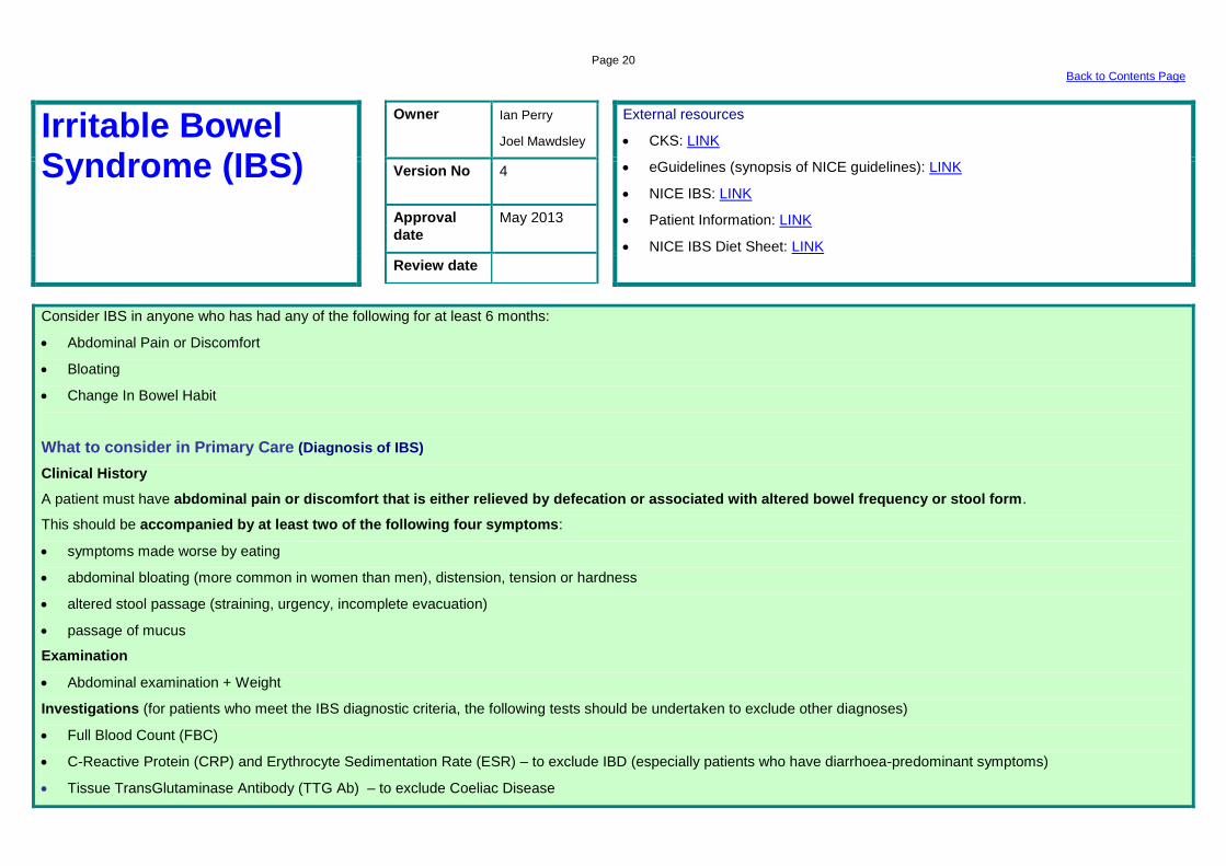
Irritable Bowel Syndrome (IBS)
Owner Ian Perry
Joel Mawdsley
External resources
CKS: LINK
eGuidelines (synopsis of NICE guidelines): LINK
NICE IBS: LINK
Patient Information: LINK
NICE IBS Diet Sheet: LINK
Version No 4
Approval
date
May 2013
Review date
Consider IBS in anyone who has had any of the following for at least 6 months:
Abdominal Pain or Discomfort
Bloating
Change In Bowel Habit
What to consider in Primary Care (Diagnosis of IBS)
Clinical History
A patient must have abdominal pain or discomfort that is either relieved by defecation or associated with altered bowel frequency or stool form.
This should be accompanied by at least two of the following four symptoms:
symptoms made worse by eating
abdominal bloating (more common in women than men), distension, tension or hardness
altered stool passage (straining, urgency, incomplete evacuation)
passage of mucus
Examination
Abdominal examination + Weight
Investigations (for patients who meet the IBS diagnostic criteria, the following tests should be undertaken to exclude other diagnoses)
Full Blood Count (FBC)
C-Reactive Protein (CRP) and Erythrocyte Sedimentation Rate (ESR) – to exclude IBD (especially patients who have diarrhoea-predominant symptoms)
Tissue TransGlutaminase Antibody (TTG Ab) – to exclude Coeliac Disease
Page 21
Back to Contents Page
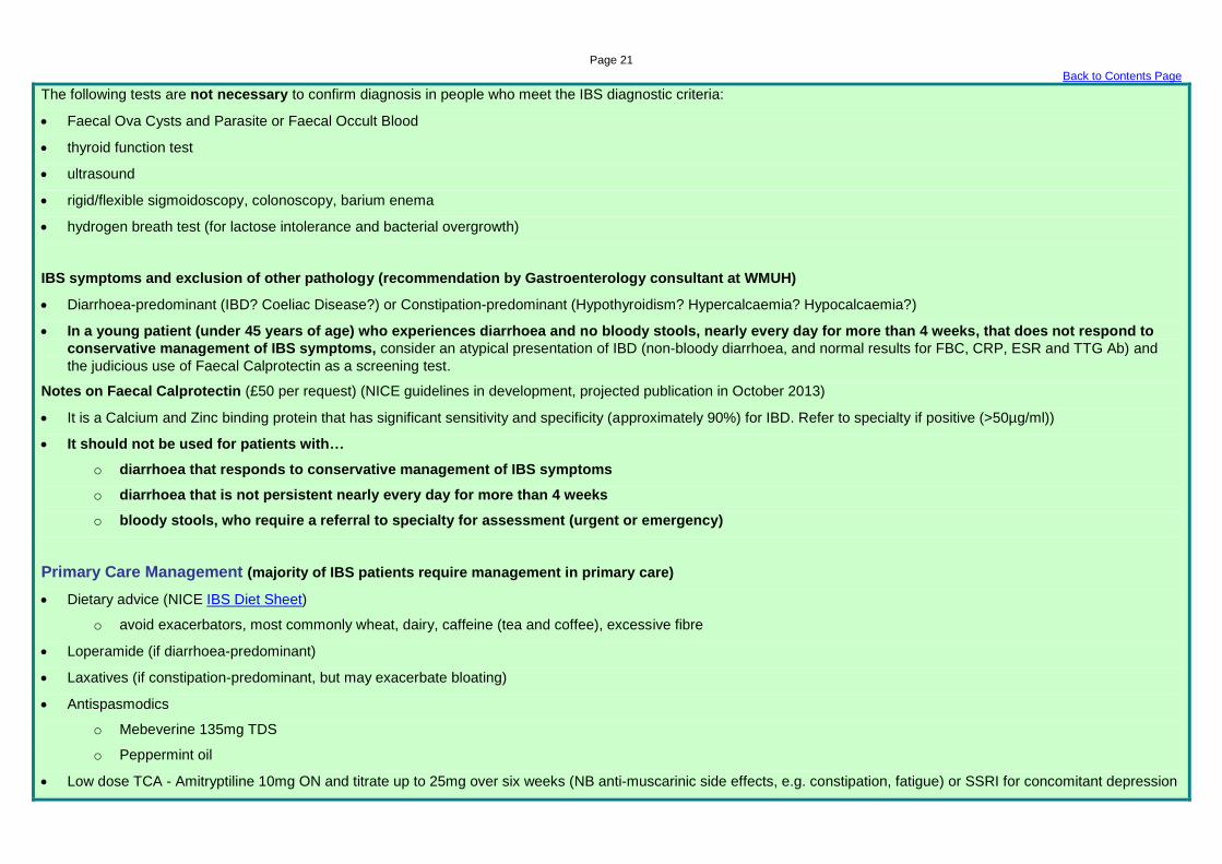
The following tests are not necessary to confirm diagnosis in people who meet the IBS diagnostic criteria:
Faecal Ova Cysts and Parasite or Faecal Occult Blood
thyroid function test
ultrasound
rigid/flexible sigmoidoscopy, colonoscopy, barium enema
hydrogen breath test (for lactose intolerance and bacterial overgrowth)
IBS symptoms and exclusion of other pathology (recommendation by Gastroenterology consultant at WMUH)
Diarrhoea-predominant (IBD? Coeliac Disease?) or Constipation-predominant (Hypothyroidism? Hypercalcaemia? Hypocalcaemia?)
In a young patient (under 45 years of age) who experiences diarrhoea and no bloody stools, nearly every day for more than 4 weeks, that does not respond to
conservative management of IBS symptoms, consider an atypical presentation of IBD (non-bloody diarrhoea, and normal results for FBC, CRP, ESR and TTG Ab) and
the judicious use of Faecal Calprotectin as a screening test.
Notes on Faecal Calprotectin (£50 per request) (NICE guidelines in development, projected publication in October 2013)
It is a Calcium and Zinc binding protein that has significant sensitivity and specificity (approximately 90%) for IBD. Refer to specialty if positive (>50µg/ml))
It should not be used for patients with…
o diarrhoea that responds to conservative management of IBS symptoms
o diarrhoea that is not persistent nearly every day for more than 4 weeks
o bloody stools, who require a referral to specialty for assessment (urgent or emergency)
Primary Care Management (majority of IBS patients require management in primary care)
Dietary advice (NICE IBS Diet Sheet)
o avoid exacerbators, most commonly wheat, dairy, caffeine (tea and coffee), excessive fibre
Loperamide (if diarrhoea-predominant)
Laxatives (if constipation-predominant, but may exacerbate bloating)
Antispasmodics
o Mebeverine 135mg TDS
o Peppermint oil
Low dose TCA - Amitryptiline 10mg ON and titrate up to 25mg over six weeks (NB anti-muscarinic side effects, e.g. constipation, fatigue) or SSRI for concomitant depression
Page 22
Back to Contents Page
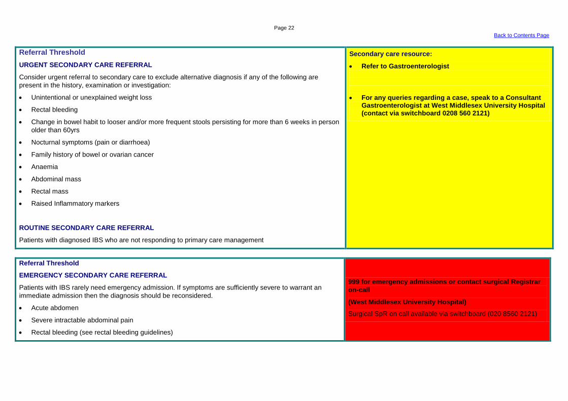
Referral Threshold
URGENT SECONDARY CARE REFERRAL
Consider urgent referral to secondary care to exclude alternative diagnosis if any of the following are
present in the history, examination or investigation:
Unintentional or unexplained weight loss
Rectal bleeding
Change in bowel habit to looser and/or more frequent stools persisting for more than 6 weeks in person
older than 60yrs
Nocturnal symptoms (pain or diarrhoea)
Family history of bowel or ovarian cancer
Anaemia
Abdominal mass
Rectal mass
Raised Inflammatory markers
ROUTINE SECONDARY CARE REFERRAL
Patients with diagnosed IBS who are not responding to primary care management
Secondary care resource:
Refer to Gastroenterologist
For any queries regarding a case, speak to a Consultant Gastroenterologist at West Middlesex University Hospital (contact via switchboard 0208 560 2121)
Referral Threshold
EMERGENCY SECONDARY CARE REFERRAL
Patients with IBS rarely need emergency admission. If symptoms are sufficiently severe to warrant an
immediate admission then the diagnosis should be reconsidered.
Acute abdomen
Severe intractable abdominal pain
Rectal bleeding (see rectal bleeding guidelines)
999 for emergency admissions or contact surgical Registrar
on-call
(West Middlesex University Hospital)
Surgical SpR on call available via switchboard (020 8560 2121)
Page 23
Back to Contents Page
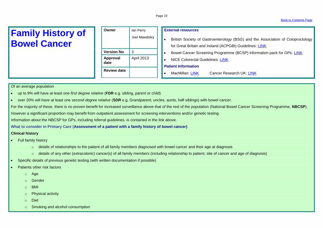
Family History of Bowel Cancer
Owner Ian Perry
Joel Mawdsley
External resources
British Society of Gastroenterology (BSG) and the Association of Coloproctology
for Great Britain and Ireland (ACPGBI) Guidelines: LINK
Bowel Cancer Screening Programme (BCSP) information pack for GPs: LINK
NICE Colorectal Guidelines: LINK
Patient Information
MacMillan: LINK Cancer Research UK: LINK
Version No 3
Approval
date
April 2013
Review date
Of an average population
up to 9% will have at least one first degree relative (FDR e.g. sibling, parent or child)
over 20% will have at least one second degree relative (SDR e.g. Grandparent, uncles, aunts, half siblings) with bowel cancer.
For the majority of these, there is no proven benefit for increased surveillance above that of the rest of the population (National Bowel Cancer Screening Programme, NBCSP).
However a significant proportion may benefit from outpatient assessment for screening interventions and/or genetic testing.
Information about the NBCSP for GPs, including referral guidelines, is contained in the link above.
What to consider in Primary Care (Assessment of a patient with a family history of bowel cancer)
Clinical history
Full family history
o details of relationships to the patient of all family members diagnosed with bowel cancer and their age at diagnosis
o details of any other (extracolonic) cancer(s) of all family members (including relationship to patient, site of cancer and age of diagnosis)
Specific details of previous genetic testing (with written documentation if possible)
Patients other risk factors
o Age
o Gender
o BMI
o Physical activity
o Diet
o Smoking and alcohol consumption
Page 24
Back to Contents Page
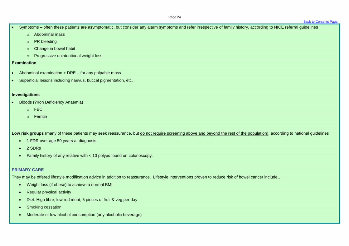
Symptoms – often these patients are asymptomatic, but consider any alarm symptoms and refer irrespective of family history, according to NICE referral guidelines
o Abdominal mass
o PR bleeding
o Change in bowel habit
o Progressive unintentional weight loss
Examination
Abdominal examination + DRE – for any palpable mass
Superficial lesions including naevus, buccal pigmentation, etc.
Investigations
Bloods (?Iron Deficiency Anaemia)
o FBC
o Ferritin
Low risk groups (many of these patients may seek reassurance, but do not require screening above and beyond the rest of the population), according to national guidelines
1 FDR over age 50 years at diagnosis.
2 SDRs
Family history of any relative with < 10 polyps found on colonoscopy.
PRIMARY CARE
They may be offered lifestyle modification advice in addition to reassurance. Lifestyle interventions proven to reduce risk of bowel cancer include…
Weight loss (if obese) to achieve a normal BMI
Regular physical activity
Diet: High fibre, low red meat, 5 pieces of fruit & veg per day
Smoking cessation
Moderate or low alcohol consumption (any alcoholic beverage)
Page 25
Back to Contents Page
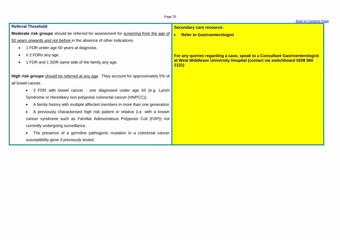
Referral Threshold
Moderate risk groups should be referred for assessment for screening from the age of
50 years onwards and not before in the absence of other indications.
1 FDR under age 50 years at diagnosis.
≥ 2 FDRs any age.
1 FDR and 1 SDR same side of the family any age.
High risk groups should be referred at any age. They account for approximately 5% of
all bowel cancer.
3 FDR with bowel cancer - one diagnosed under age 50 (e.g. Lynch
Syndrome or Hereditary non-polyposis colorectal cancer (HNPCC)).
A family history with multiple affected members in more than one generation
A previously characterised high risk patient or relative (i.e. with a known
cancer syndrome such as Familial Adenomatous Polyposis Coli (FAP)) not
currently undergoing surveillance.
The presence of a germline pathogenic mutation in a colorectal cancer
susceptibility gene if previously tested.
Secondary care resource:
Refer to Gastroenterologist
For any queries regarding a case, speak to a Consultant Gastroenterologist at West Middlesex University Hospital (contact via switchboard 0208 560 2121)
Page 26
Back to Contents Page
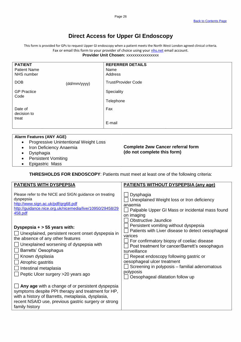
Direct Access for Upper GI Endoscopy
This form is provided for GPs to request Upper GI endoscopy when a patient meets the North West London agreed clinical criteria.
Fax or email this form to your provider of choice using your nhs.net email account. Provider Unit Chosen: xxxxxxxxxxxxxxxx
PATIENT REFERRER DETAILS
Patient Name Name NHS number Address
DOB
(dd/mm/yyyy) Trust/Provider Code
GP Practice Code
Speciality
Telephone
Date of decision to treat
Fax
Alarm Features (ANY AGE)
Progressive Unintentional Weight Loss
Iron Deficiency Anaemia
Dysphagia
Persistent Vomiting
Epigastric Mass
Complete 2ww Cancer referral form (do not complete this form)
THRESHOLDS FOR ENDOSCOPY: Patients must meet at least one of the following criteria:
PATIENTS WITH DYSPEPSIA Please refer to the NICE and SIGN guidance on treating dyspepsia http://www.sign.ac.uk/pdf/qrg68.pdf http://guidance.nice.org.uk/nicemedia/live/10950/29458/29458.pdf
Dyspepsia + > 55 years with:
Unexplained, persistent recent onset dyspepsia in the absence of any other features
Unexplained worsening of dyspepsia with
Barretts’ Oesophagus
Known dysplasia
Atrophic gastritis
Intestinal metaplasia
Peptic Ulcer surgery >20 years ago
Any age with a change of or persistent dyspepsia symptoms despite PPI therapy and treatment for HP, with a history of Barretts, metaplasia, dysplasia, recent NSAID use, previous gastric surgery or strong family history
PATIENTS WITHOUT DYSPEPSIA (any age)
Dysphagia Unexplained Weight loss or Iron deficiency
anaemia Palpable Upper GI Mass or incidental mass found
on imaging Obstructive Jaundice Persistent vomiting without dyspepsia Patients with Liver disease to detect oesophageal
varices For confirmatory biopsy of coeliac disease Post treatment for cancer/Barrett’s oesophagus
surveillance Repeat endoscopy following gastric or
oesophageal ulcer treatment Screening in polyposis – familial adenomatous
polyposis Oesophageal dilatation follow up

Page 27
Back to Contents Page
ADDITIONAL INFORMATION REQUIRED 1. Special Nursing Requirements
None Transport required: wheelchair stretcher Unable to Consent (Form 4) Interpreter required
please state language required……………….
Hoist Large bed Special Mattress (pressure sores)
2. Investigations Performed
H Pylori test result (stool antigen or breath test) Result and Date………………………………
Previous Gastroscopy Result and date…………………….............
(in Iron Deficiency Anaemia) Haemoglobin level (g/dl) AND Ferritin level
Result and date………………………………
3. Comorbidities and medication
None of the below
Warfarin Indication........................................ Clopidogrel Indication......................................... Aspirin Indication………………………………
Valvular heart disease NYHA Heart Failure >II Diabetic (Insulin / Tablet, diet) Severe respiratory disease Cognitive impairment/Communication difficulties
Any Additional information …………………………………………………………………………………………………………………………………………………………………………………………………………………………………………………………………………………………………………………………………………… Provider Use only
Meets criteria, book endoscopy Does not meet criteria, send back to GP with
advice below ………………………………………………………………………………………………………………………………………………………………………………………………………………………………………………………………………………………………………………………………………………………………………………………………………………
Page 28
Back to Contents Page
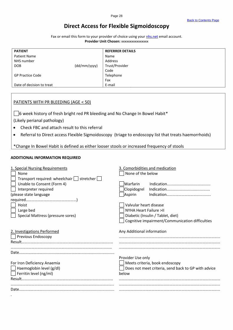
Direct Access for Flexible Sigmoidoscopy
Fax or email this form to your provider of choice using your nhs.net email account. Provider Unit Chosen: xxxxxxxxxxxxxxxx
PATIENT REFERRER DETAILS
Patient Name Name NHS number Address DOB
(dd/mm/yyyy) Trust/Provider Code
GP Practice Code Telephone Fax Date of decision to treat E-mail
PATIENTS WITH PR BLEEDING (AGE < 50)
6 week history of fresh bright red PR bleeding and No Change In Bowel Habit*
(Likely perianal pathology)
Check FBC and attach result to this referral
Referral to Direct access Flexible Sigmoidoscopy (triage to endoscopy list that treats haemorrhoids) *Change In Bowel Habit is defined as either looser stools or increased frequency of stools
ADDITIONAL INFORMATION REQUIRED 1. Special Nursing Requirements
None Transport required: wheelchair stretcher Unable to Consent (Form 4) Interpreter required
(please state language required……………………………………………)
Hoist Large bed Special Mattress (pressure sores)
2. Investigations Performed
Previous Endoscopy Result………………….............…………………………………………………………………………………………………………………………………………… Date…………………….............……………………...............................
For Iron Deficiency Anaemia
Haemoglobin level (g/dl) Ferritin level (ng/ml)
Result………………….............…………………………………………………………………………………………………………………………………………….. Date…………………….............……………………................................
3. Comorbidities and medication
None of the below
Warfarin Indication........................................ Clopidogrel Indication........................................ Aspirin Indication………………………………........
Valvular heart disease NYHA Heart Failure >II Diabetic (Insulin / Tablet, diet) Cognitive impairment/Communication difficulties
Any Additional information ……………………………………………………………………………………………………………………………………………………………………………………………………………………………………………………………………………… Provider Use only
Meets criteria, book endoscopy Does not meet criteria, send back to GP with advice
below ………………………………………………………………………………………………………………………………………………………………………………………………………………………………………………………………………………
Page 29
Back to Contents Page
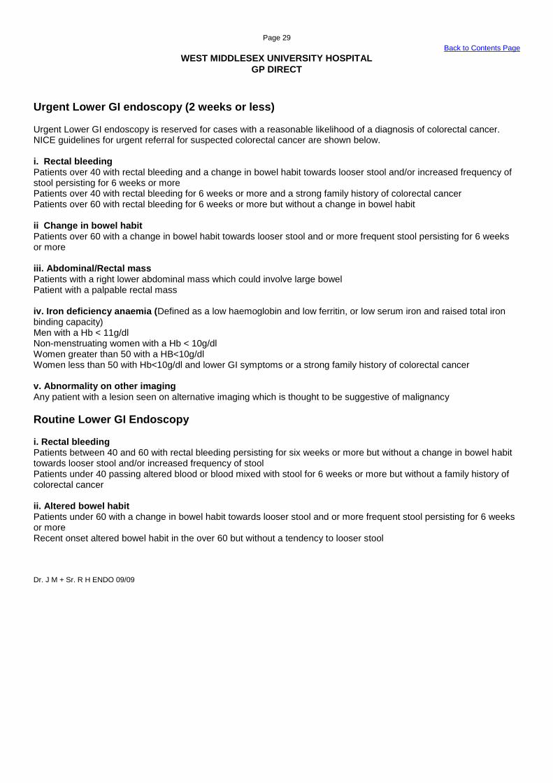
WEST MIDDLESEX UNIVERSITY HOSPITAL
GP DIRECT
Urgent Lower GI endoscopy (2 weeks or less) Urgent Lower GI endoscopy is reserved for cases with a reasonable likelihood of a diagnosis of colorectal cancer. NICE guidelines for urgent referral for suspected colorectal cancer are shown below. i. Rectal bleeding Patients over 40 with rectal bleeding and a change in bowel habit towards looser stool and/or increased frequency of stool persisting for 6 weeks or more Patients over 40 with rectal bleeding for 6 weeks or more and a strong family history of colorectal cancer Patients over 60 with rectal bleeding for 6 weeks or more but without a change in bowel habit ii Change in bowel habit Patients over 60 with a change in bowel habit towards looser stool and or more frequent stool persisting for 6 weeks or more iii. Abdominal/Rectal mass Patients with a right lower abdominal mass which could involve large bowel Patient with a palpable rectal mass iv. Iron deficiency anaemia (Defined as a low haemoglobin and low ferritin, or low serum iron and raised total iron binding capacity) Men with a Hb < 11g/dl Non-menstruating women with a Hb < 10g/dl Women greater than 50 with a HB<10g/dl Women less than 50 with Hb<10g/dl and lower GI symptoms or a strong family history of colorectal cancer v. Abnormality on other imaging Any patient with a lesion seen on alternative imaging which is thought to be suggestive of malignancy
Routine Lower GI Endoscopy i. Rectal bleeding Patients between 40 and 60 with rectal bleeding persisting for six weeks or more but without a change in bowel habit towards looser stool and/or increased frequency of stool Patients under 40 passing altered blood or blood mixed with stool for 6 weeks or more but without a family history of colorectal cancer ii. Altered bowel habit Patients under 60 with a change in bowel habit towards looser stool and or more frequent stool persisting for 6 weeks or more Recent onset altered bowel habit in the over 60 but without a tendency to looser stool Dr. J M + Sr. R H ENDO 09/09

Page 30
Back to Contents Page
Stoma Care Service at West Middlesex University Hospital Indications for referral The service is happy to see any problems related to stoma care. Typical examples of problems suitable for referral to this service include: appliance failure, leakage, sore skin / erythema, para-stomal herniation, stomal prolapse and symptoms indicative of recurrent disease. Referral sources The Nurse Specialists who lead the service recognise that timely help and support reduces the negative effects on self esteem and quality of life. With this in mind the service has been created to allow easy access for referrers. Referrals from General Practitioners, Community Nurses, Community Pharmacists, other Specialist Colleagues, and all Allied Health Care Professionals are accepted. There is also an open access policy for existing stoma patients, who are encouraged to self refer back to a nurse led stoma care assessment clinic when any problems arise. Early referral is encouraged as more costly interventions may be required if referrals are delayed. Possible interventions Following referral, advice can be given over the telephone or patients can be seen in the stoma/colorectal follow up clinic or at home depending on the most appropriate setting. A three-day standard is offered to accommodate for Friday referrals. However many patients are seem on the same day if deemed necessary. Contact details: Tracey Virgin-Elliston Lead Nurse Specialist – Stoma Care. Susan Firth Nurse Specialist – Stoma Care Stoma Care Department Clinic 4 The West Middlesex Hospital Twickenham Road Isleworth Middlesex TW7 6AF Office: 0208 321 5822 (24 hour answer phone) Fax: 0202 321 5024. Email: [email protected] [email protected] (Please send emails to both addresses in case of annual leave.)
Page 31
Back to Contents Page
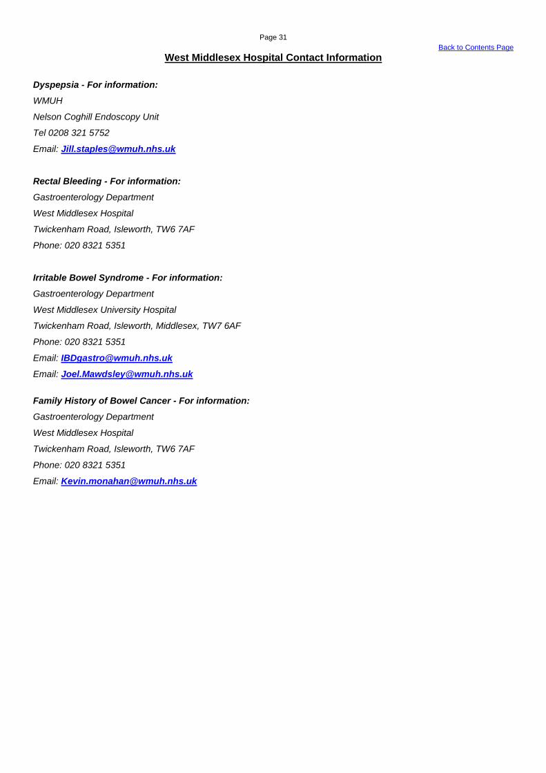
West Middlesex Hospital Contact Information
Dyspepsia - For information:
WMUH
Nelson Coghill Endoscopy Unit
Tel 0208 321 5752
Email: [email protected]
Rectal Bleeding - For information:
Gastroenterology Department
West Middlesex Hospital
Twickenham Road, Isleworth, TW6 7AF
Phone: 020 8321 5351
Irritable Bowel Syndrome - For information:
Gastroenterology Department
West Middlesex University Hospital
Twickenham Road, Isleworth, Middlesex, TW7 6AF
Phone: 020 8321 5351
Email: [email protected]
Email: [email protected]
Family History of Bowel Cancer - For information:
Gastroenterology Department
West Middlesex Hospital
Twickenham Road, Isleworth, TW6 7AF
Phone: 020 8321 5351
Email: [email protected]
Page 32
Back to Contents Page
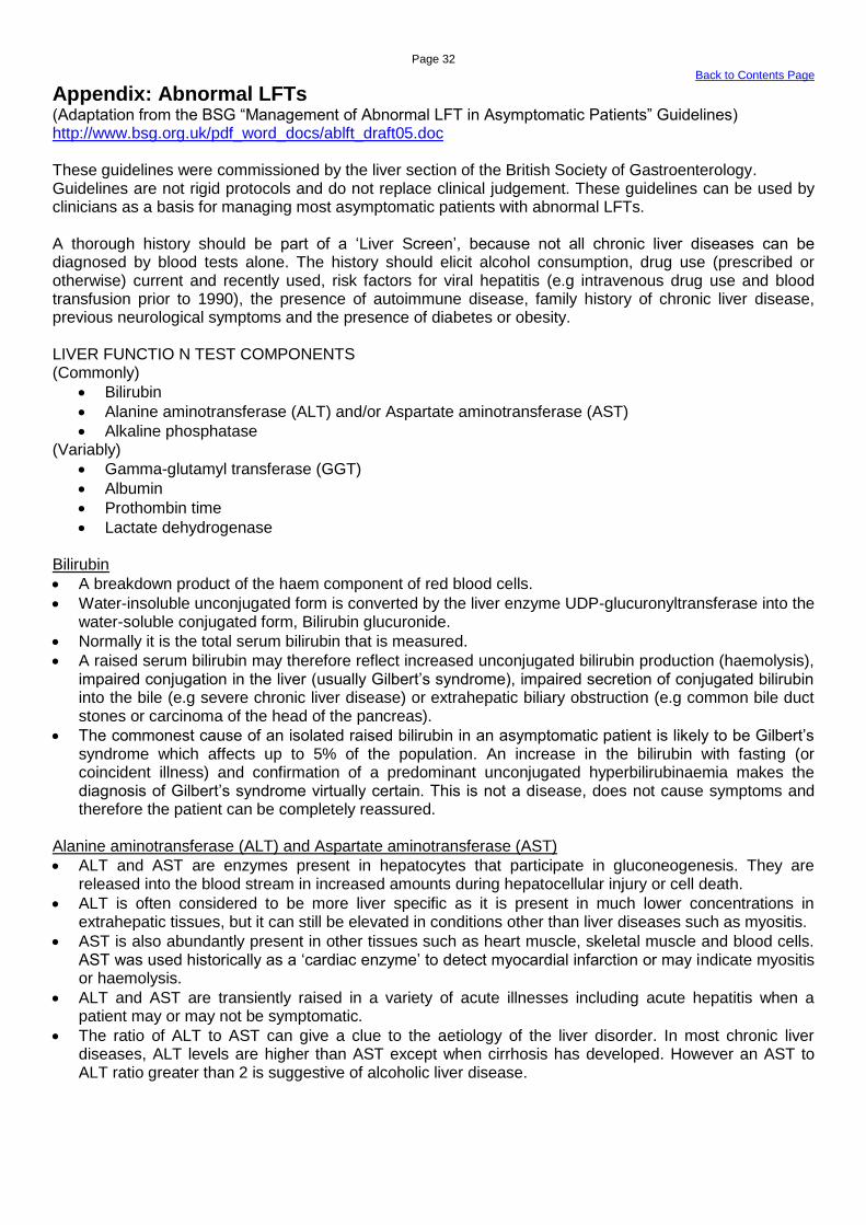
Appendix: Abnormal LFTs (Adaptation from the BSG “Management of Abnormal LFT in Asymptomatic Patients” Guidelines) http://www.bsg.org.uk/pdf_word_docs/ablft_draft05.doc These guidelines were commissioned by the liver section of the British Society of Gastroenterology. Guidelines are not rigid protocols and do not replace clinical judgement. These guidelines can be used by clinicians as a basis for managing most asymptomatic patients with abnormal LFTs. A thorough history should be part of a ‘Liver Screen’, because not all chronic liver diseases can be diagnosed by blood tests alone. The history should elicit alcohol consumption, drug use (prescribed or otherwise) current and recently used, risk factors for viral hepatitis (e.g intravenous drug use and blood transfusion prior to 1990), the presence of autoimmune disease, family history of chronic liver disease, previous neurological symptoms and the presence of diabetes or obesity. LIVER FUNCTIO N TEST COMPONENTS (Commonly)
Bilirubin
Alanine aminotransferase (ALT) and/or Aspartate aminotransferase (AST)
Alkaline phosphatase (Variably)
Gamma-glutamyl transferase (GGT)
Albumin
Prothombin time
Lactate dehydrogenase Bilirubin
A breakdown product of the haem component of red blood cells.
Water-insoluble unconjugated form is converted by the liver enzyme UDP-glucuronyltransferase into the water-soluble conjugated form, Bilirubin glucuronide.
Normally it is the total serum bilirubin that is measured.
A raised serum bilirubin may therefore reflect increased unconjugated bilirubin production (haemolysis), impaired conjugation in the liver (usually Gilbert’s syndrome), impaired secretion of conjugated bilirubin into the bile (e.g severe chronic liver disease) or extrahepatic biliary obstruction (e.g common bile duct stones or carcinoma of the head of the pancreas).
The commonest cause of an isolated raised bilirubin in an asymptomatic patient is likely to be Gilbert’s syndrome which affects up to 5% of the population. An increase in the bilirubin with fasting (or coincident illness) and confirmation of a predominant unconjugated hyperbilirubinaemia makes the diagnosis of Gilbert’s syndrome virtually certain. This is not a disease, does not cause symptoms and therefore the patient can be completely reassured.
Alanine aminotransferase (ALT) and Aspartate aminotransferase (AST)
ALT and AST are enzymes present in hepatocytes that participate in gluconeogenesis. They are released into the blood stream in increased amounts during hepatocellular injury or cell death.
ALT is often considered to be more liver specific as it is present in much lower concentrations in extrahepatic tissues, but it can still be elevated in conditions other than liver diseases such as myositis.
AST is also abundantly present in other tissues such as heart muscle, skeletal muscle and blood cells. AST was used historically as a ‘cardiac enzyme’ to detect myocardial infarction or may indicate myositis or haemolysis.
ALT and AST are transiently raised in a variety of acute illnesses including acute hepatitis when a patient may or may not be symptomatic.
The ratio of ALT to AST can give a clue to the aetiology of the liver disorder. In most chronic liver diseases, ALT levels are higher than AST except when cirrhosis has developed. However an AST to ALT ratio greater than 2 is suggestive of alcoholic liver disease.
Page 33
Back to Contents Page
Alkaline phosphatase (ALP)
ALP is produced mainly in the liver and bone but also in smaller quantities by the intestines, kidneys, and white blood cells.
Physiologically higher in childhood and pregnancy where it comes from the placenta.
Pathologically higher in cholestatic liver disease (e.g. primary biliary cirrhosis, primary sclerosing cholangitis, common bile duct obstruction, intrahepatic duct obstruction (metastasis) and drug induced cholestasis), bone disease (e.g Paget’s disease and fractures), and right-side heart failure.
There is no data on the most likely causes of a raised ALP in an asymptomatic population. In a hospitalised population, a raised ALP often resolves spontaneously or if persists is most often associated with significant extra-hepatic disease.
Gamma glutamyltransferase (GGT)
GGT is abundant in the liver and other tissues such as kidney, intestine, prostate and pancreas.
It is not present in bone, however, hence the usefulness of this enzyme in determining the source of an elevated ALP. GGT should be measured to establish the hepatic origin of the ALP.
A raised GGT is not confined to alcoholic liver disease as it can be induced by drugs and is also elevated in NAFLD. It is not always raised in chronic alcoholics; hence it is not sensitive for occult alcohol abuse. Furthermore, patients drinking excess alcohol who have an isolated raised GGT are also unlikely to have significant liver damage.
In asymptomatic patients with a raised GGT alone who do not drink excessively, hepatic fibrosis is present in only 5% of cases.
WHY INVESTIGATE ASYMPTOMATIC PATIENTS WITH ABNORMAL LFT?
The rationale for considering investigation of asymptomatic patients with abnormal LFTs is based upon knowledge that a significant number of patients with chronic liver disease have no symptoms or non-specific symptoms. Specific symptoms may only occur once cirrhosis has developed, yet cirrhosis is usually irreversible and associated with reduced life expectancy.
Increasing death rates from cirrhosis almost certainly reflect increasing rates of alcoholic liver disease and the burden of chronic hepatitis C. The obesity epidemic will also cause non-alcoholic fatty liver disease (NAFLD) associated cirrhosis to further increase liver disease mortality in the future.
Cirrhosis is associated with a reduced life expectancy, cirrhosis is caused by chronic liver diseases and abnormal LFTs may the only indication of chronic liver disease. There is now some evidence that increasing LFT levels (ALT and AST) are directly associated with increased liver related mortality rates.
In the UK there is evidence that the causes of abnormal LFTs are often not investigated. When thorough investigation is performed however, the majority of patients are found to have significant disease warranting treatment or follow up.
DO ABNORMAL LIVER FUNCTION TESTS OUTSIDE THE NORMAL RANGE ALWAYS INDICATE LIVER DISEASE?
By definition some healthy people will have abnormal LFTs, because the normal range for any LFT is based on the mean + 2 standard deviations of the mean from a healthy population. 2.5% of healthy people will therefore have any particular LFT above the normal range when tested.
Consider physiological and non-hepatic pathological causes of abnormal LFT (pregnancy, bone disease, presence of macroenzymes, haemolysis of blood sample)
DO NORMAL LIVER FUNCTION TESTS INDICATE ABSENSE OF LIVER DISEASE?
Normal LFTs do not exclude the presence of significant liver disease, thus patients still require assessment in the presence of signs of liver disease or significant risk factors for chronic liver disease (e.g exposure to hepatitis C or family history of haemochromatosis).
It is well-established that certain liver disease patients can have normal LFTs and still have significant liver disease (e.g chronic hepatitis C, haemochromatosis, and primary sclerosing cholangitis) which may progress to cirrhosis.