ganoderma lucidum polysaccharides target a...
TRANSCRIPT
Asian Pacific Journal of Cancer Prevention, Vol 15, 2014 3981
DOI:http://dx.doi.org/10.7314/APJCP.2014.15.9.3981Ganoderma Lucidum Polysaccharides Target a Fas/Caspase Dependent Pathway to Induce Apoptosis
Asian Pac J Cancer Prev, 15 (9), 3981-3986
Introduction
Colon cancer, which caused by cellular pathology in the appendix, colon, or in the rectum, is the third most common cancer in the world. Clinically, chemotherapy and radiation are typically used before or after surgery in colorectal treatment. Unfortunately, even with modern intensive medical therapy, a high ratio of recidivations and formation of metastases lead to a low cure rate and scarcely improve long-term survival rate (Luigi et al., 2009). Furthermore, the toxicity produced by chemotherapy inflicts on immune system of cancer patients in the course of treatment. Currently, compounds from herbaceous plants are recognized as pharmacological agents to inhibit cancer growth and lessen the side effects of radiation and chemotherapy (Chen et al., 2009; Chen et al., 2010). GLP, isolated from Ganoderma lucidum, have been used in East Asian countries to remedy conditions like aging, oxidation, radiation and hypertension for thousands of years (Chen et al., 2009; Pillai et al., 2010; Li et al., 2011). Previous studies have showed that GLP can mediate cytotoxicity in number of cancer cell lines including lung cancer, prostate cancer and breast cancer. Further studies showed that
Hunan Provincial Key Laboratory of Crop Germplasm Innovation and Utilization, Hunan Agricultural University, Changsha, China *For correspondence: [email protected]; [email protected]
Abstract
Ganoderma lucidum polysaccharides (GLP) extracted from Ganoderma lucidum have been shown to induce cell death in some kinds of cancer cells. This study investigated the cytotoxic and apoptotic effect of GLP on HCT-116 human colon cancer cells and the molecular mechanisms involved. Cell proliferation, cell migration, lactate dehydrogenase (LDH) levels and intracellular free calcium levels ([Ca2+]i) were determined by MTT, wound-healing, LDH release and fluorescence assays, respectively. Cell apoptosis was observed by scanning and transmission electron microscopy. For the mechanism studies, caspase-8 activation, and Fas and caspase-3 expression were evaluated. Treatment of HCT-116 cells with various concentrations of GLP (0.625-5 mg/mL) resulted in a significant decrease in cell viability (P< 0.01). This study showed that the antitumor activity of GLP was related to cell migration inhibition, cell morphology changes, intracellular Ca2+ elevation and LDH release. Also, increase in the levels of caspase-8 activity was involved in GLP-induced apoptosis. Western blotting indicated that Fas and caspase-3 protein expression was up-regulated after exposure to GLP. This investigation demonstrated for the first time that GLP shows prominent anticancer activities against the HCT-116 human colon cancer cell line through triggering intracellular calcium release and the death receptor pathway. Keywords: Apoptosis - caspase - cell migration - Fas - human colon cancer cells
RESEARCH ARTICLE
Ganoderma Lucidum Polysaccharides Target a Fas/Caspase Dependent Pathway to Induce Apoptosis in Human Colon Cancer CellsZengenni Liang, Yu-Tong Guo, You-Jin Yi, Ren-Cai Wang*, Qiu-Long Hu, Xing-Yao Xiong*
GLP programmed cell death through endothelial growth factor overexpression and tumor angiogenesis inhibition in highly metastatic mouse melanoma B16F10 cells (Sun et al., 2011), through endotoxin-induced intercellular cell adhesion molecule-1 (ICAM-1) upregulation in the neointima in mice (Liu et al., 2010), through cell differentiation and p53 activation in human monocytic leukemia THP-1 cells (Hsu et al., 2011), through activation of mitochondrial signaling pathway in MCF-7 breast cancer cells (Shang et al., 2011). GLP can also boost anticancer immunity by changing lymphocyte phenotype in H22 hepatoma mice (Zhou et al., 2009), by enhancing CD56+ NK-cell cytotoxicity in cord blood (Chien et al., 2004), by promoting the proliferation of B cells in mice (Jiang et al., 2003). Although GLP have been demonstrably effective in fighting cancer by reducing cell growth and boosting cell apoptosis, the anti-cancer effects of GLP have not been reported in human colon cancer cell line HCT-116. In this study, we isolated and purified polysaccharides from Ganoderma lucidum (Leyss. et Fr.) Karst. Thereafter, HCT-116 cell line was used as a test model to measure the hypothesis that GLP (>10 kDa) affected anticancer effects by MTT assay, migration assay and morphologic observation. We also examined the roles of lactate
Zengenni Liang et al
Asian Pacific Journal of Cancer Prevention, Vol 15, 20143982
dehydrogenase (LDH), cellular intracellular Ca2+, Fas and caspase cascades in GLP-induced apoptosis in HCT-116 cells.
Materials and Methods
Preparation of GLP Slices of Ganoderma lucidum (Leyss. et Fr.) Karst. were provided by State Key Laboratory of Sub-health Intervention Technology, State Administration of Traditional Chinese Medicine. GLP were isolated and purified using a procedure as described (Guo, 2012). Crude polysaccharides were gained through derosination, removal of oligosaccharides, hot water extraction, deproteinization, decolorization, concentration under vacuum, and freeze-dried. GLP solution (10 mg/mL in water) was ultrafiltered through 10, 30, and 50 kDa molecular weight cut off membranes (MWCO, Millipore, US), respectively, in an Amicon 8200 stirred cell (Millipore Corporation Bedford, MA). The filtrate (molecular weight of polysaccharides >10 kDa) was freeze-dried, dissolved in high glucose DMEM with 10% FBS to get a stock solution of 10 mg/mL, passed through 0.22 μm filter and stored at 4°C which was prepared for subsequent analysis.
Cell Culture Colon cancer HCT-116 cell line was obtained from Institute of Basic Medical Sciences (Beijing, China). The cells was planted in DMEM medium with high glucose supplemented with 10% FBS in 25 cm2 tissue culture flasks under a humidified atmosphere of 5% CO2 at 37°C. Frozen cells were thawed and passaged 3 times, which were subsequently used for this trial.
Cell viability assay HCT-116 cells (3×10⁴ cells/well) were seeded onto 96-well plates. After a 12 h recovery period, cells were incubated in fresh medium without or with various concentrations of GLP (0.313, 0.625, 1.25, 2.5 and 5 mg/mL) for 24, 48 and 72 h, respectively, then added 5 mg/mL of MTT solution (20 μL/well) to each well for an additional 4 h. Subsequently removing the supernatant, water-insoluble formazan was dissolved in 150 μL DMSO to each well for 10 min. The absorbance at wavelength of 492 nm was recorded using a microplate reader (MK3, Thermo, USA). Cells with starch (0-10 mg/mL) treatment were used as control. The inhibitory rate was calculated using the following formula: Inhibitory rate (%) = (1 –mean Atreatment/mean Acontrol) × 100%.
Wound-healing assay in vitro A 24-well plate was spread by monolayer HCT-116 cells and scratched wounds by 10 μL pipette tips. Cells were washed twice with PBS to remove loose cells, and then exposed to 0.625, 1.25, 2.5, 5 and 10 mg/mL of GLP. Following incubation for 48 h, the wounds were photographed under an inverted microscope (BX60, Olympus, Japan). The number of migrant cells was analyzed with Image J software from three visual fields in each group.
Morphologic observation To directly assess the effects of GLP on HCT-116 cells, GLP-induced morphological change was examined by two types of electron microscopes. Transmission electron microscope: GLP-treated cells and untreated cells were trypsinized and suspension-fixed in 1% glutaraldehyde for 2 h. After rinsing with phosphate buffered solution (PBS, pH7.4), cells were postfixed with 1% osmium tetroxide (OsO4) for another 2 h. Dehydration was carried out in an ascending grade of ethanol (50–100%, v/v). Samples were infiltrated with 50% quetol in ethanol for 1 h and additional 1 h incubation with 100% quetol for 6 h, followed by polymerized at 60°C. After 39 h, all groups were cut into ultra-thin sections, stained with 4% uranyl acetate for 10 min. Photographs were taken under a transmission electron microscope (JEM-1230, Tokyo, Japan). Scanning electron microscopy: After GLP inoculation, cells were processed and analyzed as previously described (Wahab et al., 2009). Cells were fixed in 1% glutaraldehyde, post-fixed in 1% OsO4, dehydrated in a graded of ethanol (50–100%, v/v), critical-point dried, affixed, gold-sputtered and finally viewed by a scanning electron microscope (JSM-6380LV, Tokyo, Japan).
Lactate Dehydrogenase (LDH) Release Assay The apoptotic effect of GLP was examined by measurement of LDH release (Nakagawa et al., 2005). 1.5×10⁵ cells per well were plated in 24-well cell culture plate, followed by overnight incubation. Supernatants were collected and added in a black 96-well culture plates (200 μL per well) after cells were exposed to increasing concentrations of GLP (1.25, 2.5 and 5 mg/mL) for 48 h. LDH release was measured using LDH cytotoxicity assay kit (Beyotime, China) as per the manufacturer’s instructions. Absorbance at a wavelength of 450 nm in each well was determined using an enzyme linked immunoassay instrument.
Detection of Intracellular Ca2+ level GLP-treated cells were rinsed twice with PBS, resuspended in Hanks’ buffer (pH 7.4) containing 0.2% bovine serum albumin (BSA), and treated with 10 μmol/L of fura-2 acetoxymethyl ester (Fura-2 AM) at 37℃ for 30 min in the dark. Cells were then harvested, washed three times and resuspended in hanks’ buffer. The fluorescence were monitored by recording in the ratio mode excitation wavelength at 340/380 nm and 510 nm emission wavelength using a varioskan flash (Thermo, USA). Maximal fluorescence (Fmax) was obtained in the presence of 10% Triton X-100 (final concentration 0.1%) and followed by minimum fluorescence (Fmin) with 5 mmol/L of EDTA. Intracellular Ca2+ was calculated from the fluo-2 fluoresce intensity using the equation: [Ca2+]i = Kd [ (F – Fmin)/ (Fmax – F)], where Kd of fluo-2 for Ca2+ is 224 nM (Grynkiewicz et al., 1985; Hirst et al., 1999).
Detection of caspase-8 Activity The caspase-8 activity was determined using the caspase-8 activity kit (Beyotime, China) according to the manufacture’s recommendations. Briefly, cells (1×10⁶ cells/well) were exposed to 0, 2.5 and 5 mg/mL
Asian Pacific Journal of Cancer Prevention, Vol 15, 2014 3983
DOI:http://dx.doi.org/10.7314/APJCP.2014.15.9.3981Ganoderma Lucidum Polysaccharides Target a Fas/Caspase Dependent Pathway to Induce Apoptosis
of GLP for 24 h. Cells were centrifuged at 10,000 g for 20 min at 4°C, following 15 min lysate treatment on ice. Subsequently, 10 μL of supernatant was incubated with 80 μL of reaction buffer (1% NP-40, 20 mM Tris-HCl, pH 7.5, 137 mM NaCl and 10% glycerol) and 10 μL of 2 mM substrate at 37°C for 2 h. At the end of the incubation period, the samples were read as optical density at 405 nm with multiskan mircoplate reader (MK3, Thermo, USA).
Western blot analysis Cells were treated with 5 mg/mL GLP for different time points (0, 12, 24, 36, and 48 h). After washing three times in PBS, cells were lysed in RIPA lysis buffer (Applygen, China) on ice for 30 min. The protein concentration was determined utilizing the Bradford assay (Bio-Rad, CA). Subsequently, proteins were separated by 10%-12% SDS-PAGE, electrotransferred to polyvinylidene fluoride (PVDF) membranes (Millipore, US), and blocked in 5% non-fat dehydrated milk. The membranes were incubated with β-actin (1:4000, Proteintech, US), Fas (1:1000, Proteintech, US), caspase-3 (1:500, Proteintech, US) primary antibodies, respectively, followed by secondary antibodies (1:3000, Santa Cruz Biotechnology, US). The signal was detected as described (Suresh et al., 2013).
Statistical Analysis All experiments were performed in triplicate and the data were expressed as mean ± S.D. Statistical analyse was performed using the statistical software SPSS 18.0, with highly significance at P< 0.01.
Results
Isolation and structural analysis The yield of crude polysaccharides was about 3.08%.
After ultrafiltering the GLP solution through membranes, the freeze-dried fractions were weighed. The distribution of polysaccharides as followed: 10-30 kDa, 32.1%; 30-50 kDa, 21.8%; >50 kDa, 46.1%. The three fractions were pooled to further studies. Polysaccharides had no absorption at 260 and 280 nm in the UV spectrum, suggesting the absence of nucleic acid and protein. IR spectra (the band at 847 cm−1) and optical rotation ([α]20 D + 192°) were shown α-glycosidic linkages in GLP (Zhao et al., 2005). The percentage of total sugar was determined to be 89%. The uronic acid content was 11%. High-performance anion exchange chromatography (HPAEC) showed that GLP was composed of arabinose, glucose, galactose and cellose in the molar ratios of 11:3:3:1.
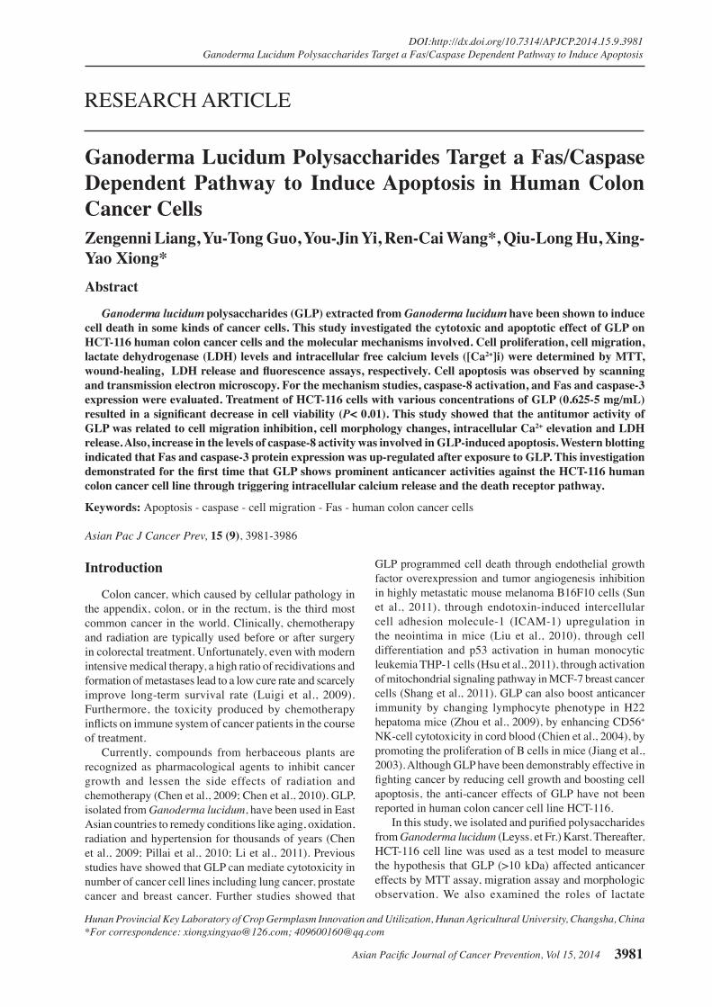
GLP dose- and time-dependently inhibit HCT-116 cell growth Primary, different doses of GLP (0-5 mg/mL) and various cultivation times were applied in order to detect the cytotoxicity of GLP in HCT-116 cells. As shown in Figure 1A, GLP inhibited the viability of HCT-116 cells in a dose- and time-dependent manner (P<0.01). The rate of cell proliferation were markedly decreased in human colon cancer cells treated with doses above 2.5 mg/mL after 72 h (P<0.01), whereas there was no significant influences at dose of 0.313 mg/mL. The inhibitory concentration of 50% (IC50) for 24, 48 and 72 h were 9.25, 5.72 and 3.69 mg/mL, respectively. The MTT assay also showed that starch incubation (0-5 mg/mL) promoted weak proliferation of HCT-116 cells (data not shown).
GLP delayed HCT-116 cell migration To determine whether GLP impact the migratory capacity of HCT-116 cells, scratch wound assays were performed. As shown in Figure 1B, GLP concentration dependently inhibited the migratory capacity of human colon cancer contrast with control group. By 48 h incubation with 0.625, 1.25, 2.5, 5 and 10 mg/mL GLP, the number of migratory cells decreased by 22.7%, 29.1%, 39.9%, 63.1% and 84.4%, respectively (P<0.01). Scanning electron microscopy To investigate whether the observed cell viability inhibition was caused by apoptosis, the surface ultrastructure of GLP-treated cells was examined by SEM electron microscopy. Untreated cells showed
Figure 1. GLP Suppressed Cell Growth and Migratory. A. Growth-inhibiting effects of GLP on HCT-116 cells. Cells were treated with 0-5 mg/mL GLP for 24, 48 and 72 h, respectively, and then determined by MTT assay. Experiment was repeated three times and similar results obtained. B. GLP delayed motility of HCT-116 cells. After wounding with pipette tips, cells were treated with 0-10 mg/mL GLP for 48 h. The number of migratory cells in the wound edge migration was quantitated by ImageJ software. *P<0.01, represents significant difference compared with the untreated control. Data are presented as mean ± SD (n=4)
B
A
0
25.0
50.0
75.0
100.0
New
ly d
iagn
osed
with
out
trea
tmen
t
New
ly d
iagn
osed
with
tre
atm
ent
Pers
iste
nce
or r
ecur
renc
e
Rem
issi
on
Non
e
Chem
othe
rapy
Radi
othe
rapy
Conc
urre
nt c
hem
orad
iatio
n
10.3
0
12.8
30.025.0
20.310.16.3
51.7
75.051.1
30.031.354.2
46.856.3
27.625.033.130.031.3
23.738.0
31.3
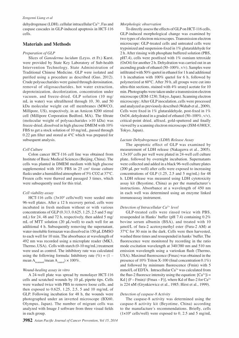
Figure 2. GLP Induced Morphological Changes in HCT-116 Cells Detected by Scanning Electron Microscopy. A&D. control group; B&E. 2.5 mg/mL GLP group; C&F. 5 mg/mL GLP group. A-C was magnified 1000 times; D-F was magnified 5000 times
A B C
D E F
Zengenni Liang et al
Asian Pacific Journal of Cancer Prevention, Vol 15, 20143984
uniform cellular distribution and numerous microvilli on their surface. In GLP-treated cells, 2.5 and 5 mg/mL of GLP were significant changes in cell morphology that corresponded to a typical cellular surface morphology of apoptosis, including cell membrane blebbing, cellular density and microvilli reduction, apoptotic body formation (as shown in white arrow) (Figure 2).
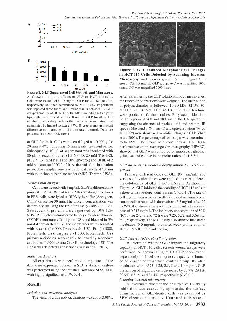
Transmission electron microscopy Induction of apoptosis was also inspected using transmission electron microscopy. Control cells had a well regular cell shape, many microvilli protruding from their surfaces, homogeneous chromatin distribution (Figure 3A), rough endoplasmic reticula and healthy-looking mitochondrion (arrow) (Figure 3B). On the contrary, 5 mg/mL GLP lead to irregular cell shape, a decreased microvillar density, mitochondria swelling, chromatin concentration and marginalization (arrow) (Figure 3C), cytoplasm vacuolization and apoptotic bodies formation (arrow) (Figure 3D). Treatment with 10 mg/mL GLP induced collapse of plasma membrane, resulting in cell decomposition and death (Figure 3E).
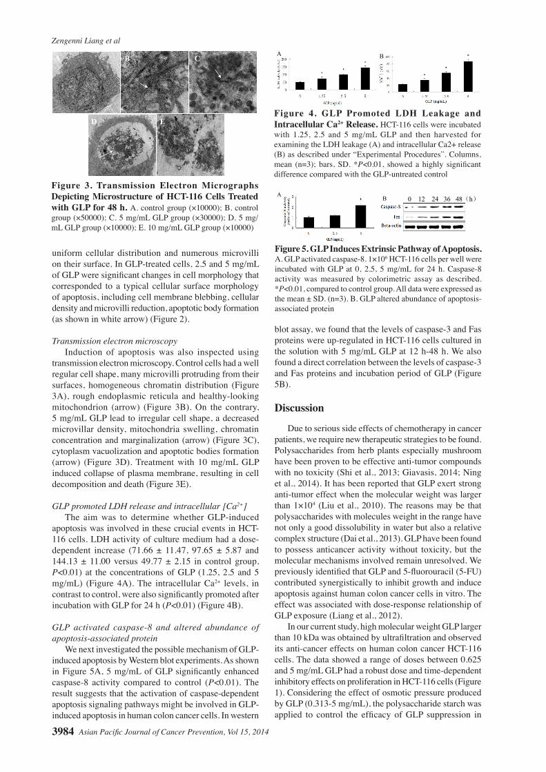
GLP promoted LDH release and intracellular [Ca2+] The aim was to determine whether GLP-induced apoptosis was involved in these crucial events in HCT-116 cells. LDH activity of culture medium had a dose-dependent increase (71.66 ± 11.47, 97.65 ± 5.87 and 144.13 ± 11.00 versus 49.77 ± 2.15 in control group, P<0.01) at the concentrations of GLP (1.25, 2.5 and 5 mg/mL) (Figure 4A). The intracellular Ca2+ levels, in contrast to control, were also significantly promoted after incubation with GLP for 24 h (P<0.01) (Figure 4B).
GLP activated caspase-8 and altered abundance of apoptosis-associated protein We next investigated the possible mechanism of GLP-induced apoptosis by Western blot experiments. As shown in Figure 5A, 5 mg/mL of GLP significantly enhanced caspase-8 activity compared to control (P<0.01). The result suggests that the activation of caspase-dependent apoptosis signaling pathways might be involved in GLP-induced apoptosis in human colon cancer cells. In western
blot assay, we found that the levels of caspase-3 and Fas proteins were up-regulated in HCT-116 cells cultured in the solution with 5 mg/mL GLP at 12 h-48 h. We also found a direct correlation between the levels of caspase-3 and Fas proteins and incubation period of GLP (Figure 5B).
Discussion
Due to serious side effects of chemotherapy in cancer patients, we require new therapeutic strategies to be found. Polysaccharides from herb plants especially mushroom have been proven to be effective anti-tumor compounds with no toxicity (Shi et al., 2013; Giavasis, 2014; Ning et al., 2014). It has been reported that GLP exert strong anti-tumor effect when the molecular weight was larger than 1×10⁴ (Liu et al., 2010). The reasons may be that polysaccharides with molecules weight in the range have not only a good dissolubility in water but also a relative complex structure (Dai et al., 2013). GLP have been found to possess anticancer activity without toxicity, but the molecular mechanisms involved remain unresolved. We previously identified that GLP and 5-fluorouracil (5-FU) contributed synergistically to inhibit growth and induce apoptosis against human colon cancer cells in vitro. The effect was associated with dose-response relationship of GLP exposure (Liang et al., 2012).
In our current study, high molecular weight GLP larger than 10 kDa was obtained by ultrafiltration and observed its anti-cancer effects on human colon cancer HCT-116 cells. The data showed a range of doses between 0.625 and 5 mg/mL GLP had a robust dose and time-dependent inhibitory effects on proliferation in HCT-116 cells (Figure 1). Considering the effect of osmotic pressure produced by GLP (0.313-5 mg/mL), the polysaccharide starch was applied to control the efficacy of GLP suppression in
Figure 3. Transmission Electron Micrographs Depicting Microstructure of HCT-116 Cells Treated with GLP for 48 h. A. control group (×10000); B. control group (×50000); C. 5 mg/mL GLP group (×30000); D. 5 mg/mL GLP group (×10000); E. 10 mg/mL GLP group (×10000)
A B
D E
C
Figure 4. GLP Promoted LDH Leakage and Intracellular Ca2+ Release. HCT-116 cells were incubated with 1.25, 2.5 and 5 mg/mL GLP and then harvested for examining the LDH leakage (A) and intracellular Ca2+ release (B) as described under “Experimental Procedures”. Columns, mean (n=3); bars, SD. *P<0.01, showed a highly significant difference compared with the GLP-untreated control
A B
Figure 5. GLP Induces Extrinsic Pathway of Apoptosis. A. GLP activated caspase-8. 1×10⁶ HCT-116 cells per well were incubated with GLP at 0, 2.5, 5 mg/mL for 24 h. Caspase-8 activity was measured by colorimetric assay as described. *P<0.01, compared to control group. All data were expressed as the mean ± SD. (n=3). B. GLP altered abundance of apoptosis-associated protein
0 12 24 36 48(h)
B A
Asian Pacific Journal of Cancer Prevention, Vol 15, 2014 3985
DOI:http://dx.doi.org/10.7314/APJCP.2014.15.9.3981Ganoderma Lucidum Polysaccharides Target a Fas/Caspase Dependent Pathway to Induce Apoptosis
HCT-116 cells. The results also showed that starch (0.313 -10 mg/mL) dose-dependently promoted cell growth, suggesting the osmotic pressure in the cell culture with these concentrations of GLP did not markedly induce cell death. Cell migration is a critical process for sustained maintenance and development of every type of cancer cells (Condeelis and Pollard, 2006). Especially, malignant cancer cells put into use their own migratory capacity to invade adjacent healthy tissues and the vasculature, then ultimately to inflict devastating organizer destruction (Yamaguchi and Condeelis, 2007). Thus, limitation of this process is a novel therapeutic strategy for controlling tumor growth and deterioration. In the present study, we reported that GLP mediated dose-dependent inhibitory effects on cell migration of human colon cancer HCT-116 cells (P<0.01). The data showed that GLP may cause antitumor effects by suppression cell growth and migration in HCT-116 cells.
Cell apoptosis triggered by most of cancer chemopreventive agents and anticancer drugs is an essential process in prevention of tumor promotion (Núñez et al., 2010). It is commonly measured by the increase in release of LDH, TUNEL staining, intracellular Ca2+ concentration, cytoplasm condensation, DNA and nuclear fragmentation (Liu et al., 2006). Under SEM and TEM observation, vacuoles, apoptotic bodies, chromatin margination and cell degradation appeared in GLP -incubated cells (Figure 2 and Figure 3). As further proof of the apoptotic effects of GLP, LDH release and intracellular Ca2+ concentration were also determined. The permeabilization of the plasma membrane is a key signature for apoptotic cells, which can be quantified in tissue culture settings by measuring the release of the intracellular enzyme LDH (Sun et al., 2013). As an important messengers and a key regulator, Ca2+ can control apoptosis in response to a variety of pathological conditions from external and internal environment though a wide range of Ca2+ sensitive factors that are compartmentalized in various intracellular organelles including the mitochondria, ER and cytoplasm. GLP has been reported to promote calcium levels in the brain of D-gal-treated mice (Li et al., 2011). In this study, GLP also induced apoptosis by promoting the release of LDH (Figure 4A) and increased the level of intracellular Ca2+ (Figure 4B). These results clearly indicated that GLP significantly induced apoptosis in HCT-116 cells.
Next, we investigated the possible mechanisms of GLP-mediated apoptosis. Intrinsic (mitochondrial) and extrinsic (death receptor) apoptotic pathways are two major ways to stimulate cell apoptosis. In extrinsic pathway, ligand-bound death receptors such as Fas and TRAIL receptors have been well characterized to be a crucial regulator, which mediate activation of caspase cascades in some types of cancer cells (Sayers, 2011). It is demonstrated that caspase-3/8 activation is physically associated with the signaling complex during Fas-induced apoptosis (Mandal et al., 2005). In addition to the caspase-dependent pathway, cell apoptosis also has been reported to be stimulated via caspase-independent pathway (Liu and Chang, 2011). In this study, GLP activated caspase-8 (Figure 5A) and up-regulated caspase-3 (Figure 5B),
indicating that caspase-dependent pathway is related to GLP-induce apoptosis. It also increased the level of Fas protein (Figure 5B). Additionally, the addition of GLP treatment is attributed to enhanced active caspase-8 signaling and levels of Fas or caspase-3. These findings implied that GLP-induced apoptosis might activate Fas-mediated caspase-dependent pathway in HCT-116 cells. Interestingly, we found cytosolic Ca2+ was released during the cell apoptosis induced by GLP, which is believed to be a key activation signal in mitochondrial signaling pathway. Therefore, whether mitochondrial pathway participates in GLP-stimulated apoptosis remains need further investigation.
In conclusion, the present study offers a novel insight into the cytotoxicity and apoptosis of GLP in human colon cancer HCT-116 cells, suggesting that these findings provide potential perspectives for further research on pharmacology of GLP as a possible candidate for the cancer prevention or treatment of colon cancer.
Acknowledgements
We are grateful to Dongbo Liu and Zhilan Xia for providing Ganoderma lucidum (Leyss. et Fr.) Karst. We also thank Zebin Huang for supplying the experimental cancer cell line.
References
Chen XP, Chen Y, Li SB, et al (2009). Free radical scavenging of Ganoderma lucidum polysaccharides and its effect on antioxidant enzymes and immunity activities in cervical carcinoma rats. Carbohyd Polym, 77, 389-93.
Chen XP, Wang WX, Li SB, et al (2010). Optimization of ultrasound-assisted extraction of Lingzhi polysaccharides using response surface methodology and its inhibitory effects on cervical cancer cells. Carbohyd Polym, 80, 944-8.
Chen YG, Shen ZJ, Chen XP (2009). Modulatory effect of Ganoderma lucidum polysaccharides on serum antioxidant enzymes activities in ovarian cancer rats. Carbohyd Polym, 78, 258-62.
Chien CM, Cheng JL, Chang WT, et al (2004). Polysaccharide of Ganoderma lucidum alter cell immunophenotypic expression and enhance CD56+ NK-cell cytotoxicity in cord blood. Bioorgan Med Chem, 12, 5603-9.
Condeelis J, Pollard JW (2006). Macrophages: Obligate partners for tumor cell migration, invasion, and metastasis. Cell, 124, 263-6.
Dai Q, Yu M, Lang L, Ji Y (2013). Research on structural modification and structure-activity relationship about anti-tumor of polysaccharides from plants. AMM, 411-414, 3232-6.
Giavasis L (2014). Bioactive fungal polysaccharides as potential functional ingredients in food and nutraceuticals. Curr Opin Biotech, 26, 162-73.
Grynkiewicz G, Poenie M, Tsien RY (1985). A new generation of Ca2+ indicators with greatly improved fluorescence properties. J Biol Chem, 60, 3440-50.
Guo YT (2012). Inhibitory effect of Ganoderma lucidum polysaccharides on human colon cancer cells proliferation. Hunan Agriculture University, 12-5 (in Chinese).
Hirst RA, Harrison C, Hirota K, Lambert DG (1999). Measurement of [Ca2+]i in whole cell suspensions using Fura-2. Methods Mol Biol, 114, 31-9.
Zengenni Liang et al
Asian Pacific Journal of Cancer Prevention, Vol 15, 20143986
Hsu JW, Huang HC, Chen ST, et al (2011). Ganoderma lucidum polysaccharides induce macrophage-like differentiation in human leukemia THP-1 cells via caspase and p53 activation. Evid-Based Compl Alt, 2011, 358-717.
Jiang ZY, Lin C, Liu XC, et al (2003). Effects of Ganoderma lucidum polysaccharides on the immune function in chickens. J Jinan Univ (Natural Science & Medicine Edition), 24, 51-3 (in Chinese).
Li FL, Zhang YM, Zhong ZJ (2011). Antihyperglycemic effect of Ganoderma lucidum polysaccharides on streptozotocin-induced diabetic mice. Int J Mol Sci, 12, 6135-45.
Li WJ, Nie SP, Xie MY, et al (2011). Ganoderma atrum polysaccharide attenuates oxidative stress induced by D-galactose in mouse brain. Life Sci, 88, 713-8.
Liang Z, Yi YJ, Guo YT, et al (2012). Effect of combined Ganoderma lucidum polysacchrides and fluorouracil on proliferation and apoptosis in human colon carcinoma HCT-116 cells. Food Sci, 33, 310-4 (in Chinese).
Lin CY, Chen YH, Lin CY, et al (2010). Ganoderma lucidum polysaccharides attenuate endotoxin-induced intercellular cell adhesion molecule-1 expression in cultured smooth muscle cells and in the neointima in mice. J Agric Food Chem, 58, 9563-71.
Liu L, Cao Y, Chen C, et al (2006). Sorafenib blocks the RAF/MEK/ERK pathway, inhibits tumor angiogenesis, and induces tumor cell apoptosis in hepatocellular carcinoma model PLC/PRF/5. Cancer Res, 66, 11851-8.
Liu W, Wang HY, Pang XB, et al (2010). Characterization and antioxidant activity of two low-molecular-weight polysaccharides purified from the fruiting bodies of Ganoderma lucidum. Int J Biol Macromol, 46, 451-7.
Liu WH, Chang LS (2011). Fas/FasL-dependent and -independent activation of caspase-8 in doxorubicin-treated human breast cancer MCF-7 cells: ADAM10 down-regulation activates Fas/FasL signaling pathway. Int J Biochem Cell Biol, 43, 1708-19.
Luigi S, Calogero C, Fabio F, et al (2009). Minor hepatic resection using heat coagulative necrosis. Am Surg, 75, 1213-9.
Mandal D, Mazumder A, Das P, et al (2005). Fas-, caspase 8-, and caspase 3-dependent signaling regulates the activity of the aminophospholipid translocase and phosphatidylserine externalization in human erythrocytes. J Biol Chem, 280, 39460-7.
Nakagawa T, Shimizu S, Watanabe T, et al (2005). Cyclophilin D-dependent mitochondrial permeability transition regulates some necrotic but not apoptotic cell death. Nature, 434, 652-8.
Ning X, Liu Q, Li C, et al (2014). Inhibitory effects of a polysaccharide extract from the Chaga medicinal mushroom, Inonotus obliquus (higher basidiomycetes), on the proliferation of human neurogliocytoma cells. Int J Med Mushrooms, 16, 29-36.
Núñez R, Sancho-Martínez SM, Novoa JML, López-Hernández J (2010). Apoptotic volume decrease as a geometric determinant for cell dismantling into apoptotic bodies. Cell Death Differ, 17, 1665-71.
Pillai TG, Nair CKK, Janardhanan KK (2010). Enhancement of repair of radiation induced DNA strand breaks in human cells by Ganoderma mushroom polyssaccharides. Food Chem, 119, 1040-3.
Sayers TJ (2011). Targeting the extrinsic apoptosis signaling pathway for cancer therapy. Cancer Immunol Immunother, 2011, 60, 1173-80.
Shang D, Li Y, Wang C, et al (2011). A novel polysaccharide from se-enriched Ganoderma lucidum induces apoptosis of human breast cancer cells. Oncol Rep, 25, 267-72.
Shi X, Zhao Y, Jiao Y, et al (2013). ROS-dependent mitochondria molecular mechanisms underlying antitumor activity of Pleurotus abalonus acidic polysaccharides in human breast cancer MCF-7 cells. Plos One, 8, e64266.
Sun B, Cai YY, Li YS, et al (2013). The nonstructural protein NP1 of human bocavirus 1 induces cell cycle arrest and apoptosis in Hela cells. Virology, 440, 75-83.
Sun LX, Lin ZB, Duan XS, et al (2011). Ganoderma lucidum polysaccharides antagonize the suppression on lymphocytes induced by culture supernatants of B16F10 melanoma cells. J Pharm Pharmacol, 63, 725-35.
Suresh S, Raaghu D, Karunagaran D (2013). Menadione (Vitamin K3) induces apoptosis of human oral cancer cells and reduces their metastatic potential by modulating the expression of epithelial to mesenchymal transition markers and inhibiting migration. Asian Pac J Cancer Prev, 14, 5461-5.
Wahab SIA, Abdul AB, Alzubairi AS, et al (2009). In vitro ultramorphological assessment of apoptosis induced by Zerumbone on (Hela). J Biomed Biotechnol, 2009, 769568.
Yamaguchi H, Condeelis J (2007). Regulation of the actin cytoskeleton in cancer cell migration and invasion. BBA-Mol Cell Res, 1773, 642-52.
Zhao GH, Kan IQ, Li ZX, Chen ZD (2005). Structural features and immunological activity of a polysaccaride from Dioscorea opposita thunb root. Carbohyd Res, 61, 125-31.
Zhou GQ, Zhao HY, Lu C, et al (2009). Effect of Ganoderma lucidum polysaccharides on intestinal mucosal immune system in H22 liver cancer bearing mice. Chin J Integr Med, 29, 335-9.