gacat3 promoted proliferation of osteoarthritis ... · gacat3 promoted proliferation of...
TRANSCRIPT
5114
Abstract. – OBJECTIVE: Osteoarthritis (OA) is one of the most common chronic joint diseas-es, caused by lesions in articular cartilage and synovial membranes. Synovitis is a major char-acteristic of OA, due to the proliferation of sy-noviocytes. Long noncoding RNAs (lncRNAs) have implicated to play an important role in ma-ny different diseases. The aim of this study was to verify the role of lncRNA gastric cancer-asso-ciated transcript 3 (GACAT3) in osteoarthritis.
MATERIALS AND METHODS: We utilized the qRT-PCR to detect the expression of lncRNA GACAT3 in osteoarthritis synoviocytes (OAS) and normal synoviocytes (NS). The cell prolifer-ation in NS and OAS after transfection with ln-cRNA-NC or lncRNA-GACAT3 was detected. The cell cycle and apoptosis rate in NS and OAS were measured by the Flow cytometry analysis. West-ern blot was used to analyze the possible related mechanism that GACAT3 regulated the cells pro-liferation in osteoarthritis.
RESULTS: We found that GACAT3 expression was significantly increased in OAS compared with NS. GACAT3 expression was decreased in OAS after transfection with siRNA and the cell proliferation in OAS after transfection with siR-NA was significantly inhibited. The cell cycle was arrested in G0/G1 phase and the apopto-sis rate was increased in OAS after transfection with siRNA. Moreover, GACAT3 could impact the proliferation of OAS by interleukin-6/signal transducer and activator of transcription-3 (IL-6/STAT3) signaling pathway.
CONCLUSIONS: In this study, we found that lncRNA GACAT3 was closely related to the os-teoarthritis. GACAT3 may be involved in the de-velopment and progression of osteoarthritis and become a potential target for treating.
Key Words:GACAT3, Osteoarthritis, Synoviocytes, IL-6/STAT3
signaling pathway.
Introduction
Osteoarthritis (OA) is a common chronic joint disease, leading to degradation of articular car-tilage and disability1,2. Most researches on OA
have paid attention to recover the function of chondrocytes, but the effect on treatment was not ideal. Recently, scatter reports indicated that synovitis is the major characteristic of OA and that, reducing the number of osteoarthritis syno-viocytes (OAS), is a key factor for curing the di-sease. Activated OAS could secrete some cytoki-nes such as interleukin-1, tumor necrosis factor α, and interleukin-6, resulting in the damage on bone and cartilage3,4. Limiting the proliferation of OAS has turned to a focus on treating osteoar-thritis. Long noncoding RNAs (lncRNAs) are an emerging class of molecules, with a length than around 200 nucleotides (nt). They play important roles in different diseases including tumors, and could regulate various physiological and patho-logical activities; indeed, abnormal expression of lncRNAs causes in proliferation, migration and invasion in tumor5,6. At the present, accumulating evidence showed that lncRNAs involved in pro-liferation, apoptosis and inflammatory response in osteoarthritis. Zhang et al7 reported that ln-cRNA-UFC1 facilitates proliferation and inhibi-ts apoptosis in a miR-34a-dependent manner in OA chondrocytes. Li et al8 found that that lncR-NA-cartilage injury-related/miR-27/matrix me-talloproteinase-13 axis involved in the degrada-tion of the extracellular matrix of chondrocyte in OA. Kang et al9 indicated that lncRNA-prostate cancer gene expression marker 1 (PCGEM1) acts as sponge lncRNA for miR-770, which regulates proliferation/apoptosis and autophagy. These re-sults showed that lncRNAs could regulate the bio-logical behavior in osteoarthritis but the detailed mechanisms of lncRNAs still need to be explored.
Gastric cancer-associated transcript 3 (GA-CAT3) is a novel lncRNA, which was previously named AC130710. Nowadays it has been officially named as GACAT3 by the HUGO Gene Nomen-clature Committee. In recent years, there have been more and more researches on lncRNA GA-CAT310. Our study aims at investigating whether lncRNA GACAT3 is related to the osteoarthritis.
European Review for Medical and Pharmacological Sciences 2018; 22: 5114-5120
X. LI, W. REN, Z.-Y. XIAO, L.-F. WU, H. WANG, P.-Y. GUO
Department of Orthopedics, First Affiliated Hospital of Kunming Medical University, Kunming, China
Corresponding Author: Peiyu Guo, MD; e-mail: [email protected]
GACAT3 promoted proliferation of osteoarthritis synoviocytes by IL-6/STAT3 signaling pathway
GACAT3 promoted proliferation of osteoarthritis synoviocytes by IL-6/STAT3 signaling pathway
5115
In our study, we first detect the expression of GACAT3 in the normal synoviocyte and osteoar-thritis synoviocytes. Then, the effect of GACAT3 on the proliferation of two cell lines was also inve-stigated. Finally, we investigated the probable me-chanism of GACAT3 in impacting activity of OAS.
Materials and Methods
Cell Culture and TreatmentOsteoarthritis synoviocytes (OAS) and normal
synoviocytes (NS) were obtained from the Shanghai Institute of Biochemistry and Cell Biology (Shan-ghai, China). All the cells were maintained under the recommended conditions; they were cultured with 9 ml complete culture medium and incubated at 37°C in a humidified environment with 5% CO2. Two days later, the medium was reclaimed to remo-ve supernatant cells and it was replaced with fresh medium. The culture dishes were observed closely for 48 hours to monitor the state of adherent cells. The cells were overspread in a monolayer culture.
RNA Extraction and Real-Time Quantitative PCR Assays
Total RNAs were severally extracted from cel-ls using a TRIzol kit (Invitrogen, Carlsbad, CA, USA) according to manufacturer’s instructions. The concentration of RNA was detected and the RNA solution was stored at -80°C for further use. Then, cDNA was obtained by reverse transcrip-tion using the TaKaRa Reverse Transcriptase kit (TaKaRa, Otsu, Shiga, Japan). The expression level of GACAT3 in NS and OAS was detected and quantified using Real-time qPCR with SYBR Premix Ex Taq (TaKaRa, Otsu, Shiga, Japan). GAPDH was used as an endogenous control.
Cell Proliferation AssayCell proliferation was evaluated by the CCK-8
assay. The cells were plated in 96-well plates at density of 103 per well with 200 ul cell suspension. Respectively, 10 μL CCK-8 solution (Dojindo La-boratories, Tokyo, Japan) were added to each well and the plate was kept for 2 hours at 37°C. Then, they were detected in absorbance at 450 nm. All the experiments were repeated 3 times.
Caspase-3 Activity AssaysWe used the Caspase-3 Colorimetric Activity
Assay Kit (Millipore, Billerica, MA, USA) to de-tect the apoptosis radio of cells via the Standard Assay Instructions.
Cell Cycle Analysis and Apoptosis Analysis
NS and OAS were seeded into six-well plates with a concentration of 3×10⁵ cells/well. After that, cells were collected with low-speed centri-fugation (1200 rpm, 5 min) at 4°C and cell pellets were re-suspended in 1 ml of PBS solution, settled with 75% of ice-cold alcohol and stored at -20°C for 48 h. Before the analysis of flow cytometry (FCM), cells were lysed, centrifuged and re-su-spended in propidium iodide (PI, Sigma-Aldrich, St. Louis, MO, USA) staining buffer with 50 μl/ml of PI and 250 μl/ml of RNase A. Lastly, the cell mixture was detected cell cycle and stained with 5 μ L of annexin V-FITC and detected apoptosis by fluorescence activated cell sorting (FACS) te-chnique (Fullerton, CA, USA) incubating for 30 mins at 4°C avoiding light. All the experiments were repeated 3 times.
Plasmid TransfectionThe cells were seeded into 6-well plates at 60-
80% confluency and placed in a fresh culture me-dium without fetal bovine serum (FBS) 2 hours before transfection. Plasmids were transfected into cells with lipofectamine 2000 (Invitrogen, Carlsbad, CA, USA) for 8 hours following the manufacturer’s protocol. Respectively, 1 ug of plasmid and 1 ul of lipofectamine 2000 were ad-ded into 250 μl of medium without fetal bovine serum (FBS) and incubated for 5-10 minutes. Di-luted plasmid and lipofectamine 2000 were mixed and incubated for 20-30 minutes. Finally, the pla-smid-lipid complex was added to the cells.
Western BlotThe cells were put in lysis buffering the pre-
sence of aprotinin, leupeptin, phenylmethanesul-fonyl fluoride (PMSF, Sigma-Aldrich, St. Louis, MO, USA) and phosphatase inhibitor mix II and III (Sigma-Aldrich, St. Louis, MO, USA). 10% resolving gel and 5% stacking gel were used for immune-blotting experiments. Then, loading pre-pared samples in the gel and electrophoresis were performed through the stacking gel at 60 V for 30 min and through the resolving gel at 110 V for 80 min. The membrane was dealt with bovine se-rum albumin (BSA, HyClone, South-Logan, UT, USA) for 1 h, incubated at 4°C in a primary anti-body overnight and developed and imaged using a gel documentation system (Bio-Rad, Hercules, CA, USA). Finally, the secondary antibody was used to incubate it for 1 hour at room temperature and the results were emerged.

X. Li, W. Ren, Z.-Y. Xiao, L.-F. Wu, H. Wang, P.-Y. Guo
5116
Statistical AnalysisAll the data were expressed as mean±SD
(standard deviation) and all the statistical analy-sis was performed using the software Graphpad 6 (GraphPad, La Jolla, CA, USA). The Student’s t-test or one-way ANOVA followed by Stu-dent-Newman-Keuls post-hoc test was used to analyze the data to assess whether there was si-gnificant different in each group. If p-value<0.05, we considered it as statistically significant.
Results
GACAT3 was Highly Expressed in the OAS and Increased the Proliferation of Cells
To explore the role of lncRNA GACAT3 in OA, we detected the expression of GACAT3 in OAS and OS by qRT-PCR. The result showed that the expression of GACAT3 was highly expressed in OAS, compared with the NS. Furthermore, we examined the proliferation of cell lines and we found that OAS had a much greater proliferation rate compared to NS (Figure 1A-B). These data suggested that increased expression of GACAT3 may be responsible for the progression of OS; however, the mechanism remains unclear.
Alter the Expression of GACAT3 Influences the Proliferation of NS and OAS
To confirm whether GACAT3 indeed exerts an influence on proliferation of synoviocytes, GA-CAT3 was overexpressed in NS and down-regula-ted GACAT3 expression in OAS; their varieties of
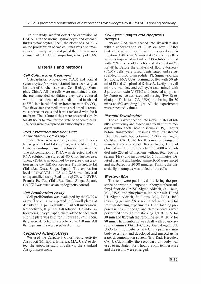
proliferation were observed. The results showed that the overexpression of GACAT3 in NS incre-ased the proliferation of cells and that the knock-down of the expression of GACAT3 inhibited the proliferation in OAS (Figure 2). This indicated that changing the expression of GACAT3 may af-fect the proliferation in synoviocytes.
The Cell Cycle was Arrested and the Apoptosis Rate was Increased in OAS after the Expression of GACAT3 was Knockdown
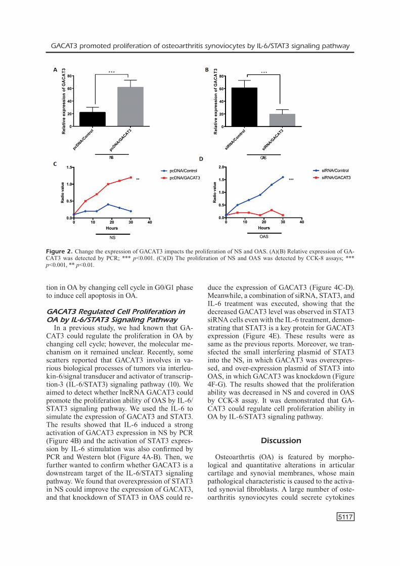
In order to investigate the apoptosis rate in NS and OAS, we used the Caspase-3 to detect the cel-ls and found that the activity of Caspase-3 was decreased after the expression of GACAT3 was overexpressed in NS and that the Caspase-3 was increased after the expression of GACAT3 was knockdown in OAS (Figure 3A-B). The results indicated that the expression of GACAT3 was re-lated to the apoptosis of cells. Furthermore, the cell cycle distribution and apoptosis rate were me-asured in NS and OAS after the expression of GA-CAT3 was altered. Flow cytometry analysis was carried out to study the proliferation mechanism of GACAT3 in OA. These data indicated that the cell percentage in G0/G1 phase was significant-ly decreased and cell percentage in S-phase was significantly increased and reduced cell apop-tosis in NS after GACAT3 was overexpressed. Furthermore, the cell percentage in G0/G1 phase was significantly increased and cell percentage in S-phase was significantly decreased and promp-ted cell apoptosis in OAS after GACAT3 was knockdown (Figure 3C-D). The results demon-strated that GACAT3 regulated the cell prolifera-
Figure 1. GACAT3 was highly expressed in the OAS and promoted the proliferation of cells. (A) The expression of GACAT3 in the NS and OAS was detected by qRT-PCR assay; ***p<0.001. (B) The proliferation of NS and OAS was detected by CCK-8 assays; ***p<0.001.
GACAT3 promoted proliferation of osteoarthritis synoviocytes by IL-6/STAT3 signaling pathway
5117
tion in OA by changing cell cycle in G0/G1 phase to induce cell apoptosis in OA.
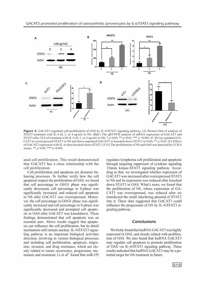
GACAT3 Regulated Cell Proliferation in OA by IL-6/STAT3 Signaling Pathway
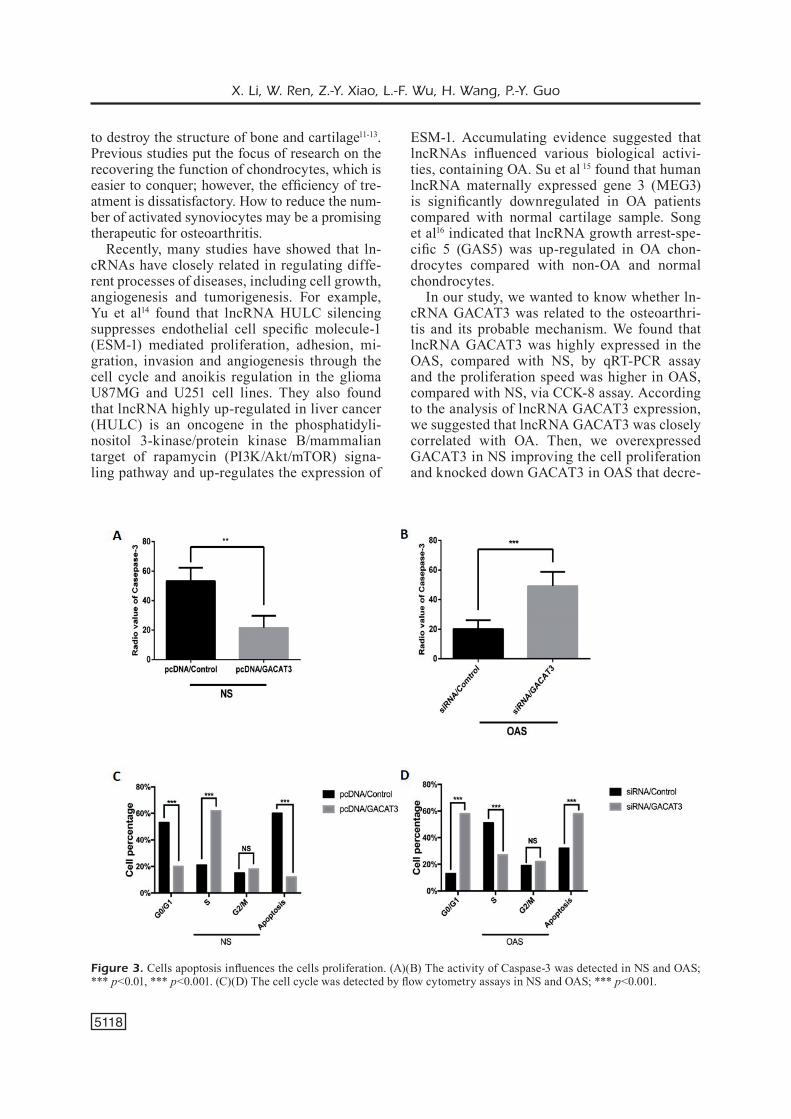
In a previous study, we had known that GA-CAT3 could regulate the proliferation in OA by changing cell cycle; however, the molecular me-chanism on it remained unclear. Recently, some scatters reported that GACAT3 involves in va-rious biological processes of tumors via interleu-kin-6/signal transducer and activator of transcrip-tion-3 (IL-6/STAT3) signaling pathway (10). We aimed to detect whether lncRNA GACAT3 could promote the proliferation ability of OAS by IL-6/STAT3 signaling pathway. We used the IL-6 to simulate the expression of GACAT3 and STAT3. The results showed that IL-6 induced a strong activation of GACAT3 expression in NS by PCR (Figure 4B) and the activation of STAT3 expres-sion by IL-6 stimulation was also confirmed by PCR and Western blot (Figure 4A-B). Then, we further wanted to confirm whether GACAT3 is a downstream target of the IL-6/STAT3 signaling pathway. We found that overexpression of STAT3 in NS could improve the expression of GACAT3, and that knockdown of STAT3 in OAS could re-
duce the expression of GACAT3 (Figure 4C-D). Meanwhile, a combination of siRNA, STAT3, and IL-6 treatment was executed, showing that the decreased GACAT3 level was observed in STAT3 siRNA cells even with the IL-6 treatment, demon-strating that STAT3 is a key protein for GACAT3 expression (Figure 4E). These results were as same as the previous reports. Moreover, we tran-sfected the small interfering plasmid of STAT3 into the NS, in which GACAT3 was overexpres-sed, and over-expression plasmid of STAT3 into OAS, in which GACAT3 was knockdown (Figure 4F-G). The results showed that the proliferation ability was decreased in NS and covered in OAS by CCK-8 assay. It was demonstrated that GA-CAT3 could regulate cell proliferation ability in OA by IL-6/STAT3 signaling pathway.
Discussion
Osteoarthrtis (OA) is featured by morpho-logical and quantitative alterations in articular cartilage and synovial membranes, whose main pathological characteristic is caused to the activa-ted synovial fibroblasts. A large number of oste-oarthritis synoviocytes could secrete cytokines
Figure 2. Change the expression of GACAT3 impacts the proliferation of NS and OAS. (A)(B) Relative expression of GA-CAT3 was detected by PCR; *** p<0.001. (C)(D) The proliferation of NS and OAS was detected by CCK-8 assays; *** p<0.001, ** p<0.01.
X. Li, W. Ren, Z.-Y. Xiao, L.-F. Wu, H. Wang, P.-Y. Guo
5118
to destroy the structure of bone and cartilage11-13. Previous studies put the focus of research on the recovering the function of chondrocytes, which is easier to conquer; however, the efficiency of tre-atment is dissatisfactory. How to reduce the num-ber of activated synoviocytes may be a promising therapeutic for osteoarthritis.
Recently, many studies have showed that ln-cRNAs have closely related in regulating diffe-rent processes of diseases, including cell growth, angiogenesis and tumorigenesis. For example, Yu et al14 found that lncRNA HULC silencing suppresses endothelial cell specific molecule-1 (ESM-1) mediated proliferation, adhesion, mi-gration, invasion and angiogenesis through the cell cycle and anoikis regulation in the glioma U87MG and U251 cell lines. They also found that lncRNA highly up-regulated in liver cancer (HULC) is an oncogene in the phosphatidyli-nositol 3-kinase/protein kinase B/mammalian target of rapamycin (PI3K/Akt/mTOR) signa-ling pathway and up-regulates the expression of
ESM-1. Accumulating evidence suggested that lncRNAs influenced various biological activi-ties, containing OA. Su et al 15 found that human lncRNA maternally expressed gene 3 (MEG3) is significantly downregulated in OA patients compared with normal cartilage sample. Song et al16 indicated that lncRNA growth arrest-spe-cific 5 (GAS5) was up-regulated in OA chon-drocytes compared with non-OA and normal chondrocytes.
In our study, we wanted to know whether ln-cRNA GACAT3 was related to the osteoarthri-tis and its probable mechanism. We found that lncRNA GACAT3 was highly expressed in the OAS, compared with NS, by qRT-PCR assay and the proliferation speed was higher in OAS, compared with NS, via CCK-8 assay. According to the analysis of lncRNA GACAT3 expression, we suggested that lncRNA GACAT3 was closely correlated with OA. Then, we overexpressed GACAT3 in NS improving the cell proliferation and knocked down GACAT3 in OAS that decre-
Figure 3. Cells apoptosis influences the cells proliferation. (A)(B) The activity of Caspase-3 was detected in NS and OAS; *** p<0.01, *** p<0.001. (C)(D) The cell cycle was detected by flow cytometry assays in NS and OAS; *** p<0.001.
GACAT3 promoted proliferation of osteoarthritis synoviocytes by IL-6/STAT3 signaling pathway
5119
ased cell proliferation. This result demonstrated that GACAT3 has a close relationship with the cell proliferation.
Cell proliferation and apoptosis are dynamic ba-lancing processes. To further verify how the cell apoptosis impact the proliferation of OAS, we found that cell percentage in G0/G1 phase was signifi-cantly decreased; cell percentage in S-phase was significantly increased, and reduced cell apoptosis in NS after GACAT3 was overexpressed. Moreo-ver, the cell percentage in G0/G1 phase was signifi-cantly increased and cell percentage in S-phase was significantly decreased and prompted cell apopto-sis in OAS after GACAT3 was knockdown. These findings demonstrated that cell apoptosis was an essential part. Above results suggest that apopto-sis can influence the cell proliferation, but its detail mechanism still remain unclear. IL-6/STAT3 signa-ling pathway is an important biological activities direction, involving in various biological processes and including cell proliferation, apoptosis, migra-tion, invasion, and drug resistance, which are clo-sely related to tumor occurrence, progression, me-tastasis and treatment. Li et al17 found that miR-155
regulates lymphoma cell proliferation and apoptosis through targeting suppressor of cytokine signaling 3/Janus kinase-STAT3 signaling pathway. Accor-ding to that, we investigated whether expression of GACAT3 was increased after overexpressed STAT3 in NS and its expression was reduced after knocked down STAT3 in OAS. What’s more, we found that the proliferation of NS, whose expression of GA-CAT3 was overexpressed, was reduced after we transfected the small interfering plasmid of STAT3 into it. These data suggested that GACAT3 could influence the progression of OA by IL-6/STAT3 si-gnaling pathway.
Conclusions
We firstly found that lncRNA GACAT3 was highly expressed in OAS, and closely related with prolifera-tion of OAS. We also found that lncRNA GACAT3 may regulate cell apoptosis to promote proliferation of OAS via IL-6/STAT3 signaling pathway. These results indicated that lncRNA GACAT3 may be a po-tential target for OA treatment in future.
Figure 4. GACAT3 regulated cell proliferation of OAS by IL-6/STAT3 signaling pathway. (A) Western blot of analysis of STAT3 treatment with IL-6 (0, 2, or 4 ng/ml) in NS. (B)(C) The qRT-PCR analysis of mRNA expression of GACAT3 and STAT3 after 24 h of treatment with IL-6 (0, 2, or 4 ng/ml) in NS; * p<0.05; ** p<0.01; *** p <0.001. (C-D) Up-regulated GA-CAT3 in overexpressed STAT3 in NS and down-regulated GACAT3 in knocked-down STAT3 in OAS; ** p<0.01. (E) Effects of GACAT3 expression with IL-6 after knocked-down STAT3. (F-G) The proliferation of NS and OAS was detected by CCK-8 assays. ** p<0.01; *** p<0.001.
X. Li, W. Ren, Z.-Y. Xiao, L.-F. Wu, H. Wang, P.-Y. Guo
5120
Conflict of InterestThe Authors declare that they have no conflict of interest.
References
1) Moulton SG, Bhatia S, CivitareSe DM, Frank rM, Dean CS, laPraDe rF. Surgical techniques and outcomes of repairing meniscal radial tears: a systematic review. Arthroscopy 2016; 32: 1919-1925.
2) SaCitharan Pk, vinCent tl. Cellular ageing mecha-nisms in osteoarthritis. Mamm Genome 2016; 27: 421-429.
3) Sun S, Bay-JenSen aC, karSDal Ma, SieBuhr aS, ZhenG Q, MakSyMowyCh wP, ChriStianSen tG, henrikSen k. The active form of MMP-3 is a marker of synovial inflammation and cartilage turnover in inflamma-tory joint diseases. BMC Musculoskelet Disord 2014; 15: 93.
4) laMBert C, DuBuC Je, Montell e, verGéS J, Munaut C, noël a, henrotin y. Gene expression pattern of cells from inflamed and normal areas of osteo-arthritis synovial membrane. Arthritis Rheumatol 2014; 66: 960-968.
5) Shi D, lianG l, ZhenG h, Cai G, li X, Xu y, Cai S. Silencing of long non-coding RNA SBDSP1 sup-presses tumor growth and invasion in osteosarco-ma. Biomed Pharmacother 2017; 85: 355-361.
6) Jia l, Sun Z, wu X, MiSteli t, SharMa v. Gene expres-sion analysis upon lncRNA DDSR1 knockdown in human fibroblasts. Genom Data 2015; 6: 277-279.
7) ZhanG G, wu y, Xu D, yan X. Long noncoding RNA UFC1 promotes proliferation of chondrocyte in osteoarthritis by acting as a sponge for miR-34a. DNA Cell Biol 2016; 35: 691-695.
8) li yF, li Sh, liu y, luo yt. Long noncoding RNA CIR promotes chondrocyte extracellular matrix
degradation in osteoarthritis by acting as a spon-ge for mir-27b. Cell Physiol Biochem 2017; 43: 602-610.
9) kanG y, SonG J, kiM D, ahn C, Park S, Chun Ch, Jin eJ. PCGEM1 stimulates proliferation of oste-oarthritic synoviocytes by acting as a sponge for miR-770. J Orthop Res 2016; 34: 412-418.
10) Shen w, yuan y, Zhao M, li J, Xu J, lou G, ZhenG J, Bu S, Guo J, Xi y. Novel long non-coding RNA GACAT3 promotes gastric cancer cell prolifera-tion through the IL-6/STAT3 signaling pathway. Tumour Biol 2016; 37: 14895-14902.
11) ChaGin aS. Effectors of mTOR-autophagy pa-thway: targeting cancer, affecting the skeleton. Curr Opin Pharmacol 2016; 28: 1-7.
12) DiBartola aC, wriGht BM, MaGnuSSen ra, FlaniGan DC. Clinical outcomes after autologous chon-drocyte implantation in adolescents’ knees: a systematic review. Arthroscopy 2016; 32: 1905-1916.
13) MalFait aM. Osteoarthritis year in review 2015: biology. Osteoarthritis Cartilage 2016; 24: 21-26.
14) yu Z, ZhanG X, Qi l, Cai y, yanG P, Xuan G, JianG y. HULC long noncoding RNA silencing suppresses angiogenesis by regulating ESM-1 via the PI3K/Akt/mTOR signaling pathway in human gliomas. Oncotarget 2016; 7: 14429-14440.
15) Su w, Xie w, ShanG Q, Su B. The long noncoding RNA MEG3 is downregulated and inversely asso-ciated with VEGF levels in osteoarthritis. Biomed Res Int 2015; 2015: 356893.
16) SonG J, ahn C, Chun Ch, Jin eJ. A long non-coding RNA, GAS5, plays a critical role in the regulation of miR-21 during osteoarthritis. J Orthop Res 2014; 32: 1628-1635.
17) li XD, li XM, Gu Jw, Sun XC. MiR-155 regula-tes lymphoma cell proliferation and apoptosis through targeting SOCS3/JAK-STAT3 signaling pathway. Eur Rev Med Pharmacol Sci 2017; 21: 5153-5159.