fungus part i
TRANSCRIPT
MYCOLOGY IPRAVEG GUPTA
INTRODUCTION• Eukaryotes• Don’t possess chlorophyll• Unicellular/multicellular• Mostly soil saprophytes• Role in degradation of organic compounds• About 250000 fungal species are identified.• Out of them only about 150-200 are known to cause
human infections. • Fungi cause infections in debilitated patients, eg.
Immunocompromised patients like AIDS patients. • Useful fungi: edible mushrooms, yeasts used in
fermentation, fungi producing antibiotics (eg penicillium).
STRUCTURE
• Fungal cells are eukaryotic cells containing cell wall, cell membrane, true nuclei, nuclear membrane, mitochondria, vaculoes, reticular endothelium, ribosomes etc like other eukaryotic cells.
• Cell division – sexual/asexual
• Cell wall – made up of chitin, glucans, mannans and complex polysaccharides.
• Cell membrane contains ergosterol in contrast to mammalian cells which contain cholesterol.
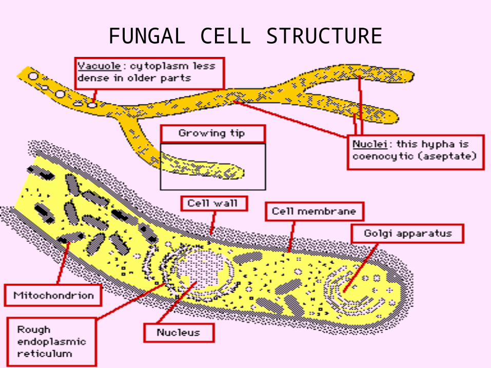
FUNGAL CELL STRUCTURE
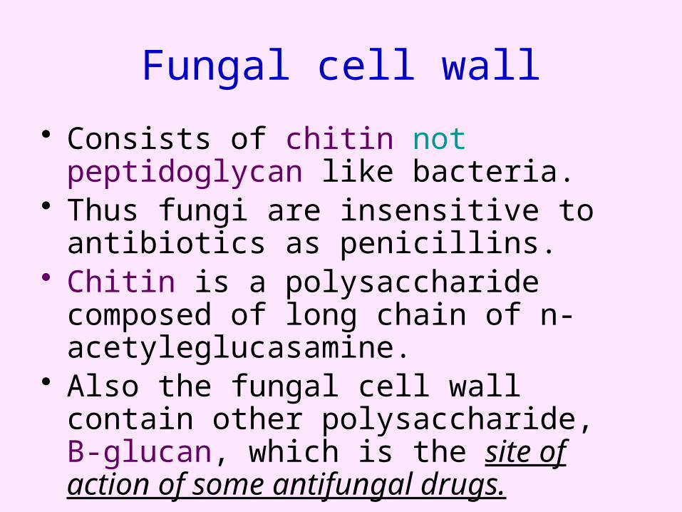
Fungal cell wall
• Consists of chitin not peptidoglycan like bacteria.
• Thus fungi are insensitive to antibiotics as penicillins.
• Chitin is a polysaccharide composed of long chain of n-acetyleglucasamine.
• Also the fungal cell wall contain other polysaccharide, B-glucan, which is the site of action of some antifungal drugs.
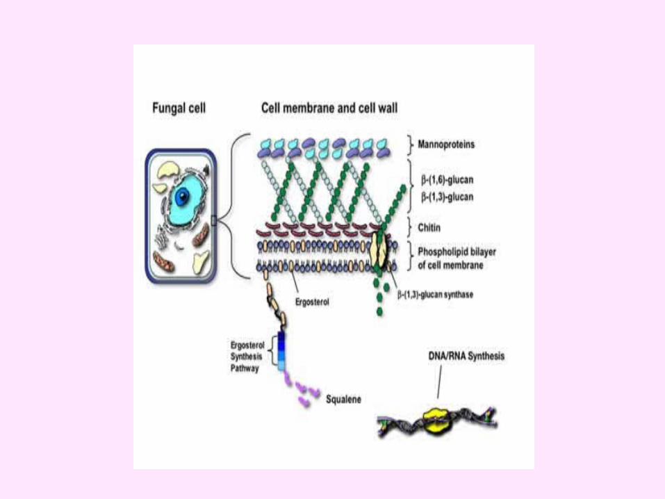
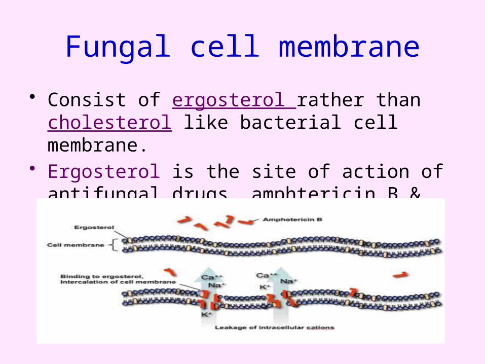
Fungal cell membrane
• Consist of ergosterol rather than cholesterol like bacterial cell membrane.
• Ergosterol is the site of action of antifungal drugs, amphtericin B & azole group
Atmospheric & carbon source requirements
• Most fungi are obligatory aerobes, some are facultative anaerobes, but none are obligatory anaerobes.
• All fungi require a performed organic source of carbon –association with decaying matter.
• Natural habitat• The environment.
Opportunistic Fungi In addition to those species which are
generally recognized as pathogenic to man it is firmly established that under unusual circumstances of abnormal susceptibility of patient, or the traumatic implantation of the fungus, other fungi are capable of causing lesions. Those are called (Opportunistic Fungi.)

These circumstances may be :
1. A debilitating condition of the host, as Diabetes.
2. A concurrent disease such as leukaemia.3. Prolonged treatment with corticosteroids. 4.Immunosuppressive drugs or an antibiotic
for long duration.
CLASSIFICATION
Taxonomical classification:• Phylum class • Zygomycota zygomycetes• Ascomycota ascomycetes • Basidiomycota basidiomycetes• Deuteromycota deuteromycetes (fungi
imperfecti)
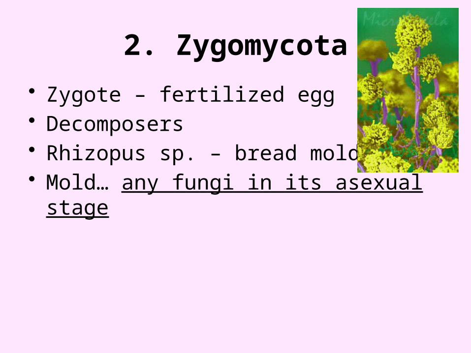
2. Zygomycota
• Zygote – fertilized egg• Decomposers • Rhizopus sp. – bread mold• Mold… any fungi in its asexual stage
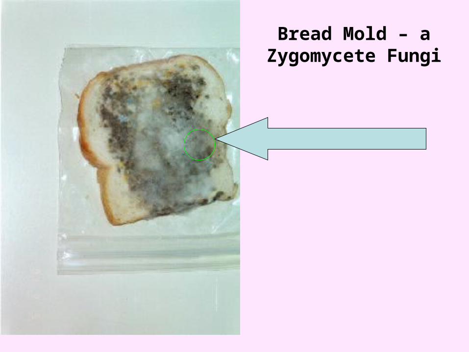
Bread Mold – a Zygomycete Fungi
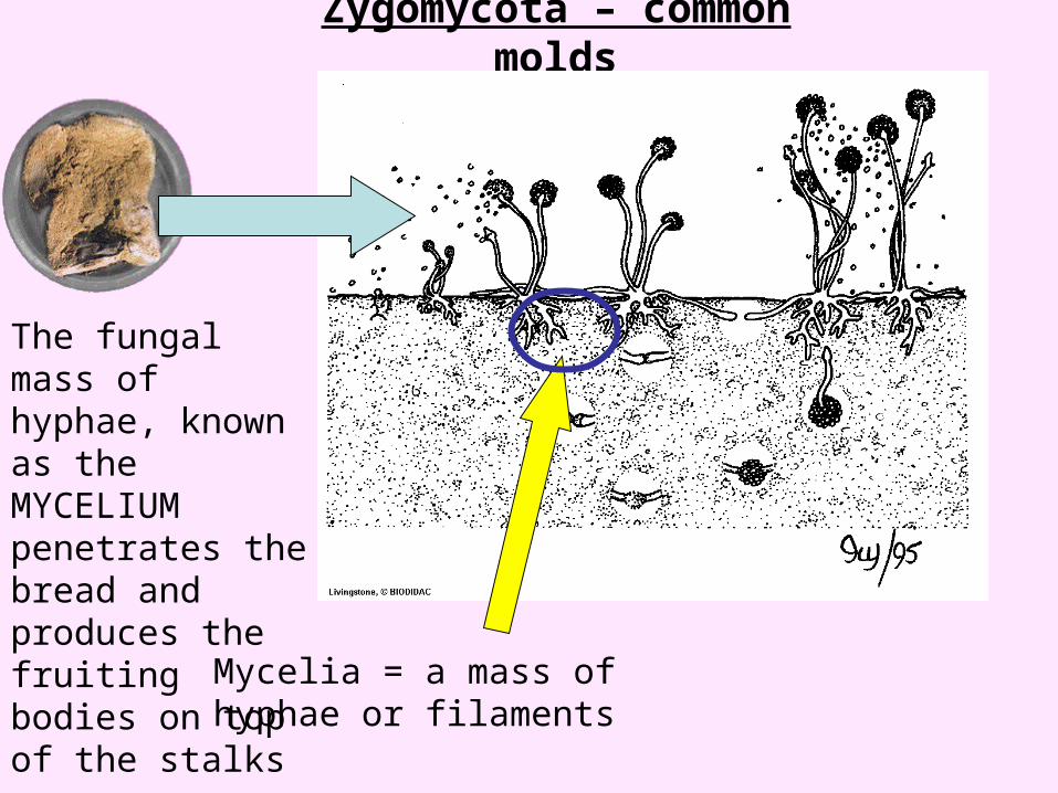
Zygomycota – common molds
The fungal mass of hyphae, known as the MYCELIUM penetrates the bread and produces the fruiting bodies on top of the stalks
Mycelia = a mass of hyphae or filaments

3. Ascomycota
• Cup shaped reproductive structures• Yeast – unicellular exception
Morel Fruiting Bodies
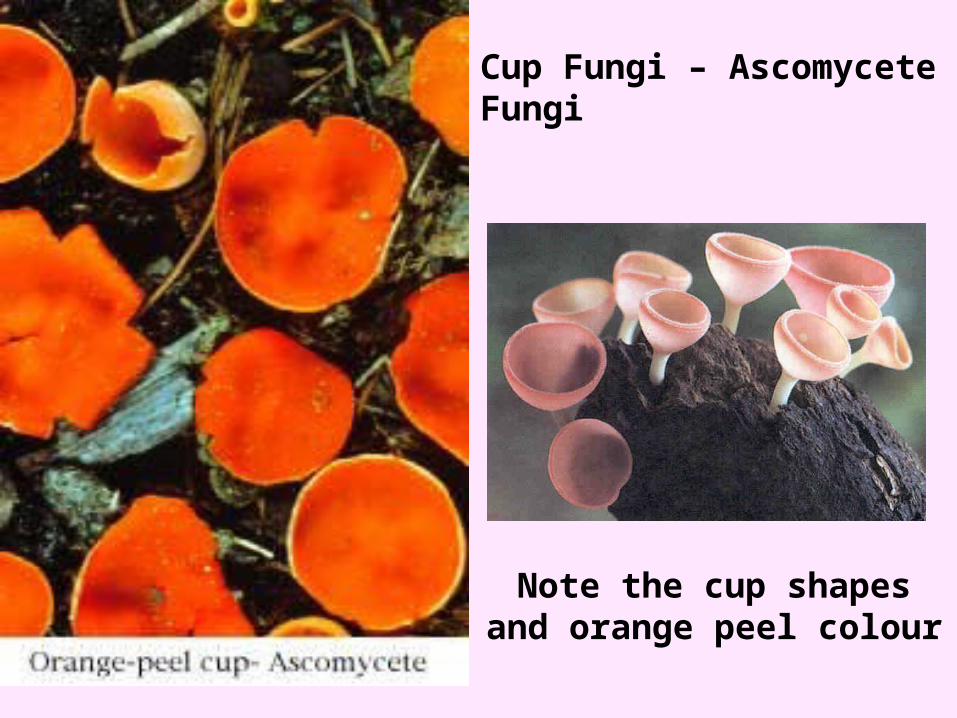
Cup Fungi – Ascomycete Fungi
Note the cup shapes and orange peel colour
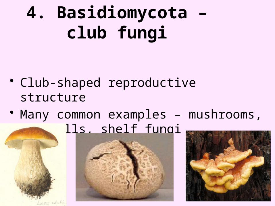
4. Basidiomycota – club fungi
• Club-shaped reproductive structure• Many common examples – mushrooms,
puffballs, shelf fungi
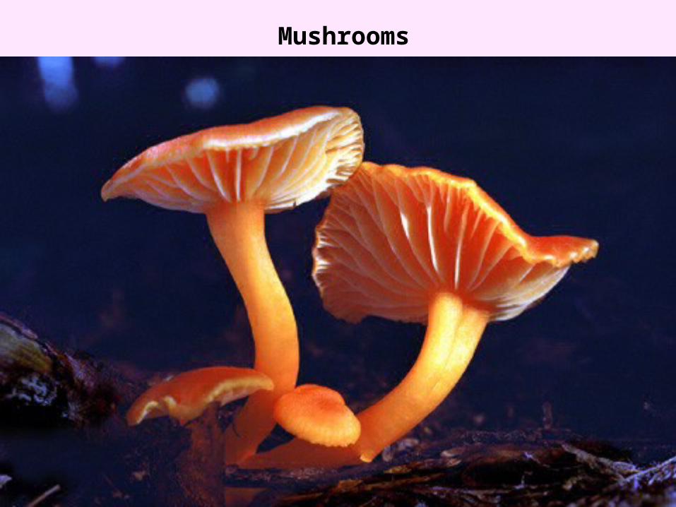
Mushrooms
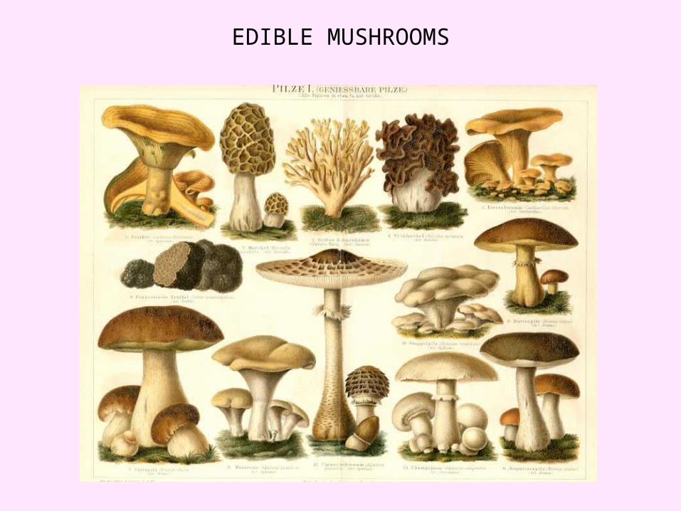
EDIBLE MUSHROOMS
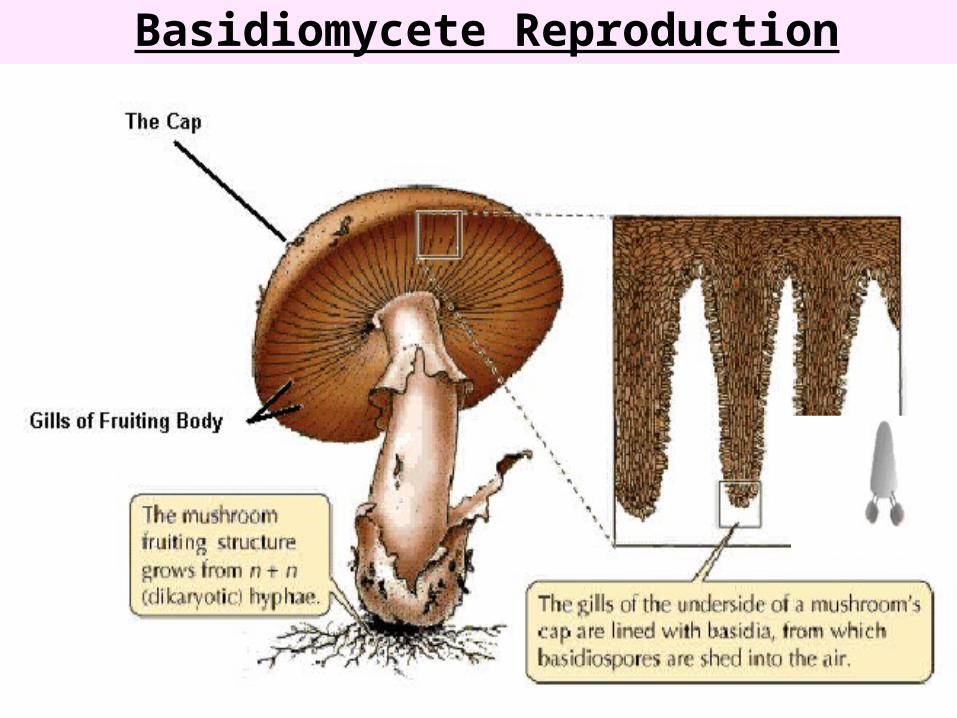
Basidiomycete Reproduction
-Regarded as imperfect because no sexual stage has been observed in their life cycle
-Members are not closely related and are not necessarily similar in structure or appearance; do not share a common ancestry
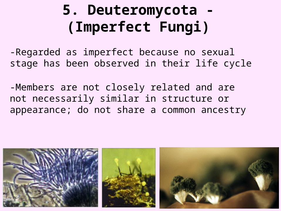
5. Deuteromycota - (Imperfect Fungi)

Morphological classification:1. Yeasts2. Yeast like fungi3. Moulds/ filamentous fungi/ mycelial fungi4. Dimorphic fungi
Description: Yeasts are round to oval unicellular fungi which
reproduce by budding or binary fission eg cryptococcus. Yeasts like fungi – some yeasts grow partly as yeasts
and partly as chains of elongated budding cells joined end to end forming pseudohyphae and pseudomycelium eg candida.

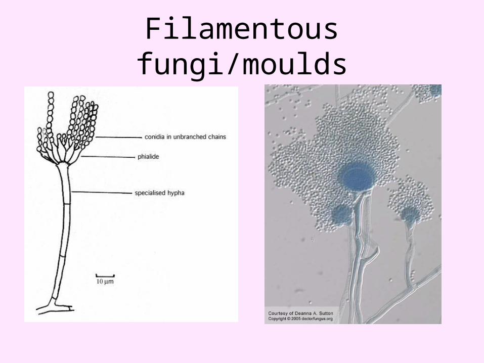
Filamentous fungi/moulds: • They grow as multicellular branching filaments.• Hypha = each filament is called hypha.• Mycelia = tangled masses of hyphae are known as
mycelia.• Thallus = body of fungus.• Septa = transverse walls dividing hyphae at irregular
intervals.• The septate hyphae are morphologically coenocytic
because septae have holes through which free flow of nuclei and other cytoplasmic material can occur.
• Aerial mycelium = the part of mycelium projecting above the culture medium.
• Vegetative mycelium = the part of mycelium growing in the culture medium.
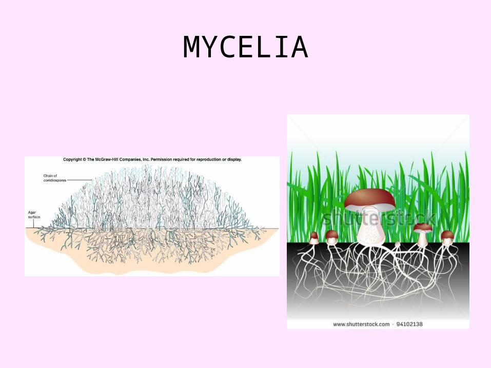
MYCELIA
• There is abundant formation of spores on aerial mycelium that affects their airborne transmission.
• Eg dermatophytes, aspergillus, zygomycetes, penicillium.
Dimorphic fungi:• They exist as both yeast form and filamentous form
depending on conditions of growth.• Yeast form (parasitic phase) occurs in host tissues and in
cultures at 37°C.• Filamentous form (saprophytic phase) occurs in soil and
in cultures at 22-25°C.• Eg fungi responsible for systemic mycoses like
histoplasma, blastomyces, coccidiodes, paracoccidiodes.

Fungal structures
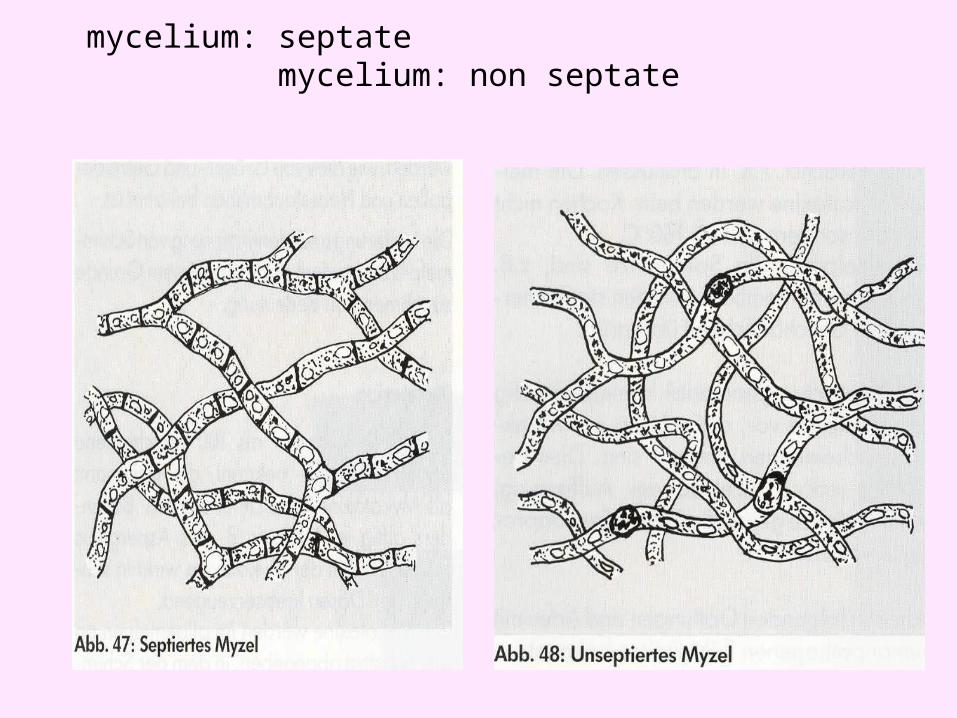
mycelium: septate mycelium: non septate
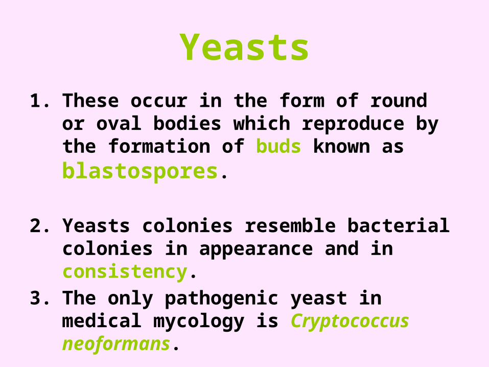
Yeasts1. These occur in the form of round or oval
bodies which reproduce by the formation of buds known as blastospores.
2. Yeasts colonies resemble bacterial colonies in appearance and in consistency.
3. The only pathogenic yeast in medical mycology is Cryptococcus neoformans.
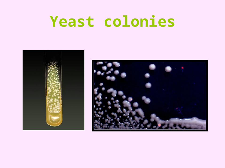
Yeast colonies
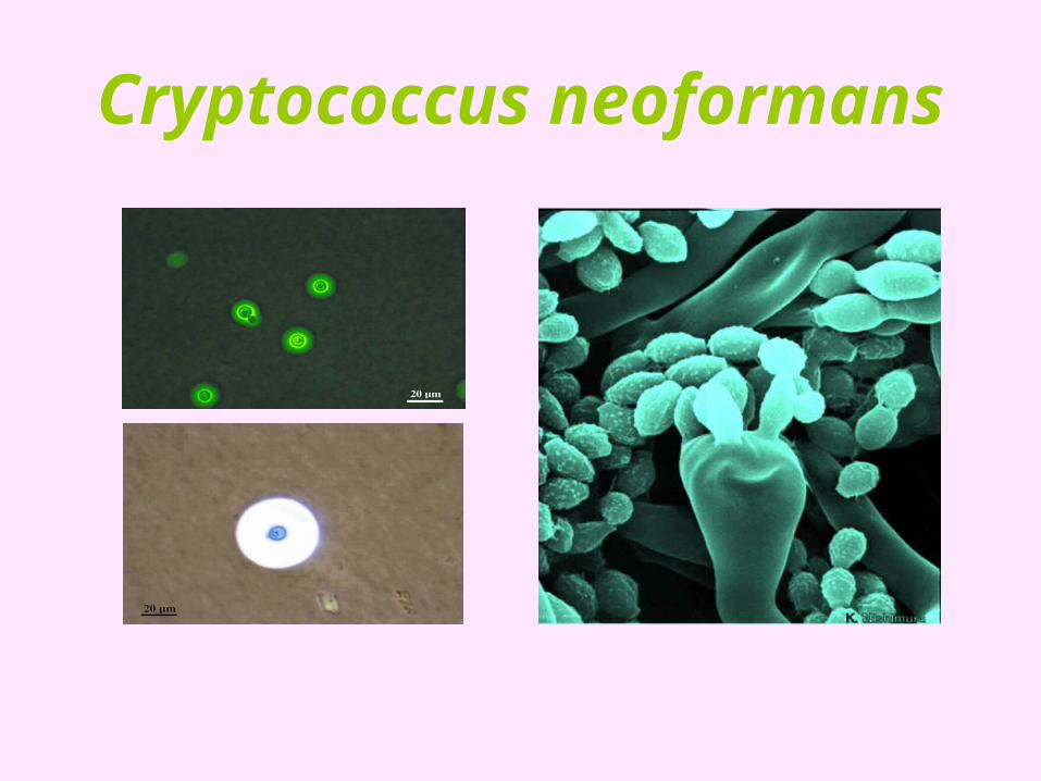
Cryptococcus neoformans
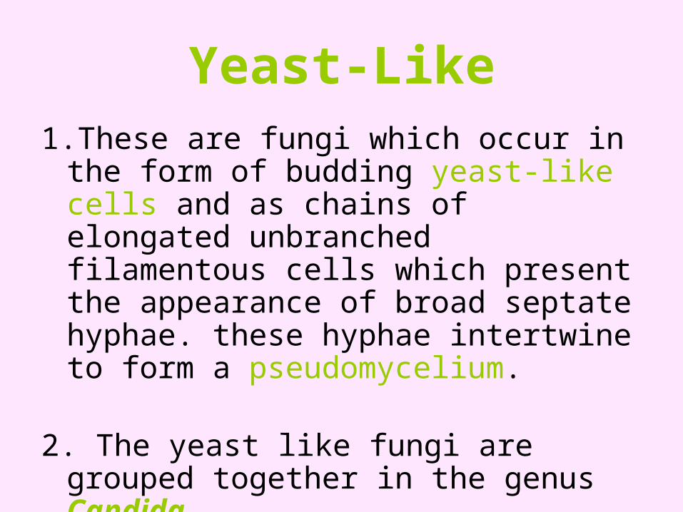
Yeast-Like1.These are fungi which occur in the form of
budding yeast-like cells and as chains of elongated unbranched filamentous cells which present the appearance of broad septate hyphae. these hyphae intertwine to form a pseudomycelium.
2. The yeast like fungi are grouped together in the genus Candida.
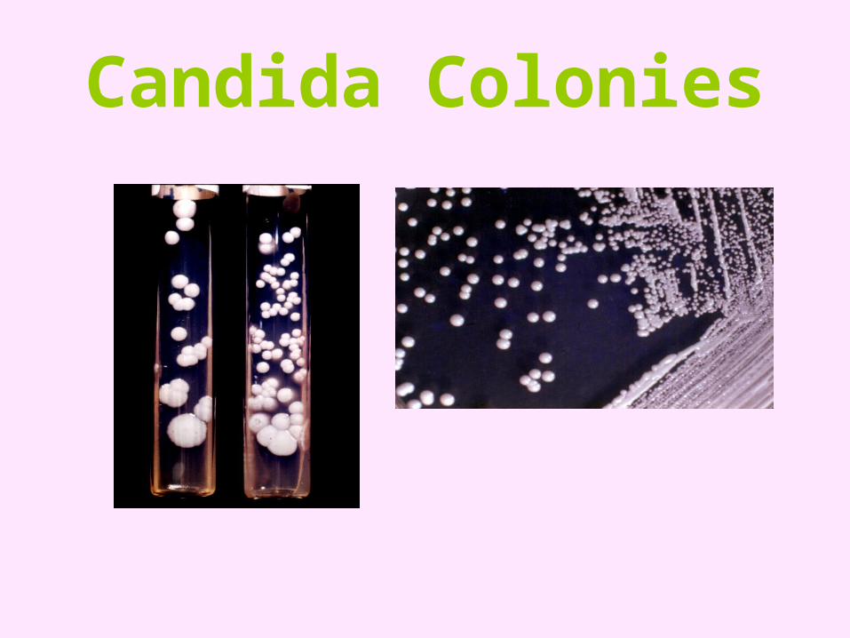
Candida Colonies
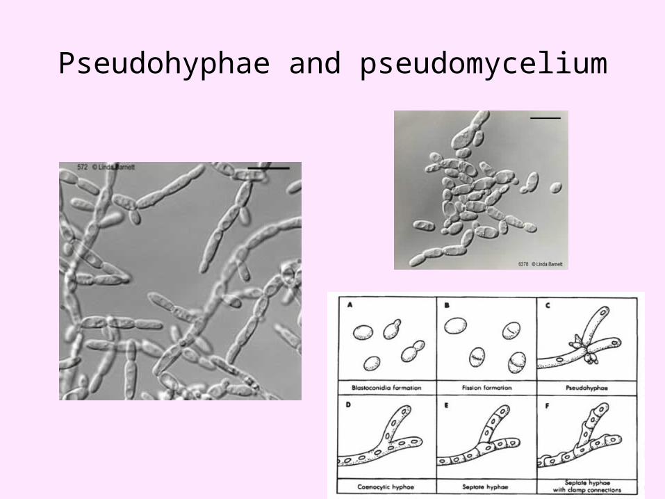
Pseudohyphae and pseudomycelium
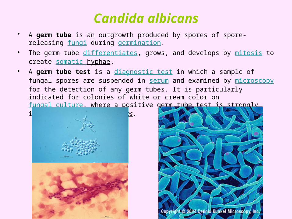
Candida albicans• A germ tube is an outgrowth produced by spores of spore-releasing fungi during
germination.• The germ tube differentiates, grows, and develops by mitosis to create somatic
hyphae.• A germ tube test is a diagnostic test in which a sample of fungal spores are
suspended in serum and examined by microscopy for the detection of any germ tubes. It is particularly indicated for colonies of white or cream color on fungal culture, where a positive germ tube test is strongly indicative of Candida albicans.
Filamentous fungi/moulds
Thermally Dimorphic Fungi
These are fungi which exhibit a filamentous mycelial morphology (saprophytic phase) when grown at room temperature 27oC, but have a typical yeast morphology (parasitic phase) inside the body and when grown at 37oC in the laboratory (e.g. Histoplasmosis).
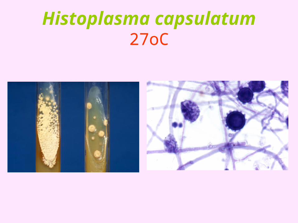
Histoplasma capsulatum 27oC
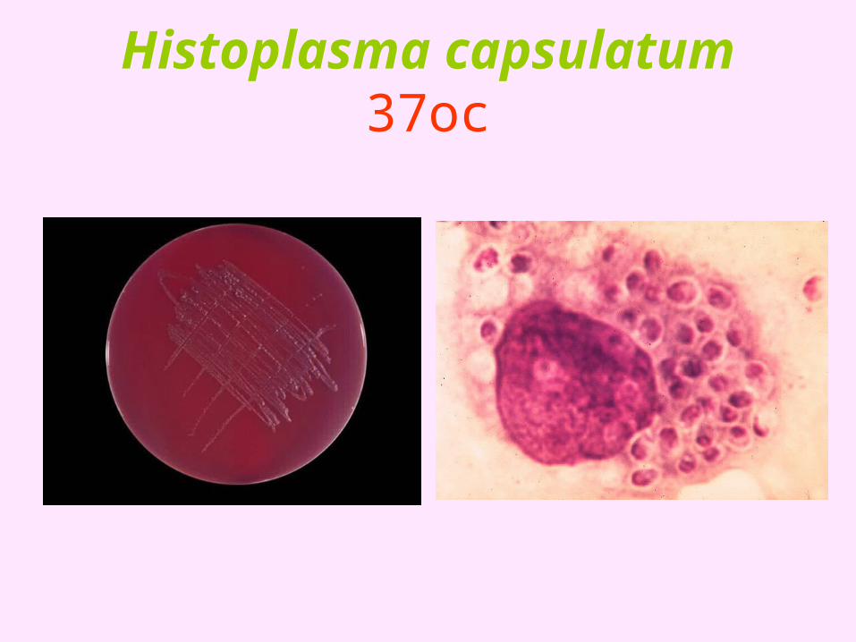
Histoplasma capsulatum 37oc
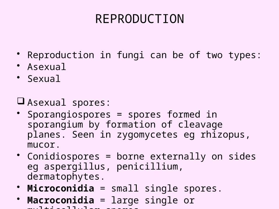
REPRODUCTION
• Reproduction in fungi can be of two types:• Asexual• Sexual
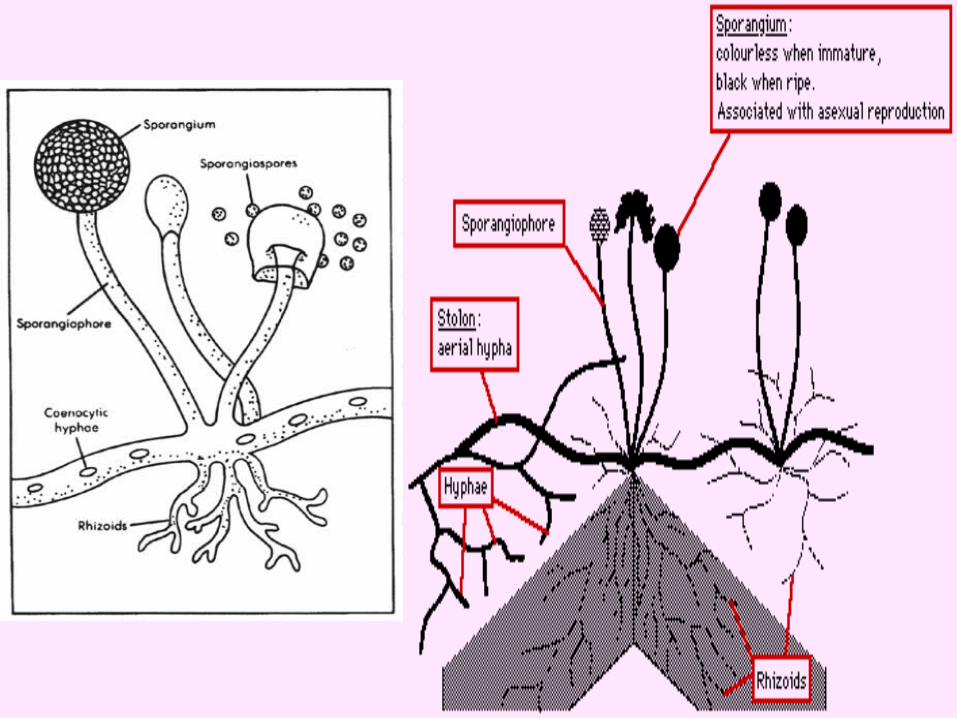
Asexual spores:• Sporangiospores = spores formed in sporangium by
formation of cleavage planes. Seen in zygomycetes eg rhizopus, mucor.
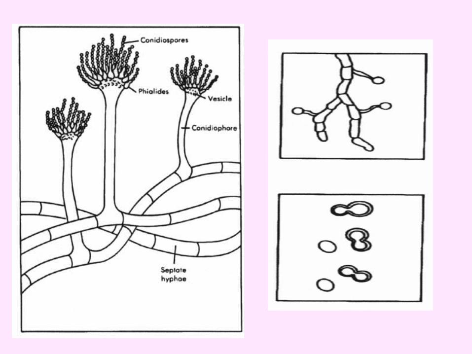
• Conidiospores = borne externally on sides eg aspergillus, penicillium, dermatophytes.
• Microconidia = small single spores.• Macroconidia = large single or multicellular spores.
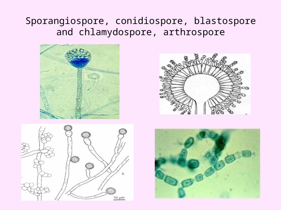
Sporangiospore, conidiospore, blastospore and chlamydospore, arthrospore
• Sexual spores: four types have been identified• Oospores• Zygospores• Ascospores• Basidiospores
• Other vegetative spores: • Blastospores – a fungal spore that arises by budding.• Arthrospores - one of a number of spores of various fungi and
certain blue-green algae, united in the form of a string of beads, formed by fission.
• Chlamydospores - a thick-walled intercalary or terminal asexual fungal spore formed by the rounding-up of a cell; it is not shed. Formed by differentiation of hyphae; seen in Candida and Histoplasma spp.
• Phialospores - A type of conidium found, for example, in many of the Eurotiales and Hypocreales. Phialospores develop at the tips of specialized finger-like cells termed phialides.
FUNGI IMPERFECTI (DEUTEROMYCETES)
• All those fungi whose sexual or perfect state is not known.
• They form septate hyphae and asexual spores or no spores.
• Most fungi causing human infections belong to this group.
INFECTION Fungal infections are of 4 types based on target tissue:• Superficial mycoses = surface infections limited to
outermost layers of skin and hair. • Cutaneous mycoses = fungal infections extending
deeper into the epidermis and its integuments. • Subcutaneous mycoses = infections involving dermis,
subcutaneous tissue, muscles and fascia. • Systemic mycoses = infections originating primarily in
the lungs ( acquired by inhalation) and spreading to other organs.
• Opportunistic mycoses = besides the above four mentioned types, this category includes infections in which fungi of no significance or low virulence infect humans with compromised immune system.
LABORATORY DIAGNOSIS (main points)
Specimens: • Skin scrapings, nail clippings, hairs• Scrapings from mucous membrane• Scrapings, crusts, aspirated pus, tissue biopsy.• Blood, CSF etc in systemic mycoses.
Microscopy:• KOH mount – KOH dissolves keratin and cellular
material but does not affect fungi. Specimen is placed on a slide, a drop of 10-20% KOH is added and covered with a coverslip, left for 20 min in incubator at 37°C to digest keratin. Then examined microscopically.
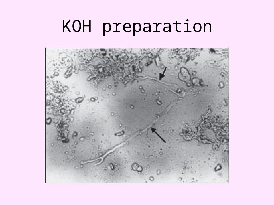
KOH preparation
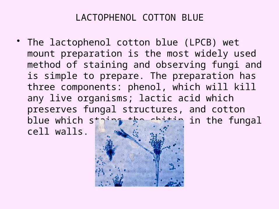
LACTOPHENOL COTTON BLUE
• The lactophenol cotton blue (LPCB) wet mount preparation is the most widely used method of staining and observing fungi and is simple to prepare. The preparation has three components: phenol, which will kill any live organisms; lactic acid which preserves fungal structures, and cotton blue which stains the chitin in the fungal cell walls.
• Stains: gram stain, papanicoulau stain, periodic acid schiff stain (PAS), methenamine sliver stain, giemsa stain etc.
• Direct immunofluorescence test
• Histology
• Antigen detection tests eg cryptococcal antigen in CSF.
Culture:• Sabouraud’s dextrose agar is commonly used for fungal
culture.• pH =5.6 does not allow bacterial growth.
• Drugs like chloramphenicol, cyclohexamide and other antibiotics are added to prevent bacterial or saprophytic fungal infection.
• Cultures are incubated at two temperatures:• One tube at 25°C (room temperature)• One tube at 37°C (incubator).• This helps reveal fungal dimorphism.
• Cultures are incubated for atleast 2-3 weeks and in some cases upto 6 weeks.
• Cultures are examined macroscopically for colony morphology, and microscopically for fungal morphology.
• Czapek-Dox agar• Cornmeal agar

THANK YOU