function and dysfunction of prefrontal brain circuitry in ... · frontal brain circuitry....
TRANSCRIPT
REVIEW
Function and Dysfunction of Prefrontal Brain Circuitryin Alcoholic Korsakoff’s Syndrome
Marlene Oscar-Berman
Received: 6 February 2012 /Accepted: 4 April 2012# Springer Science+Business Media, LLC (outside the USA) 2012
Abstract The signature symptom of alcohol-induced persist-ing amnestic disorder, more commonly referred to as alcoholicKorsakoff’s syndrome (KS), is anterograde amnesia, or mem-ory loss for recent events, and until the mid 20th Century, theputative brain damage was considered to be in diencephalicand medial temporal lobe structures. Overall intelligence, asmeasured by standardized IQ tests, usually remains intact.Preservation of IQ occurs because memories formed beforethe onset of prolonged heavy drinking—the types of informa-tion and abilities tapped by intelligence tests—remain relative-ly well preserved compared with memories recently acquired.However, clinical and experimental evidence has shown thatneurobehavioral dysfunction in alcoholic patients with KSdoes include nonmnemonic abilities, and further brain damageinvolves extensive frontal and limbic circuitries. Among theabnormalities are confabulation, disruption of elements of
executive functioning and cognitive control, and emotionalimpairments. Here, we discuss the relationship between neuro-behavioral impairments in KS and alcoholism-related braindamage. More specifically, we examine the role of damage toprefrontal brain systems in the neuropsychological profile ofalcoholic KS.
Keywords Alcoholism . Korsakoff’s syndrome . Frontalbrain circuitry . Executive functions . Emotion
Introduction
In vivo neuroimaging studies and postmortem analyses ofthe brains of alcoholics have revealed abnormalities associ-ated with prolonged and extensive alcohol consumption.Likewise, results of neuropsychological tests sensitive toimpaired mental processes after damage to particular brainsystems have disclosed abnormalities in cognitive abilitiesand emotional functioning in chronic alcoholics (Oscar-Berman and Marinkovic 2007; Chanraud et al. 2010). Oneof the striking and tragic legacies of decades of chronicalcoholism, often accompanied by poor nutrition, is thesevere neuropsychiatric condition, alcohol-induced persist-ing amnestic disorder (American Psychiatric Association2000), commonly known as alcoholic Korsakoff’s syn-drome (KS). Although the condition most often is associatedwith protracted alcohol abuse and concomitant thiamine(vitamin B1) deficiency, other ailments such as prolongedgastrointestinal disturbances can deplete thiamine and leadto KS (Charness 2010; Oscar-Berman and Evert 1997).Additional rare causes of KS have been reported in theabsence of thiamine deficiency: intraventricular hemor-rhage; thalamic infarction; T-cell lymphoma; Creutzfeldt-Jakob disease; and multiple sclerosis (see Oscar-Berman
M. Oscar-BermanDepartment of Neurology and Division of Psychiatry,Boston University School of Medicine,Boston, MA, USA
M. Oscar-BermanDepartment of Anatomy & Neurobiology,Boston University School of Medicine,Boston, MA, USA
M. Oscar-BermanPsychology Research Service,Department of Veterans Affairs Medical Center,Boston, MA, USA
M. Oscar-Berman (*)Laboratory of Neuropsychology, L-815,Boston University School of Medicine,72 East Concord Street,Boston, MA 02118, USAe-mail: [email protected]
Neuropsychol RevDOI 10.1007/s11065-012-9198-x

and Evert 1997). The focus of the present review, however,is alcoholic KS.
Alcoholic KS usually is preceded by Wernicke’s enceph-alopathy, an acute, transient stage of neurological symptomsthat include confusion, impairments of consciousness, diffi-culties moving eye muscles, and problems with gross mus-cle control. With thiamine treatment, good nutrition, andalcohol abstinence, Wernicke’s encephalopathy can resolve;it tends to develop into the more stable and chronic condi-tion of alcohol-induced persisting amnestic disorder, i.e.,alcoholic KS (Sechi and Serra 2007; Victor 1994; Zahr etal. 2011). However, many authors continue to refer to alco-holic KS as Wernicke-Korsakoff syndrome. Caine et al.(1997) suggested an operational definition of Wernicke-Korsakoff syndrome. This definition includes unremittinganterograde amnesia and “disorientation in the absence of anacute confusional state,” and two of the following: dietarydeficiency, occulomotor abnormalities, cerebellar dysfunc-tion and “either an altered mental state or mild memoryimpairment” (page 54). Note that this terminology describesa broad combination of symptoms of the acute clinicalpresentation of Wernicke’s encephalopathy together withthe subsequent chronic and stable disorder of alcohol-induced persisting amnestic disorder. In the present review,our primary focus is on the permanent KS condition, unlessotherwise indicated.
Alcoholic KS is characterized most dramatically by am-nesia, but the amnesia does not encompass all memoriesequally. Instead, memory loss for recent events (anterogradeamnesia) is considerably more severe than loss of memoriesfor information learned prior to the onset of alcoholism(retrograde amnesia). Thus, many early memories are intact,and general intelligence scores remain within the normalrange (Oscar-Berman et al. 1993). For decades subsequentto Korsakoff’s initial description of the disorder named afterhim (Victor and Yakovlev 1955), anterograde amnesia wasthe dominant focus of clinical assessment and researchinquiry, and damage within Papez (1937) circuit, especiallydiencephalic damage, was thought to be the source of theamnesia (Brion 1969; Talland 1965; Victor et al. 1971). Toaccount for additional cognitive and emotional symptomsobserved in KS patients, several writers included corticaldamage as well (Barbizet 1963; Talland 1965), althoughthere was no consensus on the locus. Investigators stressedthe idea that the diencephalic damage in KS patients wascaused by thiamine deficiency, and patients with acute,alcoholic Wernicke’s encephalopathy who do not receivethiamine treatment, have shown evidence of hemorrhagiclesions within the region around the diencephalon (Kashi etal. 2009). However, other researchers suggested that thecortical abnormalities were caused by alcohol neurotoxicityor by other conditions associated with alcoholism (e.g., liverdisease or head trauma) (Charness 2010).
There still are conflicting opinions about the pathogen-esis of KS, with much of the debate revolving around therelative contributions of nutritional deficiency and alcoholneurotoxicity (Ribeiro and Pereira 2010). In any case, itshould be noted that Korsakoff’s own account of theneuropathology of the syndrome also included damageto large regions of the brain, including cerebral cortexand structures deep within the brain; however precisecortical and subcortical loci were not detailed (Victorand Yakovlev 1955). Rather, Korsakoff described mainlythe neuropsychological characteristics that he observed inassociation with various etiologies. In fact, he reportedvirtually all of the important cognitive and emotionalimpairments that might accompany or contribute to theanterograde amnesia (Banks 1996), some of which remainunderstudied to this day. The diencephalic brain damagethat has come to be implicated in the amnesia of KS wasdetailed much later by investigators such as Brion (1969)and Victor et al. (1971, 1989) who examined postmortempathology but used differing neuropsychological criteriato classify their patients. To date, controversy remainsregarding the critical lesion sites (Harper 2009; Visser etal. 1999). Therefore, the idea that the syndrome can beconfirmed with postmortem neuropathological evidencealone without precise knowledge of ante mortem neuro-psychological symptomatology and a clinical picture, maybe misleading when attempting to reconcile differingaccounts of the neuropathology. A possible solution wasproposed by Caine et al. (1997), who suggested an oper-ational definition of KS, that is, unremitting anterogradeamnesia, a clear sensorium, and two of the following:dietary deficiency, occulomotor abnormalities, and cere-bellar dysfunction.
Nevertheless, with increased sensitivity of neurobeha-vioral assessment tools and the availability of functionaland structural brain imaging techniques, the true complex-ity of the disorder is being revealed. In addition to ante-rograde amnesia, other domains of cognitive impairmenthave been described. These include deficits in attention,abstract thinking, cognitive flexibility, and behavior reg-ulation (Oscar-Berman and Evert 1997). Additionally, KSpatients often appear to have diminished volition, theyseem emotionally apathetic, and they give an impressionof being unaware of their disabilities (Talland 1965;Victor and Yakovlev 1955; Warner 1934). Thus, the com-bined picture clearly began to point to dysfunctionalfrontal brain circuitry. Consequently, systematic studyensued, and now frontal-system dysfunction is recognizedas a primary contributor to the behavioral characteristicsof the disorder. In this review, we first describe the pre-frontal neuroanatomical networks that are vulnerable inalcoholic KS. We then summarize key domains of asso-ciated neurobehavioral impairment.
Neuropsychol Rev
Neuroanatomical Circuitry of Prefrontal Brain Systems
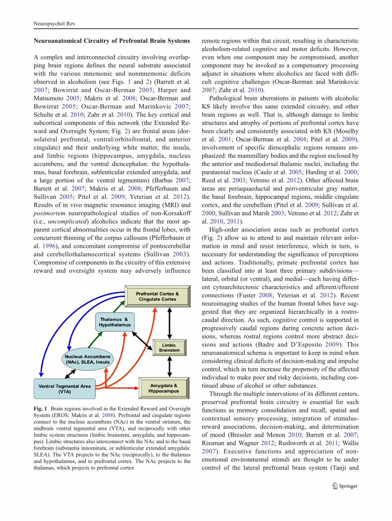
A complex and interconnected circuitry involving overlap-ping brain regions defines the neural substrate associatedwith the various mnemonic and nonmnemonic deficitsobserved in alcoholism (see Figs. 1 and 2) (Barrett et al.2007; Bowirrat and Oscar-Berman 2005; Harper andMatsumoto 2005; Makris et al. 2008; Oscar-Berman andBowirrat 2005; Oscar-Berman and Marinkovic 2007;Schulte et al. 2010; Zahr et al. 2010). The key cortical andsubcortical components of this network (the Extended Re-ward and Oversight System; Fig. 2) are frontal areas (dor-solateral prefrontal, ventral/orbitofrontal, and anteriorcingulate) and their underlying white matter, the insula,and limbic regions (hippocampus, amygdala, nucleusaccumbens, and the ventral diencephalon: the hypothala-mus, basal forebrain, sublenticular extended amygdala, anda large portion of the ventral tegmentum) (Barbas 2007;Barrett et al. 2007; Makris et al. 2008; Pfefferbaum andSullivan 2005; Pitel et al. 2009; Yeterian et al. 2012).Results of in vivo magnetic resonance imaging (MRI) andpostmortem neuropathological studies of non-Korsakoff(i.e., uncomplicated) alcoholics indicate that the most ap-parent cortical abnormalities occur in the frontal lobes, withconcurrent thinning of the corpus callosum (Pfefferbaum etal. 1996), and concomitant compromise of pontocerebellarand cerebellothalamocortical systems (Sullivan 2003).Compromise of components in the circuitry of this extensivereward and oversight system may adversely influence
remote regions within that circuit, resulting in characteristicalcoholism-related cognitive and motor deficits. However,even when one component may be compromised, anothercomponent may be invoked as a compensatory processingadjunct in situations where alcoholics are faced with diffi-cult cognitive challenges (Oscar-Berman and Marinkovic2007; Zahr et al. 2010).
Pathological brain aberrations in patients with alcoholicKS likely involve this same extended circuitry, and otherbrain regions as well. That is, although damage to limbicstructures and atrophy of portions of prefrontal cortex havebeen clearly and consistently associated with KS (Moselhyet al. 2001; Oscar-Berman et al. 2004; Pitel et al. 2009),involvement of specific diencephalic regions remains em-phasized: the mammillary bodies and the region enclosed bythe anterior and mediodorsal thalamic nuclei, including theparataenial nucleus (Caulo et al. 2005; Harding et al. 2000;Reed et al. 2003; Vetreno et al. 2012). Other affected brainareas are periaquaeductal and periventricular gray matter,the basal forebrain, hippocampal regions, middle cingulatecortex, and the cerebellum (Pitel et al. 2009; Sullivan et al.2000; Sullivan and Marsh 2003; Vetreno et al. 2012; Zahr etal. 2010, 2011).
High-order association areas such as prefrontal cortex(Fig. 2) allow us to attend to and maintain relevant infor-mation in mind and resist interference, which in turn, isnecessary for understanding the significance of perceptionsand actions. Traditionally, primate prefrontal cortex hasbeen classified into at least three primary subdivisions—lateral, orbital (or ventral), and medial—each having differ-ent cytoarchitectonic characteristics and afferent/efferentconnections (Fuster 2008; Yeterian et al. 2012). Recentneuroimaging studies of the human frontal lobes have sug-gested that they are organized hierarchically in a rostro-caudal direction. As such, cognitive control is supported inprogressively caudal regions during concrete action deci-sions, whereas rostral regions control more abstract deci-sions and actions (Badre and D’Esposito 2009). Thisneuroanatomical schema is important to keep in mind whenconsidering clinical deficits of decision-making and impulsecontrol, which in turn increase the propensity of the affectedindividual to make poor and risky decisions, including con-tinued abuse of alcohol or other substances.
Through the multiple innervations of its different centers,preserved prefrontal brain circuitry is essential for suchfunctions as memory consolidation and recall, spatial andcontextual sensory processing, integration of stimulus-reward associations, decision-making, and determinationof mood (Bressler and Menon 2010; Barrett et al. 2007;Rissman and Wagner 2012; Rushworth et al. 2011; Wallis2007). Executive functions and appreciation of non-emotional environmental stimuli are thought to be undercontrol of the lateral prefrontal brain system (Tanji and
Fig. 1 Brain regions involved in the Extended Reward and OversightSystem (EROS; Makris et al. 2008). Prefrontal and cingulate regionsconnect to the nucleus accumbens (NAc) in the ventral striatum, themidbrain ventral tegmental area (VTA), and reciprocally with otherlimbic system structures (limbic brainstem, amygdala, and hippocam-pus). Limbic structures also interconnect with the NAc and to the basalforebrain (substantia innominata, or sublenticular extended amygdala:SLEA). The VTA projects to the NAc (reciprocally), to the thalamusand hypothalamus, and to prefrontal cortex. The NAc projects to thethalamus, which projects to prefrontal cortex
Neuropsychol Rev

Hoshi 2008), with connections that include parietal cortex,the ventral striatum, and the hippocampus (Oscar-Bermanand Bowirrat 2005; Santarelli et al. 2003), and theirinterconnected fiber pathways (Schmahmann and Pandya2006). Response inhibition, emotional expression, andmemory for social cues are controlled by a ventral/orbitalfrontal circuit, with strong amygdala influence (Barbas et al.2003; LoPresti et al. 2008). The medial prefrontal cortex,most notably the anterior cingulate region, essential forinhibitory control and error-monitoring, has a role in emo-tions as well, specializing in the expression of emotionsthrough pathways to autonomic structures (Barbas et al.2003; Jackson et al. 2006; Schulte et al. 2010). Figure 3represents an example of one such model of a large-scalenetwork involving prefrontal brain circuitry and multipleconnections (Bressler and Menon 2010).
Together, these cortical-subcortical networks controlhigh-level cognitive and emotional processes important for
learning reward-values and affective-properties of stimuli,and they allow us to modulate our responses (Rushworth etal. 2011; Wood and Grafman 2003). Thus, this system isstrongly involved in many bio-behavioral functions im-paired in alcoholics, and its breakdown and dysfunctionare responsible for a variety of abnormalities, e.g., impairedmaintenance and monitoring of spatial and object informa-tion (Müller et al. 2002); disruption of decision-making(Bechara 2003; Bolla et al. 2005; Brand et al. 2005; LeDoux2000; Pandya and Yeterian 2002; Patterson et al. 2002;Poldrack et al. 1999); insensitivity to feedback or rewards(Brand et al. 2009; Wrase et al. 2007); impairments inemotional control and behavioral inhibition (Ochsner andGross 2007); and initiating drug use or relapse after pro-tracted abstinence (Oscar-Berman and Bowirrat 2005;Goldstein and Volkow 2011). Indeed, Goldstein and Volkow(2002) proposed that disrupted function of the system—withemphasis on prefrontal cortex—leads to a syndrome of
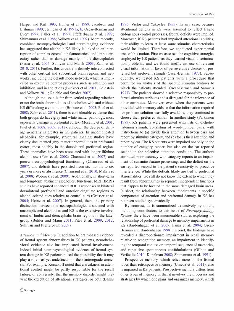
Fig. 2 Components of prefrontal brain circuitry (Barrett et al. 2007).Figure 2 highlights its cortical components. The ventral systemincludes two closely connected circuits that are anchored in the orbito-frontal cortex (OFC; Fig. 2c). The more sensory system involves thelateral sector of the OFC (a, c, purple). It is closely connected to theanterior insula (d, yellow) and the basolateral complex in the amygdala(2d, rose, ventral aspect). The visceromotor circuitry includes theventral portion of the ventromedial prefrontal cortex, which lies inthe medial sector of the OFC (2a, b, c, blue) where the medial andlateral aspects of OFC connect; ventromedial prefrontal cortex isclosely connected to the amygdala (including the central nucleus, d,
rose, dorsal aspect) and the subgenual parts of the anterior cingulatecortex on the medial wall of the brain (2b, copper and peach). Thedorsal system is associated with mental state attributions including thedorsal aspect of the ventromedial prefrontal cortex corresponding tothe frontal pole (b, maroon), the anterior cingulate (2b, peach), and thedorsomedial prefrontal cortex (2a, b, green). Ventrolateral prefrontalcortex is shown in red (2a). Structures in the reward circuitry includethe OFC, dorsolateral prefrontal (2a, orange) and cingulate cortex (2b,copper and tan), the thalamus (b, light pink), the ventral striatum (2d,green), the amygdala (2d, rose) and hippocampus (2d, gray), and thelimbic brainstem
Neuropsychol Rev

Impaired Response Inhibition and Salience Attribution in alladdictions. This syndrome is characterized by “attributingexcessive salience to the drug and drug-related cues, de-creased sensitivity to non-drug reinforcers, and decreasedability to inhibit maladaptive or disadvantageous behaviors”(Goldstein and Volkow 2011, p. 652). The authors providedan informative schematic figure, based upon neuroimagingfindings, that shows differences in prefrontal brain activitybetween addicted and healthy individuals involving functionaldomains such as attention and memory, decision-making andinhibitory control, and emotion and motivation. These samefunctional domains are the predominant dysfunctionalcharacteristics of alcoholic KS patients, and here wereview literature related to them. However, we cautionthat our use of the terminology representing the domains isgeneric, general, and atheoretical. We acknowledge thatthe definitions of the major constructs may differ acrosssubfields within the cognitive neurosciences and addic-tions (Alvarez and Emory 2006; Goldstein and Volkow2011; Knudsen 2007; Posner 2011; Rolls 2008); this isreflected in large part in the complex and widespreadunderlying neurocircuitry.
Functional Domains of Prefrontal System Abnormalitiesin Alcoholic Korsakoff’s Syndrome
Before the advent of sophisticated neuroimaging techniques,damage within the diencephalon was believed to be respon-sible for the amnesia characteristically associated with KS.However, a few investigators, including Korsakoff himself,considered the syndrome to be accompanied by a combina-tion of cortical and diencephalic lesions (Barbizet 1963;Talland 1965; Victor and Yakovlev 1955; Warner 1934)but neuroanatomical specificity was absent. Specific empha-sis on frontal involvement was apparent, however, whenVictor et al. (1971) reported mild cortical atrophy primarilyinvolving the frontal regions in 27 % of cases withWernicke-Korsakoff syndrome; the frontal atrophy was con-current with histopathological evidence showing 88 % ofthe cases having damage in the medial dorsal thalamicnucleus, and 100 % of cases involving the medial mammil-lary nucleus. Over the next two decades, there were addi-tional reports of frontal brain abnormalities in conjunctionwith diencephalic pathology associated with alcoholic KS(Adams et al. 1993; Cala et al. 1978; Cave and Squire 1992;
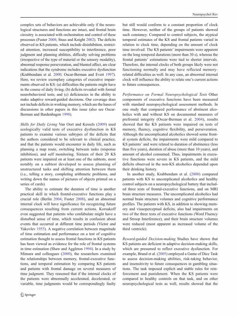
Fig. 3 An example of a model of a large-scale network involvingprefrontal brain circuitry with multiple connections. This model isbased upon a review of findings from structural and functional neuro-imaging studies (Bressler and Menon 2010). It hypothesizes that thesalience network (middle) initiates dynamic switching between thecentral-executive (right) and default-mode (left) networks, and medi-ates between attention to endogenous and exogenous events. Sensoryand limbic inputs are processed by the anterior insula, which detectssalient events and initiates appropriate control signals to regulate
behavior via the anterior cingulate cortex (and homeostatic states viathe mid and posterior insular cortex). Key nodes of the saliencenetwork include the anterior cingulate cortex and the anterior insula;the default-mode network includes the ventromedial prefrontal cortexand posterior cingulate cortex, with input from the amygdala; thecentral-executive network includes the dorsolateral prefrontal cortexand posterior parietal cortex, with input from the ventral striatum andhippocampus. (Adapted from Bressler and Menon 2010.)
Neuropsychol Rev
Harper and Kril 1993; Hunter et al. 1989; Jacobson andLishman 1990; Jernigan et al. 1991a, b; Oscar-Berman andEvert 1997; Paller et al. 1997; Pfefferbaum et al. 1992;Shimamura et al. 1988; Volkow et al. 1992). More recently,combined neuropsychological and neuroimaging evidencehas suggested that alcoholic KS likely is linked to an inter-ruption of complex cerebellothalamocortical and limbic cir-cuitry rather than to damage mainly of the diencephalon(Fama et al. 2004; Sullivan and Marsh 2003; Zahr et al.2010, 2011). Further, this circuitry is densely interconnectedwith other cortical and subcortical brain regions and net-works, including the default mode network, which is impli-cated in executive control processes such as attention andinhibition, and in addictions (Buckner et al. 2011; Goldsteinand Volkow 2011; Raichle and Snyder 2007).
Although the issue is far from settled regarding whetheror not the brain abnormalities of alcoholics with and withoutKS differ along a continuum (Brokate et al. 2003; Pitel et al.2008; Zahr et al. 2011), there is considerable evidence thatboth groups do have gray and white matter pathology, mostespecially damage in prefrontal cortex (Moselhy et al. 2001;Pitel et al. 2008, 2009, 2012), although the degree of dam-age generally is greater in KS patients. In uncomplicatedalcoholics, for example, structural imaging studies haveclearly documented gray matter abnormalities in prefrontalcortex, most notably in the dorsolateral prefrontal region.These anomalies have been associated with longer lifetimealcohol use (Fein et al. 2002; Chanraud et al. 2007) andpoorer neuropsychological functioning (Chanraud et al.2007), and deficits have persisted from six months to sixyears or more of abstinence (Chanraud et al. 2010; Makris etal. 2008; Wobrock et al. 2009). Additionally, in short-termand long-term abstinent alcoholics, functional MRI (fMRI)studies have reported enhanced BOLD responses in bilateraldorsolateral prefrontal and anterior cingulate regions toalcohol-related cues relative to control cues (Grüsser et al.2004; Heinz et al. 2007). In general, then, the primarydistinction between the neuropathologies associated withuncomplicated alcoholism and KS is the extensive involve-ment of limbic and diencephalic brain regions in the lattergroup (Buhler and Mann 2011; Pitel et al. 2009, 2012;Sullivan and Pfefferbaum 2009).
Attention and Memory In addition to brain-based evidenceof frontal system abnormalities in KS patients, neurobeha-vioral evidence also has implicated frontal involvement.Indeed, initial neuropsychological evidence of frontal sys-tem damage in KS patients raised the possibility that it mayplay a role—as yet undefined—in their anterograde amne-sia. For example, Korsakoff noted that a weakness in atten-tional control might be partly responsible for the recallfailure, or conversely, that the memory disorder might pre-vent the execution of attentional strategies, or both (Banks
1996; Victor and Yakovlev 1955). In any case, becauseattentional deficits in KS were assumed to reflect fragileendogenous control processes, frontal deficits were implied.Moreover, if KS patients had impaired attentional abilities,their ability to learn at least some stimulus characteristicswould be limited. Therefore, we conducted experimentaltests of this notion. First we assessed the cognitive strategiesemployed by KS patients as they learned visual discrimina-tion problems, and we found inefficient use of relevantvisual information in favor of perseverative choices of pre-ferred but irrelevant stimuli (Oscar-Berman 1973). Subse-quently, we tested KS patients with a procedure thatpermitted an analysis of the specific stimulus features towhich the patients attended (Oscar-Berman and Samuels1977). The patients showed a selective responsivity to pre-ferred stimulus attributes and a disregard for the relevance ofother attributes. Moreover, even when the patients wereprovided with memory aids so that the information requiredfor problem solution was fully available, they continued tochoose their preferred stimuli. In another study (Parkinson1979), KS patients were presented with lists of dichotic-listening stimuli, consisting of word-number pairs, withinstructions to (a) divide their attention between ears andreport by stimulus category, and (b) focus their attention andreport by ear. The KS patients were impaired not only on thenumber of category reports but also on the ear reportedsecond in the selective attention condition. The authorsattributed poor accuracy with category reports to an impair-ment of semantic feature processing, and the deficit on theear reported second to the patient’s sensitivity to proactiveinterference. While the deficits likely are tied to prefrontalabnormalities, we still do not know the extent to which theyresult from abnormalities in separate, independent functionsthat happen to be located in the same damaged brain areas.In short, the relationship between impairments in specificcomponents of attention and prefrontal damage in KS hasnot been studied systematically.
By contrast, as is summarized extensively by others,including contributors to this issue of NeuropsychologyReview, there have been innumerable studies exploring therelationship of prefrontal damage to memory impairments inKS (Bardenhagen et al. 2007; Fama et al. 2004; Oscar-Berman and Bardenhagen 1998). In brief, the findings haverevealed a disproportionate impairment in recall memoryrelative to recognition memory, an impairment in identify-ing the temporal context or temporal sequence of memories,and repetitive spontaneous confabulations (Gilboa andVerfaellie 2010; Kopelman 2008; Shimamura et al. 1991).
Prospective memory, which relies more on the frontallobes than retrospective memory (Umeda et al. 2011), alsois impaired in KS patients. Prospective memory differs fromother types of memory in that it involves the processes andstrategies by which one plans and organizes memory, which
Neuropsychol Rev

are important, in turn, for monitoring and then performingfuture actions. In one study, Shimamura et al. (1991) de-scribed a series of experiments comparing KS patients, non-KS amnesic patients, and frontal lobe patients on a varietyof mnemonic and nonmnemonic neuropsychological tests.They found that KS patients were as impaired as frontalpatients on tasks of prospective memory. However, Brunfautet al. (2000) later reported that aspects of prospective mem-ory were preserved in KS patients. Conflicting results oftenreflect differences in methodologies employed by differentlaboratories. For example, Groot et al. (2002) evaluatedprospective memory in patients with memory disorders (on-ly one of whom had KS) and found performance to becorrelated with scores on conventional memory and execu-tive function tests. Importantly, the investigators noted thattime-based tasks are more difficult than event-based tasksbecause the former place higher demands on inhibitorycontrol mechanisms, and compensatory strategies improveprospective memory functioning. Clearly, further work isneeded to understand the characteristics and scope of pro-spective memory impairments in KS, including strategicversus automatic monitoring of cueing, cue salience, andplanning.
Confabulations are memory distortions that occur inpatients with alcoholic KS, and Borsutzky et al. (2008)investigated the nature of these types of false memories inKS patients. The researchers used a standardized confabu-lation interview to investigate memory domains most affect-ed by confabulations in KS, and they found that the patientsconfabulated most within the episodic/autobiographicalmemory domain, i.e., mainly with respect to their personalpast and present. In another study of confabulation in KSpatients, Kessels and colleagues (2008) distinguished be-tween spontaneous and provoked confabulations, each hav-ing different underlying cognitive mechanisms and possibleneuroanatomical underpinnings. Provoked confabulations—related to intrusions on memory tests—were quantified bythird-party ratings; spontaneous confabulations, which maybe due to executive dysfunction or a source memory deficit,were assessed with a confabulation battery. The investiga-tors found deficits by the KS patients in source memory, inwhich the patients incorrectly assigned previously learnedwords to the wrong word list. Also, the KS patients hadextensive executive deficits, but no relationship between theseverity of these deficits and the severity of confabulation orintrusions on a memory task was found. Thus, the findingsprovided evidence for dissociation between spontaneousconfabulation, provoked confabulation, and false memories.It should be noted that basic questions remain about howconfabulation should be defined, how many types of con-fabulation there are, their underlying neurocognitive mech-anisms, and their neural bases (Gilboa and Verfaellie 2010).In any case, because confabulations are characteristic of KS,
Borsutzky et al. (2008)recommended that the screening ofconfabulation tendencies be used as a supplementary clini-cal tool for a detailed description of the memory profile ofKS patients.
Although memory researchers continue to differentiateamong aspects of the memory impairments related to damagein diencephalic, medial temporal, and prefrontal areas, resultshave remained inconsistent (Brokate et al. 2003). Nonetheless,studies by other investigators, including contributors to thepresent issue of Neuropsychology Review, have describedmemory impairments in KS patients that likely reflect theconsequences of frontal system damage to the signatureamnesia (Kopelman et al. 2009; Modirrousta and Fellows2008; Spiegel and Lim 2011; van Geldorp et al. 2012).
Executive Functions There is no definitive one-to-one rela-tionship between intact executive functions and frontal lobeintegrity (Alvarez and Emory 2006; Elliott 2003; Fuster2008). However, executive functions have been linked tofrontal brain systems, and there is abundant neurobehavioralevidence that alcoholic KS patients have impairments inexecutive functioning. As defined by Lezak (1982), “Exec-utive functions comprise those mental capacities necessaryfor formulating goals, planning how to achieve them, andcarrying out the plans effectively. They are at the heart of allsocially useful, personally enhancing, constructive, and cre-ative activities. With the executive functions intact, a personcan suffer many different kinds and combinations of senso-ry, motor, and cognitive deficits and still maintain the direc-tion of his own life and be productive as well. Impairment orloss of these functions compromises a person’s capacity tomaintain an independent, constructively self-serving, andsocially productive life no matter how well he can see andhear, walk and talk, and perform tests” (p. 281).
Theorists differ with regard to which of many diversecognitive functions are to be considered as executive func-tions (Alvarez and Emory 2006; Stuss and Knight 2002),but at the very least, the term encompasses a host of pro-cesses that act in harmony and are responsible for the high-level action of monitoring and controlling behaviors neces-sary for maintaining focus and achieving outcomes in pos-sibly adverse circumstances (Williams et al. 2009). Thus,executive skills include an array of complex mental abilitiessuch as connecting past experiences with present actions;planning future behavior when faced with novel tasks; mak-ing judgments; changing behaviors and strategies; payingattention; and remembering details to guide decision-making. Executive skills also are involved in evaluatingrisks, recognizing future consequences of our actions,choosing between good and bad actions, suppressing unac-ceptable social responses, determining similarities and dif-ferences among objects or events, initiating or postponingresponses, and prioritizing or switching tasks. These
Neuropsychol Rev
complex sets of behaviors are achievable only if the neuro-logical structures and functions are intact, and frontal braincircuitry is associated with orchestration and control of theseprocesses (Fuster 2008; Stuss and Knight 2002). The deficitsobserved in KS patients, which include disinhibition, restrict-ed attention, increased susceptibility to interference, poorjudgment and planning abilities, difficulty solving problems(irrespective of the type of material or the sensory modality),abnormal response perseveration, and blunted affect, are clearindications that the syndrome includes executive dysfunction(Krabbendam et al. 2000; Oscar-Berman and Evert 1997).Here, we review exemplary categories of executive impair-ments observed in KS: (a) difficulties the patients might havein the course of daily living; (b) deficits revealed with formalneurobehavioral tests; and (c) deficiencies in the ability tomake adaptive reward-guided decisions. Our coverage doesnot include deficits in workingmemory, which are the bases ofdiscussions in other papers in this issue (also see Oscar-Berman and Bardenhagen 1998).
Skills for Daily Living Van Oort and Kessels (2009) usedecologically valid tests of executive dysfunction in KSpatients to examine various subtypes of the deficits thatthe authors considered to be relevant to clinical practice,and that the patients would encounter in daily life, such asplanning a map route, switching between tasks (responseinhibition), and self monitoring. Sixteen of their 20 KSpatients were impaired on at least one of the subtests, mostnotably on a subtest developed to assess planning ofunstructured tasks and shifting attention between them(i.e., telling a story, completing arithmetic problems, andwriting down the names of pictures of objects printed on aseries of cards).
The ability to estimate the duration of time is anotherpractical skill in which frontal-executive functions play acrucial role (Berlin 2004; Fuster 2008), and an abnormalinternal clock will have significance for recognizing futureconsequences resulting from current actions. Korsakoffeven suggested that patients who confabulate might have adisturbed sense of time, which results in confusion aboutevents that occurred at different time periods (Victor andYakovlev 1955). A negative correlation between magnitudeof time estimation and performance on a test of cognitiveestimation thought to assess frontal functions in KS patientshas been viewed as evidence for the role of frontal systemsin time estimation (Shaw and Aggleton 1994). In a study byMimura and colleagues (2000), the researchers examinedthe relationships between memory, frontal-executive func-tions, and temporal estimation by comparing KS patientsand patients with frontal damage on several measures oftime judgment. They reasoned that if the internal clocks ofthe patients were abnormally accelerated, decelerated, orvariable, time judgments would be correspondingly faulty
but still would conform to a constant proportion of clocktime. However, neither of the groups of patients showedsuch constancy. Compared to control subjects, the atypicaltemporal estimates of the KS and frontal patients varied inrelation to clock time, depending on the amount of clocktime involved: The KS patients’ impairments were apparenton the long temporal durations (more than 30 s), whereas thefrontal patients’ estimations were tied to shorter intervals.Therefore, the internal clocks of both groups likely were notfunctioning normally and may have reflected memory-related difficulties as well. In any case, an abnormal internalclock will influence the ability to relate one’s current actionsto future consequences.
Performance on Formal Neuropsychological Tests Othercomponents of executive functions have been measuredwith standard neuropsychological assessment methods. Inone study that compared performance of abstinent alco-holics with and without KS on documented measures ofprefrontal integrity (Oscar-Berman et al. 2004), resultsshowed that the KS patients were impaired on tests ofmemory, fluency, cognitive flexibility, and perseveration.Although the uncomplicated alcoholics showed some front-al system deficits, the impairments were mild compared toKS patients’ and were related to duration of abstinence (lessthan five years), duration of abuse (more than 10 years), andamount of alcohol consumed. Thus, impairments in execu-tive functions were severe in KS patients, and the milddeficits observed in the non-KS alcoholics depended upontheir drinking history.
In another study, Krabbendam et al. (2000) comparedpatients with KS to uncomplicated alcoholics and healthycontrol subjects on a neuropsychological battery that includ-ed three tests of frontal-executive functions, and on MRIbrain structure measures. The uncomplicated alcoholics hadnormal brain structure volumes and cognitive performanceprofiles. The patients with KS, in addition to showing mem-ory and visuoperceptual deficits, also had impairments ontwo of the three tests of executive functions (Word Fluencyand Stroop Interference), and their brain structure volumeswere reduced (most apparent as increased volume of thethird ventricle).
Reward-guided Decision-making Studies have shown thatKS patients are deficient in adaptive decision-making skills,which are presumed to reflect executive dysfunction. Forexample, Brand et al. (2005) employed a Game of Dice Taskto assess decision-making abilities, risk-taking behavior,and insensitivity to future consequences in gambling situa-tions. The task imposed explicit and stable rules for rein-forcement and punishment. When the KS patients werecompared to healthy controls on that task, and on otherneuropsychological tests as well, results showed that the
Neuropsychol Rev

patients were highly impaired. Moreover, deficits on theGame of Dice Task were correlated with specific subcom-ponents of executive functions (categorization, monitoring,and using feedback), as measured by a modified WisconsinCard Sorting Test. Because the investigators wanted toensure that the decision-making deficits in the KS patientsin risky situations were caused by executive dysfunctionsrather than by impairments in processing feedback, theylater used the same gambling test and a modified versionof it, in which the feedback components were removed(Brand et al. 2009). The researchers found that KS patientsagain showed a preference for the risky alternatives linkedto reductions in executive functioning, irrespective ofwhether or not feedback was provided.
A key aspect of decision-making involves updating thevalue of behavioral options, which rely on the executivecontrolled by prefrontal cortex. In an early study (Oscar-Berman et al. 1976), we compared the performance ofalcoholic KS patients to that of uncomplicated alcoholicsand healthy control subjects on each of three differentschedules of spatial probability learning (50:50, 70:30, and30:70) using monetary reinforcement. Instructions wereminimal, and to earn money, the subjects simply had tochoose between two left-right alternatives that were differ-entially rewarded according to the three schedules. Althoughon all three schedules, choice ratios by the nonalcoholic sub-jects approximated the reinforcement ratios, the choice ratiosof KS patients on the second and third schedules remainedclose to the reinforcement ratio acquired with the first sched-ule. In addition, the KS patients made an abnormal number ofperseverative errors. On most measures, performance by theuncomplicated alcoholics fell between that of the other twogroups. A more recent study has used fMRI in conjunctionwith differential-reinforcement learning tasks to investigatethe integrity of neural mechanisms underlying reward-guided decision-making in uncomplicated alcohol-dependentpatients (Park et al. 2010). The results showed that functionalconnectivity between striatum and dorsolateral prefrontal cor-tex predicted impairments in behavior and the magnitude ofalcohol craving. Although KS patients have yet to be includedin brain imaging studies of decision-making, results in theuncomplicated alcoholics are in line with other findings ofabnormalities in prefrontal circuitry in addictions and thepivotal role that frontal brain systems have in adaptive updat-ing of action values and behavioral regulation (Goldstein andVolkow 2011; Koob and Volkow 2010; Wrase et al. 2007).
A somatic marker hypothesis has been proposed, whichsuggests that somatic or internal bodily signals guide adaptivedecision-making processes when we are faced with complexor conflicting cognitive choices such as those often studied ingambling tasks (Bechara et al. 2005; Damasio 1996). Process-ing these internal “emotional” signals can influence our deci-sions. Further, the relationship between decision-making and
emotion (see next section) is emphasized by results showingthat different negative feelings can influence judgments andlead to dissimilar choices (Bechara 2004; Lerner and Keltner2001). Studies using gambling tasks in substance abusers haveshown decision-making impairments in these patients (Fein etal. 2006; Verdejo et al. 2004). Alcoholism also can lead todeficits in sensitivity to reward (Oscar-Berman et al. 1976), animportant determinant of effective decision-making (Becharaet al. 2003; Fein et al. 2006). Studies have suggested thatchanges in the reward system (Makris et al. 2008) lead tothe creation of an allostatic state, which represents a chronicdeviation from reward set point, and one hypothesis holdsthat, in an allostatic state, loss of reward leads to disregulationof brain neurotransmitters (Koob and LeMoal 2008a, b; Kooband Volkow 2010). Several studies have provided support forthis hypothesis; they point to lower brain activity in alcoholicsin regions such as the frontal lobes, ventral striatum, andlimbic system (Bowirrat and Oscar-Berman 2005;Marinkovicet al. 2009; Phan et al. 2004). However, the architecturesupporting intact executive functions and top-down controlof behavior is complex and likely involves interconnectedfunctional networks that include—in addition to prefrontalcortex, ventral striatum, and limbic system—numerous otherregions such as the thalamus and cerebellum (Dosenbach et al.2008; Wijnia and Goossensen 2010), all reported to be com-promised in KS (Zahr and Sullivan 2008).
In sum, executive dysfunction is an indisputable charac-teristic of alcoholic KS. Because of the ubiquity of execu-tive impairments associated with this disorder, Kessels andcolleagues (2008) recommended revision of the DSM clas-sification of “alcohol-induced persisting amnestic disorder”in order to include executive dysfunction. They proposedthat, in so doing, the rehabilitation process would be facil-itated and might even be helpful in reducing memoryimpairments and confabulatory behavior.
Emotion Executive functioning and emotional functioningcan be closely linked, such that different emotions willinfluence decisions and actions differently (Verdejo et al.2004). Emotions engage strong mental and physical statesand rely on a seamless coordination among multiple neuro-physiological systems spanning different levels of the neu-raxis (Davidson et al. 2003; Panksepp et al. 2002). Lesionsin distinctly different areas of the brain will disrupt emo-tional processing at different levels or stages, e.g., duringperception or evaluation of emotional stimuli, and at stageswhereby emotional responses are expressed. Therefore, acommon feature shared by theories of emotional dysfunc-tion is that multiple brain structures, encompassing wide-spread brain systems, are involved, including prefrontalcortices (which are particularly involved in the final pro-cessing of emotionally relevant stimuli), the anterior cingu-late, insula, amygdala, and basal ganglia (Davidson et al.
Neuropsychol Rev
2003; Goldstein and Volkow 2011; Koob and Le Moal2008a; Panksepp et al. 2002). Not surprisingly, therefore,emotional abnormalities accompanying long-term chronicalcoholism cover a broad spectrum. Moreover, Talland(1965) speculated that KS patients’ failure to sustain emo-tional involvement in ongoing events might be an importantmechanism in the maintenance of their severe anterogradeamnesia. However, that the emotional abnormalities in KSpatients may be separable from their memory loss, wasdemonstrated in a study by Feinstein et al. (2010) showingthat emotional feelings in patients with amnesia enduredwell beyond the conscious recollection for the events thatinitially triggered the emotion.
Korsakoff noted that KS patients showed little concernabout their condition and about events that typically wouldcause elation or grief in others. In general, the emotionallives of KS patients were bland, monotonous, and devoid ofpassion or curiosity, and they seemed to lack intentions,plans, or goals (Banks 1996; Victor and Yakovlev 1955).The apathy and emotional flatness in KS patients also arecharacteristic of patients with bilateral frontal-lobe damageunrelated to alcoholism (Lezak et al. 2004; Moselhy et al.2001). Like individuals with frontal brain damage, alco-holics have impairments in cognitive processing of emo-tional signals (Jackson et al. 2006; Marinkovic et al. 2009;Monnot et al. 2001; Oscar-Berman and Marinkovic 2007;Schulte et al. 2010; Tsuchida and Fellows 2012), and theyare impaired in social skills, which in turn, impacts theirability to implement their preferred strategies for interper-sonal interactions (Gaffney et al. 1998; Verdejo et al. 2004).One interpersonal skill is perspective taking, the ability tomake inferences about the knowledge, thoughts, and feel-ings of others. This aspect of social cognition is known to bedisrupted both with severe alcoholism (Uekermann andDaum 2008) and following prefrontal cortical damage(Bramham et al. 2009; Tranel et al. 2002). To exploreperspective-taking ability in KS patients, Oosterman et al.(2011) used a story comprehension task in which inferenceshad to be made that either relied on perspective-taking ornot, and other aspects of executive function were assessedusing an extensive neuropsychological test battery. Theperformance of KS patients declined with increasing storycomplexity, but the pattern of decline for perspective-takingand non-perspective-taking stories was similar compared tothat of a control group of nonalcoholic subjects. Further-more, the performance decline with increasing task complex-ity was directly related to the overall decline in executivefunctioning measured with the neuropsychological battery.The authors concluded that aspects of executive functionunrelated to perspective-taking per se, appeared to underliethe KS patients’ difficulties with story comprehension.
Systematic studies evaluating emotional dysfunction inKS patients have revealed deficits in labeling and recognition
of anger, fear, and surprise. KS patients also are deficient inprocessing emotional prosody and in judging the intensity ofemotions (Clark et al. 2007; Montagne et al. 2006; Oscar-Berman et al. 1990; Snitz et al. 2002).We examined the abilityof KS patients to identify different affective states using facialexpressions and voice intonations (affective prosody) as stim-uli (Oscar-Berman et al. 1990). We found that KS patientsshowed deficits in emotional perception in both modalities,but since we did not separate the different task demands ofprosody and semantic content, Snitz et al. (2002) ex-plored those aspects of the task. They examined affec-tive prosody discrimination and identification in KSpatients, and observed impairments when the semanticcontent was either neutral or incongruent with prosody.The results suggested that KS patients were impaired ininterpreting the meaning of affective prosody in theabsence of semantic cues as to the emotional contentof sentences. Montagne et al. (2006) investigated theperception and recognition of emotional expressions inKS patients by showing them video clips in which aneutral facial expression gradually changed into an emo-tional expression. The investigators gauged the amountof expression on the face that was needed for correctidentification. They found that the KS patients showedimpaired recognition of facial expressions of anger, fearand surprise, and they interpreted the pattern as evi-dence for a general expression-recognition deficit, whichlikely reflected dysfunction of frontal brain circuitry,including connections with the amygdala.
In a study that sought to differentiate KS patients’ deficitsin judgments of emotional stimuli from those of uncompli-cated alcoholics and healthy control subjects, we askedparticipants to rate stimuli according to emotional valenceand intensity (Clark et al. 2007). The primary differenceamong the groups was that the KS patients attributed themost positive valence to neutral stimuli. Overall, the patternof behavioral results implicated bi-hemispheric frontal andsubcortical involvement in the abnormalities of emotionidentification, but it was not clear whether the findings weredue to a deficiency in emotional processing itself, or wheth-er deteriorated basic processes underlying all kinds of judg-ment tasks resulted in affective judgment errors. Therefore,Brand et al. (2003) analyzed possible underlying cognitiveprocesses of emotional and nonemotional judgments in KSpatients using a cognitive estimation test consisting of fourdimensions (size, weight, quantity, and time) and an affec-tive judgment task comprising negative, neutral, and posi-tive words. In a later study in the same laboratory (Labuddaet al. 2010), the researchers also assessed valence classifi-cation performance for emotional pictures. In both studies,the KS patients’ results showed marked deficits concerningboth cognitive estimation and affective judgments. The def-icits were highly intercorrelated, and performance was
Neuropsychol Rev

related to processing speed, executive functions, and mem-ory, suggesting a common basis for cognitive estimation andaffective judgments.
The relationship between emotional impairments andabnormalities of frontal brain circuitry in KS has been basedprimarily on inferences made in the absence of neuroimag-ing data with KS patients. However, fMRI studies of emo-tional functioning in patients with focal lesions of differentsubregions within prefrontal areas have disclosed that dam-age to ventromedial prefrontal cortex impaired the detectionof subtle facial expressions of emotion (Tsuchida andFellows 2012). Such patients had difficulty distinguishingemotional from neutral expressions. In contrast, patientswith left ventrolateral prefrontal lesions were able to detectthe presence of emotional signals but had difficulty discrim-inating between specific emotions. These effects wereregionally specific: Dorsomedial prefrontal damage had noeffect on either aspect of emotion recognition. The findingssuggested that separable processes relying critically on dis-tinct regions within prefrontal cortex are responsible, on theone hand, for detecting emotional signals from facialexpressions and, on the other, for correctly classifying suchsignals.
Other studies have used fMRI scans to documentnumerous instances of emotional abnormalities in non-KS alcoholics (Marinkovic et al. 2009; Oscar-Bermanand Marinkovic 2007; Urban et al. 2007), and Schulteet al. (2010) described contributions of cortico-limbicfiber degradation to emotional dysfunction and impairedcognitive control of emotionally motivated actions inalcoholism. Thus, fiber tractography, together with func-tional neuroimaging, represent promising technologies toexplore the role of abnormalities of regional fronto-cortico-limbic-striatal-cerebellar connectivity in dysregu-lation of emotion in KS.
Summary, Conclusions, and Some Remaining Questions
Alcoholic KS occurs more often with prolonged alcoholismthan from other etiologies, and it is characterized by severeanterograde amnesia that is out of proportion to other symp-toms. Additional neuropsychological changes occur as well,including a variety of cognitive impairments and emotionalabnormalities. The amnesia in alcoholic KS patients is thoughtto be caused mainly by damage to diencephalic and limbicstructures, but cortical and cerebellar gray- and white-matterdamage contribute to aspects of memory loss. Other neuro-psychological impairments such as poor attention, difficultywith executive functions, and emotional flatness and apathylikely reflect dysfunction of a large and highly integratedfronto-cortico-striatal-cerebellar circuitry. Our understandingof KS has come a long way since Korsakoff’s initial
descriptions more than a century ago. Neuropsychologicaland neuroimaging techniques have revolutionized the wayswe can explore brain and behavioral abnormalities. By skill-fully applying these techniques, scientists now recognize thatanterograde amnesia in KS is only a piece of a much morecomplex condition. Here we focused principally on the non-mnemonic neuropsychological dysfunctions associated withbrain damage to prefrontal brain systems.
While advances have been made in understanding thecontribution of dysfunction of prefrontal circuitry to neu-robehavioral deficits in KS, many questions remain unan-swered. We do not yet know, for example, the full natureof the attentional weaknesses in KS nor the extent towhich they contribute to the memory impairments (orconversely, whether the impaired memory interferes withthe execution of attentional strategies) (Banks 1996).Additionally, we do not know precisely how emotionalchanges in KS are related to the memory defect, nor towhat extent, and in what ways emotional involvement isessential for encoding or retrieving memories (or con-versely, whether memory is essential to emotional pro-cesses) (Schulte et al. 2010).
The anterograde amnesia of KS is unremitting. Al-though there is mounting evidence from research on un-complicated alcoholism that damage to one component ofthe affected brain circuitry can lead to compensatoryfunctionality from another component (Oscar-Bermanand Marinkovic 2007; Zahr et al. 2010), this remains anopen question regarding alcoholic KS. For example,might the extensive and combined frontal and cerebellardamage in KS reduce the brain’s ability to establish func-tional compensation, thereby contributing to the perma-nence of the disorder?
Another unexplored question concerns whether or notfrontal system damage in alcoholic KS is manifesteddifferently in men and women. This is likely, becauseof the plethora of gender differences in brain and be-havioral abnormalities in alcohol use disorders generally(Medina et al. 2008; Pfefferbaum and Sullivan 2002;Urban et al. 2007).
The results of recent neurobehavioral and neuroimag-ing studies have revealed frontal system dysfunctions inalcoholic KS that parallel those reported for most addic-tions, including widespread prefrontal hypoactivity dur-ing exposure to cognitive and emotional challenges(Goldstein and Volkow 2011). Moreover, the roles as-cribed to disruption of prefrontal circuitry common toKS and the addictions are emotion disregulation andinterference with executive control. Although frontalsystem circuitry is a highly integrated system, its sub-components (dorsolateral prefrontal, ventral/orbitofrontal,and medial frontal), including their underlying connec-tions, are involved in diverse neurobehavioral functions.
Neuropsychol Rev
What remains to be determined is the relative contribu-tion of the subcomponents to the various domains ofimpairment, and whether the functions of the subcom-ponents are affected differently in KS, uncomplicatedalcoholism, and other addictions. As results of contin-ued research begin to answer these many questions,treatment and management options will expand for theafflicted individuals (Sechi and Serra 2007).
Acknowledgments Support for the writing of this review came fromthe US Department of Health and Human Services NIAAA R01-AA07112 and K05-AA00219, and from the Medical Research Serviceof the US Department of Veterans Affairs.
References
Adams, K. M., Gilman, S., Koeppe, R. A., Kluin, K. J., Brunberg, J.A., Dede, D., et al. (1993). Neuropsychological deficits are cor-related with frontal hypometabolism in positron emission tomog-raphy studies of older alcoholic patients. Alcoholism: Clinical andExperimental Research, 17(2), 205–210.
Alvarez, J. A., & Emory, E. (2006). Executive function and the frontallobes: a meta-analytic review. Neuropsychology Review, 16(1),17–42.
American Psychiatric Association. (2000). Diagnostic and statisticalmanual of mental disorders (4th edition, Text Revision). Wash-ington, DC: American Psychiatric Association.
Badre, D., & D’Esposito, M. (2009). Is the rostro-caudal axis of thefrontal lobe hierarchical? Nature Reviews Neuroscience, 10(9),659–669.
Banks, W. P. (1996). Korsakoff and amnesia. Consciousness andCognition, 5(1–2), 22–26.
Barbas, H. (2007). Flow of information for emotions through temporaland orbitofrontal pathways. Journal of Anatomy, 211(2), 237–249.
Barbas, H., Saha, S., Rempel-Clower, N., & Ghashghaei, T. (2003).Serial pathways from primate prefrontal cortex to autonomic areasmay influence emotional expression. BMC Neuroscience, 4(1),25–37.
Barbizet, J. (1963). Defect of memorizing of hippocampal-mammillaryorigin: a review. Journal of Neurology, Neurosurgery & Psychi-atry, 26, 127–135.
Bardenhagen, F. J., Oscar-Berman, M., & Bowden, S. C. (2007). Ruleknowledge aids performance on spatial and object alternationtasks by alcoholic patients with and without Korsakoff’s amnesia.Neuropsychiatric Disease and Treatment, 3(6), 907–918.
Barrett, L. F., Mesquita, B., Ochsner, K. N., & Gross, J. J. (2007). Theexperience of emotion. Annual Review of Psychology, 58, 373–403.
Bechara, A. (2003). Risky business: emotion, decision-making, andaddiction. Journal of Gambling Studies, 19(1), 23–51.
Bechara, A. (2004). The role of emotion in decision-making: evidencefrom neurological patients with orbitofrontal damage. Brain andCognition, 55(1), 30–40.
Bechara, A., Damasio, H., & Damasio, A. R. (2003). Role of theamygdala in decision-making. Annals of the NY Academy ofSciences, 985, 356–369.
Bechara, A., Damasio, H., Tranel, D., & Damasio, A. R. (2005). TheIowa Gambling Task and the somatic marker hypothesis: somequestions and answers. Trends in Cognitive Science, 9(4), 159–164.
Berlin, H. A. (2004). Impulsivity, time perception, emotion and rein-forcement sensitivity in patients with orbitofrontal cortex lesions.Brain, 127(5), 1108–1126.
Bolla, K. I., Eldreth, D. A., Matochik, J. A., & Cadet, J. L. (2005).Neural substrates of faulty decision-making in abstinent marijua-na users. NeuroImage, 26(2), 480–492.
Borsutzky, S., Fujiwara, E., Brand, M., & Markowitsch, H. J. (2008).Confabulations in alcoholic Korsakoff patients. Neuropsycholo-gia, 46(13), 3133–3143.
Bowirrat, A., & Oscar-Berman, M. (2005). Relationship between dopa-minergic neurotransmission, alcoholism, and Reward DeficiencySyndrome. American Journal of Genetics Part B: NeuropsychiatricGenetics, 132B(1), 29–37.
Bramham, J., Morris, R. G., Hornak, J., Bullock, P., & Polkey, C. E.(2009). Social and emotional functioning following bilateral andunilateral neurosurgical prefrontal cortex lesions. Journal of Neu-ropsychology, 3(1), 125–143.
Brand, M., Fujiwara, E., Kalbe, E., Steingass, H.-P., Kessler, J., &Markowitsch, H. J. (2003). Cognitive estimation and affectivejudgments in alcoholic Korsakoff patients. Journal of Clinicaland Experimental Neuropsychology, 25(3), 324–334.
Brand, M., Fujiwara, E., Borsutzky, S., Kalbe, E., Kessler, J., &Markowitsch, H. J. (2005). Decision-making deficits of Korsakoffpatients in a new gambling task with explicit rules: associationswith executive functions. Neuropsychology, 19(3), 267–277.
Brand, M., Pawlikowski, M., Labudda, K., Laier, C., von Rothkirch,N., & Markowitsch, H. J. (2009). Do amnesic patients withKorsakoff’s syndrome use feedback when making decisions un-der risky conditions? An experimental investigation with theGame of Dice Task with and without feedback. Brain and Cog-nition, 69(2), 279–290.
Bressler, S. L., & Menon, V. (2010). Large-scale brain networks incognition: emerging methods and principles. Trends in CognitiveSciences, 14(6), 277–290.
Brion, S. (1969). Korsakoff’s syndrome: Clinico-anatomical and physio-pathological considerations. In G. A. Talland&N. C.Waugh (Eds.),The pathology of memory (pp. 22–29). New York: Academic.
Brokate, B., Hildebrandt, H., Eling, P., Fichtner, H., Runge, K., &Timm, C. (2003). Frontal lobe dysfunctions in Korsakoff’s syn-drome and chronic alcoholism: continuity or discontinuity? Neu-ropsychology, 17(3), 420–428.
Brunfaut, E., Vanoverberghe, V., & d’Ydewalle, G. (2000). Prospectiveremembering of Korsakoffs and alcoholics as a function of theprospective-memory and on-going tasks. Neuropsychologia, 38(7), 975–984.
Buckner, R. L., Krienen, F. M., Castellanos, A., Diaz, J. C., & Yeo, B.T. T. (2011). The organization of the human cerebellum estimatedby intrinsic functional connectivity. Journal of Neurophysiology,106(5), 2322–2345.
Buhler, M., & Mann, K. (2011). Alcohol and the human brain: asystematic review of different neuroimaging methods. Alcohol-ism: Clinical and Experimental Research, 35(10), 1771–1793.
Caine, D., Halliday, G. M., Kril, J. J., & Harper, C. G. (1997).Operational criteria for the classification of chronic alcoholics:identification of Wernicke’s encephalopathy. Journal of Neurolo-gy, Neurosurgery & Psychiatry, 62(1), 51–60.
Cala, L. A., Jones, B., Mastaglia, F. L., & Wiley, B. (1978). Brainatrophy and intellectual impairment in heavy drinkers—a clinical,psychometric and computerized tomography study. Australianand New Zealand Journal of Medicine, 8(2), 147–153.
Caulo, M., Van Hecke, J., Toma, L., Ferretti, A., Tartaro, A., Colosimo,C., et al. (2005). Functional MRI study of diencephalic amnesia inWernicke-Korsakoff syndrome. Brain, 128(7), 1584–1594.
Cave, C. B., & Squire, L. R. (1992). Intact verbal and nonverbal short-term memory following damage to the human hippocampus.Hippocampus, 2(2), 151–163.
Chanraud, S., Martelli, C., Delain, F., Kostogianni, N., Douaud, G.,Aubin, H. J., et al. (2007). Brain morphometry and cognitiveperformance in detoxified alcohol-dependents with preserved
Neuropsychol Rev

psychosocial functioning. Neuropsychopharmacology, 32(2),429–438.
Chanraud, S., Pitel, A. L., & Sullivan, E. V. (2010). Structural imagingof alcoholic abuse. In M. E. Shenton & B. I. Turetsky (Eds.),Understanding neuropsychiatric disorders. New York: Cam-bridge University Press.
Charness, M. E. (2010). Overview of the chronic neurologic compli-cations of alcohol. uptodate.com, 1–12
Clark, U. S., Oscar-Berman, M., Shagrin, B., & Pencina, M. (2007).Alcoholism and judgments of affective stimuli. Neuropsychology,21(3), 346–362.
Damasio, A. R. (1996). The somatic marker hypothesis and the possi-ble functions of the prefrontal cortex. Philosophical Transactionsof the Royal Society of London. Series B, Biological Sciences, 351(1346), 1413–1420.
Davidson, R. J., Scherer, K. R., & Goldsmith, H. H. (Eds.). (2003).Handbook of affective sciences (Series in affective science). NewYork: Oxford University Press.
Dosenbach, N. U. F., Fair, D. A., Cohen, A. L., Schlaggar, B. L., &Petersen, S. E. (2008). A dual-networks architecture of top-downcontrol. Trends in Cognitive Sciences, 12(3), 99–105.
Elliott, R. (2003). Executive functions and their disorders. BritishMedical Bulletin, 65, 49–59.
Fama, R., Marsh, L., & Sullivan, E. V. (2004). Dissociation of remoteand anterograde memory impairment and neural correlates inalcoholic Korsakoff syndrome. Journal of the International Neu-ropsychological Society, 10, 427–441.
Fein, G., Di Sclafani, V., Cardenas, V. A., Goldmann, H., Tolou-Shams,M., & Meyerhoff, D. J. (2002). Cortical gray matter loss intreatment-naive alcohol dependent individuals. Alcoholism: Clinicaland Experimental Research, 26(4), 558–564.
Fein, G., Landman, B., Tran, H., McGillivray, S., Finn, P., Barakos, J.,et al. (2006). Brain atrophy in long-term abstinent alcoholics whodemonstrate impairment on a simulated gambling task. Neuro-Image, 32(3), 1465–1471.
Feinstein, J. S., Duff, M. C., & Tranel, D. (2010). Sustained experience ofemotion after loss of memory in patients with amnesia. Proceedingsof the National Academy of Sciences, 107(17), 7674–7679.
Fuster, J. M. (2008). The prefrontal cortex (4th ed.). London:Academic.
Gaffney, L. R., Thorpe, K., Young, R., Collett, R., & Occhipinti, S.(1998). Facial skills, expectancies and drinking in adolescents.Addictive Behaviors, 23(5), 587–599.
Gilboa, A., & Verfaellie, M. (2010). Introduction—telling it like itisn’t: the cognitive neuroscience of confabulation. Journal of theInternational Neuropsychological Society, 16(06), 961–966.
Goldstein, R. Z., & Volkow, N. D. (2002). Drug addiction and itsunderlying neurobiological basis: neuroimaging evidence for theinvolvement of the frontal cortex. The American Journal of Psy-chiatry, 159(10), 1642–1652.
Goldstein, R. Z., & Volkow, N. D. (2011). Dysfunction of the prefron-tal cortex in addiction: neuroimaging findings and clinical impli-cations. Nature Reviews Neuroscience, 12, 652–669.
Groot, Y. C. T., Wilson, B. A., Evans, J., & Watson, P. (2002).Prospective memory functioning in people with and without braininjury. Journal of the International Neuropsychological Society, 8(5), 645–654.
Grüsser, S. M., Wrase, J., Klein, S., Hermann, D., Smolka, M. N., Ruf,M., et al. (2004). Cue-induced activation of the striatum and medialprefrontal cortex is associated with subsequent relapse in abstinentalcoholics. Psychopharmacology (Berlin), 175(3), 296–302.
Harding, A., Halliday, G., Caine, D., & Kril, J. J. (2000). Degenerationof anterior thalamic nuclei differentiates alcoholics with amnesia.Brain, 123(1), 141–154.
Harper, C. (2009). The neuropathology of alcohol-related brain dam-age. Alcohol and Alcoholism, 44(2), 136–140.
Harper, C. G., & Kril, J. J. (1993). Neuropathological changes inalcoholics. In Alcohol-induced brain damage (pp. 39–69). Rock-ville, MD: NIH Publications.
Harper, C. G., & Matsumoto, I. (2005). Ethanol and brain damage.Current Opinion in Pharmacology, 5, 73–78.
Heinz, A., Wrase, J., Kahnt, T., Beck, A., Bromand, Z., Grüsser, S. M.,et al. (2007). Brain activation elicited by affectively positivestimuli is associated with a lower risk of relapse in detoxifiedalcoholic subjects. Alcoholism: Clinical and Experimental Re-search, 31(7), 1138–1147.
Hunter, R., McLuskie, R., Wyper, D., Patterson, J., Christie, J. E.,Brooks, D. N., et al. (1989). The pattern of function-relatedregional cerebral blood flow investigated by single photon emis-sion tomography with 99mTc-HMPAO in patients with presenileAlzheimer’s disease and Korsakoff’s psychosis. PsychologicalMedicine, 19(4), 847–855.
Jackson, P. L., Brunet, E., Meltzoff, A. N., & Decety, J. (2006).Empathy examined through the neural mechanisms involved inimagining how I feel versus how you feel pain. Neuropsycholo-gia, 44(5), 752–761.
Jacobson, R. R., & Lishman, W. A. (1990). Cortical and diencephaliclesions in Korsakoff’s syndrome: a clinical and CT scan study.Psychological Medicine, 20(1), 63–75.
Jernigan, T. L., Butters, N., DiTraglia, G., Schafer, K., Smith, T., Irwin,M., et al. (1991a). Reduced cerebral grey matter observed inalcoholics using magnetic resonance imaging. Alcoholism: Clin-ical and Experimental Research, 15(3), 418–427.
Jernigan, T. L., Schafer, K., Butters, N., & Cermak, L. S. (1991b).Magnetic resonance imaging of alcoholic Korsakoff patients.Neuropsychopharmacology, 4(3), 175–186.
Kashi, M. R., Henderson, G. I., & Schenker, S. (2009). Wernicke’sencephalopathy. In D. W. McCandless (Ed.), Metabolic encepha-lopathy (pp. 281–301). New York: Springer.
Kessels, R. P. C., Kortrijk, H. E., Wester, A. J., & Nys, G. M. S. (2008).Confabulation behavior and false memories in Korsakoff’s syn-drome: role of source memory and executive functioning. Psychi-atry and Clinical Neurosciences, 62(2), 220–225.
Knudsen, E. I. (2007). Fundamental components of attention. AnnualReview of Neuroscience, 30, 57–78.
Koob, G. F., & Le Moal, M. (2008a). Addiction and the brain antire-ward system. Annual Review of Psychology, 59, 29–53.
Koob, G. F., & Le Moal, M. (2008b). Review. Neurobiological mech-anisms for opponent motivational processes in addiction. Philo-sophical Transactions of the Royal Society of London. Series B,Biological Sciences, 363(1507), 3113–3123.
Koob, G. F., & Volkow, N. D. (2010). Neurocircuitry of addiction.Neuropsychopharmacology, 35(1), 217–238.
Kopelman, M. D. (2008). Alcohol and frontal lobe impairment: fasci-nating findings. Addiction, 103, 736–737.
Kopelman, M. D., Thomson, A. D., Guerrini, I., & Marshall, E. J.(2009). The Korsakoff syndrome: clinical aspects, psychologyand treatment. Alcohol and Alcoholism, 44(2), 148–154.
Krabbendam, L., Visser, P. J., Derix, M. M., Verhey, F., Hofman, P.,Verhoeven, W., et al. (2000). Normal cognitive performance inpatients with chronic alcoholism in contrast to patients withKorsakoff’s syndrome. Journal of Neuropsychiatry and ClinicalNeuroscience, 12(1), 44–50.
Labudda, K., von Rothkirch, N., Pawlikowski, M., Laier, C., &Brand, M. (2010). Categorization abilities for emotional andnonemotional stimuli in patients with alcohol-related Korsak-off syndrome. Cognitive and Behavioral Neurology, 23(2),89–97.
LeDoux, J. E. (2000). Emotion circuits in the brain. Annual Review ofNeuroscience, 23, 155–184.
Lerner, J. S., & Keltner, D. (2001). Fear, anger, and risk. Journal ofPersonality and Social Psychology, 81(1), 146–159.
Neuropsychol Rev
Lezak, M. D. (1982). The problem of assessing executive functions.International Journal of Psychology, 17(1–4), 281–297.
Lezak, M. D., Howieson, D. B., & Loring, D. W. (2004). Neuropsycho-logical assessment (4th ed.). New York: Oxford University Press.
LoPresti, M. L., Schon, K., Tricarico, M. D., Swisher, J. D., Celone, K.A., & Stern, C. E. (2008). Working memory for social cues recruitsorbitofrontal cortex and amygdala: a functional magnetic resonanceimaging study of delayed matching to sample for emotional expres-sions. Journal of Neuroscience, 28(14), 3718–3728.
Makris, N., Oscar-Berman, M., Jaffin, S. K., Hodge, S. M., Kennedy, D.N., Caviness, V. S., et al. (2008). Decreased volume of the brainreward system in alcoholism. Biological Psychiatry, 64(3), 192–202.
Marinkovic, K., Oscar-Berman, M., Urban, T., O’Reilly, C. E.,Howard, J. A., Sawyer, K., et al. (2009). Alcoholism and damp-ened temporal limbic activation to emotional faces. Alcoholism:Clinical and Experimental Research, 33(11), 1880–1892.
Medina, K. L., McQueeny, T., Nagel, B. J., Hanson, K. L., Schweinsburg,A. D., & Tapert, S. F. (2008). Prefrontal cortex volumes in adoles-cents with alcohol use disorders: unique gender effects. Alcoholism:Clinical and Experimental Research, 32(3), 386–394.
Mimura, M., Kinsbourne, M., & O’Connor, M. (2000). Time estima-tion by patients with frontal lesions and by Korsakoff amnesics.Journal of the International Neuropsychological Society, 6(5),517–528.
Modirrousta, M., & Fellows, L. K. (2008). Medial prefrontal cortexplays a critical and selective role in ‘feeling of knowing’ meta-memory judgments. Neuropsychologia, 46(12), 2958–2965.
Monnot, M., Nixon, S. J., Lovallo, W. R., & Ross, E. (2001). Alteredemotional perception in alcoholics: deficits in affective prosodycomprehension. Alcoholism: Clinical and Experimental Re-search, 25(3), 362–369.
Montagne, B., Kessels, R. P. C., Wester, A. J., & de Haan, E. H. F.(2006). Processing of emotional facial expressions in Korsakoff’ssyndrome. Cortex, 42(5), 705–710.
Moselhy, H. F., Georgiou, G., & Kahn, A. (2001). Frontal lobe changesin alcoholism: a review of the literature. Alcohol and Alcoholism,36(5), 357–368.
Müller, N. G., Machado, L., & Knight, R. T. (2002). Contributions ofsubregions of the prefrontal cortex to working memory: evidencefrom brain lesions in humans. Journal of Cognitive Neuroscience,14(5), 673–686.
Ochsner, K. N., & Gross, J. J. (2007). The neural architecture ofemotional regulation. In Handbook of emotion regulation (pp.87–109, Vol. 5). New York, NY: Guilford Press.
Oosterman, J. M., de Goede, M., Wester, A. J., van Zandvoort, M. J.E., & Kessels, R. P. C. (2011). Perspective taking in Korsakoff’ssyndrome: the role of executive functioning and task complexity.Acta Neuropsychiatrica, 23(6), 302–308.
Oscar-Berman, M. (1973). Hypothesis testing and focusing behaviorduring concept formation by amnesic Korsakoff patients. Neuro-psychologia, 11(2), 191–198.
Oscar-Berman, M., & Bardenhagen, F. (1998). Nonhuman primatemodels of memory dysfunction in neurodegenerative disease:Contributions from Comparative Neuropsychology. In A. I. Trös-ter (Ed.), Memory in neurodegenerative disease (pp. 3–20). Cam-bridge: Cambridge University Press.
Oscar-Berman, M., & Bowirrat, A. (2005). Genetic influences inemotional dysfunction and alcoholism-related brain damage. Neu-ropsychiatric Disease and Treatment, 1(3), 211–229.
Oscar-Berman, M., & Evert, D. (1997). Alcoholic Korsakoff’s syn-drome. In P. D. Nussbaum (Ed.), Handbook of neuropsychologyand aging (Critical Issues in Neuropsychology). New York: Ple-num Press.
Oscar-Berman, M., & Marinkovic, K. (2007). Alcohol: effects onneurobehavioral functions and the brain. Neuropsychology Re-view, 17(3), 239–257.
Oscar-Berman, M., & Samuels, I. (1977). Stimulus-preference andmemory factors in Korsakoff’s syndrome. Neuropsychologia, 15(1), 99–106.
Oscar-Berman, M., Sahakian, B. J., & Wikmark, G. (1976). Spatial prob-ability learning by alcoholic Korsakoff patients. Journal of Experi-mental Psychology: Human Learning and Memory, 2, 215–222.
Oscar-Berman, M., Hancock, M., Mildworf, B., & Hutner, N. (1990).Emotional perception and memory in alcoholism and aging. Al-coholism: Clinical and Experimental Research, 14(3), 383–393.
Oscar-Berman, M., Clancy, J. P., & Weber, D. A. (1993). Discrepanciesbetween IQ and memory scores in alcoholism and aging. ClinicalNeuropsychologist, 7(3), 281–296.
Oscar-Berman, M., Kirkley, S. M., Gansler, D. A., & Couture, A.(2004). Comparisons of Korsakoff and non-Korsakoff alcoholicson neuropsychological tests of prefrontal brain functioning. Alco-holism: Clinical and Experimental Research, 28(4), 667–675.
Paller, K. A., Acharya, A., Richardson, B. C., Plaisant, O., Shimamura,A. P., Reed, B. R., et al. (1997). Functional neuroimaging ofcortical dysfunction in alcoholic Korsakoff’s syndrome. Journalof Cognitive Neuroscience, 9(2), 277–293.
Pandya, D. N., & Yeterian, E. H. (2002). The anatomical substrates ofemotional behavior: The role of the cerebral cortex. In J. Grafman& F. Boller (Eds.), The frontal lobes, Vol. 7 (2nd ed.). New York:Elsevier.
Panksepp, J., Knutson, B., & Burgdorf, J. (2002). The role of brainemotional systems in addictions: a neuro-evolutionary perspectiveand new ‘self-report’ animal model. Addiction, 97(4), 459–469.
Papez, J. W. (1937). A proposed mechanism of emotion. Archives ofNeurology and Psychiatry, 38(4), 725.
Park, S. Q., Kahnt, T., Beck, A., Cohen, M. X., Dolan, R. J., Wrase, J.,et al. (2010). Prefrontal cortex fails to learn from reward predic-tion errors in alcohol dependence. The Journal of Neuroscience,30(22), 7749–7753.
Parkinson, S. R. (1979). The amnesic Korsakoff syndrome: a study ofselective and divided attention. Neuropsychologia, 17(1), 67–75.
Patterson, J. C., Ungerleider, L. G., & Bandettini, P. A. (2002). Task-independent functional brain activity correlation with skin conduc-tance changes: an fMRI study. NeuroImage, 17(4), 1797–1806.
Pfefferbaum, A., & Sullivan, E. V. (2002). Microstructural but notmacrostructural disruption of white matter in women with chronicalcoholism. NeuroImage, 15(3), 708–718.
Pfefferbaum, A., & Sullivan, E. V. (2005). Disruption of brain whitematter microstructure by excessive intracellular and extracellularfluid in alcoholism: evidence from diffusion tensor imaging.Neuropsychopharmacology, 30(2), 423–432.
Pfefferbaum, A., Lim, K. O., Zipursky, R. B., Mathalon, D. H.,Rosenbloom, M. J., Lane, B., et al. (1992). Brain gray and whitematter volume loss accelerates with aging in chronic alcoholics: aquantitative MRI study. Alcoholism: Clinical and ExperimentalResearch, 16(6), 1078–1089.
Pfefferbaum, A., Lim, K. O., Desmond, J. E., & Sullivan, E. V. (1996).Thinning of the corpus callosum in older alcoholic men: a mag-netic resonance imaging study. Alcoholism: Clinical and Experi-mental Research, 20(4), 752–757.
Phan, K. L., Wager, T. D., Taylor, S. F., & Liberzon, I. (2004).Functional neuroimaging studies of human emotions. CNS Spec-troscopy, 9(4), 258–266.
Pitel, A.-L., Beaunieux, H., Witkowski, T., Vabret, F., de la Sayette, V.,Viader, F., et al. (2008). Episodic and working memory deficits inalcoholic Korsakoff patients: the continuity theory revisited. Alco-holism: Clinical and Experimental Research, 32(7), 1229–1241.
Pitel, A.-L., Aupée, A.-M., Chételat, G., Mézenge, F., Beaunieux, H.,de la Sayette, V., et al. (2009). Morphological and glucose me-tabolism abnormalities in alcoholic Korsakoff’s syndrome: groupcomparisons and individual analyses. PLoS ONE, 4(11), e7748.doi:10.1371/journal.pone.0007748.
Neuropsychol Rev
Pitel, A. L., Chételat, G., Le Berre, A. P., Desgranges, B., Eustache, F., &Beaunieux, H. (2012). Macrostructural abnormalities in Korsakoff’ssyndrome compared to uncomplicated alcoholism. Neurology.
Poldrack, R. A., Wagner, A. D., Prull, M. W., Desmond, J. E., Glover,G. H., & Gabrieli, J. D. (1999). Functional specialization forsemantic and phonological processing in the left inferior prefron-tal cortex. NeuroImage, 10(1), 15–35.
Posner, M. I. (2011). Imaging attention networks. NeuroImage.Raichle, M. E., & Snyder, A. Z. (2007). A default mode of brain
function: a brief history of an evolving idea. NeuroImage, 37(4),1083–1090. discussion 1097–1089.
Reed, L., Lasserson, D., Marsden, P., Stanhope, N., Stevens, T., Bello,F., et al. (2003). FDG-PET Findings in the Wernicke-KorsakoffSyndrome. Cortex, 39(4–5), 1027–1045.
Ribeiro, A. M., & Pereira, S. R. C. (2010). Animal models of alcohol-induced dementia. In P. P. De Deyn & D. Van Dam (Eds.), Animalmodels of dementia (pp. 665–683). New York: Springer.
Rissman, J., & Wagner, A. D. (2012). Distributed representations inmemory: insights from functional brain imaging. Annual Reviewof Psychology, 63, 101–128.
Rolls, E. T. (2008). Memory, attention, and decision-making. NewYork: Oxford University Press.
Rushworth, M. F. S., Noonan, M. P., Boorman, E. D., Walton, M. E., &Behrens, T. E. (2011). Frontal cortex and reward-guided learningand decision-making. Neuron, 70(6), 1054–1069.
Santarelli, L., Saxe, M., Gross, C., Surget, A., Battaglia, F., Dulawa, S., etal. (2003). Requirement of hippocampal neurogenesis for the be-havioral effects of antidepressants. Science, 301(5634), 805–809.
Schmahmann, J. D., & Pandya, D. N. (2006). Fiber pathways of thebrain. New York: Oxford University Press, USA.
Schulte, T., Mũller-Oehring, E. M., Pfefferbaum, A., & Sullivan, E. V.(2010). Neurocircuitry of emotion and cognition in alcoholism:contributions from white matter fiber tractography. Dialogues inClinical Neuroscience, 12(4), 554–560.
Sechi, G. P., & Serra, A. (2007). Wernicke’s encephalopathy: newclinical settings and recent advances in diagnosis and manage-ment. Lancet Neurology, 6, 442–455.
Shaw, C., & Aggleton, J. P. (1994). The ability of amnesic subjects toestimate time intervals. Neuropsychologia, 32(7), 857–873.
Shimamura, A. P., Jernigan, T. L., & Squire, L. R. (1988). Korsakoff’ssyndrome: radiological (CT) findings and neuropsychologicalcorrelates. Journal of Neuroscience, 8(11), 4400–4410.
Shimamura, A. P., Janowsky, J. S., & Squire, L. S. (1991). What is therole of frontal lobe damage in memory disorders? In H. S. Levin,H. M. Eisenber, & A. L. Benton (Eds.), Frontal lobe function anddysfunction. New York: Oxford University Press.
Snitz, B. E., Hellinger, A., & Daum, I. (2002). Impaired process-ing of affective prosody in Korsakoff’s syndrome. Cortex, 38(5), 797–803.
Spiegel, D. R., & Lim, K.-J. (2011). A case of probable Korsakoff’ssyndrome: a syndrome of frontal lobe and diencephalic structuralpathogenesis and a comparison with medial temporal lobe demen-tias. Innovations in Clinical Neuroscience, 8(6), 15–19.
Stuss, D. T., & Knight, R. T. (Eds.). (2002). Principles of frontal lobefunction (1st ed.). New York: Oxford University Press.
Sullivan, E. V. (2003). Compromised pontocerebellar and cerebello-thalamocortical systems: speculations on their contributions tocognitive and motor impairment in nonamnesic alcoholism. Alco-holism: Clinical and Experimental Research, 27(9), 1409–1419.
Sullivan, E. V., & Marsh, L. (2003). Hippocampal volume deficits inalcoholic Korsakoff’s syndrome. Neurology, 61(12), 1716–1719.
Sullivan, E. V., & Pfefferbaum, A. (2009). Neuroimaging of theWernicke-Korsakoff Syndrome. Alcohol and Alcoholism, 44(2),155–165.
Sullivan, E. V., Deshmukh, A., Desmond, J. E., Lim, K. O., & Pfefferbaum,A. (2000). Cerebellar volume decline in normal aging, alcoholism,
andKorsakoff s syndrome: relation to ataxia.Neuropsychology, 14(3),x341–352.
Talland, G. A. (1965). Deranged memory: A psychonomic study of theamnesic syndrome. New York: Academic.
Tanji, J., & Hoshi, E. (2008). Role of the lateral prefrontal cortex inexecutive behavioral control. Physiological Reviews, 88(1), 37–57.
Tranel, D., Bechara, A., & Denburg, N. L. (2002). Asymmetric func-tional roles of right and left ventromedial prefrontal cortices insocial conduct, decision-making, and emotional processing. Cor-tex, 38(4), 589–612.
Tsuchida, A., & Fellows, L. K. (2012). Are you upset? Distinct rolesfor orbitofrontal and lateral prefrontal cortex in detecting anddistinguishing facial expressions of emotion. Cerebral Cortex,22. doi:10.1093/cercor/bhr370.
Uekermann, J., & Daum, I. (2008). Social cognition in alcoholism: alink to prefrontal cortex dysfunction? Addiction, 103(5), 726–735.
Umeda, S., Kurosaki, Y., Terasawa, Y., Kato, M., & Miyahara, Y.(2011). Deficits in prospective memory following damage to theprefrontal cortex. Neuropsychologia, 49(8), 2178–2184.
Urban, T., Oscar-Berman, M., Marinkovic, K., Sawyer, K., & Harris,G. (2007). Neural correlates of gender differences in the process-ing of emotional stimuli. Paper presented at the Society for Neu-roscience, San Diego, CA, November.
van Geldorp, B., Bergmann, H. C., Robertson, J., Wester, A. J., &Kessels, R. P. C. (2012). The interaction of working memoryperformance and episodic memory formation in patients withKorsakoff’s amnesia. Brain Research, 1433(C), 98–103.
Van Oort, R., & Kessels, R. P. C. (2009). Executive dysfunction inKorsakoff’s syndrome: time to revise the DSM criteria foralcohol-induced persisting amnestic disorder? International Jour-nal of Psychiatry in Clinical Practice, 13(1), 78–81.
Verdejo, A., Aguilar de Arcos, F., & Perez Garcia, M. (2004). Alterations inthe decision making processes linked to the ventromedialprefrontal cortex in drug-abusing patients. Review of Neurology,38(7), 601–606.
Vetreno, R. P., Ramos, R. L., Anzalone, S., & Savage, L. M. (2012).Brain and behavioral pathology in an animal model of Wernicke’sencephalopathy and Wernicke-Korsakoff syndrome. Brain Re-search, 1436, 178–192.
Victor, M. (1994). Alcoholic dementia. Canadian Journal of Neuro-logical Science, 21, 88–99.
Victor, M., & Yakovlev, P. I. (1955). S.S. Korsakoff’s psychicdisorder in conjunction with peripheral neuritis. Neurology, 5,394–406.
Victor,M., Adams, R. D., &Collins, G. H. (1971). TheWernicke-Korsakoffsyndrome: A clinical and pathological study of 245 patients, 82 withpost-mortem examinations. Philadelphia: Davis, F. A.
Victor, M., Adams, R. D., & Collins, G. H. (1989). TheWernicke-Korsakoff syndrome and related neurologic disor-ders due to alcoholism and malnutrition (2nd ed.). Philadel-phia: F.A. Davis.
Visser, P. J., Krabbendam, L., Verhey, F. R. J., Hofman, P. A. M.,Verhoeven, W. M. A., Tuinier, S., et al. (1999). Brain corre-lates of memory dysfunction in alcoholic Korsakoff’s syn-drome. Journal of Neurology, Neurosurgery & Psychiatry, 67(6), 774–778.
Volkow, N. D., Hitzemann, R., Wang, G. J., Fowler, J. S., Burr, G.,Pascani, K., et al. (1992). Decreased brain metabolism in neuro-logically intact healthy alcoholics. American Journal of Psychia-try, 149(8), 1016–1022.
Wallis, J. D. (2007). Orbitofrontal cortex and its contribution todecision-making. The Annual Review of Neuroscience, 30,31–56.
Warner, F. J. (1934). The brain changes in chronic alcoholism andKorsakoff’s psychosis. The Journal of Nervous and Mental Dis-ease, 80(6), 629.
Neuropsychol Rev

Wijnia, J. W., & Goossensen, A. (2010). Cerebellar neurocognition andKorsakoff’s syndrome: a hypothesis. Medical Hypotheses, 75(2),266–268.
Williams, P. G., Suchy, Y., & Rau, H. K. (2009). Individual differencesin executive functioning: implications for stress regulation. An-nals of Behavioral Medicine, 37(2), 126–140.
Wobrock, T., Falkai, P., Schneider-Axmann, T., Frommann, N.,Wölwer, W., & Gaebel, W. (2009). Effects of abstinence on brainmorphology in alcoholism. European Archives of Psychiatry andClinical Neuroscience, 259(3), 143–150.
Wood, J. N., & Grafman, J. (2003). Human prefrontal cortex: process-ing and representational perspectives. Nature Reviews Neurosci-ence, 4(2), 139–147.
Wrase, J., Schlagenhauf, F., Kienast, T., Wustenberg, T., Bermpohl, F.,Kahnt, T., et al. (2007). Dysfunction of reward processing
correlates with alcohol craving in detoxified alcoholics. Neuro-Image, 35(2), 787–794.
Yeterian, E. H., Pandya, D. N., Tomaiuolo, F., & Petrides, M. (2012).The cortical connectivity of the prefrontal cortex in the monkeybrain. Cortex, 48(1), 58–81.
Zar, N. M., & Sullivan, E. V. (2008). Translational studies of alcohol-ism: bridging the gap. Alcohol Research & Health, 31(3), 215–230.
Zahr, N. M., Pitel, A. L., Chanraud, S., & Sullivan, E. V. (2010).Contributions of studies on alcohol use disorders to under-standing cerebellar function. Neuropsychology Review, 20(3),280–289.
Zahr, N. M., Kaufman, K. L., & Harper, C. G. (2011). Clinical andpathological features of alcohol-related brain damage. NatureReviews Neurology, 7(5), 284–294.
Neuropsychol Rev