fully motorized optical-resolution photoacoustic...
TRANSCRIPT
Fully motorized optical-resolutionphotoacoustic microscopy
Lei Li,1,2 Chenghung Yeh,1,2 Song Hu,2,3 Lidai Wang,2 Brian T. Soetikno,2 Ruimin Chen,4
Qifa Zhou,4 K. Kirk Shung,4 Konstantin I. Maslov,2 and Lihong V. Wang1,2,*1Department of Electrical and System Engineering, Washington University in St. Louis One Brookings
Dr., St. Louis, Missouri 63130, USA2Optical Imaging Laboratory, Department of Biomedical Engineering, Washington University in St. Louis,
One Brookings Dr., St. Louis, Missouri 63130, USA3Department of Biomedical Engineering, University of Virginia, P.O. Box 800759, Charlottesville, Virginia 22908, USA
4Resource Center for Medical Ultrasonic Transducer Technology, Department of Biomedical Engineering,University of Southern California, 1042 Downey Way, DRB 136, Los Angeles, California 90089-1111, USA
*Corresponding author: [email protected]
Received December 11, 2013; accepted February 8, 2014;posted March 3, 2014 (Doc. ID 202913); published March 28, 2014
We have developed fully motorized optical-resolution photoacoustic microscopy (OR-PAM), which integrates fivecomplementary scanning modes and simultaneously provides a high imaging speed and a wide field of view (FOV)with 2.6 μm lateral resolution. With one-dimensional (1D) motion-mode mechanical scanning, we measured theblood flow through a cross section of a blood vessel in vivo. With two-dimensional (2D) optical scanning at a laserrepetition rate of 40 kHz, we achieved a 2 kHz B-scan rate over a range of 50 μm with 20 A-lines and 50 Hzvolumetric-scan rate over a FOV of 50 μm × 50 μm with 400 A-lines, which enabled real-time tracking of cellulardynamics in vivo. With synchronized 1D optical and 2D mechanical hybrid scanning, we imaged a 10 mm × 8 mmFOVwithin three minutes, which is 20 times faster than the conventional mechanical scan in our second-generationOR-PAM. With three-dimensional mechanical contour scanning, we maintained the optimal signal-to-noise ratioand spatial resolution of OR-PAM while imaging objects with uneven surfaces, which is essential for quantitativestudies. © 2014 Optical Society of AmericaOCIS codes: (170.3880) Medical and biological imaging; (170.5120) Photoacoustic imaging; (180.5810) Scanning
microscopy; (180.6900) Three-dimensional microscopy.http://dx.doi.org/10.1364/OL.39.002117
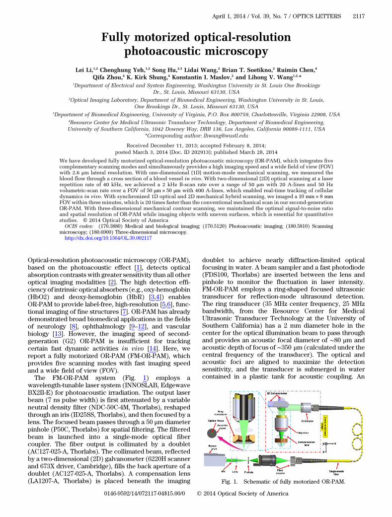
Optical-resolution photoacoustic microscopy (OR-PAM),based on the photoacoustic effect [1], detects opticalabsorption contrasts with greater sensitivity than all otheroptical imaging modalities [2]. The high detection effi-ciencyof intrinsic optical absorbers (e.g., oxy-hemoglobin(HbO2) and deoxy-hemoglobin (HbR) [3,4]) enablesOR-PAM to provide label-free, high-resolution [5,6], func-tional imaging of fine structures [7]. OR-PAM has alreadydemonstrated broad biomedical applications in the fieldsof neurology [8], ophthalmology [9–12], and vascularbiology [13]. However, the imaging speed of second-generation (G2) OR-PAM is insufficient for trackingcertain fast dynamic activities in vivo [14]. Here, wereport a fully motorized OR-PAM (FM-OR-PAM), whichprovides five scanning modes with fast imaging speedand a wide field of view (FOV).The FM-OR-PAM system (Fig. 1) employs a
wavelength-tunable laser system (INNOSLAB, EdgewaveBX2II-E) for photoacoustic irradiation. The output laserbeam (7 ns pulse width) is first attenuated by a variableneutral density filter (NDC-50C-4M, Thorlabs), reshapedthrough an iris (ID25SS, Thorlabs), and then focused by alens. The focused beam passes through a 50 μm diameterpinhole (P50C, Thorlabs) for spatial filtering. The filteredbeam is launched into a single-mode optical fibercoupler. The fiber output is collimated by a doublet(AC127-025-A, Thorlabs). The collimated beam, reflectedby a two-dimensional (2D) galvanometer (6220H scannerand 673X driver, Cambridge), fills the back aperture of adoublet (AC127-025-A, Thorlabs). A compensation lens(LA1207-A, Thorlabs) is placed beneath the imaging
doublet to achieve nearly diffraction-limited opticalfocusing in water. A beam sampler and a fast photodiode(FDS100, Thorlabs) are inserted between the lens andpinhole to monitor the fluctuation in laser intensity.FM-OR-PAM employs a ring-shaped focused ultrasonictransducer for reflection-mode ultrasound detection.The ring transducer (35 MHz center frequency, 25 MHzbandwidth, from the Resource Center for MedicalUltrasonic Transducer Technology at the University ofSouthern California) has a 2 mm diameter hole in thecenter for the optical illumination beam to pass throughand provides an acoustic focal diameter of ∼80 μm andacoustic depth of focus of ∼350 μm (calculated under thecentral frequency of the transducer). The optical andacoustic foci are aligned to maximize the detectionsensitivity, and the transducer is submerged in watercontained in a plastic tank for acoustic coupling. An
Fig. 1. Schematic of fully motorized OR-PAM.
April 1, 2014 / Vol. 39, No. 7 / OPTICS LETTERS 2117
0146-9592/14/072117-04$15.00/0 © 2014 Optical Society of America

imaging window at the bottom of the water tank is sealedwith a polyethylene membrane for optical and acoustictransmission.FM-OR-PAM’s lateral resolution (quantified in FWHM),
determined by the numerical aperture of the water-immersed imaging doublet, was calculated to be 2.1 μmat 532 nm. It was experimentally quantified by imagingthe sharp-edged metal square [Fig. 2(a)]. The edge-spreadfunction (ESF) was estimated by averaging the edge ofthe metal square along the y axis, and was fitted to anerror function (p � Aerf ��x − x0�∕σ
���
2p
� � B, R2 of0.995) based on the assumption that the beam profilewas Gaussian. The line-spread function (LSF) was thencalculated as the derivative of the ESF. The lateralresolution, defined as the FWHM of the LSF, was2.6 μm [Fig. 2(b)], which is slightly worse than thetheoretical value due to the imperfect compensation ofaberration.The whole scanning head is mounted on a three-axis
motorized translation stage (x and y axes, PLS-85, PImiCos GmbH; Z axis, KR15, THK. CO., LTD). The three-dimensional (3D) translation stage for mechanicalscanning, in combination with the 2D galvanometer foroptical scanning, provides five scanning modes: (1) 1Dmotion-mode (M-mode) mechanical scanning, (2) 2Dmechanical scanning, (3) 3D mechanical contour scan-ning, (4) 2D optical scanning, and (5) synchronized 1Doptical and 2D mechanical hybrid scanning.One-dimensional M-mode mechanical scanning (mode
1) and 2D mechanical scanning (mode 2) are inheritedfrom previously realized G2-OR-PAM as the basic func-tions of OR-PAM. Mode 1 was developed to measuretransverse blood flow based on the photoacousticDoppler broadening of bandwidth. In mode 1, the motorscans across the vessel of interest to acquire 2D B-scanimages with a step size of 2.5 μm over 300 μm interval. Ateach A-line position, 200 photoacoustic A-line signals areacquired in M-mode, with a 10-kHz laser repetition rate,before the motor translates to the next position. Then thespeed and direction of the transverse blood flow alongthe cross section can be calculated by Fourier analysis[15]. With mode 2 scanning (2.5 μm step size in both xaxis and y axis directions over a FOV of 5 mm×10 mm), FM-OR-PAM can acquired 3D images to revealthe anatomy and hemoglobin oxygen saturation (sO2) ofthe mouse ear vasculature with capillary-level resolution,as illustrated in Figs. 3(a) and 3(b), respectively; Fig. 3(c)
depicts the 3D vasculature of the whole mouse ear(Media 1). Figure 3(d) shows the blood flow measuredacross the selected vessel pair in mode 1. From the flowspeed and sO2 of the mouse ear, the metabolic rate ofoxygen (MRO2) can be calculated [16,17]. The calculatedMRO2 of this vessel pair is 0.27 ml∕100 g∕min, which isin agreement with the data previously measured in amouse ear [17]. The measurement of flow and sO2 invivo proves the capability of FM-OR-PAM for quantitativefunctional imaging of small animals.
To get optimal sensitivity and resolution of OR-PAM,the features of interest should be in the optical andacoustic confocal region (generally 30 μm in length inthe axial direction). However, due to uneven tissue sur-faces (e.g., tumors or brain), mode 2 scanning cannotcapture all the features of interest within the confocal re-gion. Here, 3D mechanical contour scanning (raster scan-ning with axial adjustment [18], mode 3) was integratedin FM-OR-PAM to overcome this limitation by acquiringoptimally focused 3D images. The sample was firstscanned in mode 2 with a coarse step size (2.5 μm x axismotor step size, 50 μm y axis motor step size). The maxi-mum-amplitude position in each A-line was identified foreach B-scan, and then a polynomial function was em-ployed to fit the set of maxima for the 2D curved surfaceinto a coarse map. The coarse map is further linearlyinterpolated along the y-direction to reduce the intervalto 5 μm and keeps the same x axis step size (2.5 μm). Last,the three-axis motorized translation stage scans accord-ing to the refined contour map. The acquisition time of
Fig. 2. Quantification of the lateral resolution of the FM-OR-PAM system. (a) Maximum-amplitude-projection (MAP) imageof a sharp-edged metal square acquired with FM-OR-PAM.(b) Edge spread function (ESF) extracted from (a) and linespread function (LSF) obtained by taking the derivative ofthe ESF. The ESF data were averaged along y.
Fig. 3. In vivo MAP images of (a) total hemoglobin, (b) sO2(measured at 532 and 559 nm) in a mouse ear acquired withFM-OR-PAM in mode 2, and (c) 3D vasculature of the mouseear (Media 1). (d) Blood flow distribution across the vessel pairmarked with an arrow in (b) imaged in mode 1.
2118 OPTICS LETTERS / Vol. 39, No. 7 / April 1, 2014

the coarse map is about 1 min. The contour scanning canbe finished in 25 minutes. Therefore, the total data ac-quisition time is about 26 minutes. A tumorous mouseear with uneven surfaces [Fig. 4(a)] was imaged in vivoin modes 2 and 3 to prove the capability of contour scan-ning. The tumor region does not show up in the imageacquired in mode 2 [Fig. 4(b)] due to the out-of-focuseffect. Comparatively, the image acquired in mode 3[Fig. 4(c)] reveals the vasculature of both the mouseear and the tumor with resolved capillaries; and the3D images [Fig. 4(d)] show the contour of the tumorousear (Media 2). The signal to noise ratio (SNR) of the in-focus region using mode 2 scanning [s1, highlighted bywhite rectangle in Fig. 4(b)] is 18.1� 2.1, the SNR ofthe tumor region [s2, highlighted by the white circle inFig. 4(b)] is 1.5� 0.8. Comparatively, the SNR of the tu-mor region imaged by mode 3 scanning [s3, highlightedby the white circle in Fig. 4(c)] is 15.6� 4.7. We exper-imentally demonstrated that the mode 3 scanning canalways keep the features of interest in the optical andacoustic confocal region with maintained SNR.High-speed label-free imaging of single red blood cells
(RBCs) in vivo with millisecond-scale temporal resolu-tion and micrometer-scale spatial resolution holds thekey to uncovering the fundamental mechanisms of cellu-lar metabolism [19]. To this end, a voice coil-basedOR-PAM system with 100 Hz B-scan rate was invented[19], and a fiber bundle-based OR-PAM system with600 Hz B-scan rate was created [20]. Alternatively, a2D galvanometer is employed in FM-OR-PAM to achievehigh-speed optical imaging within the acoustic focus(mode 4) at a laser repetition rate of 40 kHz. In vivo sin-gle cell tracking is enabled by the resulting B-scan imag-ing over a range of 50 μmwith 20 A-lines at a rate of 2 kHzor volumetric (3D) imaging over a FOV of 50 μm × 50 μmwith 400 A-lines at a rate of 50 Hz. The optical scanningrange was set to 50 μm, smaller than the acoustic focus,to maintain a high SNR. Figure 5(a) shows part of the earvascular anatomy of a living nude mouse (Hsd: Athymic
Nude–Foxn1nu, Harlan Co.), imaged in mode 2. With thesite map, mode 4 is further used to monitor a small regionof interest, and Fig. 5(b) shows a single RBC imaged invivo in mode 4. A zoom-in sequence of RBCs flowingalong the capillaries was captured in real-time video witha 20 Hz frame rate (Media 3). Mode 4 has achieved aB-scan imaging speed 20 times higher than voice-coilPAM, at the expense of FOV, which however is often nota concern if the target is cellular or capillary dynamics.
To achieve an optimal tradeoff between imaging speedand FOV, we implemented synchronized 1D optical and2D mechanical hybrid scanning (mode 5). The scanningmechanism of mode 5 is illustrated in Fig. 6(a). In mode 5,the laser was working at a 40 kHz repetition rate. The yaxis galvanometer repeatedly scanned with a line-scaninterval of 50 μm and a line-scan rate of 2 kHz. The x axismotor was synchronized with the y axis galvanometerand moved one-step forward after the y axis galvanom-eter finished one line scan. After the x axis motorfinished one B-scan, the y axis motor moved in a largestep size of 50 μm (in comparison with a step size of2.5–5.0 μm in mode 2). Figure 6(b) shows the image ofa mouse ear with a 10 mm × 8 mm FOV acquired
Fig. 4. Comparison of 2D mechanical scanning at one depth(mode 2) and 3D mechanical contour scanning (mode 3) of amouse ear in vivo. The white circles outline the tumor region.(a) Photo of a mouse ear with a growing tumor. In vivo MAPimages acquired with FM-OR-PAM in (b) mode 2 and (c) mode3; (d) 3D image of the tumorous ear vasculature (Media 2).
Fig. 5. In vivo real-time imaging of single red blood cells(RBCs) in a mouse ear. (a) MAP image acquired with FM-OR-PAM in mode 2. (b) Tracking single RBCs in a selectedcapillary in vivo in mode 4 (2D optical scanning) (Media 3).
Fig. 6. Mouse ear imaging in vivo with FM-OR-PAM in mode 5(synchronized 1D optical and 2D mechanical hybrid scanning).(a) Mechanism of the mode 5 scanning. (b) MAP image of themouse ear vasculature acquired in mode 5 (Media 4).
April 1, 2014 / Vol. 39, No. 7 / OPTICS LETTERS 2119
within 150 seconds in mode 5. After mode 5 scanning, wefirst found the maximum amplitude (MA) point of eachA-line signal. Then the MA points were rearranged fol-lowing the scanning process. The rearranged MA pointsfor each B-scan were shown in the form of animation(Media 4). With the much enlarged scanning step ofthe y axis motor, the imaging speed of mode 5 is 20 timesfaster than that of G2 OR-PAM [14].In summary, FM-OR-PAM provides five scanning
modes with high resolution, fast imaging, and a wideFOV. Its capability of tracking in vivo cell activities inreal time and imaging uneven surfaces will be invaluablefor fast and quantitative microscopic studies of tumorsand the brain as long as the region of interest is opticallyaccessible.
The authors appreciate professor James Ballard’sclose reading of the manuscript and thank Jinyang Liang,Chiye Li, and Yong Zhou for helpful discussions andexperimental assistance. This work was sponsored inpart by National Institutes of Health (NIH) grantsDP1 EB016986 (NIH Director’s Pioneer Award), R01CA159959, and R01 CA134539. L.V. Wang has a financialinterest in Microphotoacoustics Inc., and Endra Inc.,which, however, did not support this work. K. I. Maslovhas a financial interest in Microphotoacoustics Inc.
References
1. K. Maslov, H. F. Zhang, S. Hu, and L. V. Wang, Opt. Lett. 33,929 (2008).
2. L. V. Wang and S. Hu, Science 335, 1458 (2012).3. Y. Jiang, A. Forbrich, T. Harrison, and R. J. Zemp,
J. Biomed. Opt. 17, 036012 (2012).4. A. Ray, J. R. Rajian, Y. E. Lee, X. Wang, and R. Kopelman, J.
Biomed. Opt. 17, 057004 (2012).
5. R. L. Shelton and B. E. Applegate, Biomed. Opt. Express 1,676 (2010).
6. R. L. Shelton, S. P. Mattison, and B. E. Applegate, “Volumet-ric imaging of erythrocytes using label-free multiphotonphotoacoustic microscopy,” J. Biophotonics, doi: 10.1002/jbio.201300059.
7. R. J. Paproski, A. E. Forbrich, K. Wachowicz, M. M. Hitt, andR. J. Zemp, Biomed. Opt. Express 2, 771 (2011).
8. G. Suffredini, J. E. East, and L. M. Levy, “New applicationsof nanotechnology for neuroimaging,” Am. J. Neuroradiol.(to be published).
9. S. Hu, B. Rao, K. Maslov, and L. V. Wang, Opt. Lett. 35, 1(2010).
10. S. L. Jiao, M. S. Jiang, J. M. Hu, A. Fawzi, Q. F. Zhou, K. K.Shung, C. A. Puliafito, and H. F. Zhang, Opt. Express 18,3967 (2010).
11. T. Liu, H. Li, W. Song, S. L. Jiao, and H. F. Zhang, Curr. EyeRes. 38, 1229 (2013).
12. W. Song, Q. Wei, S. Jiao, and H. F. Zhang, J. Visualized Exp.71, e4390 (2013).
13. S. S. Oladipupo, S. Hu, A. C. Santeford, J. J. Yao, J. R.Kovalski, R. V. Shohet, K. Maslov, L. V. Wang, and J. M.Arbeit, Blood 117, 4142 (2011).
14. S. Hu, K. Maslov, and L. V. Wang, Opt. Lett. 36, 269(2011).
15. J. J. Yao, K. I. Maslov, Y. F. Shi, L. A. Taber, and L. H. V.Wang, Opt. Lett. 35, 1419 (2010).
16. S. Hu and L. V. Wang, Front. Neuroenerg. 2, 10 (2010).17. J. J. Yao, K. I. Maslov, Y. Zhang, Y. N. Xia, and L. V. Wang, J.
Biomed. Opt. 16, 076003 (2011).18. H. F. Zhang, K. Maslov, M. L. Li, G. Stoica, and L. H. V. Wang,
Opt. Express 14, 9317 (2006).19. L. D. Wang, K. Maslov, and L. H. V. Wang, Proc. Natl. Acad.
Sci. USA 110, 5759 (2013).20. P. Hajireza, W. Shi, and R. J. Zemp, Opt. Lett. 36, 4107
(2011).
2120 OPTICS LETTERS / Vol. 39, No. 7 / April 1, 2014