ft-ir, ft-raman and uv-visible spectral analysis on (e)-n ......ft-ir, ft-raman and uv-visible...
TRANSCRIPT
2017Vol. 1 No. 2: 7
1
iMedPub Journals
Research Article
http://www.imedpub.com
© Under License of Creative Commons Attribution 3.0 License | This article is available in: http://www.imedpub.com/archives-in-chemical-research/
Archives in Chemical Research ISSN 2572-4657
DOI: 10.21767/2572-4657.100007
Bharanidharan S1, Saleem H1, Subashchandrabose S2, Suresh M3 and Ramesh Babu N4
1 Department of Physics, Annamalai University, Annamalainagar-608002, Tamil Nadu, India
2 Centre for Research and Development, PRIST University, Thanjavur-613403, Tamil Nadu, India
3 Department of Chemistry, College of Engineering, Guindy, Anna Universtiy, Chennai, 620 025, India
4 Dept. of Physics, M.I.E.T. Engineering College, Trichy-620007, Tamil Nadu, India
Corresponding author: Saleem H
Department of Physics, Annamalai University, Tamil Nadu, India.
Tel: +91 9443879295
Citation: Bharanidharan S, Saleem H, Subashchandrabose S, et al. FT-IR, FT-Raman and UV-Visible Spectral Analysis on (E)-N′-(thiophen-2-ylmethylene) Nicotinohydrazide. Arch Chem Res. 2017, 1:2.
IntroductionGenerally, Pyridine ring is a heterocyclic organic compound with the chemical formula C5H5N. Pyridine is structurally related to benzene, with one methine group (=CH-) replaced by a nitrogen atom. It occurs in many important compounds, including azines and the vitamins niacin and pyridoxal. The precursor of pyridine is used to agrochemicals, pharmaceuticals and is also an important solvent and reagent. Mostly, it is used in the in vitro synthesis of DNA, sulfa pyridine (a drug against bacterial and viral infections), antihistaminic drugs tripelennamine and mepyramine, as well as water repellents, bactericides, and herbicides. Some chemical compounds, although not synthesized from pyridine, contain its ring structure. They include B vitamins niacin and pyridoxal, an anti-tuberculosis drug isoniazide, nicotine and other nitrogen-containing plant products [1].
The ring of Thiophene and its derivatives have been reported to possess broad spectrum of biological properties including anti-inflammatory, analgesic, antidepressant, antimicrobial and anticonvulsant activities [2-4]. Antiepileptic drugs (AEDs) like tiagabine, etizolam, brotizolam are containing thiophene moiety
FT-IR, FT-Raman and UV-Visible Spectral Analysis on (E)-N′-(thiophen-2-ylmethylene)
Nicotinohydrazide
in their structures as active pharmacophore [5,6]. In addition, thiophene and its derivatives functionalized with the formyl group are versatile building blocks for the synthesis of donor–acceptor substituted p-conjugated systems for several optical applications.
The hydrazone group in the organic compound brings out several physical and chemical properties. The hydrazones are bearing the >C=N-N< which leads the molecule towards nucleophilic and electrophilic in nature. In the hydrazone moiety, the nitrogen atom behaves as nucleophilic and carbon atom behaves as nucleophilic as well as electrophilic in nature [7-9]. The benzohydrazide derivatives shows wide spectrum of
Received: December 21, 2016; Accepted: January 02, 2017; Published: January 09, 2017
AbstractVibrational analysis of the (E)-N′-(thiophen-2-ylmethylene) nicotinohydrazide (T2CNH) compound was carried out in solid phase using FTIR and FT-Raman spectroscopic techniques in the ranges: 400-4000 cm-1 and 100-4000 cm-1, respectively. The molecular geometries and harmonic vibrational frequencies were calculated using DFT/6-311++G(d,p) basis set. A detailed interpretation of the IR and Raman spectra, based on the total energy distribution (TED) of the normal modes. The bond parameters such as bond lengths, bond angles and dihedral angles were calculated at the same level of theory. The natural bonding orbital (NBO) study reveals that inter and intra-molecular charge transfer of the molecule. The electronic transition was studied using UV-Vis spectrum. The NLO, band gap energy, MEP map, Mulliken atomic charges were calculated using the same level of basis set. In addition the thermodymanic properties were also calculated.
Keywords: FT-IR; FT-Raman; TED; NBO; T2CNH
2017Vol. 1 No. 2: 7
2 This Article is Available in: http://www.imedpub.com/archives-in-chemical-research/
Archives in Chemical Research ISSN 2572-4657
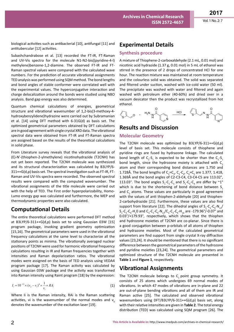
Experimental Details Synthesis procedureA mixture of Thiophene-2-carboxaldehyde (2.1 mL, 0.01 mol) and nicotinic acid hydrazide (1.37 g, 0.01 mol) in 5 mL of ethanol was stirred in the presence of 2 drops of concentrated HCl for one hour. The reaction mixture was maintained at room temperature and the colourless solid was obtained. The solid was separated and filtered under suction, washed with ice-cold water (50 ml). The precipitate was washed with water and filtered and again washed with petroleum ether (40-60%) and dried over in a vacuum desicator then the product was recrystallized from hot ethanol.
NH
O
N
H2N HCl NNH
O
N
SOS
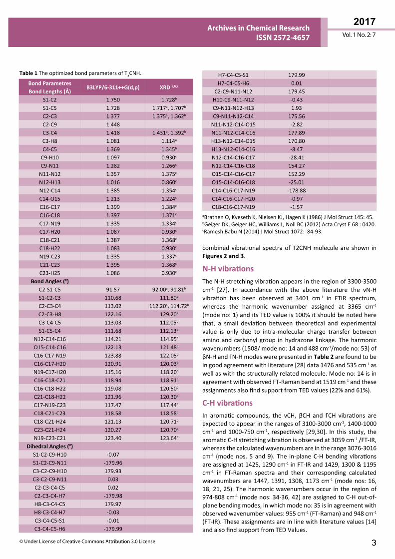
Results and DiscussionMolecular Geometry The T2CNH molecule was optimized by B3LYP/6-311++G(d,p) level of basis set. This molecule consists of thiophene and pyridine rings are fused by hydrazone linkage. The calculated bond length of C2-S1 is expected to be shorter than the C5-S1 bond length, since the hydrozone moiety is attached with C2 atom and their corresponding bond distances are 1.750 and 1.728Å. The bond lengths of C2=C3, C3-C4, C4=C5 are 1.377, 1.418, 1.369Å and the bond angles of C2-C3-C4, C3-C4-C5 are 113.02°, 113.03°. The bond angles S1-C2-C3 and S1-C5-C4 are differ by ~1° which is due to the shortening of bond distance between S1 and C2 atoms. These values are particularly in good agreement with the values of anti thiophen-2-aldehyde [20] and thiophen-2-carbohydrazide [21]. Furthermore, these values are also find support from literature [22]. The dihedral angles of S1-C2-C9-N11/S1-C2-C9-H10 and C3-C2-C9-N11/C3-C2-C9-H10 are -179.96°/-0.07° and 0.03°/+179.93°, respectively, which shows that the thiophen and hydrozone moieties of T2CNH are co-planar. Since there is a good conjugation between p-orbitals of all atoms of thiophen and hydrazone moieties. Most of the calculated geometrical parameters are find support from single crystal X-ray diffraction values [23,24]. It should be mentioned that there is no significant difference between the geometrical parameters of the hydrozone and pyridine moieties [23,24]. The geometrical parameters and optimized structure of the T2CNH molecule are presented in Table 1 and Figure 1, respectively.
Vibrational AssignmentsThe T2CNH molecule belongs to C1 point group symmetry. It consists of 25 atoms which undergoes 69 normal modes of vibrations. In which 47 modes of vibrations are in-plane and 22 are out-of-plane bending vibrations and all of them are IR and Raman active [25]. The calculated and observed vibrational wavenumbers using DFT/B3LYP/6-311++G(d,p) basis set, along with their relative intensities are given in Table 2. The total energy distribution (TED) was calculated using SQM program [26]. The
biological activities such as antibacterial [10], antifungal [11] and antitubercular [12] activities.
Subashchandrabose et al. [13] recorded the FT-IR, FT-Raman and UV-Vis spectra for the molecule N1-N2-bis((pyridine-4-l)methylene)benzene-1,2-diamine. The observed FT-IR and FT-Raman spectral values were compared with the calculated wave numbers. For the prediction of accurate vibrational assignments TED analysis was performed using SQM method. The bond lengths and bond angles of stable conformer were correlated well with the experimental values. The hyperconjugative interaction and charge delocalization around the bonds were studied using NBO analysis. Band gap energy was also determined.
Quantum chemical calculations of energies, geometrical structure and vibrational wavenumber of 1,2-bis(3-methoxy-4-hydroxybenzylidene)hydrazine were carried out by Subramanian et al. [14] using DFT method with 6-31G(d) as basis set. The optimized geometrical parameters obtained by DFT calculations are in good agreement with single crystal XRD data. The vibrational spectral data were obtained from FT-IR and FT-Raman spectra are assigned based on the results of the theoretical calculations in solid phase.
From Literature survey reveals that the vibrational analysis of (E)-N′-(thiophen-2-ylmethylene) nicotinohydrazide (T2CNH) has not yet been reported. The T2CNH molecule was synthesized and its structural characterization was calculated by B3LYP/6-311++G(d,p) basis set. The spectral investigation such as FT-IR, FT-Raman and UV-Vis spectra were recorded. The observed spectral results were compared with the computed wavenumber. The vibrational assignments of the title molecule were carried out with the help of TED. The First order hyperpolarizability, Homo-Lumo energy gap was calculated and furthermore, the MEP and thermodynamic properties were also calculated.
Computational Details The entire theoretical calculations were performed DFT method at B3LYP/6-311++G(d,p) basis set to using Gaussian 03W [15] program package, invoking gradient geometry optimization [15,16]. The geometrical parameters were used in the vibrational frequency calculations at the same level to characterize all the stationary points as minima. The vibrationally averaged nuclear positions of T2CNH were used for harmonic vibrational frequency calculations resulting in IR and Raman frequencies together with intensities and Raman depolarization ratios. The vibrational modes were assigned on the basis of TED analysis using VEDA4 program package [17]. The Raman activity was calculated by using Gaussian 03W package and the activity was transformed into Raman intensity using Raint program [18] by the expression:
12 4 110 ( )i o i ii
I v v RAv
−= × − × × (1)
Where Ii is the Raman intensity, RAi is the Raman scattering activities, νi is the wavenumber of the normal modes and ν0 denotes the wavenumber of the excitation laser [19].
3© Under License of Creative Commons Attribution 3.0 License
Vol. 1 No. 2: 7
2017Archives in Chemical Research
ISSN 2572-4657
Bond ParametresB3LYP/6-311++G(d,p) XRD a,b,c
Bond Lengths (Å)S1-C2 1.750 1.728b
S1-C5 1.728 1.717a, 1.707b
C2-C3 1.377 1.375a, 1.362b
C2-C9 1.448C3-C4 1.418 1.431a, 1.392b
C3-H8 1.081 1.114a
C4-C5 1.369 1.345b
C9-H10 1.097 0.930c
C9-N11 1.282 1.266c
N11-N12 1.357 1.375c
N12-H13 1.016 0.860c
N12-C14 1.385 1.354c
C14-O15 1.213 1.224c
C16-C17 1.399 1.384c
C16-C18 1.397 1.371c
C17-N19 1.335 1.334c
C17-H20 1.087 0.930c
C18-C21 1.387 1.368c
C18-H22 1.083 0.930c
N19-C23 1.335 1.337c
C21-C23 1.395 1.368c
C23-H25 1.086 0.930c
Bond Angles (°)C2-S1-C5 91.57 92.00a, 91.81b
S1-C2-C3 110.68 111.80a
C2-C3-C4 113.02 112.20a, 114.72b
C2-C3-H8 122.16 129.20a
C3-C4-C5 113.03 112.05b
S1-C5-C4 111.68 112.13b
N12-C14-C16 114.21 114.95c
O15-C14-C16 122.13 121.48c
C16-C17-N19 123.88 122.05c
C16-C17-H20 120.91 120.03c
N19-C17-H20 115.16 118.20c
C16-C18-C21 118.94 118.91c
C16-C18-H22 119.08 120.50c
C21-C18-H22 121.96 120.30c
C17-N19-C23 117.47 117.44c
C18-C21-C23 118.58 118.58c
C18-C21-H24 121.13 120.71c
C23-C21-H24 120.27 120.70c
N19-C23-C21 123.40 123.64c
Dihedral Angles (°)S1-C2-C9-H10 -0.07S1-C2-C9-N11 -179.96C3-C2-C9-H10 179.93C3-C2-C9-N11 0.03C2-C3-C4-C5 0.02C2-C3-C4-H7 -179.98H8-C3-C4-C5 179.97H8-C3-C4-H7 -0.03C3-C4-C5-S1 -0.01C3-C4-C5-H6 -179.99
Table 1 The optimized bond parameters of T2CNH.
aBrathen O, Kveseth K, Nielsen KJ, Hagen K (1986) J Mol Struct 145: 45.bGeiger DK, Geiger HC, Williams L, Noll BC (2012) Acta Cryst E 68 : 0420.cRamesh Babu N (2014) J Mol Struct 1072: 84-93.
H7-C4-C5-S1 179.99H7-C4-C5-H6 0.01
C2-C9-N11-N12 179.45H10-C9-N11-N12 -0.43C9-N11-N12-H13 1.93C9-N11-N12-C14 175.56
N11-N12-C14-O15 -2.82N11-N12-C14-C16 177.89H13-N12-C14-O15 170.80H13-N12-C14-C16 -8.47N12-C14-C16-C17 -28.41N12-C14-C16-C18 154.27O15-C14-C16-C17 152.29O15-C14-C16-C18 -25.01C14-C16-C17-N19 -178.88C14-C16-C17-H20 -0.97C18-C16-C17-N19 -1.57
combined vibrational spectra of T2CNH molecule are shown in Figures 2 and 3.
N-H vibrations The N-H stretching vibration appears in the region of 3300-3500 cm-1 [27]. In accordance with the above literature the νN-H vibration has been observed at 3401 cm-1 in FTIR spectrum, whereas the harmonic wavenumber assigned at 3365 cm-1 (mode no: 1) and its TED value is 100% it should be noted here that, a small deviation between theoretical and experimental value is only due to intra-molecular charge transfer between amino and carbonyl group in hydrazone linkage. The harmonic wavenumbers (1508/ mode no: 14 and 488 cm-1/mode no: 53) of βN-H and ΓN-H modes were presented in Table 2 are found to be in good agreement with literature [28] data 1476 and 535 cm-1 as well as with the structurally related molecule. Mode no: 14 is in agreement with observed FT-Raman band at 1519 cm-1 and these assignments also find support from TED values (22% and 61%).
C-H vibrations In aromatic compounds, the νCH, βCH and ΓCH vibrations are expected to appear in the ranges of 3100-3000 cm-1, 1400-1000 cm-1 and 1000-750 cm-1, respectively [29,30]. In this study, the aromatic C-H stretching vibration is observed at 3059 cm-1 /FT-IR, whereas the calculated wavenumbers are in the range 3076-3016 cm-1 (mode nos. 5 and 9). The in-plane C-H bending vibrations are assigned at 1425, 1290 cm-1 in FT-IR and 1429, 1300 & 1195 cm-1 in FT-Raman spectra and their corresponding calculated wavenumbers are 1447, 1391, 1308, 1173 cm-1 (mode nos: 16, 18, 21, 25). The harmonic wavenumbers occur in the region of 974-808 cm-1 (mode nos: 34-36, 42) are assigned to C-H out-of-plane bending modes, in which mode no: 35 is in agreement with observed wavenumber values: 955 cm-1 (FT-Raman) and 948 cm-1 (FT-IR). These assignments are in line with literature values [14] and also find support from TED Values.
2017Vol. 1 No. 2: 7
4 This Article is Available in: http://www.imedpub.com/archives-in-chemical-research/
Archives in Chemical Research ISSN 2572-4657
Figure 1 The Optimized structure of (E)-N′-(thiophen-2-ylmethylene) nicotinohydrazide (T2CNH).
ModeNo
Calculated Frequencies
(cm-1)
Observed Frequencies cm-1) IR Intensity Raman Intensity
ReducedMasses
ForceConsts Vibrational assignments≥10% (TED)d
Un Scaled Scaleda FT-IR FT-Raman Abs. Rel.b Abs. Rel.c
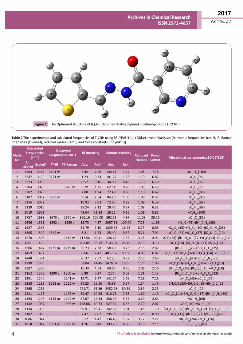
1 3503 3365 3401 w 7.42 2.06 134.41 1.67 1.08 7.78 νN12H13(100)2 3247 3120 3171 w 1.23 0.34 101.27 1.26 1.10 6.82 νC5H6(94)3 3223 3096 0.57 0.16 34.50 0.43 1.10 6.70 νC3H8(97)4 3204 3078 3079 w 6.29 1.75 62.54 0.78 1.09 6.59 νC4H7(96)5 3202 3076 7.00 1.94 72.48 0.90 1.10 6.62 νC18H22(99)6 3187 3062 3059 w 9.14 2.54 84.20 1.05 1.09 6.53 νC21H24(95)7 3156 3032 10.92 3.03 72.05 0.89 1.09 6.39 νC23H25(92)8 3139 3016 29.24 8.12 29.47 0.37 1.09 6.32 νC17H20(99)9 3019 2901 42.64 11.84 29.11 0.36 1.09 5.83 νC9H10(100)
10 1757 1688 1673 s 1670 w 360.16 100.00 392.18 4.87 11.08 20.16 νO15C14(85)11 1656 1591 1592 s 1589 s 15.73 4.37 8057.31 100.00 7.23 11.68 νN11C9(75)+βH10C9N11(20)12 1627 1563 32.79 9.10 1339.53 16.63 5.75 8.96 νC18C21(30)+νN19C23(30)+βC21C23N19(25)13 1605 1542 1549 w 6.21 1.73 25.40 0.32 5.21 7.91 νC21C23(31)+νC16C18(27)+βC17N19C23(10)14 1570 1508 1519 w 217.56 60.41 126.25 1.57 3.38 4.91 νC2C3(28)+βH13N12N11(22)+νC4C5(13)+νC2C9(11)15 1551 1490 202.85 56.32 2133.94 26.48 2.19 3.10 νC2C3(14)+βH13N12N11(41)+νC4C5(12)16 1506 1447 1425 m 1429 m 26.23 7.28 58.60 0.73 2.15 2.87 βH24C21C23(27)+βH20C17C16(25)17 1459 1402 8.66 2.41 4817.87 59.80 4.83 6.07 νC2C3(13)+νC4C5(32)+βH7C4C5(14)+νC3C4(15)18 1448 1391 26.37 7.32 62.23 0.77 2.18 2.69 βH25C23N19(43)+βC16C17N19(23)19 1390 1335 52.04 14.45 1478.54 18.35 1.75 1.99 νC4C5(13)+βH10C9N11(38)+βH6C5C4(13)20 1387 1333 16.34 4.54 60.17 0.75 2.08 2.36 βH10C9N11(25)+βH6C5C4(15)+νC3C4(18)21 1362 1308 1290 s 1300 m 0.98 0.27 2.27 0.03 1.31 1.43 βH20C17C16(45)+βH22C18C21(23)22 1291 1240 1241 w 19.71 5.47 141.74 1.76 7.28 7.15 νN19C23(43)+νC16C18(27)23 1268 1219 1218 m 1225 w 94.23 26.16 45.69 0.57 1.54 1.46 βH7C4C5(23)+βH8C3C4(29)+βH6C5C4(13)24 1265 1215 221.71 61.56 2432.18 30.19 2.39 2.25 νC14C16(22)25 1221 1173 1195 w 39.47 10.96 610.76 7.58 1.60 1.40 νC18C21(11)+βH24C21C23(17)+βH25C23N19(30)26 1195 1148 1145 m 1149 w 87.67 24.34 456.90 5.67 3.39 2.85 νN11N12(60)27 1142 1097 1096 w 146.68 40.73 527.62 6.55 2.70 2.07 νC2C9(10)+νN12C14(40)28 1134 1090 69.91 19.41 242.18 3.01 1.45 1.10 βH24C21C23(20)+βC16C17N19(12)+βH22C18C21(36)29 1102 1059 7.47 2.07 295.96 3.67 1.26 0.90 νC4C5(15)+βH7C4C5(22)+βH6C5C4(47)30 1086 1044 5.12 1.42 134.46 1.67 3.17 2.21 νN11N12(24)+νN12C14(16)31 1058 1017 1031 w 1030 w 1.76 0.49 393.20 4.88 3.19 2.11 βC16C18C21(45)
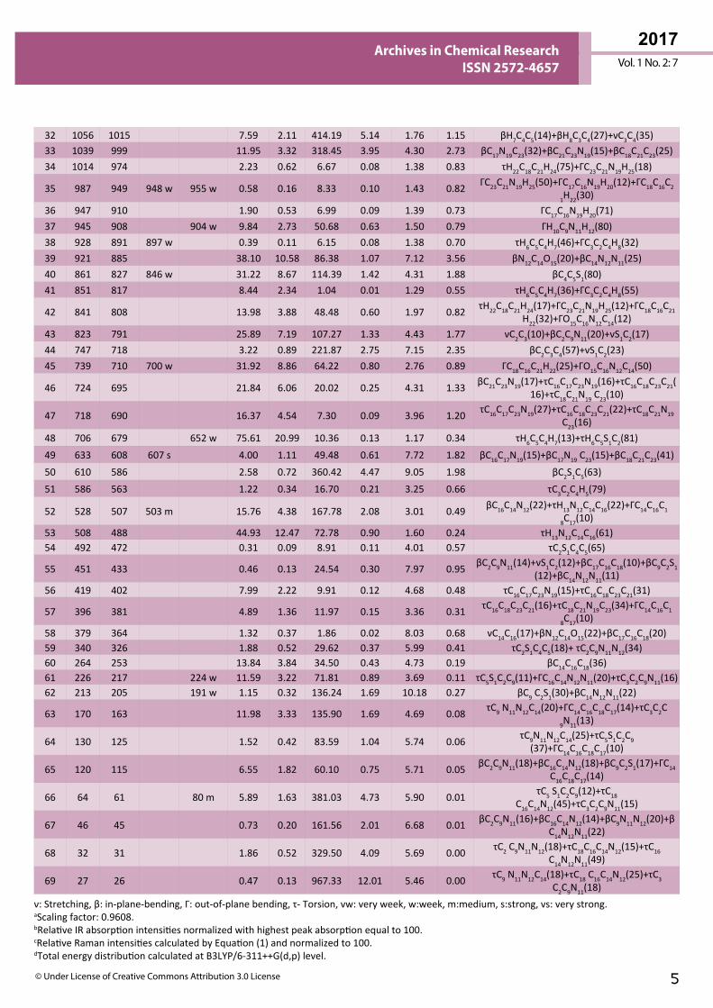
Table 2 The experimental and calculated frequencies of T2CNH using B3LYP/6-311++G(d,p) level of basis set [harmonic frequencies (cm−1), IR, Raman intensities (Km/mol), reduced masses (amu) and force constants (mdynA°−1)].
5© Under License of Creative Commons Attribution 3.0 License
Vol. 1 No. 2: 7
2017Archives in Chemical Research
ISSN 2572-4657
32 1056 1015 7.59 2.11 414.19 5.14 1.76 1.15 βH7C4C5(14)+βH8C3C4(27)+νC3C4(35)33 1039 999 11.95 3.32 318.45 3.95 4.30 2.73 βC17N19C23(32)+βC21C23N19(15)+βC18C21C23(25)34 1014 974 2.23 0.62 6.67 0.08 1.38 0.83 τH22C18C21H24(75)+ΓC23C21N19H25(18)
35 987 949 948 w 955 w 0.58 0.16 8.33 0.10 1.43 0.82 ΓC23C21N19H25(50)+ΓC17C16N19H20(12)+ΓC18C16C2
1H22(30)36 947 910 1.90 0.53 6.99 0.09 1.39 0.73 ΓC17C16N19H20(71)37 945 908 904 w 9.84 2.73 50.68 0.63 1.50 0.79 ΓH10C9N11H12(80)38 928 891 897 w 0.39 0.11 6.15 0.08 1.38 0.70 τH6C5C4H7(46)+ΓC3C2C4H8(32)39 921 885 38.10 10.58 86.38 1.07 7.12 3.56 βN12C14O15(20)+βC14N12N11(25)40 861 827 846 w 31.22 8.67 114.39 1.42 4.31 1.88 βC4C5S1(80)41 851 817 8.44 2.34 1.04 0.01 1.29 0.55 τH6C5C4H7(36)+ΓC3C2C4H8(55)
42 841 808 13.98 3.88 48.48 0.60 1.97 0.82 τH22C18C21H24(17)+ΓC23C21N19H25(12)+ΓC18C16C21H22(32)+ΓO15C16N12C14(12)
43 823 791 25.89 7.19 107.27 1.33 4.43 1.77 νC2C3(10)+βC2C9N11(20)+νS1C2(17)44 747 718 3.22 0.89 221.87 2.75 7.15 2.35 βC2C3C4(57)+νS1C2(23)45 739 710 700 w 31.92 8.86 64.22 0.80 2.76 0.89 ΓC18C16C21H22(25)+ΓO15C16N12C14(50)
46 724 695 21.84 6.06 20.02 0.25 4.31 1.33 βC21C23N19(17)+τC16C17C23N19(16)+τC16C18C23C21(16)+τC18C21N19 C23(10)
47 718 690 16.37 4.54 7.30 0.09 3.96 1.20 τC16C17C23N19(27)+τC16C18C23C21(22)+τC18C21N19 C23(16)
48 706 679 652 w 75.61 20.99 10.36 0.13 1.17 0.34 τH6C5C4H7(13)+τH6C5S1C2(81)49 633 608 607 s 4.00 1.11 49.48 0.61 7.72 1.82 βC16C17N19(15)+βC17N19 C23(15)+βC18C21C23(41)50 610 586 2.58 0.72 360.42 4.47 9.05 1.98 βC2S1C5(63)51 586 563 1.22 0.34 16.70 0.21 3.25 0.66 τC3C2C4H5(79)
52 528 507 503 m 15.76 4.38 167.78 2.08 3.01 0.49 βC16C14N12(22)+τH13N12C14C16(22)+ΓC14C16C1
8C17(10)53 508 488 44.93 12.47 72.78 0.90 1.60 0.24 τH13N12C14C16(61)54 492 472 0.31 0.09 8.91 0.11 4.01 0.57 τC2S1C4C5(65)
55 451 433 0.46 0.13 24.54 0.30 7.97 0.95 βC2C9N11(14)+νS1C2(12)+βC17C16C18(10)+βC9C2S1 (12)+βC14N12N11(11)
56 419 402 7.99 2.22 9.91 0.12 4.68 0.48 τC16C17C23N19(15)+τC16C18C23C21(31)
57 396 381 4.89 1.36 11.97 0.15 3.36 0.31 τC16C18C23C21(16)+τC18C21N19C23(34)+ΓC14C16C1
8C17(10)58 379 364 1.32 0.37 1.86 0.02 8.03 0.68 νC14C16(17)+βN12C14O15(22)+βC17C16C18(20)59 340 326 1.88 0.52 29.62 0.37 5.99 0.41 τC2S1C4C5(18)+ τC2C9N11N12(34)60 264 253 13.84 3.84 34.50 0.43 4.73 0.19 βC14C16C18(36)61 226 217 224 w 11.59 3.22 71.81 0.89 3.69 0.11 τC5S1C2C9(11)+ΓC16C14N12N11(20)+τC3C2C9N11(16)62 213 205 191 w 1.15 0.32 136.24 1.69 10.18 0.27 βC9 C2S1(30)+βC14N12N11(22)
63 170 163 11.98 3.33 135.90 1.69 4.69 0.08 τC9 N11N12C14(20)+ΓC14C16C18C17(14)+τC3C2C9N11(13)
64 130 125 1.52 0.42 83.59 1.04 5.74 0.06 τC9N11N12C14(25)+τC5S1C2C9 (37)+ΓC14C16C18C17(10)
65 120 115 6.55 1.82 60.10 0.75 5.71 0.05 βC2C9N11(18)+βC16C14N12(18)+βC9C2S1(17)+ΓC14C16C18C17(14)
66 64 61 80 m 5.89 1.63 381.03 4.73 5.90 0.01 τC5 S1C2C9(12)+τC18 C16C14N12(45)+τC3C2C9N11(15)
67 46 45 0.73 0.20 161.56 2.01 6.68 0.01 βC2C9N11(16)+βC16C14N12(14)+βC9N11N12(20)+βC14N12N11(22)
68 32 31 1.86 0.52 329.50 4.09 5.69 0.00 τC2 C9N11N12(18)+τC18C16C14N12(15)+τC16 C14N12N11(49)
69 27 26 0.47 0.13 967.33 12.01 5.46 0.00 τC9 N11N12C14(18)+τC18 C16C14N12(25)+τC3 C2C9N11(18)
ν: Stretching, β: in-plane-bending, Γ: out-of-plane bending, τ- Torsion, vw: very week, w:week, m:medium, s:strong, vs: very strong. aScaling factor: 0.9608.bRelative IR absorption intensities normalized with highest peak absorption equal to 100. cRelative Raman intensities calculated by Equation (1) and normalized to 100.dTotal energy distribution calculated at B3LYP/6-311++G(d,p) level.
2017Vol. 1 No. 2: 7
6 This Article is Available in: http://www.imedpub.com/archives-in-chemical-research/
Archives in Chemical Research ISSN 2572-4657
made in the present study. These vibrational assignments are further supported by literature [28] and TED output.
C=C, C-C vibrations In the pyridine ring, the ν(C-C) stretching vibrations are usually occur in the ranges of 1590-1640, 1560-1580 and 1470-1510 cm-1 [33]. The computed wavenumber for ν(C-C) modes are lies at 1563, 1542, 1240 and 1173 cm-1 (mode nos: 12, 13, 22 and 25) with TED values. In the present study it has been established well and the FTIR band at 1549 cm-1 and FT-Raman bands at 1241, 1195 cm-1 are designated as νC-C vibrations. These assignments are find support from the literature [34] and also from TED values.
The bands arising from βCCC and ΓCCC of pyridine moiety are ascribed to bands observed at 1031, 607 and 503 cm-1 respectively in FTIR spectrum. These vibrational assignments find support from harmonic bands: 1017, 999, 608 and 507, 381, 163 cm-1 (mode nos: 31, 33, 49 and 52, 57, 63) in addition to literature values [35].
4000 3500 3000 2500 2000 1500 1000 500
0.00
0.05
0.10
0.15
0.20
0.25
0.30
IR/B3LYP/6-311++G(d,p)
IR In
tens
ity
Wavenumber (cm-1)
4000 3500 3000 2500 2000 1500 1000 500
1.0
0.9
0.8
0.7
0.6
FT-IR/EXPERIMENTAL
IR In
tens
ity
Wavenumber (cm-1)
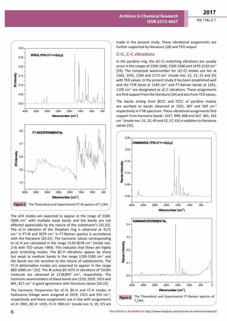
Figure 2 The Theoretical and Experimental FT-IR spectra of T2CNH. 3500 3000 2500 2000 1500 1000 500
0.00
0.05
0.10
0.15
0.20
0.25
RAMAN/B3LYP/6-311++G(d,p)
Ram
an In
tens
ity
Wavenumber (cm-1)
3500 3000 2500 2000 1500 1000 500 0
0.0
0.1
0.2
0.3
0.4RAMAN/EXPERIMENTAL
Ram
an In
tens
ity
Wavenumber (cm-1)
Figure 3 The Theoretical and Experimental FT-Raman spectra of T2CNH.
The νCH modes are expected to appear in the range of 3100-3000 cm-1 with multiple weak bands and the bands are not affected appreciably by the nature of the substituent’s [31,32]. The νC-H vibration of the thiophen ring is observed at 3171 cm-1 in FT-IR and 3079 cm-1 in FT-Raman spectra in accordance with the literature [20-22]. The harmonic values corresponding to νC-H are calculated in the range 3120-3078 cm-1 (mode nos: 2-4) with TED values >96%. This indicates that these are highly pure stretching modes. The βC-H vibrations appear by sharp but weak to medium bands in the range 1100-1500 cm-1 and the bands are not sensitive to the nature of substitutents. The ΓC-H deformation modes are expected to appear in the range 800-1000 cm-1 [32]. The IR active βC-H/ΓC-H vibrations of T2CNH molecule are observed at 1218/897 cm-1, respectively. The harmonic wavenumbers of these bands are 1219, 1059, 1015 and 891, 817 cm-1 in good agreement with literature values [20-22].
The harmonic frequencies for νC-H, βC-H and ΓC-H modes in hydrozone linkage were assigned at 2919, 1315 and 910 cm-1, respectively and these assignments are in line with assignments νC-H: 2901, βC-H: 1335, ΓC-H: 908 cm-1 (mode nos: 9, 19, 37) are
7© Under License of Creative Commons Attribution 3.0 License
Vol. 1 No. 2: 7
2017Archives in Chemical Research
ISSN 2572-4657
The ring carbon-carbon stretching vibrations in thiophen ring are reported in the ranges of 1329-1431, 1420-1501 and 1419-1519 cm-1, respectively [20-22]. For the same mode the computed wavenumbers are: 1508 (C=C), 1490 (C=C) and 1402 cm-1 (C-C). These assignments are having considerable TED values ≥ 15% and its corresponding mode no: 14 is further supported by observed Raman band 1519 cm-1. The βCCC mode of thiophene moiety is ascribed to mode no: 44 (718 cm-1), this supported by TED value (57%) also. The mode nos: 27 and 24 are attributed to νC2-C9 and νC14-C16 mode and are in agreement with literature [28].
C=N, C-N vibrations The identifications of C=N and C-N vibrations is a difficult task, since the mixing of vibrations is possible in this region [36]. However, with the help of Gauss View (3.0) software and TED results, those vibrations are described and assigned in this study. The C=N and C-N stretching vibrations appear in the ranges of 1670-1600 cm-1 and 1382-1266 cm-1 respectively [36]. In hydrozone linkage, the νC9=N11 and νC14-N12 stretching vibrations are assigned respectively at 1592 cm-1 (FTIR) /1589 cm-1 (FT-Raman) and at 1096 cm-1 (FT-Raman). The TED results show that these vibrations are mixed with βHCN and νC-C modes and their corresponding harmonic frequencies are 1591 (mode no: 11) and 1097 cm-1 (mode no: 27) well correlated with experimental observations. These vibrational assignments are also supported by literature [28] in addition to TED output (75% and 40%).
In pyridine ring, the harmonic/observed bands at 1563 (mode no: 12), 1240 (mode no: 22)/1241 cm-1 in FT-Raman spectrum can be assigned to C-N stretching vibration and TED also predict that these vibrations are mixed with νC-C modes. These assignments are in good agreement with our earlier study [28] and also find support from TED values (30%, 43%). The βC21-C23-N19, βC16-C17-N19, βC17-N19-C23 and βC2-C9=N11, βC16-C14-N12 deformations belong to pyridine and hydrozone moieties, respectively assigned to 1563, 1391, 999 and 791, 507 cm-1 (mode nos: 12, 18, 33 and 43, 52). Similarly the τC16C17C23N19, τC18C21N19C23, τC3C2C9N11, ΓC16C14N12N11 modes are assigned to wavenumbers: 690, 381, 217, 217 cm-1 (mode nos: 47, 57, 61, 61).
N-N vibrations The νN-N stretching was observed at 1145 cm-1 in FTIR is undoubtedly assigned to νN11-N12 vibration and the value of this band is calculated at 1148 cm-1 (mode no: 26) with a TED of (60%) [28]. This assignment is well correlated with observed FT-Raman band 1149 cm-1. The in-plane bending vibrations of C2-C9=N11, C9=N11-N12 & C14-N12-N11 are assigned to wavenumbers: 791, 45 & 885, having considerable TED values (≥ 20%). The wavenumbers 163, 326 cm-1 (mode nos: 63, 59) are assigned to τC9=N11-N12-C14, τC2-C9=N11-N12 modes.
C-S vibrations In T2CNH the scaled wavenumbers 791 and 718 cm-1 (mode nos: 43 and 44) are assigned to νC-S modes of thiophen ring moiety. This assignment is in agreement with the assignments proposed by various authors [20,21]. This mode is well known to mix with neighboring modes (νCC, βCCN/mode no: 43 and βCCC/mode no: 44) as reported in the literature [37]. The harmonic frequencies of βCSC and ΓCSC vibrations are ascribed to wavenumbers: 586 cm-1 (mode no: 50) and 125 cm-1 (mode no: 64) with 62% and 37% of TED values, respectively. Further the wavenumbers 827 cm-1 (mode no: 40) and 205 cm-1 (mode no: 62) are assigned to βC4C5S1 and βC9C2S1 modes, respectively, which are in line with the observed bands (846 cm-1: FTIR and 191 cm-1: FT-Raman) in addition to support the TED values [80% and 30%].
C=O vibrations The C=O is formed by Pπ-Pπ bonding between carbon and oxygen atoms. Carbonyl (C=O) group stretching vibration is expected to appear in the region of 1680-1715 cm-1 [38]. In this study, the carbonyl group stretching vibration appear at 1670 cm-1 as strong band in FT-IR and at 1673 cm-1 as weak band in FT-Raman spectra. The values of νC=O band is calculated at 1688 cm-1 (mode no: 10) with a TED of 85%. The βC=O and ΓC=O vibrations are assigned respectively to mode nos: 39 and 45, in which the predicted wavenumber related to ΓC=O mode is found to be in moderate agreement with the observed FTIR band at 700 cm-1.
NLO PropertyAnalysis of organic compounds having conjugated π-electron systems and large hyperpolarizability using IR and Raman spectroscopy has evolved as a subject of scientific research. The application of the title molecule in the field of non-linear optics demands the investigation of its structural and bonding features
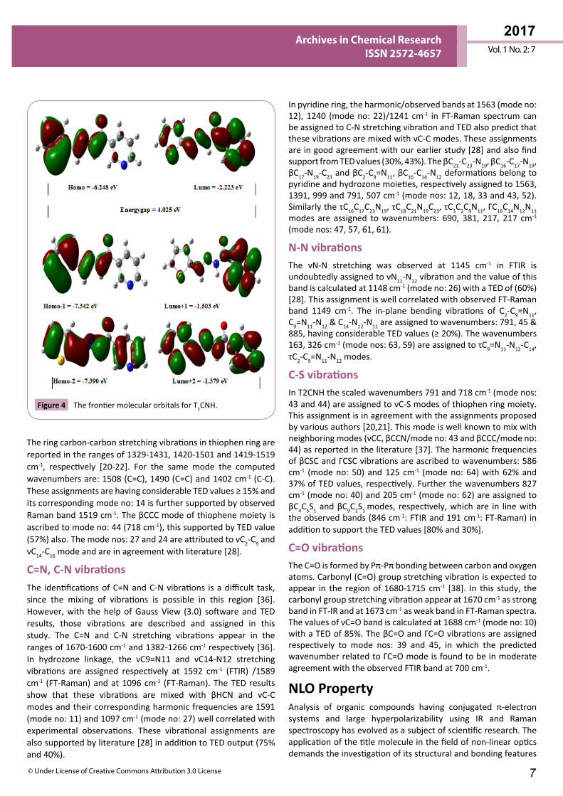
Figure 4 The frontier molecular orbitals for T2CNH.
2017Vol. 1 No. 2: 7
8 This Article is Available in: http://www.imedpub.com/archives-in-chemical-research/
Archives in Chemical Research ISSN 2572-4657
contributing to the hyperpolarizability enhancement by analyzing the vibrational modes using IR and Raman spectroscopy. The DFT/B3LYP/6-311++G(d,p) basis set has been used for the prediction of first hyperpolarizability.
The calculated first hyperpolarizability and the total molecular dipole moment of T2CNH is 1.5861 × 10⁻³⁰ esu, 0.9906 Debye, respectively obtained by B3LYP/6-311++G(d,p) level of theory. The total dipole moment of the title molecule is moderately equal and β0 value of T2CNH is 4 times greater than that of urea, hence the molecule T2CNH has considerable non-linear optical (NLO) activity and the hyperpolarizabilities of T2CNH are given in Table 3.
NBO AnalysisThis method gives information about the intra- and inter-molecular interactions among bonds. Furthermore, it provides a convenient basis for investigating the interactions in both filled and virtual orbital spaces along with charge transfer and conjugative interactions in molecular system [39]. The natural bonding orbital (NBO) analysis was performed for T2CNH using B3LYP/6-311++G(d,p) basis set in order to elucidate the intra-molecular, hybridization and delocalization of ED within T2CNH. The strong intra-molecular hyperconjugative interaction of the σ and π electrons of C−C to the anti C−C bond of the pyridine ring leads to stabilization of some part of the pyridine ring. The NBO analysis has been carried out by B3LYP/6-311++G(d,p) basis set and the E(2) values and types of the transition are shown in Table 4.
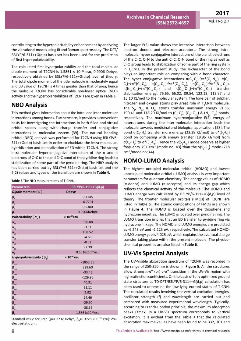
Parameters B3LYP/6-311++G(d,p)Dipole moment ( μ ) Debyeμx 0.3145μy -0.7701μz 0.5380μ 0.9906DebyePolarizability ( α0 ) × 10-30esuαxx 330.88αxy -3.11αyy 168.52αxz -4.63αyz -0.11αzz 97.39α0 0.5318x10⁻³⁰esuHyperpolarizability ( β0 ) × 10-30esuβxxx -1853.85βxxy 229.60βxyy -33.45βyyy -129.96βxxz 40.31βxyz 21.11βyyz 2.92βxzz 54.46βyzz -29.90βzzz -38.55β0 1.5861x10⁻³⁰esu
Table 3 The NLO measurements of T2CNH.
Standard value for urea (μ=1.3732 Debye, β0=0.3728 × 10⁻³⁰ esu): esu-electrostatic unit
The larger E(2) value shows the intensive interaction between electron donors and electron acceptors. The strong intra-molecular hyper conjugative interactions of the σ and π electrons of the C=C, C=N to the anti C=C, C=N bond of the ring as well as C=O group leads to stabilization of some part of the ring system in T2CNH. In the present study, the π-character of the bond plays an important role on comparing with σ bond character. The hyper conjugative interactions π(C2-C3)→π*(C9-N11), π(C4-C5)→π*(C2-C3), π(C16-C17)→π*(C18-C21), π(C18-C21)→π*(N19-C23), π(N19-C23)→π*(C16-C17) and π(C14-O15)→π*(C16-C17) transfer stabilization energy: 76.65, 66.02, 89.54, 122.13, 112.97 and 15.10 KJ/mol to the molecular system. The lone pair of sulphar, nitrogen and oxygen atoms play great role in T2CNH molecule. The S1, N12 & O15 atoms transfer maximum energy 91.55, 190.41 and 118.20 KJ/mol to (C4-C5), (C14-O15) & (N12-C14) bonds, respectively. The maximum hyperconjucative E(2) energy of heteroatoms during the inter-molecular interaction leads the molecule towards medicinal and biological applications [28]. The bond σ(C3-H8) transfer more energy (23.39 KJ/mol) to σ*(S1-C2) bond on comparing with energy transfer (18.95 KJ/mol) from σ(C4-H7) to σ*(S1-C5). Hence the ν(S1-C2) mode observe at higher frequency 791 cm-1 (mode no: 43) than the ν(S1-C5) mode (718 cm-1/mode no: 44).
HOMO-LUMO Analysis The highest occupied molecular orbital (HOMO) and lowest unoccupied molecular orbital (LUMO) analysis is very important parameters for quantum chemistry. The energy values of HOMO (π-donor) and LUMO (π-acceptor) and its energy gap which reflects the chemical activity of the molecule. The HOMO and LUMO energy was calculated by B3LYP/6-311++G(d,p) level of theory. The frontier molecular orbitals (FMOs) of T2CNH are listed in Table 5. The atomic compositions of FMOs are shown in Figure 4. The HOMO is located over the thiophene and hydrozone moieties. The LUMO is located over pyridine ring. The LUMO transition implies that an ED transfer to pyridine ring via hydrazone linkage. The HOMO and LUMO energies are predicted as -6.248 eV and -2.223 eV, respectively. The calculated HOMO-LUMO energy gap is 4.025 eV, which explains the eventual charge transfer taking place within the present molecule. The physico-chemical properties are also listed in Table 6.
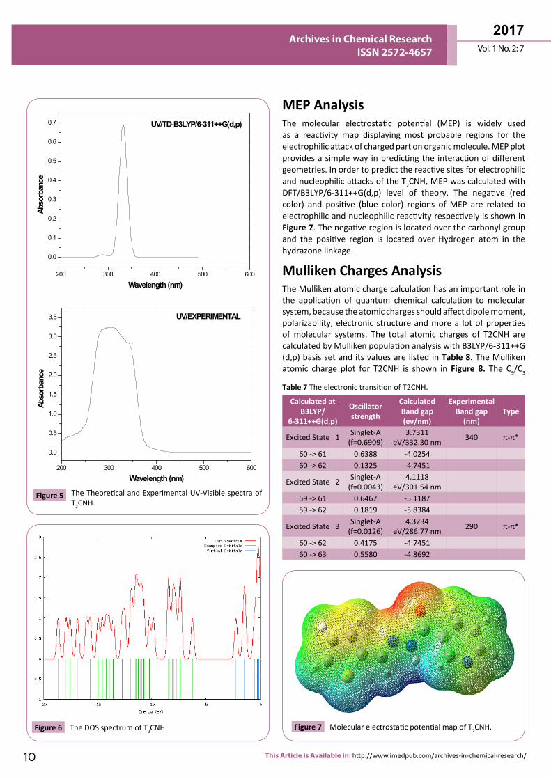
UV-Vis Spectral Analysis The UV-Visible absorption spectrum of T2CNH was recorded in the range of 250-350 nm is shown in Figure 5. All the structures allow strong π-π* (or) σ-σ* transition in the UV-Vis region with high extinction coefficients. On the basis of fully optimized ground state structure at TD-DFT/B3LYP/6-311++G(d,p) calculation has been used to determine the low-lying excited states of T2CNH. The calculated results involving the vertical excitation energies, oscillator strength (f) and wavelength are carried out and compared with measured experimental wavelength. Typically, according to Franck-Condon principle, the maximum absorption peaks (λmax) in a UV-Vis spectrum corresponds to vertical excitation. It is evident from the Table 7 that the calculated absorption maxima values have been found to be 332, 301 and

9© Under License of Creative Commons Attribution 3.0 License
Vol. 1 No. 2: 7
2017Archives in Chemical Research
ISSN 2572-4657
Type Donor NBO (i) ED/e Acceptor NBO (j) ED/e E(2) kJ/mol E(j)-E(i) a.u. F(i,j) a.u.π -π* BD (2) C2 - C3 1.795 BD*(2) C4 - C5 0.309 69.58 0.28 0.06
BD*(2) C9 - N11 0.209 76.65 0.28 0.06σ -σ* BD (1) C3 - H8 1.972 BD*(1) S1 - C2 0.028 23.39 0.73 0.06
BD*(1) C2 - C3 0.021 7.57 1.12 0.04BD*(1) C4 - C5 0.014 8.08 1.11 0.04
π -π* BD (2) C4 - C5 1.846 BD*(2) C2 - C3 0.351 66.02 0.30 0.06σ -σ* BD (1) C4 - H7 1.976 BD*(1) S1 - C5 0.017 18.95 0.75 0.05
BD*(1) C2 - C3 0.021 8.03 1.12 0.04BD*(1) C4 - C5 0.014 5.73 1.12 0.04
π -π* BD (2) C14 - O15 1.980 BD*(2) C16 - C17 0.335 15.10 0.40 0.04π -π* BD (2) C16 - C17 1.633 BD*(2) C14 - O15 0.277 65.98 0.3 0.06
BD*(2) C18 - C21 0.277 89.54 0.29 0.07BD*(2) N19 - C23 0.366 69.71 0.27 0.06
π -π* BD (2) C18 - C21 1.636 BD*(2) C16 - C17 0.335 74.85 0.28 0.06BD*(2) N19 - C23 0.366 122.13 0.27 0.08
π -π* BD (2) N19 - C23 1.706 BD*(2) C16 - C17 0.335 112.97 0.32 0.08BD*(2) C18 - C21 0.277 52.59 0.33 0.06
n -π* LP (2) S1 1.622 BD*(2) C2 - C3 0.351 87.15 0.27 0.07BD*(2) C4 - C5 0.309 91.55 0.26 0.07
n -π* LP (2) N12 1.667 BD*(2) C9 - N11 0.209 115.14 0.29 0.08BD*(2) C14 - O15 0.017 4.44 0.88 0.03BD*(2) C14 - O15 0.277 190.41 0.32 0.11
n -σ* LP (1) O15 1.855 BD*(1) N12 - C14 0.084 118.2 0.67 0.12BD*(1) C14 - C16 0.069 80.37 0.66 0.10
n -σ* LP (1) N19 1.916 BD*(1) C16 - C17 0.033 39.33 0.90 0.08
Table 4 The second order perturbation theory analysis of Fock Matrix in NBO basis for T2CNH.
Occupancy Orbital energies (a.u) Orbital energies (eV) Kinetic energies (a.u)O56 -0.292 -7.947 1.641O57 -0.284 -7.740 1.641O58 -0.271 -7.390 1.582O59 -0.269 -7.342 2.186O60 -0.229 -6.248 1.567V61 -0.081 -2.223 1.590V62 -0.055 -1.503 1.586V63 -0.055 -1.503 1.424V64 -0.019 -0.519 0.488V65 -0.009 -0.255 0.535
Table 5 The frontier molecular orbital of T2CNH.
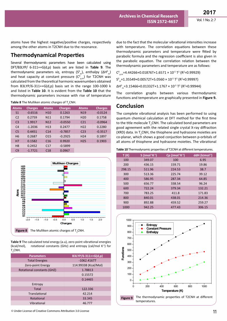
286 nm which correlates well with the experimental values 340 and 290 nm. The more intense band at 340 nm has maximum oscillator strength (f=0.6909), corresponds to Homo-Lumo transition and is mostly characterized as n-π* type. This type of transition is attributed to the presence of large no of free lone pairs of electrons available on sulphar (S1), Nitrogen (N11, N12) and oxygen (O15) atoms. The experimental and theoretical UV-Vis absorption spectrum is shown in Figure 5. The density of states spectrum of T2CNH is shown in Figure 6. It was used to calculate group contributions to the molecular orbitals (HOMO and LUMO). DOS plot shows population analysis per orbital and demonstrates a simple view of the character of the molecular orbitals (MOs) in a certain energy range.
Parameters ValuesHOMO -6.248 eVLUMO -2.223 eV
Energy gap 4.025 eVIonization potential (IP) 6.248 eV
Electron affinity (EA) 2.223 eVElectrophilicity Index (ω) 2.562
Chemical Potential (µ) 7.359Electro negativity (χ) -7.359
Hardness (η) -4.025
Table 6 The Physico-Chemical properties of T2CNH.
2017Vol. 1 No. 2: 7
10 This Article is Available in: http://www.imedpub.com/archives-in-chemical-research/
Archives in Chemical Research ISSN 2572-4657
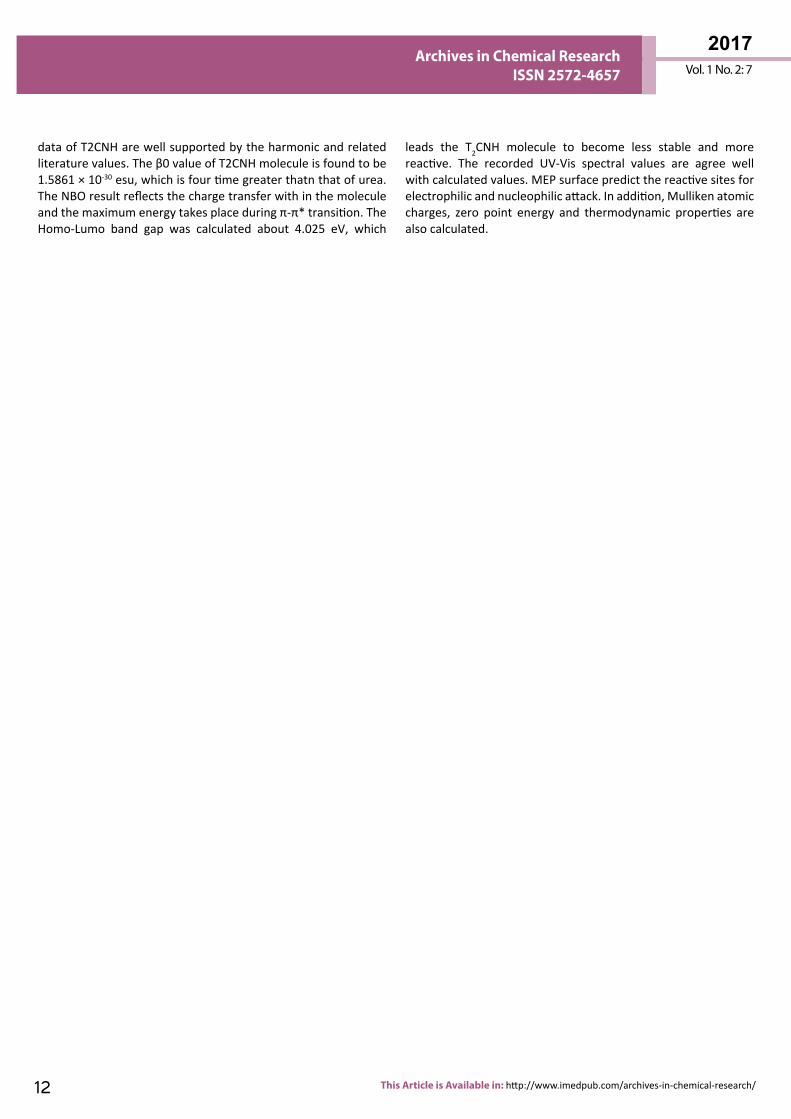
MEP Analysis The molecular electrostatic potential (MEP) is widely used as a reactivity map displaying most probable regions for the electrophilic attack of charged part on organic molecule. MEP plot provides a simple way in predicting the interaction of different geometries. In order to predict the reactive sites for electrophilic and nucleophilic attacks of the T2CNH, MEP was calculated with DFT/B3LYP/6-311++G(d,p) level of theory. The negative (red color) and positive (blue color) regions of MEP are related to electrophilic and nucleophilic reactivity respectively is shown in Figure 7. The negative region is located over the carbonyl group and the positive region is located over Hydrogen atom in the hydrazone linkage.
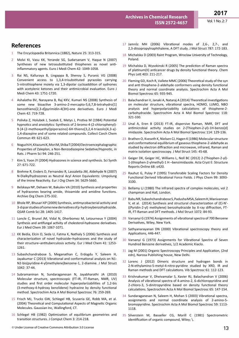
Mulliken Charges AnalysisThe Mulliken atomic charge calculation has an important role in the application of quantum chemical calculation to molecular system, because the atomic charges should affect dipole moment, polarizability, electronic structure and more a lot of properties of molecular systems. The total atomic charges of T2CNH are calculated by Mulliken population analysis with B3LYP/6-311++G (d,p) basis set and its values are listed in Table 8. The Mulliken atomic charge plot for T2CNH is shown in Figure 8. The C9/C3
200 300 400 500 600
0.0
0.1
0.2
0.3
0.4
0.5
0.6
0.7 UV/TD-B3LYP/6-311++G(d,p)
Abso
rban
ce
Wavelength (nm)
200 300 400 500 600
0.0
0.5
1.0
1.5
2.0
2.5
3.0
3.5 UV/EXPERIMENTAL
Abso
rban
ce
Wavelength (nm)
Figure 5 The Theoretical and Experimental UV-Visible spectra of T2CNH.
Figure 6 The DOS spectrum of T2CNH. Figure 7 Molecular electrostatic potential map of T2CNH.
Calculated atB3LYP/
6-311++G(d,p)
Oscillator strength
CalculatedBand gap (ev/nm)
ExperimentalBand gap
(nm)Type
Excited State 1 Singlet-A (f=0.6909)
3.7311 eV/332.30 nm 340 π-π*
60 -> 61 0.6388 -4.025460 -> 62 0.1325 -4.7451
Excited State 2 Singlet-A (f=0.0043)
4.1118 eV/301.54 nm
59 -> 61 0.6467 -5.118759 -> 62 0.1819 -5.8384
Excited State 3 Singlet-A (f=0.0126)
4.3234 eV/286.77 nm 290 π-π*
60 -> 62 0.4175 -4.745160 -> 63 0.5580 -4.8692
Table 7 The electronic transition of T2CNH.
11© Under License of Creative Commons Attribution 3.0 License
Vol. 1 No. 2: 7
2017Archives in Chemical Research
ISSN 2572-4657
due to the fact that the molecular vibrational intensities increase with temperature. The correlation equations between these thermodynamic parameters and temperature were fitted by parabolic formula and the regression coefficient is also given in the parabolic equation. The correlation relation between the thermodynamic parameters and temperature are as follows:
C0p,m=4.44266+0.01876T+1.6571 × 10⁻⁵ T2 (R2=0.99929)
S0m=1.35549+0.00572T+5.0560 × 10⁻⁵ T2 (R2=0.99997)
ΔH0m=3.15466+0.01332T+1.1767 × 10⁻⁵ T2 (R2=0.99944)
The correlation graphs between various thermodynamic functions and temperature are graphically presented in Figure 9.
ConclusionThe complete vibrational analysis has been performed to using quantum chemical calculation at DFT method for the first time to the title molecule T2CNH. The calculated bond parameters are good agreement with the related single crystal X-ray diffraction (XRD) data. In T2CNH, the thiophene and hydrazone moieties are co-planar, which shows a good conjuction between p-orbitals of all atoms of thiophene and hydrazone moieties. The vibrational
atoms have the highest negative/positive charges, respectively among the other atoms in T2CNH due to the resonance.
Thermodynamical PropertiesSeveral thermodynamic parameters have been calculated using DFT/B3LYP/ 6-311++G(d,p) basis set are listed in Table 9. The thermodynamic parameters viz, entropy (S0
m), enthalpy (ΔH0m)
and heat capacity at constant pressure (C0p,m) for T2CNH was
calculated from the theoretical harmonic wavenumbers obtained from B3LYP/6-311++G(d,p) basis set in the range 100-1000 k and listed in Table 10. It is evident from the Table 10 that the thermodynamic parameters increase with rise of temperature
S1C2C3C4C5H6H7H8C9
H10N11N12H13C14O15C16C17C18N19H20C21H22C23H24H25
-2.0 -1.5 -1.0 -0.5 0.0 0.5 1.0 1.5 2.0
Charges
Atom
s
Mulliken Charges
Figure 8 The Mulliken atomic charges of T2CNH.
0 200 400 600 800 1000
0
100
200
300
400
500
600
700
800
900
1000
Func
tions
Temperature (K)
Entropy Constant Pressure Enthalpy
Figure 9 The thermodynamic properties of T2CNH at different temperatures.
Parameters B3LYP/6-311++G(d,p)Total Energies -1062.41677
Zero-point Energy 114.99338 (Kcal/Mol)Rotational constants (GHZ) 1.78813
0.155720.14465
EntropyTotal 122.336
Translational 42.214Rotational 33.345Vibrational 46.777
Table 9 The calculated total energy (a.u), zero point vibrational energies (kcal/mol), rotational constants (GHz) and entropy (cal/mol K-1) for T2CNH.
Atoms Charges Atoms Charges Atoms ChargesS1 -0.6516 H10 0.1263 N19 -0.0124C2 0.2759 N11 0.1794 H20 0.1758C3 1.9917 N12 -0.0550 C21 -0.0964C4 -1.2036 H13 0.2477 H22 0.2280C5 0.4451 C14 -0.7857 C23 -0.3517H6 0.2687 O15 -0.2925 H24 0.1897H7 0.1582 C16 0.9920 H25 0.1903H8 0.2452 C17 -0.5899C9 -1.7721 C18 0.0967
Table 8 The Mulliken atomic charges of T2CNH.
T (K) S (Jmol-1K-1) Cp (Jmol-1K-1) ddH (kJmol-1)100 349.07 100 6.95200 436.15 159.71 19.86
298.15 511.96 224.53 38.7300 513.36 225.74 39.12400 586.95 287.34 64.85500 656.77 338.54 96.24600 722.24 379.34 132.21700 783.25 411.8 171.83800 840.01 438.01 214.36900 892.88 459.52 259.27
1000 942.25 477.43 306.15
Table 10 Thermodynamic properties of T2CNH at different temperatures.
2017Vol. 1 No. 2: 7
12 This Article is Available in: http://www.imedpub.com/archives-in-chemical-research/
Archives in Chemical Research ISSN 2572-4657
data of T2CNH are well supported by the harmonic and related literature values. The β0 value of T2CNH molecule is found to be 1.5861 × 10⁻³⁰ esu, which is four time greater thatn that of urea. The NBO result reflects the charge transfer with in the molecule and the maximum energy takes place during π-π* transition. The Homo-Lumo band gap was calculated about 4.025 eV, which
leads the T2CNH molecule to become less stable and more reactive. The recorded UV-Vis spectral values are agree well with calculated values. MEP surface predict the reactive sites for electrophilic and nucleophilic attack. In addition, Mulliken atomic charges, zero point energy and thermodynamic properties are also calculated.
13© Under License of Creative Commons Attribution 3.0 License
Vol. 1 No. 2: 7
2017Archives in Chemical Research
ISSN 2572-4657
References1 The Encyclopœdia Britannica (1882), Nature 25: 313-315.
2 Molvi KI, Vasu KK, Yerande SG, Sudarsanam V, Haque N (2007) Syntheses of new tetrasubstituted thiophenes as novel anti-inflammatory agents. Euro J Medi Chem 42: 1049-1058.
3 Rai NS, Kalluraya B, Lingappa B, Shenoy S, Puranic VG (2008) Convenient access to 1,3,4-trisubstituted pyrazoles carrying 5-nitrothiophene moiety via 1,3-dipolar cycloaddition of sydnones with acetylenic ketones and their antimicrobial evaluation. Euro J Medi Chem 43: 1751-1720.
4 Ashalatha BV, Narayana B, Raj KKV, Kumari NS (2008) Synthesis of some new bioactive 3-amino-2-mercapto-5,6,7,8-tetrahydro[1]benzothieno[2,3-d]pyrimidin-4(3H)-one derivatives. Euro J Medi Chem 42: 719-728.
5 Polivka Z, Holubek J, Svatek E, Metys J, Protiva M (1984) Potential hypnotics and anxiolytics: Synthesis of 2-bromo-4-(2-chlorophenyl)-9-[4-(2-methoxyethyl)piperazino]-6H-thieno[3,2,4-triazolo[4,3-a]-1,4-diazepine and of some related compounds. Collect Czech Chem Commun 49: 621-623.
6 Noguchi H, Kitazumi K, Mori M, Shiba T (2004) Electroencephalographic Properties of Zaleplon, a Non-Benzodiazepine Sedative/Hypnotic, in Rats. J Pharm Sci 94: 246-251.
7 Kim S, Yoon JY (2004) Hydrazones in science and synthesis. Sci Synth 27: 671-722.
8 Brehme R, Enders D, Fernandez R, Lassaletta JM, Aldehyde N (2007) N-Dialkylhydrazones as Neutral Acyl Anion Equivalents: Umpolung of the Imine Reactivity. Eur J Org Chem 34: 5629-5660.
9 Belskaya NP, Dehaen W, Bakulev VA (2010) Synthesis and properties of hydrazones bearing amide, thioamide and amidine functions. Archive Org Chem 275-332.
10 Bhole RP, Bhusari KP (2009) Synthesis, antimycobacterial activity and 3-d qsar studies of some new derivatives of p-hydroxybenzohydrazide. QSAR Comb Sci 28: 1405-1417.
11 Loncle C, Brunel JM, Vidal N, Dherbomez M, Letourneux Y (2004) Synthesis and antifungal activity of cholesterol-hydrazone derivatives. Eur J Med Chem 39: 1067-1071.
12 KK Bedia, Elcin O, Seda U, Fatma K, Nathaly S (2006) Synthesis and characterization of novel hydrazide–hydrazones and the study of their structure–antituberculosis activity. Eur J Med Chem 41: 1253-1261.
13 Subashchandrabose S, Meganathan C, Erdogdu Y, Saleem H, Jayakumar C (2013) Vibrational and conformational analysis on N1-N2-bis(pyridine-4-yl)methylene)benzene-1, 2-diamine. J Mol Struct 1042: 37-44.
14 Subramaninan N, Sundaraganesan N, Jayabharathi JA (2010) Molecular structure, spectroscopic (FT-IR, FT-Raman, NMR, UV) studies and first order molecular hyperpolarizabilities of 1,2-bis (3-methoxy-4-hydroxy benzlidene) hydrazine by density functional method. Spectrochim Acta A Mol Biomol Spectrosc 76: 259-269.
15 Frisch MJ, Trucks GW, Schlegel HB, Scuseria GE, Robb MA, et al. (2004) Theoretical and Computational Aspects of Magnetic Organic Molecules. Gaussian Inc, Wallingford, CT.
16 Schlegel HB (1982) Optimization of equilibrium geometries and transition structures. J Comput Chem 3: 214-218.
17 Jamróz MH (2006) Vibrational modes of 2,6-, 2,7-, and 2,3-diisopropylnaphthalene, A DFT study. J Mol Struct 787: 172-183.
18 Michalska D (2003) Raint Program, Wroclaw University of Technology, Poland.
19 Michalska D, Wysokinski R (2005) The prediction of Raman spectra of platinum(II) anticancer drugs by density functional theory. Chem Phys Lett 403: 211-217.
20 Fleming GD, Koch R, Vallete MMC (2006) Theoretical study of the syn and anti thiophene-2-aldehyde conformers using density functional theory and normal coordinate analysis. Spectrochim Acta A Mol Biomol Spectrosc 65: 935-945.
21 Balachandran V, Janaki A, Nataraj A (2014) Theoretical investigations on molecular structure, vibrational spectra, HOMO, LUMO, NBO analysis and hyperpolarizability calculations of thiophene-2-carbohydrazide. Spectrochim Acta A Mol Biomol Spectrosc 118: 321-330.
22 Unal A, Eren B (2013) FT-IR, dispersive Raman, NMR, DFT and antimicrobial activity studies on 2-(Thiophen-2-yl)-1H-benzo[d]imidazole. Spectrochim Acta A Mol Biomol Spectrosc 114: 129-136.
23 Brathen O, Kveseth K, Nielsen CJ, Hagen K (1986) Molecular structure and conformational equilibrium of gaseous thiophene-2-aldehyde as studied by electron diffraction and microwave, infrared, Raman and matrix isolation spectroscopy. J Mol Struct 145: 45-68.
24 Geiger DK, Geiger HC, Williams L, Noll BC (2012) 2-(Thiophen-2-yl)-1-(thiophen-2-ylmethyl)-1 H –benzimidazole. Acta Cryst E: Structure Reports Online 68: o420.
25 Rauhut G, Pulay P (1995) Transferable Scaling Factors for Density Functional Derived Vibrational Force Fields. J Phys Chem 99: 3093-3100.
26 Bellamy LJ (1980) The infrared spectra of complex molecules, vol 2 champman and Hall, London.
27 Babu NR, Subashchandrabose S, Padusha MSA, Saleem H, Manivannan V, et al. (2014) Synthesis and structural characterization of (E)-N′-((Pyridin-2-yl) methylene) benzohydrazide by X-ray diffraction, FT-IR, FT-Raman and DFT methods. J Mol Struct 1072: 84-93.
28 Varsanyi G (1974) Assignments of vibrational spectra of 700 Benzene Derivatives, Wiley, New York.
29 Sathyanarayanan DN (2000) Vibrational spectroscopy theory and Applications, 446-447.
30 Varsanyi G (1973) Assignments for Vibrational Spectra of Seven Hundred Benzene derivatives, 1/2 Academic Kiaclo.
31 Jag M (2001) Organic Spectroscopy Principles and Application, (2nd edn), Narosa Publishing house, New Delhi.
32 Lorenc J (2012) Dimeric structure and hydrogen bonds in 2-N-ethylamino-5-metyl-4-nitro-pyridine studied by XRD, IR and Raman methods and DFT calculations. Vib Spectrosc 61: 112-123.
33 Krishnakumar V, Dheivamalar S, Xavier RJ, Balachandran V (2006) Analysis of vibrational spectra of 4-amino-2, 6-dichloropyridine and 2-chloro-3, 5-dinitropyridine based on density functional theory calculations. Spectrochim Acta A Mol Biomol Spectrosc 65: 147-154.
34 Sundaraganesan N, Saleem H, Mohan S (2003) Vibrational spectra, assignments and normal coordinate analysis of 2-amino-5-bromopyridine. Spectrochim Acta A Mol Biomol Spectrosc 59: 1113-1118.
35 Silverstein M, Basseller CG, Morill C (1981) Spectrometric identification of organic compound, Wiley; \.

2017Vol. 1 No. 2: 7
14 This Article is Available in: http://www.imedpub.com/archives-in-chemical-research/
Archives in Chemical Research ISSN 2572-4657
36 Badawi HM (2009) Structural stability, C–N internal rotations and vibrational spectral analysis of non-planar phenylurea and phenylthiourea. Spectrochim Acta A Mol Biomol Spectrosc 72: 523-527.
37 James C, Ravikumar C, Sundius T, Krishnakumar V, Kesavamoorthy R, et al. (2008) FT-Raman and FTIR spectra, normal coordinate analysis
and ab initio computations of (2-methylphenoxy) acetic acid dimer. Vib Spectrosc 47: 10-20.
38 Weinhold F, Landis CR (2005) Valency and bonding: a natural bond orbital donor-acceptor perspective. Cambridge University Press.
39 Ott JB, Boerio-Goates J (2000) Calculations from Statistical Thermodynamics.