fragile x syndrome and acute lymphoblastic leukemia
TRANSCRIPT

Fragile X Syndrome and Acute Lymphoblastic Leukemia
MICHAEL CUNNINGHAM, BA’ AND JOSEPH D. DICKERMAN, MDt
Fragile X [Fra(X)] syndrome is an example of a heritable fragility syndrome associated with mental retardation. It is characterized by a fragile site on the X chromosome at Xq27-28. There have recently been three reports of malignant solid tumors associated with Fra(X) syndrome. We describe the first case of a hematologic malignancy p-cell acute lymphocytic leukemia (ALL)] in a patient with Fra(X) syndrome. The possibility of a predisposition to malignancy in Fra(X) is discussed.
Cancer 62:2383-2386, 1988.
HE COEXISTENCE OF MALIGNANT ganglioglioma, T testicular carcinoma, and colonic adenocarcinoma with Fra(X) has been reported.”* In addition, two benign tumors have been described in patients with Fra(X).334 This article represents, to our knowledge, the first reported case of a patient with Fra(X) who developed a hematologic malignancy [T-cell acute lymphocytic leukemia (ALL)]. The discovery of a transforming sequence of Xq275 and the knowledge of an increased rate of nonrandom trans- location across the fragile site6 increases the importance of recording the occurrence of malignancies in patients with Fra(X).
Case Report
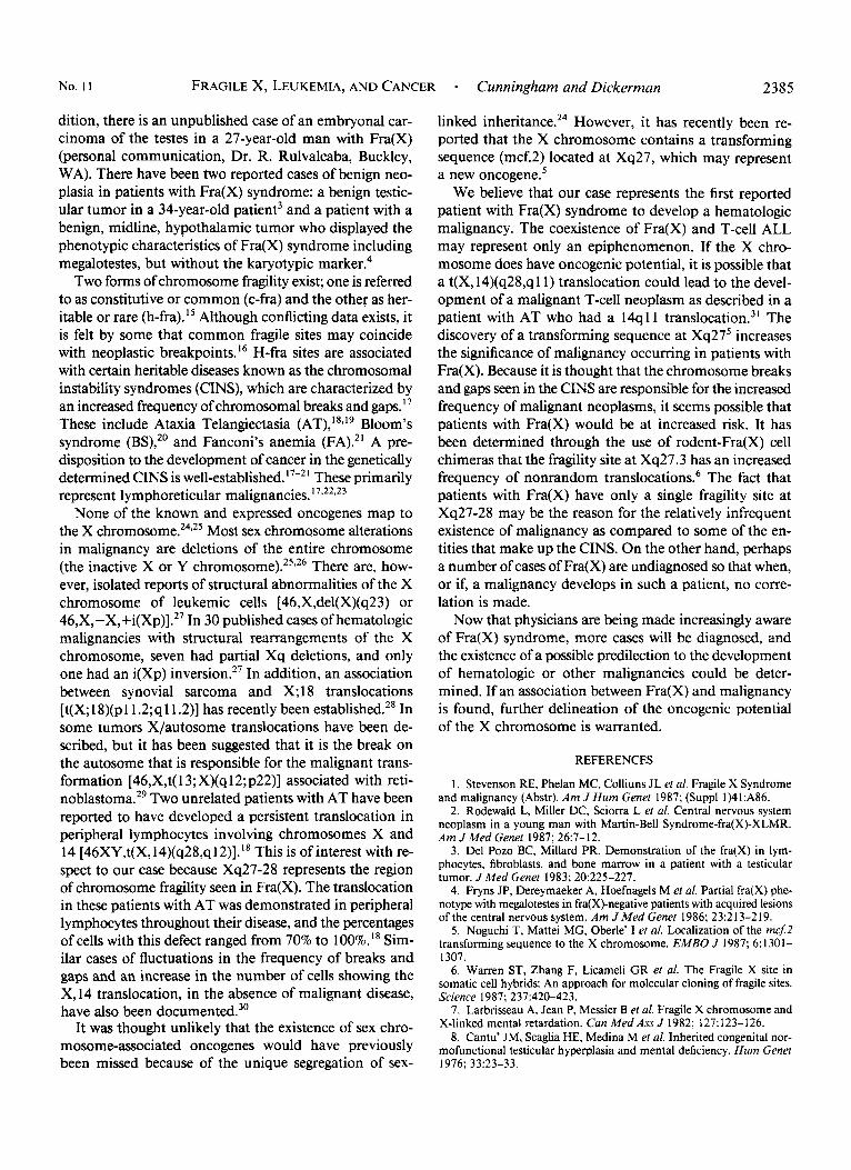
Our patient was a 3Yz-year-old white boy with a lifelong history of developmental delay. At 18 months of age he was evaluated for this problem. Significant findings on physical exam included large ears (5.8 cm, >97%) and mild nail hypoplasia. He dem- onstrated autistic characteristics including head banging, rocking, and moderate developmental delay (8 months). Giemsa-stained lymphocytes grown in folate-deficient medium demonstrated the presence of a fragile site at Xq27-28 in 16 of 50 cells ex- amined. This finding is consistent with the diagnosis of Fra(X) syndrome (Fig. 1).
Three years later he was admitted to the Medical Center Hos- pital of Vermont with a 5-day history of irritability, decreased oral intake, lethargy, low-grade fever, and increased bruisability. Physical examination was significant for a pale, irritable, dys- morphic-appearing child in mild distress. Testicular volume was
From the Departments of *Pathology and ?Pediatrics, University of Vermont, College of Medicine, Burlington, Vermont.
The authors thank Dr. Peter Nowell (University of Pennsylvania, School of Medicine) for his advice and assistance in attempting DNA probing of the formalin-fixed tissue, Drs. Alan Guttmacher and Jackson Clemmons (University of Vermont, College of Medicine) and Robert Miller (National Institutes of Health) for their advice and assistance, and Ms. Nancy Moreland for typing this manuscript.
Address for reprints: Joseph D. Dickerman, MD, Department of Pe- diatrics, University of Vermont, College of Medicine, Given Building, Burlington, VT 05405.
Accepted for publication June 9, 1988.
grossly normal to physical exam [the absence of testicular en- largement in this patient is consistent with the usual onset of macroorchidism after puberty in patients with Fra(X)].7.8 It was noticed that he had multiple petechiae, ecchymoses, and purpura over the entire body, mild, bilateral nystagmus, a small right retinal hemorrhage, prominent pinnae, generalized lymphade- nopathy, an enlarged liver and spleen, and hypoplastic nails. He alternated between sleep and restlessness, and his strength and response to tactile stimuli were normal. There was no mediastinal widening seen in a chest roentgenogram. Initial laboratory find- ings were as follows: hemoglobin, 7.4 g/dl; hematocrit, 20%; leukocyte count, 568,00O/pl(2% neutrophils, 2% lymphocytes, and 96% blasts); platelet count, 23,OOO/pl; and uric acid, 8.6 mgldl.
Six hours after admission, he vomited and had a brief, gen- eralized seizure requiring bag and mask ventilatory assistance. He was placed on 100% oxygen, and after another brief seizure, he was stabilized through the night. He was transfused with 200 cc of packed red cells and five units of platelets. Approximately 13 hours after admission, another six units of platelets were transfused, and a bone marrow aspirate and lumbar puncture were performed. Lumbar puncture demonstrated 11 erythro- cyteslpl and two leukocytes/pl. Shortly thereafter, he had a car- diac arrest, and despite aggressive resuscitative efforts, expired 15 hours after his admission. Knowledge of the previous diag- nosis of Fra(X) was not known until shortly after completion of the autopsy.
The premortem bone marrow aspirate revealed 2% granulo- cytes, 1% erythroid cells, 4% lymphocytes, and 94% blasts. Im- munologic studies of the blasts demonstrated 58% E-rosettes, 2% polyvalent surface immunoglobulins, and 12% B- 1 clones. Based on the E-rosette value of 5870, the demonstration of E- rosetting blasts on a cytocentrifuged, Wright-stained preparation and the morphologic appearance of the cells, the diagnosis of T-cell acute lymphoblastic leukemia, L2, was made. Further immunologic studies were inconclusive due to the nonviability of bone marrow cells.
Autopsy
Significant physical features included the following: height, 101 cm; weight, 12.77 kg; head circumference, 49
2383

2384 CANCER December 1 1988 Vol. 62
FIG. 1. Giemsa-stained lymphocytes grown in folate-deficient medium show a fragile site at Xq27-28.
cm; large ears (5.75 cm >75%), and a low nasal bridge. There was generalized lymphadenopathy, enlargement of the liver and spleen (liver, 625 g; spleen, 400 g), and an enlarged thymus (64 g). The examination of the lungs demonstrated pulmonary infiltration with leukemic cells and pulmonary edema. An examination of the central nervous system revealed multiple, 2- to 8-mm hemor- rhages in all coronal sections of the cerebrum and cere- bellum. In addition, an examination of the brain stem demonstrated a 2-mm hemorrhage in the region of the left basis pontis and a 4-mm hemorrhage within the area of the left motor nucleus of the vagus nerve. The histologic appearance of the hemorrhages was characterized by a central zone of leukemic cells with surrounding hemor- rhage into the white matter. All vessels within the sub- stance of the cortex were filled with leukemic cells, and their walls were in various stages of destruction. There was marked leptomeningeal infiltration with leukemic cells.
Karyotypic analysis of the premortem bone marrow cells was hampered by their nonviability. Of eight cells examined by standard Giemsa staining, all displayed a normal karyotype. The cells were not grown in folate- deficient medium because the diagnosis of Fra(X) syn-
drome had previously been established. Attempts to fur- ther characterize the malignant clone with DNA probes to detect rearrangements in DNA extracted from for- malin-fixed thymic tissue were unsuccessful.
Discussion
Fragile X [Fra(X)] syndrome is an X-linked form of mental retardation. The fragile site is present on the long arm of the X chromosome (Xq27-28) in cells grown in folate-deficient media.' The estimated prevalence is one in 1360 men/boys and one in 2073 women/girls." Phys- ical features of Fra(X) syndrome include large head cir- cumference, prominent forehead, long face, high arched palate, and long, prominent ears.791 ' The mast distinctive physical feature is enlarged testicular volume (macroor- chidism) thought to develop in 80% of patients with me& tal retardation and the Fra(X) karyotypic marker.''-I3 No ethnic predilection has been found. l4
There have been only three reported cases of malig- nancy associated with Fra(X). These include a carcinoma of the testis in a 50-year-old man, a mucin-producing adenocarcinoma of the colon in a 14-year-old boy,' and a malignant ganglioglioma in a 17-year-old man.* In ad-
No. 1 1 FRAGILE X, LEUKEMIA, AND CANCER - Cunningham and Dickerman 2385
dition, there is an unpublished case of an embryonal car- cinoma of the testes in a 27-year-old man with Fra(X) (personal communication, Dr. R. Rulvalcaba, Buckley, WA). There have been two reported cases of benign neo- plasia in patients with Fra(X) syndrome: a benign testic- ular tumor in a 34-year-old patient3 and a patient with a benign, midline, hypothalamic tumor who displayed the phenotypic characteristics of Fra(X) syndrome including megalotestes, but without the karyotypic marker.4
Two forms of chromosome fragility exist; one is referred to as constitutive or common (c-fra) and the other as her- itable or rare (h-fra).” Although conflicting data exists, it is felt by some that common fragile sites may coincide with neoplastic breakpoints.I6 H-fra sites are associated with certain heritable diseases known as the chromosomal instability syndromes (CINS), which are characterized by an increased frequency of chromosomal breaks and gaps.I7 These include Ataxia Telangiectasia (AT), I 8 , I 9 Bloom’s syndrome (BS),20 and Fanconi’s anemia (FA).2’ A pre- disposition to the development of cancer in the genetically determined CINS is well-established. 17-21 These primarily represent lymphoreticular malignancies. 17,22,23
None of the known and expressed oncogenes map to the X chromosome.24325 Most sex chromosome alterations in malignancy are deletions of the entire chromosome (the inactive X or Y chromosome).25326 There are, how- ever, isolated reports of structural abnormalities of the X chromosome of leukemic cells [46,X,del(X)(q23) or 46,X,-x,+i(X~)].*~ In 30 published cases of hematologic malignancies with structural rearrangements of the X chromosome, seven had partial Xq deletions, and only one had an i(Xp) in~ersion.~’ In addition, an association between synovial sarcoma and X; 1 8 translocations [t(X; 18)(p11.2;ql1.2)] has recently been established.28 In some tumors X/autosome translocations have been de- scribed, but it has been suggested that it is the break on the autosome that is responsible for the malignant trans- formation [46,X,t( 13; X)(q12;p22)] associated with reti- nobla~toma.’~ Two unrelated patients with AT have been reported to have developed a persistent translocation in peripheral lymphocytes involving chromosomes X and 14 [46XY,t(X,14)(q28,ql2)].l8 This is of interest with re- spect to our case because Xq27-28 represents the region of chromosome fragility seen in Fra(X). The translocation in these patients with AT was demonstrated in peripheral lymphocytes throughout their disease, and the percentages of cells with this defect ranged from 70% to loo%.’* Sim- ilar cases of fluctuations in the frequency of breaks and gaps and an increase in the number of cells showing the X, 14 translocation, in the absence of malignant disease, have also been d~curnented.~’
It was thought unlikely that the existence of sex chro- mosome-associated oncogenes would have previously been missed because of the unique segregation of sex-
linked inheritan~e.’~ However, it has recently been re- ported that the X chromosome contains a transforming sequence (mcf.2) located at Xq27, which may represent a new oncogene.’
We believe that our case represents the first reported patient with Fra(X) syndrome to develop a hematologic malignancy. The coexistence of Fra(X) and T-cell ALL may represent only an epiphenomenon. If the X chro- mosome does have oncogenic potential, it is possible that a t(X,14)(q28,qll) translocation could lead to the devel- opment of a malignant T-cell neoplasm as described in a patient with AT who had a 14qll transl~cation.~’ The discovery of a transforming sequence at Xq27’ increases the significance of malignancy occumng in patients with Fra(X). Because it is thought that the chromosome breaks and gaps seen in the CINS are responsible for the increased frequency of malignant neoplasms, it seems possible that patients with Fra(X) would be at increased risk. It has been determined through the use of rodent-Fra(X) cell chimeras that the fragility site at Xq27.3 has an increased frequency of nonrandom translocations.6 The fact that patients with Fra(X) have only a single fragility site at Xq27-28 may be the reason for the relatively infrequent existence of malignancy as compared to some of the en- tities that make up the CINS. On the other hand, perhaps a number of cases of Fra(X) are undiagnosed so that when, or if, a malignancy develops in such a patient, no corre- lation is made.
Now that physicians are being made increasingly aware of Fra(X) syndrome, more cases will be diagnosed, and the existence of a possible predilection to the development of hematologic or other malignancies could be deter- mined. If an association between Fra(X) and malignancy is found, further delineation of the oncogenic potential of the X chromosome is warranted.
REFERENCES
1. Stevenson RE, Phelan MC, Colliuns JL ef al. Fragile X Syndrome and malignancy (Abstr). Am J Hum Genet 1987; (Suppl 1)41:A86.
2. Rodewald L, Miller DC, Sciorra L et al. Central nervous system neoplasm in a young man with Martin-Bell Syndrome-fra(X)-XLMR. Am JMed Genet 1987; 26:7-12.
3. Del Pozo BC, Millard PR. Demonstration of the fra(X) in lym- phocytes, fibroblasts, and bone marrow in a patient with a testicular tumor. JMed Genet 1983; 20:225-227.
4. Fryns JP, Dereymaeker A, Hoefnagels M et a/. Partial fra(X) phe- notype with megalotestes in fra(X)-negative patients with acquired lesions of the central nervous system. Am J Med Genet 1986; 23:2 13-2 19.
5. Noguchi T, Mattei MG, Oberle’ I et al. Localization of the mcj2 transforming sequence to the X chromosome. EMBO J 1987; 6:1301- 1307.
6 . Warren ST, Zhang F, Licameli GR et al. The Fragile X site in somatic cell hybrids: An approach for molecular cloning of fragile sites. Science 1987; 237:420-423.
7. Larbrisseau A, Jean P, Messier B et al. Fragile X chromosome and X-linked mental retardation. Can Med Ass J 1982; 127: 123-126.
8. Cantu’ JM, Scaglia HE, Medina M el al. Inherited congenital nor- mofunctional testicular hyperplasia and mental deficiency. Hum Genet 1976; 33~23-33.

2386 CANCER December I 1988 Vol. 62
9. Nussbaum RL, Ledbetter DH. Fragile X syndrome: A unique mu- tation in man. Ann Rev Genet 1986; 20:109-45.
10. Webb TP, Bundey SE, Thake A1 et al. Population incidence and segregation ratios in the martin bell syndrome. Am J Med Genet 1986;
I 1. Chudley AE, Hagerman RJ. Fragile X Syndrome. J Pediatr 1987;
12. Brown WT, Mezzacappa PM, Jenkins EC. Screening for fragile X syndrome by testicular size measurement. Lancet 198 1; I : 1055.
13. Ruvalcaba RHA, Myhre SA, Roosen-Runge EC et al. X-linked mental deficiency megalotestes syndrome. JAMA 1977; 238: 1646- 1650.
14. Turner G , Opitz JM, Brown WT et al. Conference report: Second international workshop on the fragile X and on X-linked mental retar- dation. Am JMed Genet 1986; 23:ll-67.
15. Daniel A. Clinical implications of the constitutive fragile sites. Am JMed Genet 1986; 23:419-427.
16. LeBeau M. Editorial: Chromosomal fragile sites and cancer-specific breakpoints: A moderating viewpoint. Cancer Genet Cytogenet 1988;
17. Schroeder TM. Genetically determined chromosome instability syndromes. Cytogenet Cell Genet 1982; 33:119-132.
18. Beatty DW, Arens LJ, Nelson MM. Ataxia telangiectasia: X,14 translocation, progressive deterioration of lymphocyte numbers and function, and abnormal in vitro immunoglobulin production. SAf r Med
19. Bridges BA, Harnden DG. Ataxia-Telangiectasia (A-T): A model of cancer susceptibility. In: Bridges BA, Harnden DG, eds. Ataxia Tel- angiectasia: A cellular and molecular link between cancer, neuropa- thology, and immune deficiency. New York: John Wiley & Sons, 1982:
20. Kuhn EM, Therman E. Cytogenetics of Bloom’s syndrome. Cancer
23:573-580.
110:821-831.
3 155-6 1.
J198669:115-118.
3-10.
Genet Cytogenet 1986; 22:l-18.
2 I . Schweiger M, Auer B, Burtscher HJ et al. DNA repair in human cells: Biochemistry of the hereditary diseases Fanconi’s anemia and Cockayne syndrome. Biol Chem Hoppe Seyler 1986; 367:1185-1195.
22. Swift M, Sholman L, Perry M et al. Malignant neoplasms in the families of patients with Ataxia-Telangiectasia. Cancer Res 1976; 36:
23. Swift M, Caldwell RJ, Chase C. Reassessment of cancer predis- position of Fanconi anemia heterozygotes. J Natl Cancer Inst 1980; 65:
24. Hameister H, Adolph S . Oncogenes on the mammalian X chro- mosome. Hum Genet 1986; 72:241-244.
25. Mitelman F. Catalogue of Chromosome Aberrations in Cancer, 2nd ed. New York: Alan R. Liss, 1985; 609-623.
26. Mamaev N, Mamaeva S . Two c a m of acute myeloblastic leukemia (M2 type) with karyotypes 45X,-X,t(6;8)(q27;q22), inv(9) and 46,XY,t(8,2 I)(q22,922),de1(9)(q22). Cancer Genet Cytogenet 1985; 18:
27. Werner-Favre C, Beris P, Engel E. X chromosome rearrangements and leukemia. Cytogenet Cell Genet 1985; 39:80.
28. Turc-Care1 C, Dal Cin P, Limon J et al. Involvement of chro- mosome X in primary cytogenetic change in human neoplasia: Non- random translocation in synovial sarcoma. Proc Nut1 Acad Sci 1987; 84:
29. Ejima Y, Sasaki MS, Kaneko A et al. Possible inactivation of part of chromosome 13 due to 13qXp translocation associated with retino- blastoma. Clin Genet 1982; 21:357-361.
30. Taylor AMR, Oxford JM, Metcalfe. Spontaneous cytogenetic ab- normalities in lymphocytes from thirteen patients with Ataxia Telan- giectasia. Int JCancer 1981; 27:311-319.
3 I . Hollis RJ, Kennaugh AA, Butterworth SV ez al. Growth of large chromosomally abnormal T cell clones in ataxia telangiectasia patients is associated with translocation at 14qll: A model for other T cell neo- plasia. Hum Genet 1987; 655:l-7.
209-2 15.
863-867.
105-1 11.
1981-1985.