formula one study - virtualpathology.leeds.ac.uk study/docs... · cliteria of pathological findings...
TRANSCRIPT

APPENDIX 01: Assessment criteria
Formula One Study UK–Japan Joint Study for Risk Factors of Lymph Node
Metastasis in Submucosal Invasive (pT1) Colorectal Cancer
Assessment criteria of
pathological parameters
Ver.2
Cliteria of Pathological findings in the Formula One Study (version 1.22)
1
1. Table of contents
Contents Page
1. Table of contents 1
2. List of abbreviations 2
3. Assessment of the growth of tumours 3
3.1 Japanese endoscopic classification 3
3.2 Pathological classification of the growth type of tumour 3
4. Assessment of lymphatic and blood vessel invasion 5
4.1 Lymphatic vessel invasion 5
4.2 Blood vessel invasion 5
5. Assessment criteria for new pathological risk parameters 9
5.1 Tumour budding 9
5.2 Poorly differentiated clusters 11
5.3 Tumour grade at the invasive front 14
5.4 Depth of submucosal invasion [JSCCR method] 17
5.5 Quantitative factors 18
6. References 20

Cliteria of Pathological findings in the Formula One Study (version 1.22)
2
2. List of abbreviations
CRC colorectal cancer
F1 study UK–Japan Joint Study for Risk Factors of Lymph Node Metastasis in
Submucosal Invasive (pT1) Colorectal Cancer
JSCCR Japanese Society for Cancer of the Colon and Rectum
LNM lymph node metastasis
MUC mucinous adenocarcinoma
NPG nonpolypoid growth
PDC poorly differentiated cluster
PG polypoid growth
POR poorly differentiated adenocarcinoma
SIG signet-ring cell carcinoma
UK United Kingdom
Cliteria of Pathological findings in the Formula One Study (version 1.22)
3
3. Assessment of the growth of tumours
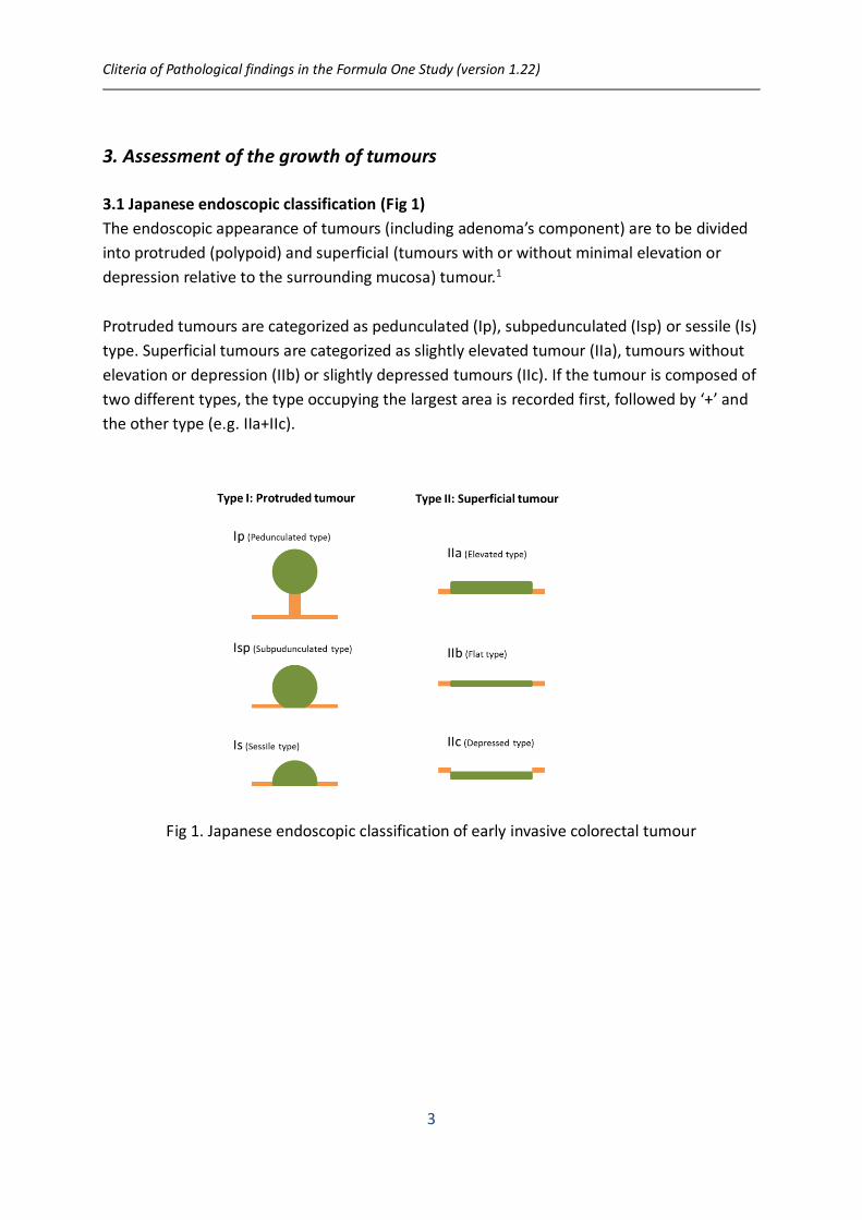
3.1 Japanese endoscopic classification (Fig 1)
The endoscopic appearance of tumours (including adenoma’s component) are to be divided
into protruded (polypoid) and superficial (tumours with or without minimal elevation or
depression relative to the surrounding mucosa) tumour.1
Protruded tumours are categorized as pedunculated (Ip), subpedunculated (Isp) or sessile (Is)
type. Superficial tumours are categorized as slightly elevated tumour (IIa), tumours without
elevation or depression (IIb) or slightly depressed tumours (IIc). If the tumour is composed of
two different types, the type occupying the largest area is recorded first, followed by ‘+’ and
the other type (e.g. IIa+IIc).
Fig 1. Japanese endoscopic classification of early invasive colorectal tumour

Cliteria of Pathological findings in the Formula One Study (version 1.22)
4
3.2 Pathological classification of the growth type of tumour (Fig 2)
Histologically, tumors are to be classified into two types: polypoid growth (PG) carcinoma
from intramucosal proliferation of adenoma or carcinoma, and nonpolypoid (NPG) growth
carcinoma without intramucosal protuberant growth.2
Furthermore, PG carcinomas are to be classified into pudunculated lesions and sessile or
broad-based lesion.
Fig 2. Pathological classification of the growth type of early invasive tumours

Cliteria of Pathological findings in the Formula One Study (version 1.22)
5
4. Assessment criteria for new pathological risk parameters
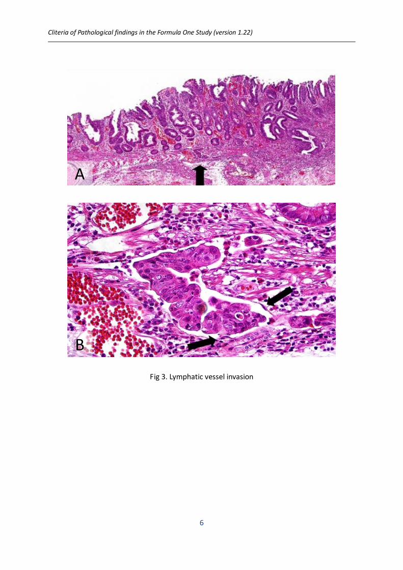
4.1 Lymphatic vessel invasion
Lymphatic vessel invasion is defined as the invasion of the tumor cells into the lymphatic vessels, and is
judged as positive only when tumor cells were identified within endothelial-lined space3). Lymphatic lacks
muscular wall and red blood cells, which are features of blood vessels. In submucosa, normal lymphatic
vessels spread along the muscularis mucosae. And tumor spreading through lymphatic vessel can be found
in lymphatic vessels around the tumor (Fig 3. A, B).
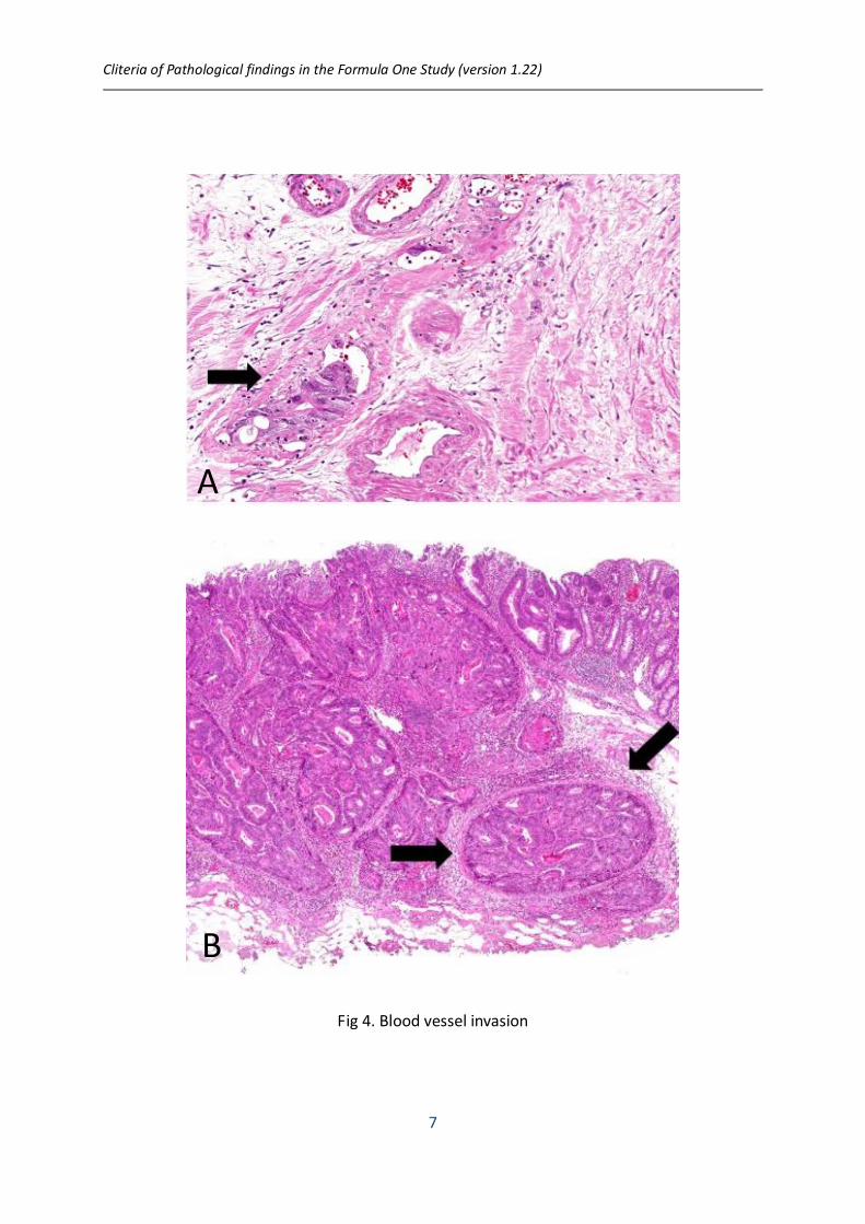
4.2 Blood vessel invasion
Blood vessel invasion is defined as the invasion of the tumor cells into the blood vessels3). Blood vessel
invasion is assessed as positive when tumor cells were within the endothelial-lined space, often containing
red blood cells, and/or were surrounded by a rim or of the smooth muscle (Fig 4. A)4). The structure of
blood vessels is often disrupted through the process of tumor’s involvement and the vessel wall can be
totally replaced by fibrous tissue (Fig 4. B)5). Even though, as mentioned above, only definite lesions in H.E
staining should be assigned as positive.
Note
# Problem in the diagnosis of small vessel invasion
pT1 colorectal cancers often shows small vessel invasion. Definite differentiation between small blood
vessel invasion and lymphatic vessel invasions can be difficult. Therefore, based on pathological findings in
this instruction, participants in this study are required to discriminate lymphatic and blood invasion as far
as possible. Only definite lesions should be assigned as positive.
# Problem of the space around tumor nests
Shrinkage artefacts and mucous lake often exhibit tissue space around the tumor nests. On the other hand,
these lesions must be discriminated from lymphatic or blood vessel invasion. Note that nuclei of
endothelial cells protrude into the vascular space which often contains lymphatic fluid or red blood cells6,7).
In colorectal cancers, shrinkage artefacts are often seen around tumor area with budding, poorly
differentiated clusters, or micropapillary component. Shrinkage seemed to be developed as a result during
processing. This space tends to occur along one margin of tumor nest, and counter of the surrounding
stroma is much similar with that of tumor nests. Spicula of slender connections often bridge the space
formed by shrinkage (Fig 5. A). Nuclei of stromal cells may be left at the margin of surrounding stroma of
shrinkage artefact. They have slender nuclei that do not protrude into the surrounding stroma (Fig 5. A,
arrow, and compare with Fig 3. B, arrow)7). Mucous lake is not lined by endothelial cells (Fig 5. B).
Cliteria of Pathological findings in the Formula One Study (version 1.22)
6
Fig 3. Lymphatic vessel invasion
A
B
Cliteria of Pathological findings in the Formula One Study (version 1.22)
7
Fig 4. Blood vessel invasion
A
B

Cliteria of Pathological findings in the Formula One Study (version 1.22)
8
Fig 5. space around tumor nests
A
B

Cliteria of Pathological findings in the Formula One Study (version 1.22)
9
5. Assessment criteria for new pathological risk parameters
5.1 Tumour budding
The assessment of tumour budding is basically performed according to the recommendations of the
International Tumour Budding Consensus Conference (ITBCC) 2016.8)
Tumour budding is defined as a cancer cell nest consisting of 1 or <5 cells that have infiltrated
the interstitium at the invasive margin of the cancer (Fig 6).9-12
Fig 6. Tumour budding
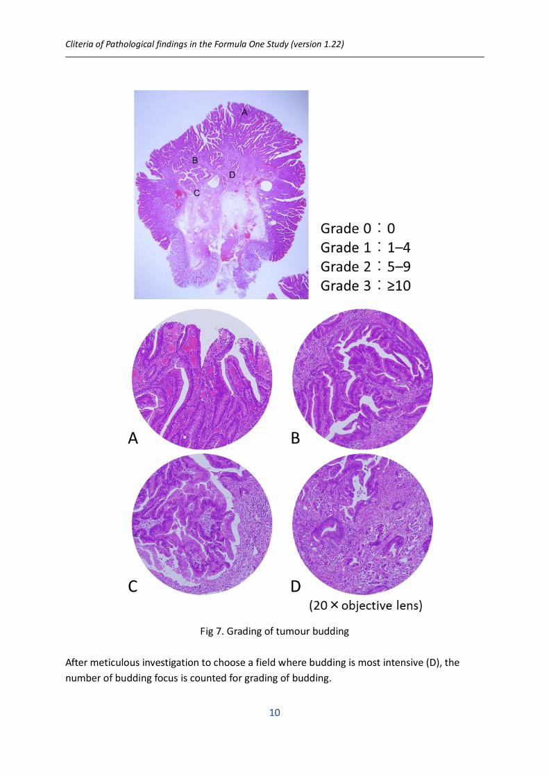
For grading of budding, the number of buddings is counted in a field observed through a 20×
objective lens (WHK 10× ocular lens; 0.785 mm2) after selecting one field wherein the
number of buddings is the greatest (Fig 7).10-12 Depending on the number of buddings, the
grade of budding is defined as follows:
Grade 0: 0
Grade 1: 1 to 4
Grade 2: 5 to 9
Grade 3: ≥10
Cliteria of Pathological findings in the Formula One Study (version 1.22)
10
Fig 7. Grading of tumour budding
After meticulous investigation to choose a field where budding is most intensive (D), the
number of budding focus is counted for grading of budding.
Cliteria of Pathological findings in the Formula One Study (version 1.22)
11
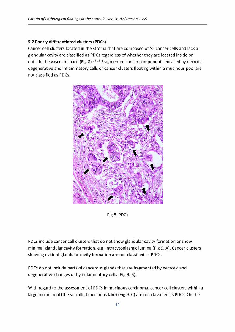
5.2 Poorly differentiated clusters (PDCs)
Cancer cell clusters located in the stroma that are composed of ≥5 cancer cells and lack a
glandular cavity are classified as PDCs regardless of whether they are located inside or
outside the vascular space (Fig 8).13-15 Fragmented cancer components encased by necrotic
degenerative and inflammatory cells or cancer clusters floating within a mucinous pool are
not classified as PDCs.
Fig 8. PDCs
PDCs include cancer cell clusters that do not show glandular cavity formation or show
minimal glandular cavity formation, e.g. intracytoplasmic lumina (Fig 9. A). Cancer clusters
showing evident glandular cavity formation are not classified as PDCs.
PDCs do not include parts of cancerous glands that are fragmented by necrotic and
degenerative changes or by inflammatory cells (Fig 9. B).
With regard to the assessment of PDCs in mucinous carcinoma, cancer cell clusters within a
large mucin pool (the so-called mucinous lake) (Fig 9. C) are not classified as PDCs. On the

Cliteria of Pathological findings in the Formula One Study (version 1.22)
12
other hand, cancer cell clusters without glandular cavity formation infiltrating the stroma
with minimal extracellular mucin formation (Fig 9. D) are classified as PDCs. In particular, the
ratio of the size of the cancer cluster to that of the mucinous area surrounding the cluster
should be considered before making the decision. Cancer cell clusters accompanied by a
mucinous area with size comparable to that of the cancer cell clusters can be classified as
PDCs.
Fig 9. Criteria for judging PDCs

Cliteria of Pathological findings in the Formula One Study (version 1.22)
13
For grading of PDCs, the number of PDCs is counted in a field observed through a 20×
objective lens (WHK 10× ocular lens; 0.785 mm2) after selecting one field wherein the
number of PDCs is the greatest (Fig 10). Depending on the number of PDCs, the grade of
PDCs is defined as follows:
Grade 0: 0
Grade 1: 1 to 4
Grade 2: 5 to 9
Grade 3: ≥10
Fig 10. Grading of PDCs
After meticulous investigation to choose a field where PDCs are most intensive, the number
of PDCs in a field observed through a 20× objective lens is counted for grading.

Cliteria of Pathological findings in the Formula One Study (version 1.22)
14
5.3 Tumour grade at the invasive front
Tumour grade at the invasive front is determined on the basis of semi-quantitative
assessment of the poorly differentiated adenocarcinoma (POR), mucinous adenocarcinoma
(MUC), and signet-ring cell carcinoma (SIG) using a microscopic field and a 40× objective lens
(Fig 11).13
Fig 11. Tumour grade at the invasive front
More specifically, the largest area of POR, MUC or SIG is determined first using a low-power
microscopic field. The designation of grade 1 is applied to tumours with no POR/MUC/SIG;
grade 2 is applied to tumours with POR/MUC/SIG that do not fill the field of a 40× objective
lens and grade 3 is applied to tumours having POR/MUC/SIG components that fully fill the
microscopic field of a 40× objective lens (Fig 12, Fig 13).

Cliteria of Pathological findings in the Formula One Study (version 1.22)
15
Fig 12. The poorly differentiated component (POR) in colorectal cancer
Figures A, B (case 1), grade 2 POR. Figures C, D (case 2), grade 3 POR.
A, The POR area existed as the least differentiated component of the tumour’s leading
front in case 1 (arrows; original magnification of the objective lens: 10x). B, Microscopic field
with a 40x objective lens of the POR area in A. C, The POR area existed at the tumor’s leading
front in case 2 (arrows; original magnification of the objective lens: 10x). D, Microscopic field
with a 40x objective lens of the POR area in C.
Note that the POR in case 1 is not extensive enough to fully fill the microscopic field with
the 40x objective lens, and the differentiated cancer lesion is intermingled with the POR area
in the microscopic field with the 40x objective lens. On the other hand, the microscopic field
with the 40x objective lens is filled with the POR in case 2.

Cliteria of Pathological findings in the Formula One Study (version 1.22)
16
Fig 13. The mucinous component (MUC) in colorectal cancer
Figures A, B (case 3), grade 2 MUC. Figure C, D (case 4), grade 3 MUC.
A, MUC area observed in the tumor invasive front region of case 3 (arrows; original
magnification of the objective lens: 4x). B, Microscopic field with a 40x objective lens of the
MUC area in A. C, MUC area observed in the tumor invasive front region of case 4 (arrows;
original magnification of the objective lens: 10x). D, Microscopic field with a 40x objective
lens of the MUC area in C.
Note that the largest area of pools of extracelluar mucin in the tumor fully occupied the
microscopic field with the 40x objective lens in case 4, which is classified in grade 3, but not
in case 3, which is classified in grade 2.
Cliteria of Pathological findings in the Formula One Study (version 1.22)
17
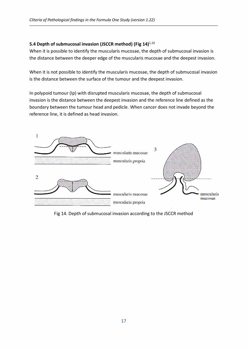
5.4 Depth of submucosal invasion (JSCCR method) (Fig 14)1,16
When it is possible to identify the muscularis mucosae, the depth of submucosal invasion is
the distance between the deeper edge of the muscularis mucosae and the deepest invasion.
When it is not possible to identify the muscularis mucosae, the depth of submucosal invasion
is the distance between the surface of the tumour and the deepest invasion.
In polypoid tumour (Ip) with disrupted muscularis mucosae, the depth of submucosal
invasion is the distance between the deepest invasion and the reference line defined as the
boundary between the tumour head and pedicle. When cancer does not invade beyond the
reference line, it is defined as head invasion.
Fig 14. Depth of submucosal invasion according to the JSCCR method
Cliteria of Pathological findings in the Formula One Study (version 1.22)
18
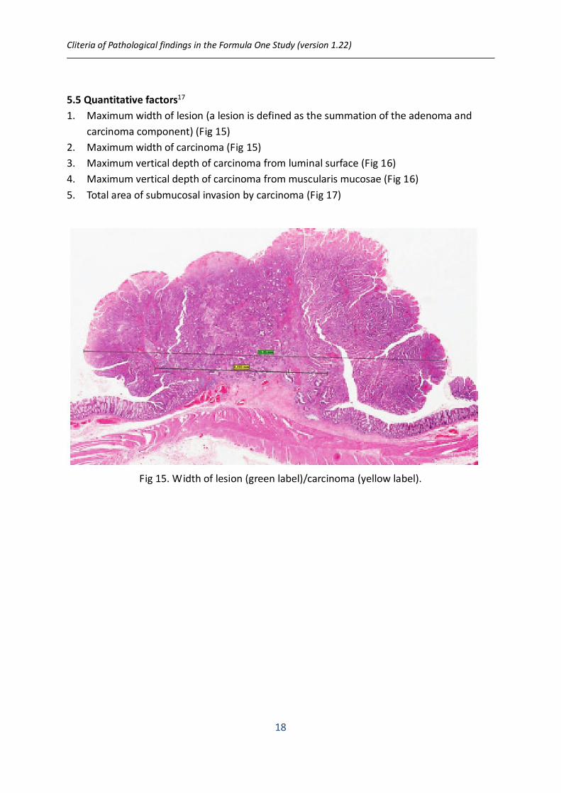
5.5 Quantitative factors17
1. Maximum width of lesion (a lesion is defined as the summation of the adenoma and
carcinoma component) (Fig 15)
2. Maximum width of carcinoma (Fig 15)
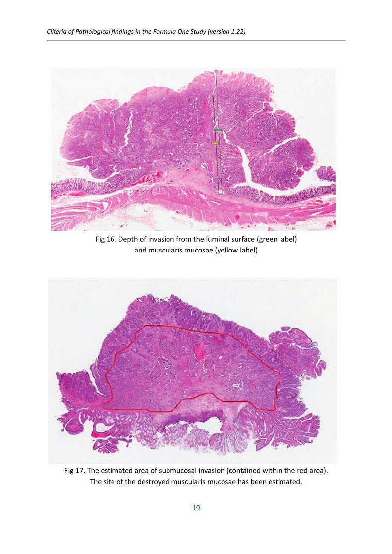
3. Maximum vertical depth of carcinoma from luminal surface (Fig 16)
4. Maximum vertical depth of carcinoma from muscularis mucosae (Fig 16)
5. Total area of submucosal invasion by carcinoma (Fig 17)
Fig 15. Width of lesion (green label)/carcinoma (yellow label).
Cliteria of Pathological findings in the Formula One Study (version 1.22)
19
Fig 16. Depth of invasion from the luminal surface (green label)
and muscularis mucosae (yellow label)
Fig 17. The estimated area of submucosal invasion (contained within the red area).
The site of the destroyed muscularis mucosae has been estimated.

Cliteria of Pathological findings in the Formula One Study (version 1.22)
20
6. References
1. Japanese Society for Cancer of the Colon and Rectum. Japanese Classification of
Colorectal Carcinoma (Second English Edition). Tokyo: Kanehara & Co., Ltd, 2009.
2. Shimoda T, Ikegami M, Fujisaki J, Matsui T, Aizawa S, Ishikaw E. Early colorectal
carcinoma with special reference to its development de novo. Cancer
1989;64:1138-1146.
3. Kojima M, Shimazaki H, Iwaya K, et al. Pathological diagnostic criterion of blood and
lymphatic vessel invasion in colorectal cancer: a framework for developing an objective
pathological diagnostic system using the Delphi method, from the pathology working
group of the Japanese Society for Cancer of the Colon and Rectum. J Clin Pathol.
2013;66:551-558.
4. Talbot IC, Richie S, Leighton M, et al. Invasion of veins by carcinoma of rectum: method
of detection, histological features and significance. Histopathology. 1981;5:141-163.
5. Kirsch R, Messenger DE, Ridell RH, et al. Venous invasion in colorectal cancer. Am J Surg
Pathol. 2013;37:200-210.
6. Mills SE, et al. Histology for pathologists, 3rd Edn. 2007. Lippincott Williams and Wilkins,
NY.
7. Rosen PP. Tumor emboli in intramammary lymphatics in breast carcinoma: pathologic
criteria for diagnosis and clinical significance. Pathol Annu. 1983;18:215-232.
8. Lugli A, Kirsch R, Ajioka Y, et al. Recommendations for reporting tumor budding in
colorectal cancer based on the International Tumor Budding Consensus Conference
(ITBCC) 2016. Mod Pathol. 2017;30:1299-1311.
9. Hase K, Shatney C, Mochizuki H, et al. Long-term results of curative resection of
"minimally invasive" colorectal cancer. Dis Colon Rectum 1995;38:19-26.
10. Ueno H, Mochizuki H, Hashiguchi Y, et al. Risk factors for an adverse outcome in early
invasive colorectal carcinoma. Gastroenterol 2004;127:385-394.
11. Kawachi H, Eishi Y, Ueno H, et al. A three-tier classification system based on the depth of
submucosal invasion and budding/sprouting can improve the treatment strategy for T1
colorectal cancer: a retrospective multicenter study. Mod Pathol 2015;28:872-879.
12. Watanabe T, Itabashi M, Shimada Y, et al. Japanese Society for Cancer of the Colon and
Rectum (JSCCR) guidelines 2010 for the treatment of colorectal cancer. Int J Clin Oncol
2009;17:1-29.
13. Ueno H, Shimazaki H, Shinto E, et al. New criteria for histologic grading of colorectal
cancer. Am J Surg Pathol 2012;36:193-201.
14. Ueno H, Hase K, Hashiguchi Y, et al. Site-specific tumor grading system in colorectal

Cliteria of Pathological findings in the Formula One Study (version 1.22)
21
cancer: multicenter pathological review of the value of quantifying poorly differentiated
clusters. Am J Surg Pathol 2014;38:197-204.
15. Ueno H, Hase K, Hashiguchi Y, et al. Novel risk factors for lymph node metastasis in early
invasive colorectal cancer: a multi-institution pathological review. J Gastroenterol
2014;49:1314-1323.
16. Fujimori T, Fujii S, Saito N, Sugihara K. Pathological diagnosis of early colorectal
carcinoma and its clinical implications. Digestion 2009;79:40-51.
17. Toh E-W, Brown P, Morris E, Botterill I, Quirke P. Area of submucosal invasion and thdth
of invasion predicts lymph node metastasis in pT1 colorectal cancers. Dis Colon Rectum
2015;58:393-400.