forensic medicine department of forensic pathology, school of forensic medicine, china medical...
TRANSCRIPT

Forensic MedicineForensic Medicine
Department of Forensic Pathology,
School of Forensic Medicine,
China Medical University, Shenyang
Department of Forensic Pathology,
School of Forensic Medicine,
China Medical University, Shenyang
Chapter 1 General Consideration on Forensic Medicine
Chapter 1 General Consideration on Forensic Medicine

1. Definition of Forensic Medicine
Forensic Medicine, also called Legal Medicine,
Medico-legal Medicine, or Medical Jurisprudence,
is a branch of medicine, which deals with the
interaction of medicine with the law by application
of theories and techniques of medical science ,
biology and the other natural sciences.

2. Interaction of forensic medicine with medicine
Basic Natural Sciences Basic Medical Sciences Subjects as Medical Basis
Medicine Clinical Medicine Applied Medicine Preventive Medicine Forensic Medicine Sports, Compensation Medicine
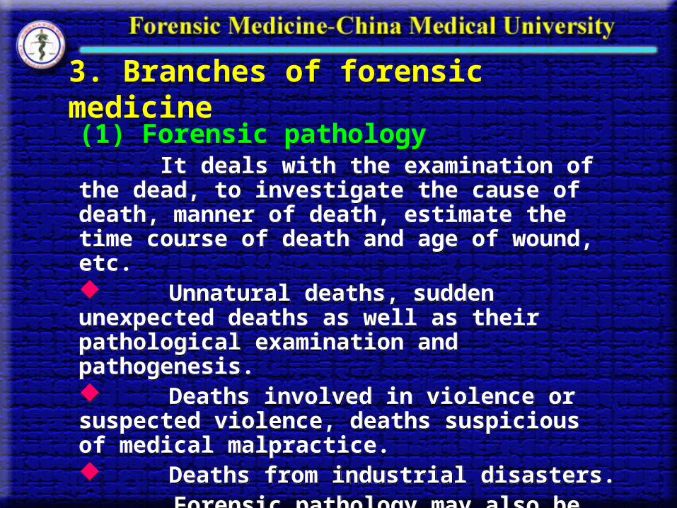
3. Branches of forensic medicine3. Branches of forensic medicine
(1) Forensic pathology It deals with the examination of the dead, to investigate the cause of death, manner of death, estimate the time course of death and age of wound, etc. Unnatural deaths, sudden unexpected deaths as well as their pathological examination and pathogenesis. Deaths involved in violence or suspected violence, deaths suspicious of medical malpractice. Deaths from industrial disasters. Forensic pathology may also be thought of as the ‘pathology of trauma’.
(1) Forensic pathology It deals with the examination of the dead, to investigate the cause of death, manner of death, estimate the time course of death and age of wound, etc. Unnatural deaths, sudden unexpected deaths as well as their pathological examination and pathogenesis. Deaths involved in violence or suspected violence, deaths suspicious of medical malpractice. Deaths from industrial disasters. Forensic pathology may also be thought of as the ‘pathology of trauma’.

(2) Clinical forensic medicine(2) Clinical forensic medicine
It deals with the examination of living individuals. Its
main tasks cover several aspects including :
Evaluation of severity of injuries and illness resulted
from wounds,
Working ability,
Investigation of physical abuse and sexual function,
Examination of victims of sexual assaults, the accused
of criminal abortion and pregnancy,
Inspection on simulated or artificial disease or injuries
and drunken status.

(3) Science of forensic material evidence (3) Science of forensic material evidence
Science of forensic material evidence, or forensic physical
evidence mainly deals with :
The studies on bloodstains, body fluid stains such as saliva
and semen;
The origin of hairs and tissue fragments;
The performance of personal identification and paternity
tests by forensic serological techniques or DNA analysis.

To date, techniques in molecular biology such as DNA
fingerprinting, examination of STR loci after PCR
procedures have been widely used in the area.
A sub-branch called forensic odontology may play an
important role in estimation of age, sex, possible
occupation by examination of denture and personal
identity by tooth marks.
Forensic anthropology is the sub-branch dealing with
personal identification by study on bones and bone
fragments, skin ridges and fingerprints as well as photo
pictures.

(4) Forensic toxicology(4) Forensic toxicology
Forensic toxicology, the study of poisons largely
devotes to mechanisms through which toxic substances
function, the cause and manner of poisoning, symptoms
and morphological findings as well as determination of
lethal dose of poisons.
Forensic toxicology, the study of poisons largely
devotes to mechanisms through which toxic substances
function, the cause and manner of poisoning, symptoms
and morphological findings as well as determination of
lethal dose of poisons.

(5) Forensic chemistry or forensic toxicological analysis
Forensic chemistry (forensic toxicological analysis)
mainly deals with qualification and quantitation of toxic
substances in the biological fluids, tissues or other
samples for toxicological purpose.
Forensic chemistry (forensic toxicological analysis)
mainly deals with qualification and quantitation of toxic
substances in the biological fluids, tissues or other
samples for toxicological purpose.

(6) Forensic psychiatry
Forensic psychiatry is one of the sub-branches of forensic
medicine, which is responsible for analysis and examination
of human behaviour, personality, responsibility and psychia
tric problems and resolution of legal matters involving psyc
hological disorders, which may offer a profile of the assailan
t to law enforcement officers.
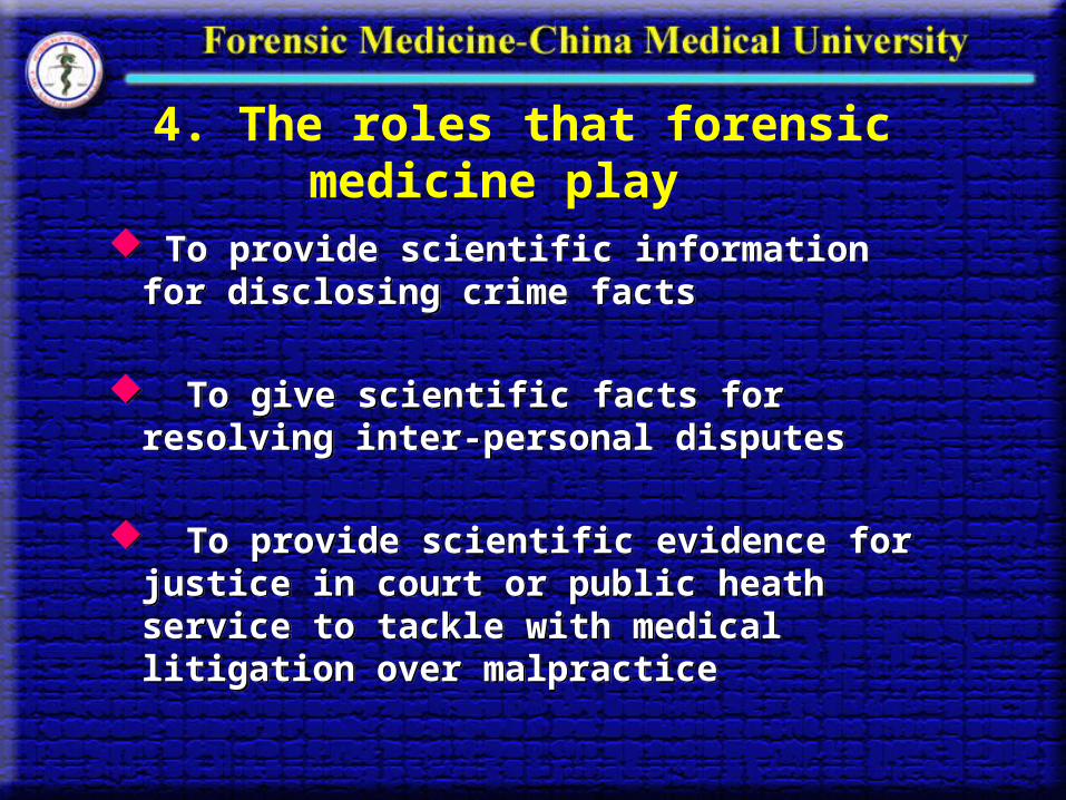
4. The roles that forensic medicine play
To provide scientific information for disclosing crime facts
To give scientific facts for resolving inter-personal disputes
To provide scientific evidence for justice in court or public heath service to tackle with medical litigation over malpractice
To provide scientific information for disclosing crime facts
To give scientific facts for resolving inter-personal disputes
To provide scientific evidence for justice in court or public heath service to tackle with medical litigation over malpractice
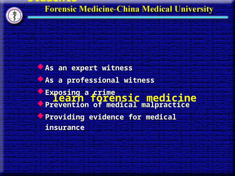
5. Purposes for which medical students learn forensic medicineAs an expert witness
As a professional witness
Exposing a crime
Prevention of medical malpractice
Providing evidence for medical insurance
As an expert witness
As a professional witness
Exposing a crime
Prevention of medical malpractice
Providing evidence for medical insurance

6. The jobs forensic practitioners undertake
Crime scene investigation
Autopsy
Examination of the living individuals
Test of the collected material evidence
Crime scene investigation
Autopsy
Examination of the living individuals
Test of the collected material evidence

(1) Crime scene investigation
Crime scene is referred as to any place where the deceased or any physical or trace evidence connected with the crime are discovered.
More than one crime scene may be encountered.
Crime scene investigation is a consecutive detective action.
Places and objects involved in crime must be investigated and examined, the suspected searched and the deceased autopsied.

Upon arrival at the scene of death, request a summary of what is known about the incident, the protection of the scene, the time of arrival as well as condition of the weather.
Work out a practical and effective plan for investigation in the light of the scene and then begin to search.
Record observations at the scene, photograph the body and the scene.
Make a sketch of the scene when necessary. Any object that had to be moved at the scene should be definitely marked in place.

After photographs are taken, scene investigation may
begin with the investigators gathering at the center of the
scene and moving out in spoke-like directions.
On arrival at the scene, the body should be examined to
determine whether it is alive or dead.
A comprehensive external examination of the deceased
may be carried out only after the crime scene investigation
is nearly complete.
During the investigation attentions should be paid to the
following aspects:
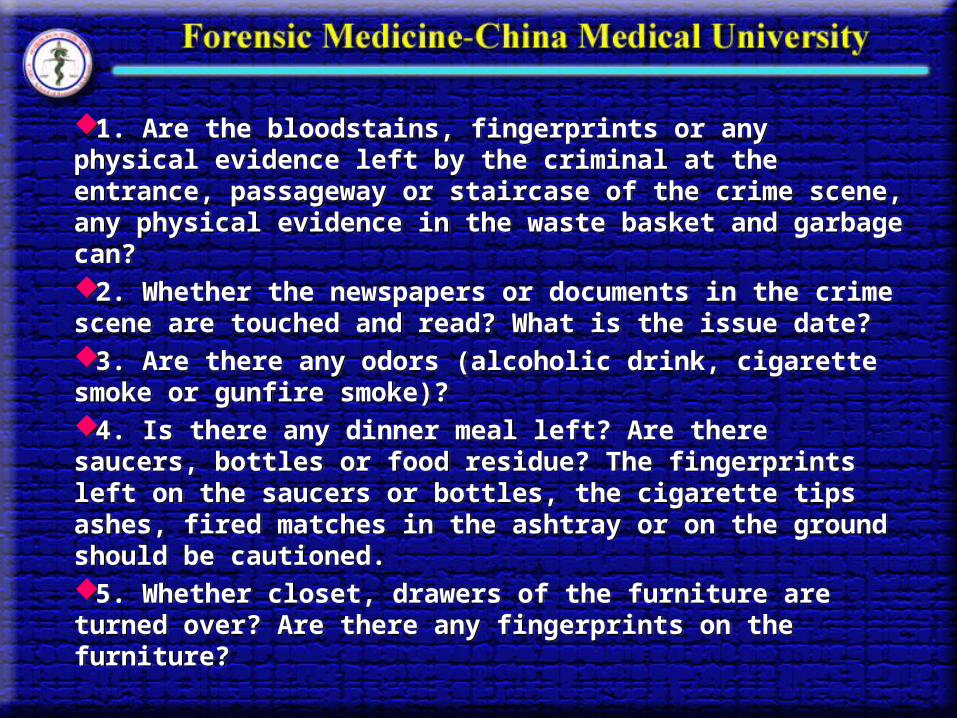
1. Are the bloodstains, fingerprints or any physical evidence left by the criminal at the entrance, passageway or staircase of the crime scene, any physical evidence in the waste basket and garbage can?2. Whether the newspapers or documents in the crime scene are touched and read? What is the issue date?3. Are there any odors (alcoholic drink, cigarette smoke or gunfire smoke)?4. Is there any dinner meal left? Are there saucers, bottles or food residue? The fingerprints left on the saucers or bottles, the cigarette tips ashes, fired matches in the ashtray or on the ground should be cautioned. 5. Whether closet, drawers of the furniture are turned over? Are there any fingerprints on the furniture?
1. Are the bloodstains, fingerprints or any physical evidence left by the criminal at the entrance, passageway or staircase of the crime scene, any physical evidence in the waste basket and garbage can?2. Whether the newspapers or documents in the crime scene are touched and read? What is the issue date?3. Are there any odors (alcoholic drink, cigarette smoke or gunfire smoke)?4. Is there any dinner meal left? Are there saucers, bottles or food residue? The fingerprints left on the saucers or bottles, the cigarette tips ashes, fired matches in the ashtray or on the ground should be cautioned. 5. Whether closet, drawers of the furniture are turned over? Are there any fingerprints on the furniture?

6. Is there any damage to the ceiling, wall and furniture at the crime scene? Is the damage connected with the assault?
7. Are there fired shells, bullet entrances present on the ground, the walls, the ceiling in the homicidal cases that the firearm is involved?
8. Where do the ligatures come from in the cases of hanging or ligature strangulation?
9. Careful searching places where weapons may be hidden should be implemented.
10.Are there posthumous paper and fingerprints on the paper left at the scene in the cases of suicide?

(2) Autopsy
Medico-legal autopsy must be conducted under one of the following circumstances:1. Autopsies must be performed on the bodies to determine the causes of death in criminal cases and on the unidentified bodies to determine the cause and manner of death;2. On the decedents who suddenly and unexpectedly died when homicide or suicide is suspected;3. On the bodies that are involved in the legal matters due to industrial or agricultural poisonings or grave infectious diseases.
Medico-legal autopsy must be conducted under one of the following circumstances:1. Autopsies must be performed on the bodies to determine the causes of death in criminal cases and on the unidentified bodies to determine the cause and manner of death;2. On the decedents who suddenly and unexpectedly died when homicide or suicide is suspected;3. On the bodies that are involved in the legal matters due to industrial or agricultural poisonings or grave infectious diseases.

The purposes of the medico-legal autopsy are:
To establish the causes of death, which is focused on
determining whether the death is violent or non-violent.
To interpret the mode or manner of death.
To estimate the time of death.
To conjecture the possible weapon inflicting wounds.
To conduct personal identification

(3) Examination of the living individuals
The jobs involved in the examination of the living individuals include:
Inspection of wounds
Medico-legal expertise on working ability
Appreciation of disease
Investigation of sex-associated problems
Psychiatric evaluation
Paternity test
Investigation of simulation and self-mutilation
The jobs involved in the examination of the living individuals include:
Inspection of wounds
Medico-legal expertise on working ability
Appreciation of disease
Investigation of sex-associated problems
Psychiatric evaluation
Paternity test
Investigation of simulation and self-mutilation

(4) Test of collected material evidenceMaterial evidence is any tangible article or trace left by the offender during perpetration, by which the criminal and his crime act may be judged to some extent.
Material evidence varies with cases and may be analyzed in the specialized laboratories.
In the medico-legal laboratory, any trace physical evidence such as bloodstain, semen stain, salivary stain, hairs, urine, feces, tooth or bone fragments can be examined.
When necessary, facial restoration or reconstruction may be used for personal identification.
Material evidence is any tangible article or trace left by the offender during perpetration, by which the criminal and his crime act may be judged to some extent.
Material evidence varies with cases and may be analyzed in the specialized laboratories.
In the medico-legal laboratory, any trace physical evidence such as bloodstain, semen stain, salivary stain, hairs, urine, feces, tooth or bone fragments can be examined.
When necessary, facial restoration or reconstruction may be used for personal identification.
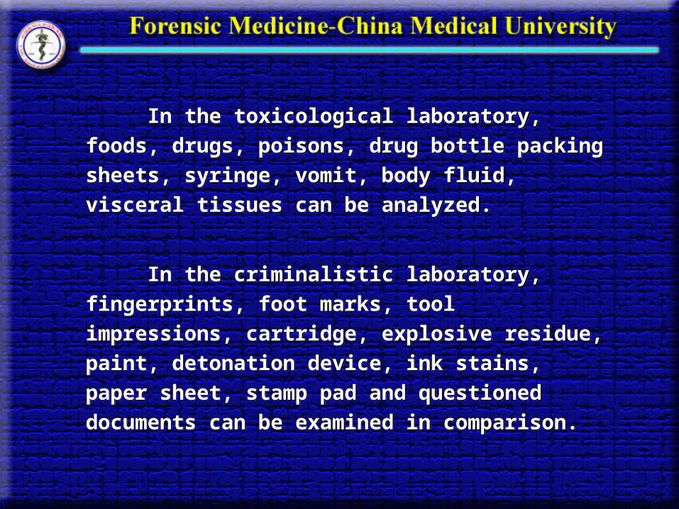
In the toxicological laboratory, foods, drugs,
poisons, drug bottle packing sheets, syringe, vomit,
body fluid, visceral tissues can be analyzed.
In the criminalistic laboratory, fingerprints, foot
marks, tool impressions, cartridge, explosive residue,
paint, detonation device, ink stains, paper sheet,
stamp pad and questioned documents can be
examined in comparison.

Chapter 2 Death and Postmortem Phenomena

Section 1 Death 1. Definition of Death
Death is always considered to be cessation of life. Traditionally, death has been defined as complete and persistent cessation of respiration and blood circulation.
Cardiac death Respiratory death Brain death
1. Definition of Death
Death is always considered to be cessation of life. Traditionally, death has been defined as complete and persistent cessation of respiration and blood circulation.
Cardiac death Respiratory death Brain death

(1) Cardiac death, which is referred to as the death resulted from stoppage of heart beating before cessation of respiration. Cardiac death is often caused by such heart diseases as
coronary heart disease
cardiomyopathy
valvular disease
pericardial effusion
cardiac conducting system abnormalities
cardiac trauma, etc.

(2) Respiratory death, which is referred to as the death that is resulted from cessation of respiration prior to stoppage of heart beating. Respiratory death is mainly due to:
obstruction of airway such as hanging, strangulation by ligature, manual strangulation, drowning, and overlay,
pulmonary diseases such as pulmonary edema, pulmonary parenchyma, pulmonary emphysema, pneumonothorax, pulmonary infarction,
respiratory center paralysis, respiratory muscular paralysis due to muscular relaxant poisoning,
hypokalemia, etc.

(3) Brain death, which is referred to as the complete cessation of the total brain function, including activities in the cerebrum, cerebellum and brainstem, although heart beating may exist and/or spinal cord may function.
i. Cause of brain death Cardiac failure due to myocardial infarction,
standstill, valve dysfunction Head trauma Cerebral apoplexy (stroke) CNS infectious diseases Intoxications Other miscellaneous conditions

ii. Respirator brain Respirator brain was created to describe a condition of
brain in that brain death has occurred in patient whose breathing is supported continuously with ventilator or respirator.
Gross pathology of “respirator brain” The brain appears dusky gray, and congested The brain reveals generalized swelling with evidence of
tonsillar herniation or the other herniations The brain is soft, often mushy, or nearly liquid The cerebellum and brainstem are often macerated and
fragments of the cerebellar tonsils may have fallen into the spinal canal
The pituitary gland appears strawberry-like

iii. Criteria for certifying brain death
(a) The patient should be in deep coma, show no response to even intensely painful stimuli.
(b) There should be no spontaneous respiration for three minutes when taken off the ventilator.
(c) There should be no reflexes; the pupils should be fixed, dilated, and unresponsive to light; no eye movement should be elicited by turning the head or by cold water irrigation of the ears; there should be no corneal reflexes; no blinking; no postural activity, pharyngeal reflexes, and no swallowing, yawning, vocalization; no deep tendon reflexes should be present.

(d) The EEG should be flat for at least 10 min of technically adequate recording; there should be no response to noise or pinching.(e) There should be no change in any of the above tests when repeated after 24 hr.(f) There should be no evidence of hypothermia, nor of CNS depressant drugs present in the blood.
2. Death Process Agonal stage Clinical death Biological death
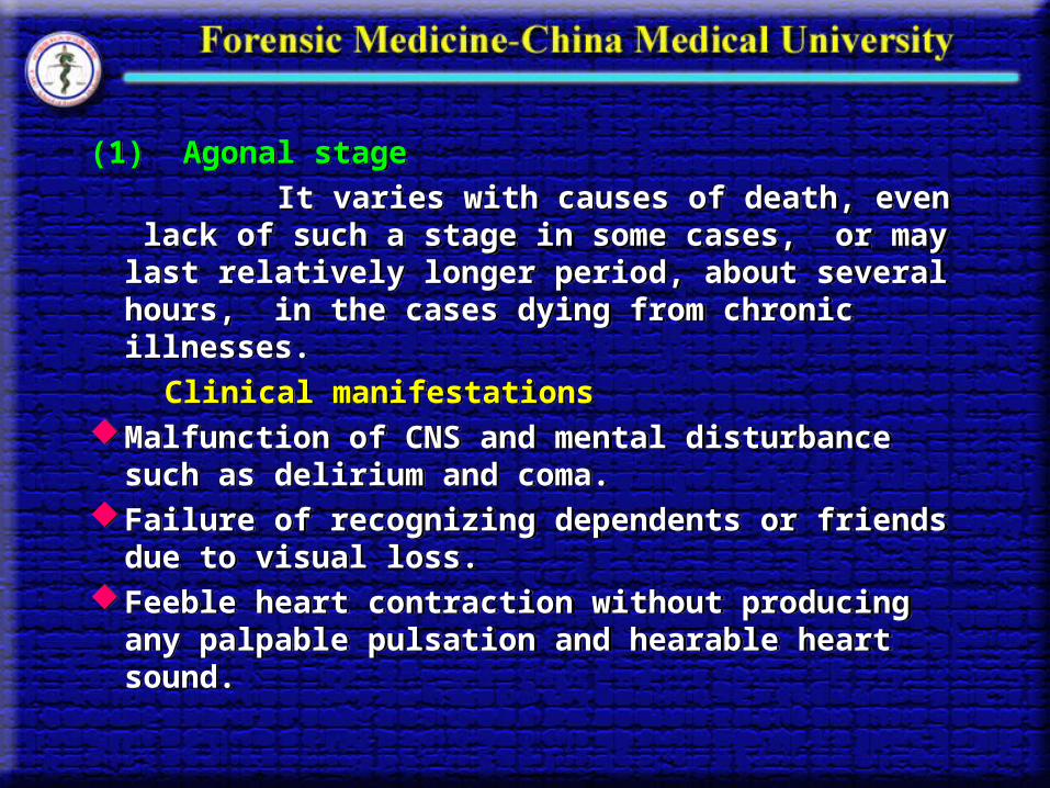
(1) Agonal stage
It varies with causes of death, even lack of such a stage in some cases, or may last relatively longer period, about several hours, in the cases dying from chronic illnesses.
Clinical manifestationsMalfunction of CNS and mental disturbance such as
delirium and coma. Failure of recognizing dependents or friends due to
visual loss. Feeble heart contraction without producing any
palpable pulsation and hearable heart sound.

(2) Clinical death
which is also called somatic death or individual death.
Characteristics :
Cessation of circulation and spontaneous respiration,
No reflexes elicited.
This stage may last 5- 6 min in which cerebral cortex might survive without oxygen or from hypoxia in essence.
(2) Clinical death
which is also called somatic death or individual death.
Characteristics :
Cessation of circulation and spontaneous respiration,
No reflexes elicited.
This stage may last 5- 6 min in which cerebral cortex might survive without oxygen or from hypoxia in essence.

(3) Biological death
Death occurs in cells and tissues in this stage. Thus, death in this stage is also called molecular death or total death.
Characteristics Complete cessation of cellular metabolisms and
irreversible damage of the cells. Early signs of death: cessation of circulation and
respiration, dilated pupils, disappearance of any reflexes
Early postmortem changes: muscular flaccidity, the cooling of the body, and the rigidity of the body, etc.
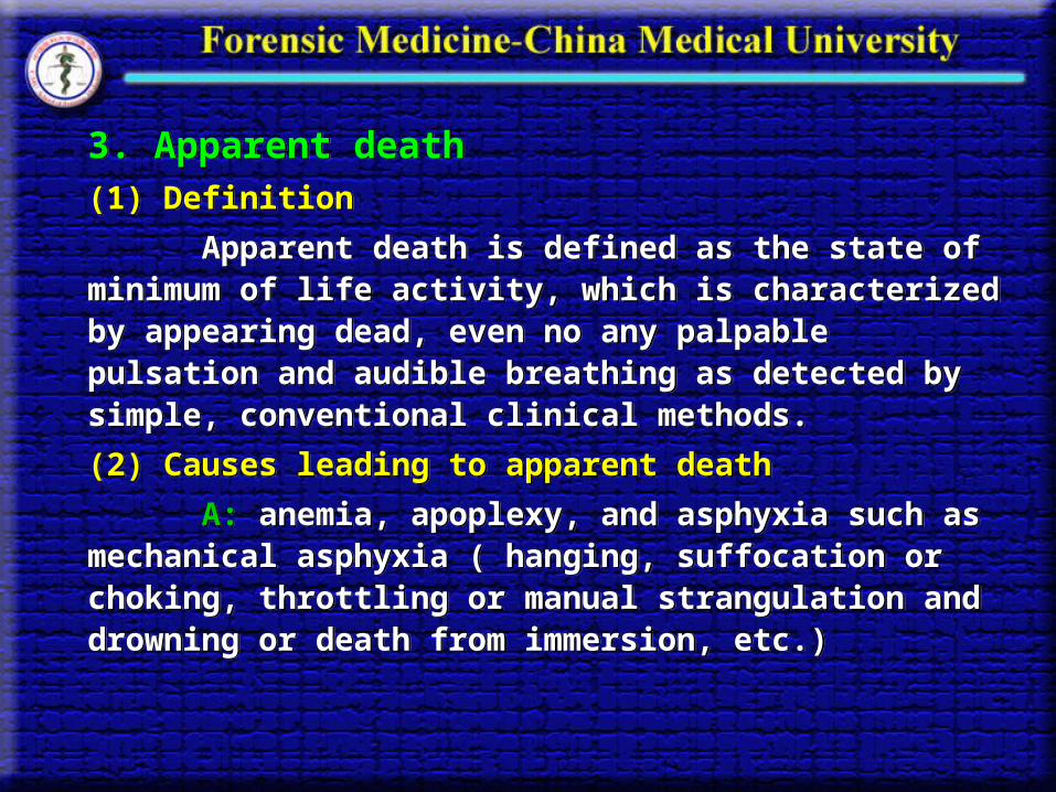
3. Apparent death(1) Definition
Apparent death is defined as the state of minimum of life activity, which is characterized by appearing dead, even no any palpable pulsation and audible breathing as detected by simple, conventional clinical methods.
(2) Causes leading to apparent death
A: anemia, apoplexy, and asphyxia such as mechanical asphyxia ( hanging, suffocation or choking, throttling or manual strangulation and drowning or death from immersion, etc.)

E: electrical injuries.
I: injuries, especially head injury.
O: opium, i.e., poisoning of opium and the other sedative
drugs such as CNS depressants.
U: uremia.
Other causes : brain concussion, hypothermia, low
environmental temperature, sunstroke, malnutrition.
E: electrical injuries.
I: injuries, especially head injury.
O: opium, i.e., poisoning of opium and the other sedative
drugs such as CNS depressants.
U: uremia.
Other causes : brain concussion, hypothermia, low
environmental temperature, sunstroke, malnutrition.

4. Classification of death in forensic medicine
(1) Violent death (a) Suicidal death or suicide, which is self-inflicted by
violent manner. (b) Homicidal death or homicide, which is caused by
others by violent ways. (c) Accidental death, which is induced by such accidents as
natural, industrial or agricultural disasters. (2) Non-violent death which is also called natural death, is
any death caused by diseases and abnormalities or aging, but it is very common that an individual dies from sickness.
Section 2 Postmortem Phenomenon
Postmortem changes are referred to as a series of physical, chemical and morphological changes that occur in cadaver due to the influences of physical, chemical and biological factors inside and outside the body as cessation of metabolism and functional activity occur after somatic death.
The appearances revealed in the cadaver as result of postmortem changes are termed postmortem phenomena.
The early postmortem phenomena The late postmortem phenomena The early postmortem phenomena are referred to
those occurring 24 hours after death, whereas the late ones beyond 24 hours after death.

1. The early postmortem phenomena (1) Muscular flaccidity
Muscular flaccidity is the condition where there is complete relaxation of the muscles developing at the time of death, which is also known as the primary muscular flaccidity.
Contact flattening of the muscles occurs during muscular flaccidity. Alterations of the position after death may be indicated by the presence of the areas of contact flattening on parts of the body which no longer lie in contact with a solid surface.

(2) Algor mortis Algor mortis, also called cooling of the body is another i
mportant phenomenon that begins to occur at the time of death.
• Three major ways by which the human body loses heat, i.e., by radiation, convection and vaporization.
• Factors affecting the rate of cooling of a body
i. The mean atmospheric temperature during period of cooling • The rate of cooling is rapid in a cold day.• The rate of cooling is rapid during the night .• Cooling of the body does not occur in the atmospheric temp
erature above 40 . ℃

• A body cools more rapidly in the open air than in a small poorly ventilated room.
• Cooling is more rapid in a humid temperature than in a dry atmosphere.
ii.The presence or absence of clothing or other coverings on the body after death
• The cooling is relatively slow in clothed bodies.
iii.The state of nutrition and development of the body
• The cooling of a large body is relatively slower than the cooling of a small body.
• The cooling is greatly retarded in obese as compared with the thin subjects.

iv. The causes of death affecting the rate of cooling of the body
The rate of cooling of the body may be slower in the cadavers with the conditions such as infectious diseases of bacterial origin, epileptica status, and pontine hemorrhage.
In cases of barbiturate intoxications, the rate of cooling of the body can be more rapid.
By recording the temperature of a body and calculating the rate of cooling of the body at crime scene, we can estimate the time of death. If combined with the other early postmortem phenomena, estimation of time since death may tend to be more accurate.
(3) Livor mortis
i. Definition:
After death, the blood gravitates to the dependent portions of the body. The dependent capillaries and veins become distended. This capillovenous distention is known as postmortem hypostasis. Livor mortis is the appeared color in the portions of the body due to capillovenous distention, which is also termed postmortem lividity.
ii. Distribution of the postmortem lividity and hypostasis (a) External appearances of the lividity
In the supine position, the lividity is distributed externally over the dorsal aspect of the trunk, the posterior aspects of the head and neck, extensor surfaces of the up limbs and the flexor surfaces of the lower limbs.
In the supine position, lividity is not observed over the back of the crown of the head, the backs of the shoulders, the buttocks and the backs of the heels because pressure prevents the filling of the veins and capillaries of the skin in these areas.
A ventral distribution of lividity is seen externally in a prone position after death. In this case, the lividity is distributed externally over the forehead, the cheeks and chin, the upper parts of the chest and abdomen, and the extensor surfaces of the lower limbs.
In the prone position, the lividity cannot be found in the area of the mouth and nose, the part of the chest because of contact pressure in these areas.
In the upright position, lividity is usually found in the dependent lower limbs, the external genitalia, the lower parts of the forearms, and the hands and the parts surrounding the waist above the waist-band.
When the position of the body is uncertain, the distribution of the lividity is different, for instance, in death from immersion, the body usually floats face downwards, with the back of the chest at the highest level in the fluid, lividity may be most marked in the face, over the front of the trunk and in the limbs.


(b) Internal appearances of the hypostasis
The site of distribution of hypostasis in the viscera
depends upon the position of the body.
In the supine position, hypostasis is seen in the posterior portions of the cerebrum and cerebellum, the dorsal portions of the lungs, the posterior wall of the stomach, the dorsal portions of the liver, the kidneys, and the lowermost coils of the intestine in the pelvic cavity.
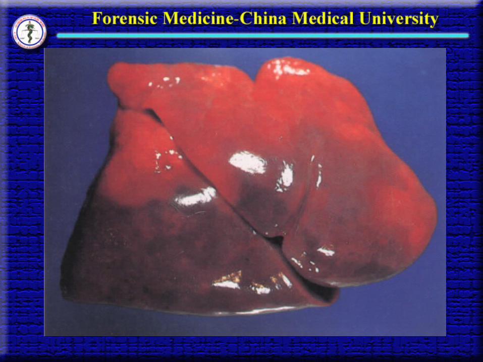
iii. The color of lividity
The intensity of the color of lividity depends upon the mount of reduced hemoglobin in the blood.
Under normal condition: purplish-blue
When there is so much reduced hemoglobin in the blood circulation before death: deep purplish-blue
In acute cyanide poisoning: bright pink color
In acute carbon monoxide poisoning: cherry-red color
In potassium chloride poisoning: chocolate- brown
In deaths from exposure to cold: bright pink
iv. The extent and the time of appearance of
postmortem lividity in the skin
The extent and the time of appearance of postmortem lividity in the skin mainly depend upon the volume of blood in circulation at the time of death and the fluidity of the blood after death.
When the volume of the blood increases at the time of death, or the blood is fluid postmortem, the extent of lividity is usually marked, and the time of its appearance is early or accelerated.
When the volume of blood decreases, the lividity is usually limited in its extent and the time of its appearance may be delayed.

In general, livor mortis begin to develop 1- 2 hours
postmortem, and reaches maximum extent 12 hours after
death. The development of livor mortis may be divided into
three stages:
gravitation stage,
diffusion stage,
infiltration (fixing) stage.
In general, livor mortis begin to develop 1- 2 hours
postmortem, and reaches maximum extent 12 hours after
death. The development of livor mortis may be divided into
three stages:
gravitation stage,
diffusion stage,
infiltration (fixing) stage.
v. The significance of postmortem lividity in
forensic practice
(a) Livor mortis is one of the early signs to certify
death.
(b) Livor mortis is helpful for estimating the time
since death.
(c) The color and extent of postmortem lividity
may indicate the possible causes of death.
(d) Postmortem lividity may suggest the position
in which a body has lain, and sometimes
movement of the body.

(4) Rigor Mortis
i. Definition
Rigor mortis is a postmortem stiffening of the voluntary and involuntary muscles of the body which develops at a variable period after death and succeeds the state of primary muscular flaccidity.
ii. Mechanisms of rigor mortis
Recent studies indicated that loss of ATP (adenosine trisphosphate) may play an important role in the development of rigor mortis.
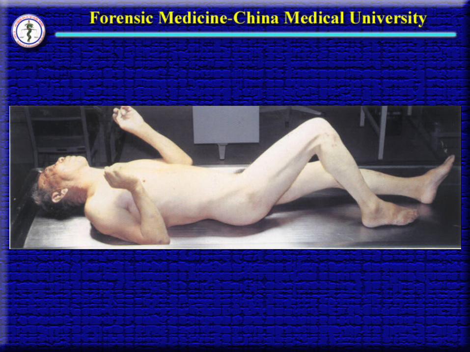

iii. Orders of appearance and disappearance of rigor mortis in striated (voluntary) muscles
Face
Hand Forearm Arm Neck and Lower jaw Arm Forearm Hand
Thorax
Foot Leg Thigh Abdomen and Pelvis Thigh Leg Foot
iii. Orders of appearance and disappearance of rigor mortis in striated (voluntary) muscles
Face
Hand Forearm Arm Neck and Lower jaw Arm Forearm Hand
Thorax
Foot Leg Thigh Abdomen and Pelvis Thigh Leg Foot

Generally, 1–3 hours after death, rigor mortis produces but sometimes as early as 10 minutes postmortem, or 7–8 hours after death.
4–6 hours after death it may spread throughout the body
12 –15 hours, it may be fully developed, and may stiffen mostly 24 hours postmortem.
The body may stiffen for 24- 48 hours and then begin to relieve.
If rigor mortis is destroyed within 4- 6 hours after death, restiffening can occur. If it is destroyed by force artificially beyond 6- 8 hours after death, the rigidity cannot recur.

Rigor mortis can involve not only skeleton muscles, but also smooth muscles and cardiac muscle.
Rigor mortis can causes contraction of the cardiac muscle, which should not be mistaken for myocardial hypertrophy.
Secondary muscular flaccidity following rigor mortis may result in distention of the atria or ventricles, which should not be mistaken for antemortem dilatation of the chambers or myocardial degeneration.
During rigor mortis the arrectoris pilorum muscles may contract and this contraction may lead to a condition of the skin known as ‘cutis anserina’ or ‘goose skin’
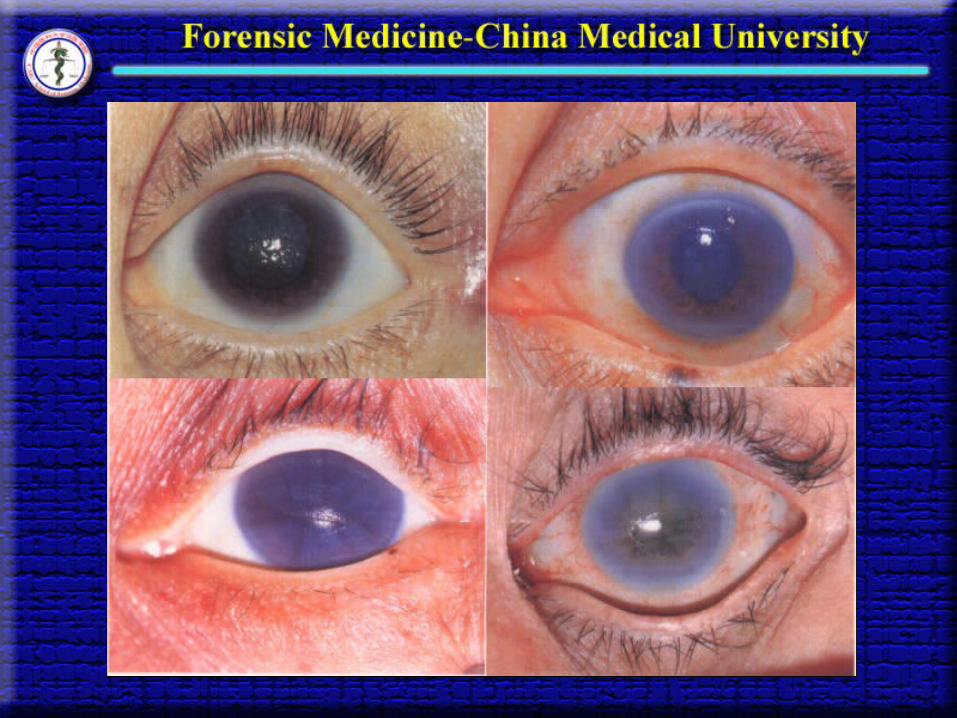
Rigor mortis may affect the iris muscles, and the contraction of the iris muscles may vary in each eye, giving rise to irregularity and inequality of the pupils.
iv. The factors influencing the rate of onset and duration of rigor mortis The onset of the rigor mortis is more rapid and the duration is shorter in
(a) high environmental temperature than in low temperature,
(b) after prolonged muscular activity antemortem in
deaths from convulsion due to poisoning of some drugs such as strychinine and diseases such as tetanus as well as in death after exhaustion in battle, and(c) in the foetus, still-borne babies, infants and children, and in aged individuals.
v. The significance of rigor mortis in forensic practice
(a) It is one of the early signs of death.(b) It is helpful for estimation of timing death.
(5) Cadaveric spasm
i. Definition
Cadaveric spasm is a condition in which those
muscles which are in a state of normal contraction at
the actual moment of somatic death persist in this state
throughout the period of molecular death when the
other muscles are in the state of primary flaccidity.
Cadaveric spasm is also known as instantaneous
cadaveric rigidity.
ii. The muscles mostly involved in cadaveric spasm and conditions in which cadaveric spasm most commonly occurs
Cadaveric spasm most commonly seen in several groups of muscles such as the muscle of the forearms, and hands.
Cadaveric spasm can be simulated if an object is placed in the hand of the deceased during the stage of primary muscular flaccidity, and if the fingers are then put into position around the object until rigor mortis is fully developed.
iii. The conditions in which cadaveric spasm is most commonly seen
(a) In a small proportion of suicidal deaths from firearms and cut-throat wounds when the revolver, pistol or knife is firmly grasped at the moment of death;(b) In certain cases of drowning when grass or weeds or other objects in the fluid medium are clutched by the deceased;(c) In certain mountain fatalities when branches of shrubs or trees are seized by the deceased; and(d) In certain cases of homicide such as rape when some portion of clothing or hair belonging to the assailant is found in the deceased hand.

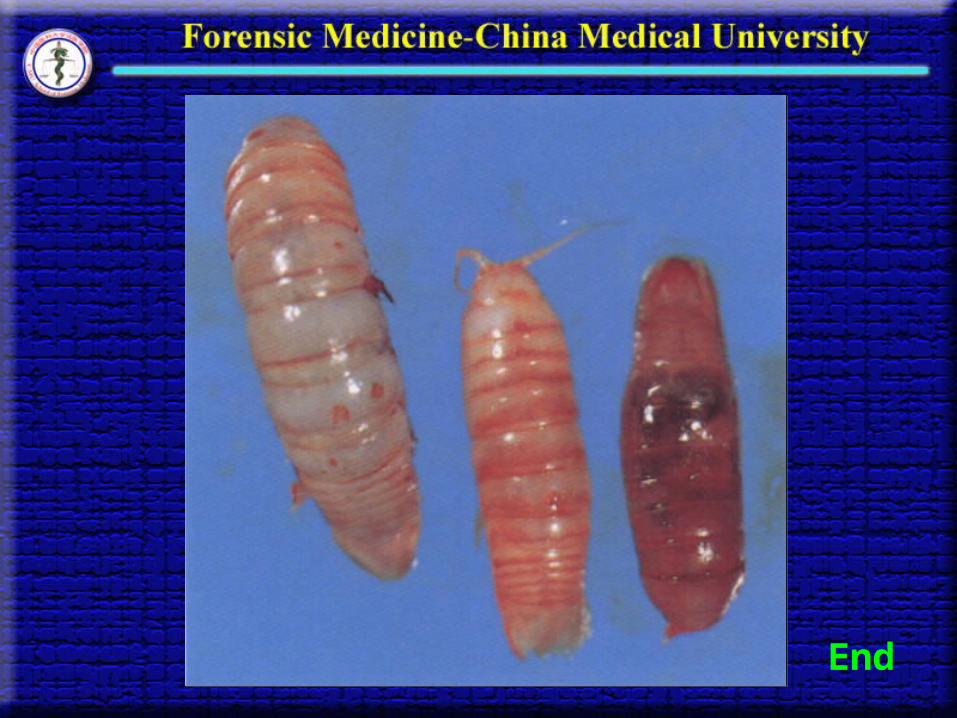
(6) The other early postmortem phenomena i. Local desiccation, which is also known as parchment- like transformation, is that part of the body surface, especially surface of injury and mucous membrane, become dry and hard with brown-like discoloration due to evaporation of water. ii. Postmortem turbidity of the cornea is that cornea will gradually turn into grayish discoloration and turbidity leading to semi-transparency or even untransparency.iii. Autolysis is another postmortem phenomenon that is of forensic interest. Autolysis can destroy structures of cells due to self-digestion by intracellular enzymes .
(6) The other early postmortem phenomena i. Local desiccation, which is also known as parchment- like transformation, is that part of the body surface, especially surface of injury and mucous membrane, become dry and hard with brown-like discoloration due to evaporation of water. ii. Postmortem turbidity of the cornea is that cornea will gradually turn into grayish discoloration and turbidity leading to semi-transparency or even untransparency.iii. Autolysis is another postmortem phenomenon that is of forensic interest. Autolysis can destroy structures of cells due to self-digestion by intracellular enzymes .


2. The Late Postmortem Phenomena
(1) Putrefaction i. Definition
Putrefaction is known as that the body undergoes a process of decomposition caused by proteolytic and the other enzymes that are produced by bacteria, which leads to the gradual dissolution of the tissues into gases, liquids and salts.
The putrefaction usually develops 24 hours after death in the normal environmental temperature.

ii. Strains of bacteria responsible for the putrefaction There are two groups of bacteria concerning with the putrefaction of the body.The first group : those frequently found in the tissues at autopsy and normally present in the respiratory or intestinal tracts. The second group: a wide variety of pathogenic and nonpathogenic strains iii. Tissue changes in putrefaction Changes developed in the tissues undergoing putrefaction include:changes in colorthe evolution of gases in the tissuesthe liquefaction of the tissues
ii. Strains of bacteria responsible for the putrefaction There are two groups of bacteria concerning with the putrefaction of the body.The first group : those frequently found in the tissues at autopsy and normally present in the respiratory or intestinal tracts. The second group: a wide variety of pathogenic and nonpathogenic strains iii. Tissue changes in putrefaction Changes developed in the tissues undergoing putrefaction include:changes in colorthe evolution of gases in the tissuesthe liquefaction of the tissues

(a) Changes in the color of the tissues
Various derivatives of putrefied hemoglobin penetrate into tissues and stain the surrounding tissues, gradually change to a green-yellow, green-blue, or greenish-black color.
The earliest external changes is a greenish discoloration of the skin over the region of the right iliac fossa.
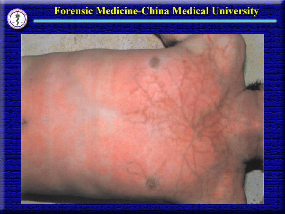
Reddish-brown arborescent markings appear in the skin in various parts of the body.
The earliest internal change is a reddish-brown discoloration of the inner surfaces of the vessels, especially of the aorta
(a) Changes in the color of the tissues
Various derivatives of putrefied hemoglobin penetrate into tissues and stain the surrounding tissues, gradually change to a green-yellow, green-blue, or greenish-black color.
The earliest external changes is a greenish discoloration of the skin over the region of the right iliac fossa.
Reddish-brown arborescent markings appear in the skin in various parts of the body.
The earliest internal change is a reddish-brown discoloration of the inner surfaces of the vessels, especially of the aorta


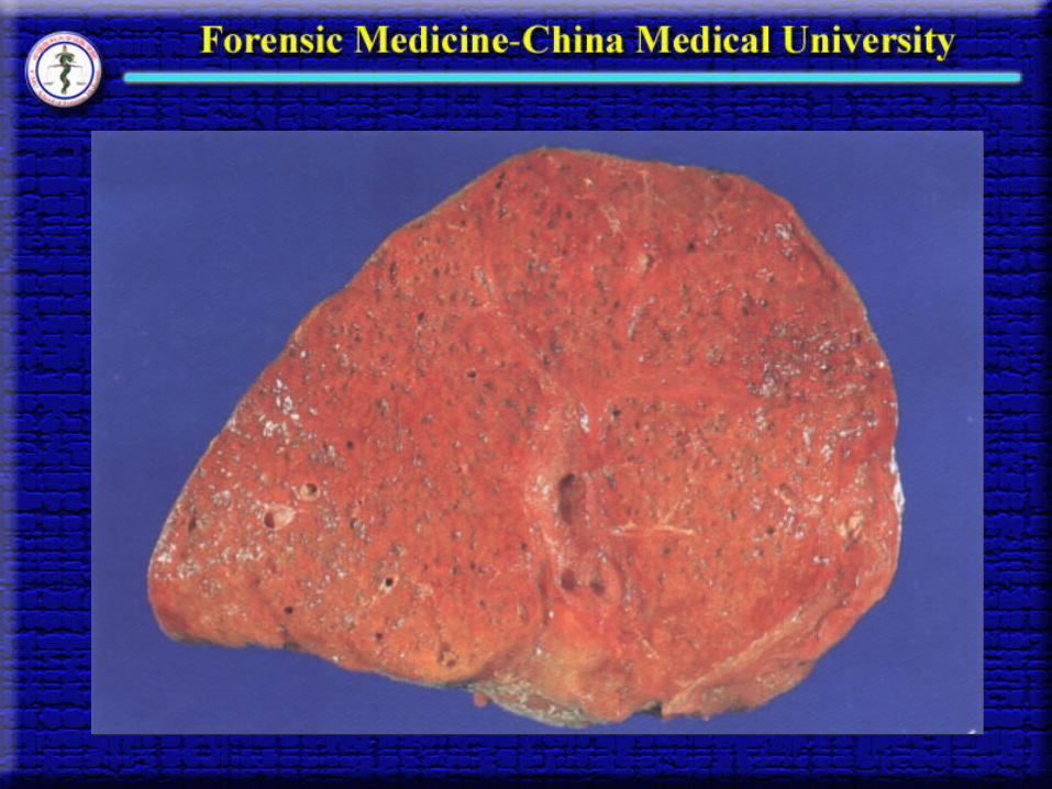
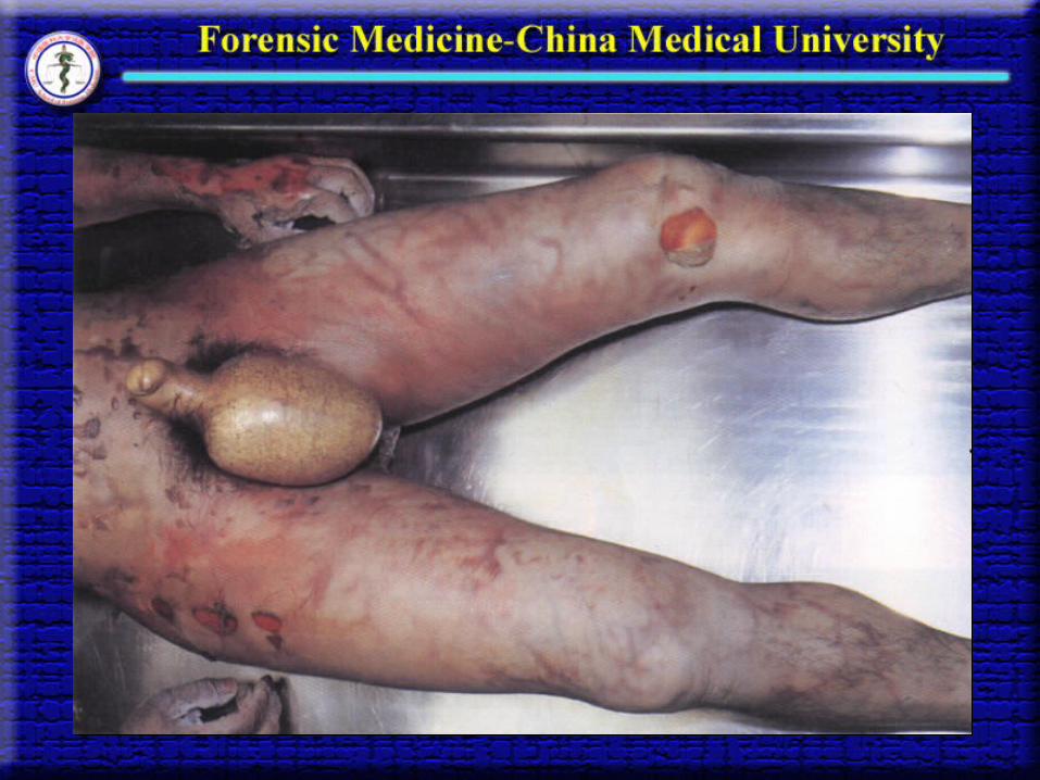
(b) The evolution of gases in the tissues An offensive odor, distention of the abdomen,Faeces forced out of the rectum and stomach contents forced through the mouth and nose,The foetus expelled from the uterus as called ‘postmortem delivery’ ,Neck and face bloated,The eyes proptosed and the tongue swollen and protruding beyond the line of the teeth,Bloodstained froth at the mouth and nostrils,Bullae in the dermis, and desquamated ‘foamy’ appearance of the organs as called foaming organs such as foaming liver, kidney, etc
(b) The evolution of gases in the tissues An offensive odor, distention of the abdomen,Faeces forced out of the rectum and stomach contents forced through the mouth and nose,The foetus expelled from the uterus as called ‘postmortem delivery’ ,Neck and face bloated,The eyes proptosed and the tongue swollen and protruding beyond the line of the teeth,Bloodstained froth at the mouth and nostrils,Bullae in the dermis, and desquamated ‘foamy’ appearance of the organs as called foaming organs such as foaming liver, kidney, etc


(c) The liquefaction of the tissues
Generally, the rate of putrefaction of the organs
depends upon the amount of muscles and fibrous tissue in an organ the organs composed of muscular and fibrous tissues resist putrefaction longer than the parenchymatous organs.
Exception is the intestines and the stomach which almost putrefy as rapidly as the spleen because their contents contain much more putrefactive organisms at the time of death though they are composed of so many fibrous tissues.

(d) Molded cadaver
(e) Skeletonized remains
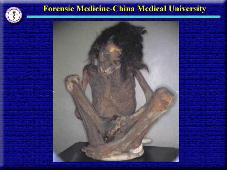
(2) Mummification (i) Definition
Mummification is a modification of putrefaction which is
characterized by the dehydration or desiccation of the tissues
and viscera after death.
(2) Mummification (i) Definition
Mummification is a modification of putrefaction which is
characterized by the dehydration or desiccation of the tissues
and viscera after death.

(ii) The conditions responsible for mummification
Mummification is not a common condition, but is seen
occasionally in bodies that have been buried in dry soil, e.g.,
in desert sand. The conditions necessary for the production
of mummification are absence of dampness and the
continuous action of a current of dry or warm air.
Dehydration begins before putrefactive changes occur.

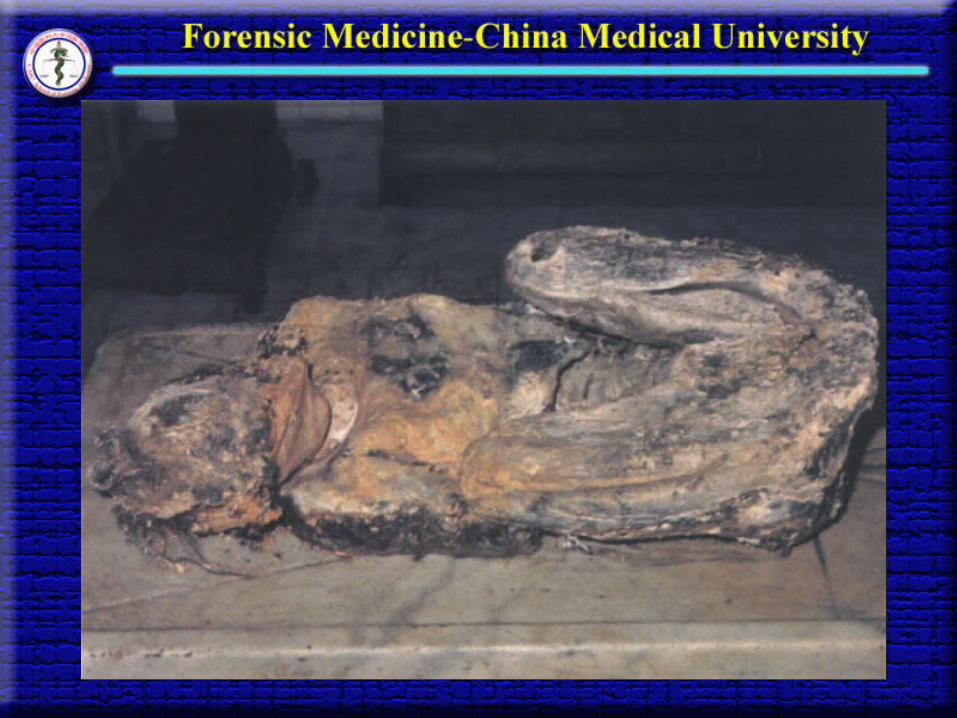
(iii) The appearances of mummification
The appearance of a mummified body is quite typical. The whole structure is hard, dry and shriveled and often black in colour, and the anatomical features are well preserved.
The time required for the complete mummification of a body varies greatly from a period of several months to a year or longer. This may persist for many years once it has developed.

(3) Saponification or adipocere formation(i) Definition
Saponification or adipocere formation is a modification of putrefaction which is characterized by the transformation of certain of the fatty tissues of the body into a substance known as adipocere that is a yellow-white greasy, wax-like substance that has a rancid odor.
(ii) The cause of saponification
The cause of saponification is the change of fatty constituents of the body into new chemical compounds, more or less stable in composition, and the process is brought about by gradual hydrogenation of pre-existing fats in the body to form higher fatty acids.
Adipocere is not common. It is seen occasionally in the tissues of bodies that have been buried in moist soil or immersed in fluid media.
(iii) The rate of adipocere formation
The time necessary for the adipocere formation
varies greatly. It usually develops over a period of many
months. Under warm condition, e.g., in the tropics, the
process may develop within a few weeks in bodies that
immersed in fluid media. Adipocere may persist for
several years after its formation.
(iv) Appearances and significance of adipocere
Adipocere develops in the subcutaneous tissues. It is not
common for the entire body to be converted into adipocere.
More generally, it is found in the fatty tissues of the cheeks,
breasts and buttocks. On rare occasions and particularly in i
nfants, the process may involve all the subcutaneous tissues
of the body. The process does not affect the viscera.
The importance of adipocere is that when the process
involves the fatty tissues of the cheeks, the features of a
deceased may remain identifiable many years after burial
or immersion. Similarly, the anatomical form of wounds
caused at the time of death may be recognizable many
months or years after burial.

End