food allergens affect the intestinal tight junction...
TRANSCRIPT
345
Original article
Food allergens affect the intestinal tight junction
permeability in inducing intestinal food allergy in rats
Tao Chen,1,2,#
Xiaoyu Liu,1,#
Li Ma,1,2,#
Weiyi He,1 Weizhong Li,
1 Yanjuan Cao
1 and Zhigang Liu
1,*
Summary
Background: The intestinal tract plays an
important role in food allergy and the intestinal
mucosa barrier is critical for maintenance of its
function. The underlying mechanisms of how
food allergens modulate the intestinal
permeability in inducing intestinal food allergy
remain elusive.
Objective: The aim of this study was to explore
the mechanism of how food allergens influence
the function of intestinal barrier and induce
intestinal food allergy.
Methods: Ovalbumin (OVA) was chosen to
establish intestinal food allergy models in
juvenile and adult rats that were confirmed by
IgE and IgG assay. Intestinal tissue morphology
was analyzed by HE staining. Intestinal
permeability was dynamically monitored using a
Lactulose (L)-Mannitol (M) assay. The
morphology of the tight junctions in the intestinal
mucosa barrier were analyzed under TEM. The
expression of key molecules in tight junction
regulation was evaluated by Real-time PCR.
Results: We found: 1) The sensitization rate in
juvenile rats was higher than in adult rats; 2)
Intestine fluff erosion was more serious in
juvenile rats than in adult rats in the duodenum
and ileum; 3) Intestinal permeability was
severely damaged, according to the results of the
Lactulose (L)-Mannitol (M) assay; 4) Tight
junction damage on the mucosal barrier was
observed; Real-time PCR results showed that the
expression of some key molecules that are
involved in tight junction regulation was also
affected. Conclusions: Our data suggested that
the allergy sensitization rate of Ovalbumin
(OVA) in the juvenile group is higher than in
adults and food allergens may increase intestinal
mucosal permeability through intestinal tight
junction regulation in inducing intestinal food
allergy. (Asian Pac J Allergy Immunol 2014;32:345-
53)
Keywords: Food allergen, ovalbumin, allergy,
intestinal permeability, tight junction
Introduction
Food allergy is a serious public health
problem,1,2 which cause severe symptoms in the
gastrointestinal tract, skin, respiratory tract and other
organ systems, and even anaphylactic shock and
death.3-5 Epidemiological studies have reported that
the incidence of food allergies (FA) has been
increasing. Previous research has contributed a lot of
information about food allergy. Its incidence is
higher in children (6% to 10%) than in adults (just
1% to 2%)5 and its pathogenesis is still not well
understood.
As the mainly organ involved in digestion, the
intestinal tract is considered to be a barrier to food
allergens in cases of food allergy. The intestinal
barrier, which can be divided into the mechanical
barrier, microbial barrier, immunological barrier
and chemical barrier etc., plays an important role in
maintaining intestinal function to prevent bacteria,
endotoxins and other toxic substances passing into
the underlying tissues.6,7 Studies suggested that
abnormal intestinal permeability is also asociated
with diabetes, celiac disease, inflammatory bowel
disease and other diseases besides food allergy.8,9
Increased intestinal permeability is also believed to
# Authors equally contributed to this work.
From 1. State Key Laboratory of Respiratory Disease for
Allergy at Shengzhen University; School of Medicine,
Shenzhen University, 3688 Nanhai Ave., Shenzhen,
Guangdong, P. R. China, 518060.
2. Shenzhen ENT Institute, Longgang Central Hospital,
Shenzhen, Guangdong, P. R. China, 518116.
Corresponding author: Zhigang Liu
E-mail: [email protected]
Submitted date: 24/9/2013
Accepted date: 3/3/2014
Asian Pac J Allergy Immunol 2014;32:345-53 DOI 10.12932/AP0443.32.4.2014
346
be significantly related to the pathogenesis of these
diseases and more and more studies are focusing on
this relationship.
It seems that the food allergy morbidity varies in
different age groups, which is possibly associated
with the development and functional status of the
intestinal mucosal barrier.10 Permeability increase
and functional damage can be found in the intestinal
barrier when food allergy occurs.7,11, 12 It is reported
that intestinal permeability increases in the early
stages of food allergy.12,13 Yet the molecular basis
of the structural changes in the intestinal barrier
associated with food allergy remain unknown. The
aim of our study was to explore the
molecular mechanisms of how food allergens affect
and damage the morphology and function of the
intestinal mucosal barrier to induce intestinal food
allergy, through OVA induced food allergy models
in juvenile or adult rats.
Methods
Animals
4 week (considered Juvenile14) and 12 week old
(considered Adult14) BN (Brown Norway) rats were
purchased from Vital River Laboratory Animal
Technology Co. Ltd. (Beijing, China) and raised in
SPF facilities. The animal studies were approved by
the School of Medicine of Shenzhen University, and
carried out as previously reported.15,16 Briefly, the
juvenile and adult rats were randomly divided into a
sensitization group and a control group. The
sensitization group was administrated 1mg/mL
OVA (in PBS, phosphate-buffered saline)
(Sigma/Flu/Ald, USA) orally everyday for 48 days,
while the control group was administrated1 mL
PBS. All the animals were challenged on the 49th
day with 100mg/mL OVA after the last sensitization
and sacrificed 8hrs later (Figure 1). Blood and
intestinal tissue samples were collected for further
study.
Intestinal permeability evaluation (Lactulose/
Mannitol assay)
Intestinal permeability was evaluated by the
ratio of lactulose and mannitol (M/L) in urine
excretion as previously reported.17, 18 Rats were put
into metabolic cages and given (i.g.) 100mg
lactulose and 50 mg mannitol (dissolving in
1mLPBS) orally once a week. Rat urine was
collected during a 24hrs fasting period (but with
water). 5% acetic acid was added to the urine
samples after centrifugation. The samples were then
boiled, centrifuged and the filtered through a
0.22μm Millipore filter to remove impurities (such
as urine proteins). Afterwards the specimen was
applied to HPLC analysis to determine the ratio of
lactulose and mannitol (L/M) [20μL was taken for
analysis (mobile phase: acetonitrile/water
(80%/20%); flow rate: 1mL/min; column
temperature: 35C)
Serum OVA-IgE/OVA-IgG analyzing
Blood was collected from the rats’ tail vein
every week. OVA-sIgE and OVA-sIgG were
assayed by ELISA as previously reported 19. In
brief, undiluted serum samples were detected using
OVA pre-coated plates (10 μg/mL OVA of
100 μL/well) and HRP-labeled rat-anti-mouse IgE
or IgG (1:1000) as the secondary antibody
(Southern Biotechnology Associates Inc.,
Birmingham, AL). After the incubation and reaction
with TMB (3,3',5,5'-Tetramethylbenzidine), the
final OD value was detected at 450 nm wavelength
using a RT-2100C Microplate reader (Rayto Life
and Analytical Sciences Co., Ltd., China ).
HE staining
Intestinal samples were collected immediately
after rat sacrifice. The specimens were fixed with
4% paraformaldehyde for 24 hours and stained with
Hematoxylln-Eosinstain after dehydration,
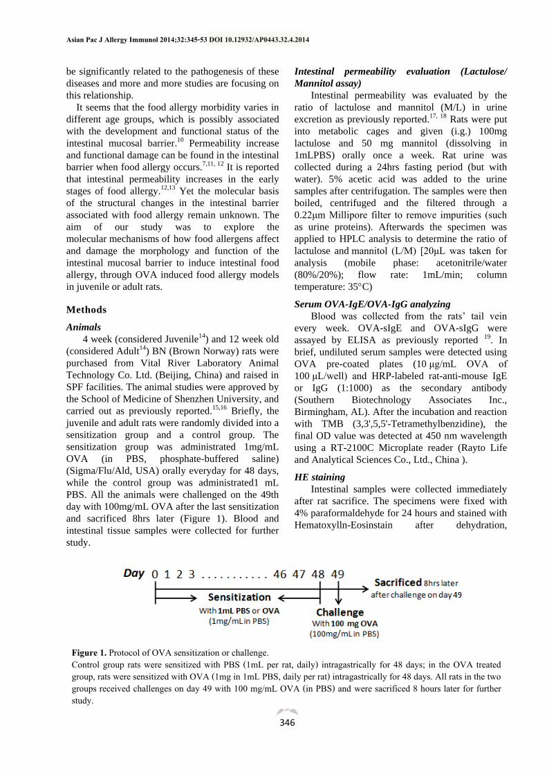
Figure 1. Protocol of OVA sensitization or challenge. Control group rats were sensitized with PBS (1mL per rat, daily) intragastrically for 48 days; in the OVA treated group, rats were sensitized with OVA (1mg in 1mL PBS, daily per rat) intragastrically for 48 days. All rats in the two groups received challenges on day 49 with 100 mg/mL OVA (in PBS) and were sacrificed 8 hours later for further study.
Allergen affects intestinal tight junction
347
embeding and slicing. The structural and
morphological changes were observed and analyzed
under a OLYMPUS BX51 microscope.
Electron microscope study
Rat intestinal samples were collected
immediately after sacrifice. Samples were cut into
1.5mm×1.5mm×2 mm pieces and put into a fixation
solution (provided by the electron microscopy room
of the School of Medicine at Sun Yat-
sen University). Samples then were kept at 4
degrees C overnight and sent to the electron
microscopy room of the School of Medicine at Sun
Yat-sen University for analysis. The structural and
morphological changes (of epithelial cells, tight
junction, etc.) were examined under the electron
microscope.
Real-time PCR
50-100 mg of intestinal tissues were collected
and ground in liquid nitrogen, then moved into a
1.5mL EP tube. The total RNA was extracted using
a TRIzol Reagent (15596018, invitrogen/Gibco/MP)
and reverse transcribed using a Reverse
Transcriptase Kit (Fermentas). Real-time PCR was
performed using C1000TM Thermal Cyclers (Bio-
Rad, USA) with SYBR green fluorescence. Samples
were run in triplicate and the cycling parameters
were: 95°C for 3 min, followed by 40 cycles of
95°C for 25s, 60°C for 20s, and a detection step at
72°C for 30 s. Human β-actin transcript was used as
an internal control for results standardization (2-
△△CT) by eliminating variations in mRNA quantity
and each RNA sample was arranged in triplicate.
All the primers were synthesized by BGI
Cooperation (Shenzhen, China) as list:
ZO-1: Forward, 5’-ACCCACGAAGTTATGAGCA
AG-3’; Reverse, 5’-AGACTGTGGTTTCATTGC
TGG-3’;
Occludin: Forward, 5’-ATTCCTCTGACCTTGTC
CGTG-3’; Reverse, 5’-CCTGTCGTGTAGTCG
GTTTCA-3’;
Claudin-2: Forward, 5’-ATTCCTCTGACCTTGTC
CGTG-3’; Reverse, 5’-AGCCAACCGCCGTCAC
AATG-3’;
Claudin-8: Forward, 5’-TGTCGTGTTTGAGAA
CCGCTGGG-3’; Reverse, 5’-ACGGACGCAG
CACACATCAGTC-3’;
Claudin-9: Forward, 5’-TTCCACTGGCCTTG
AACTCCTCG-3’; Reverse, 5’-GCTGTTGCCAA
TGAAGGCGGT-3’;
Claudin-15: Forward, 5’-AACTGCTGGGACTT
CCCGTCCAT-3’; Reverse, 5’-TCGATGTTGCCC
ACGTTGGTGC-3’;
β-actin: Forward, 5’-GTCTCACCACTGGCA
TTGTG-3’; Reverse, 5’-TCTCAGCTGTGGTGGT
GAAG-3’.
Statistical analysis
The data were analyzed using SPSS v13.0
software. All values in the study are presented as
mean±SD from at least three independent
experiments. Statistical analysis of experiments in
cell lines was evaluated using Student's t test.
Values of P < 0.05 were considered to be
statistically significant.
Result
Juvenile rats were more prone to allergy to OVA
induction compared with the adults.
Successful food allergy model induction was
determined according to a significant serum IgE rise
and behavior observations (e.g., diarrhea). We found
that it was earier to induce allergy in juvenile rats
(with a sensitization rate 69.23%) to OVA,
compared with their adult counterparts (Table 1).
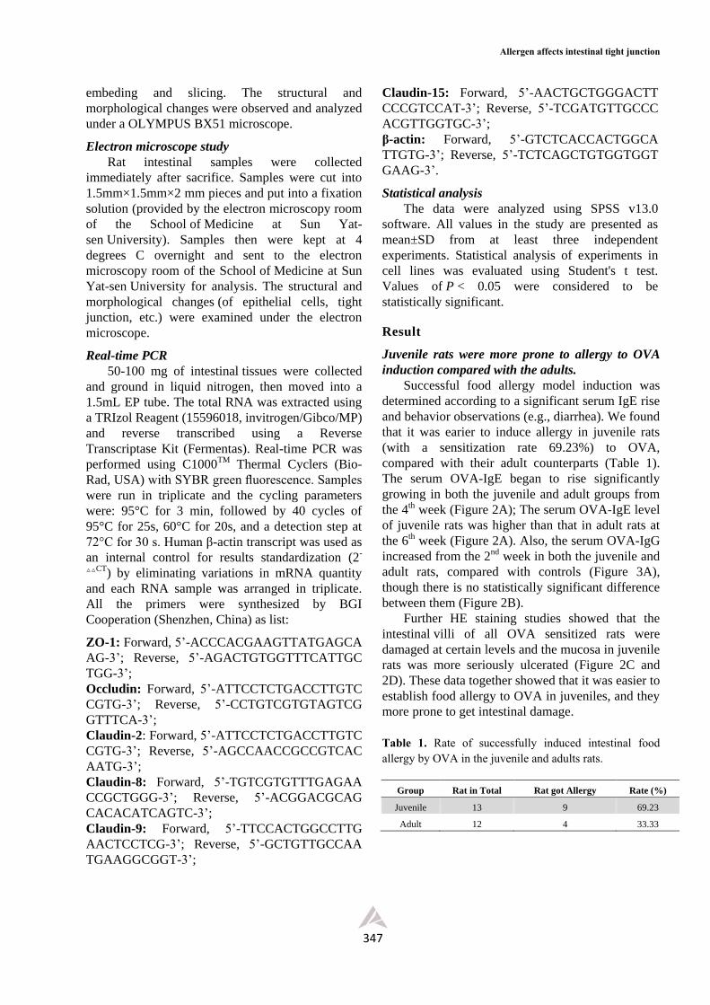
The serum OVA-IgE began to rise significantly
growing in both the juvenile and adult groups from
the 4th week (Figure 2A); The serum OVA-IgE level
of juvenile rats was higher than that in adult rats at
the 6th week (Figure 2A). Also, the serum OVA-IgG
increased from the 2nd week in both the juvenile and
adult rats, compared with controls (Figure 3A),
though there is no statistically significant difference
between them (Figure 2B).
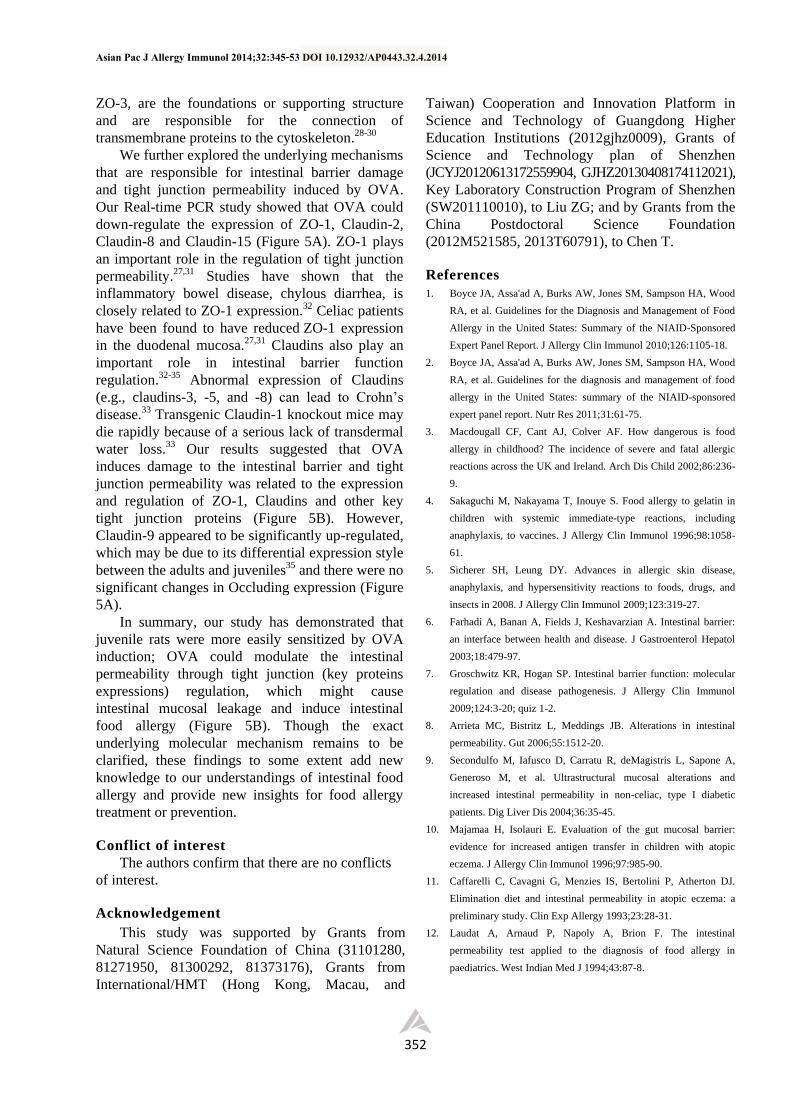
Further HE staining studies showed that the
intestinal villi of all OVA sensitized rats were
damaged at certain levels and the mucosa in juvenile
rats was more seriously ulcerated (Figure 2C and
2D). These data together showed that it was easier to
establish food allergy to OVA in juveniles, and they
more prone to get intestinal damage.
Table 1. Rate of successfully induced intestinal food allergy by OVA in the juvenile and adults rats.
Group Rat in Total Rat got Allergy Rate (%)
Juvenile 13 9 69.23
Adult 12 4 33.33
Asian Pac J Allergy Immunol 2014;32:345-53 DOI 10.12932/AP0443.32.4.2014
348
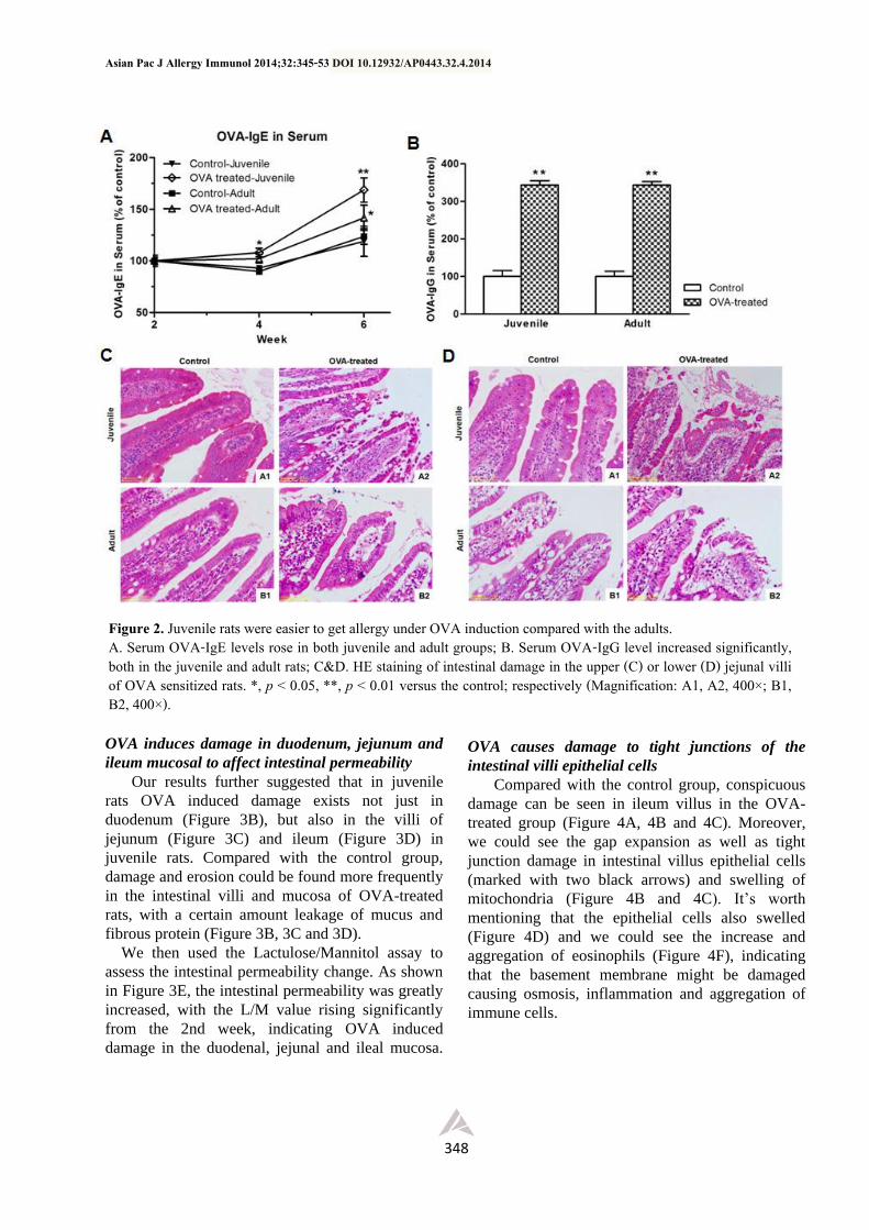
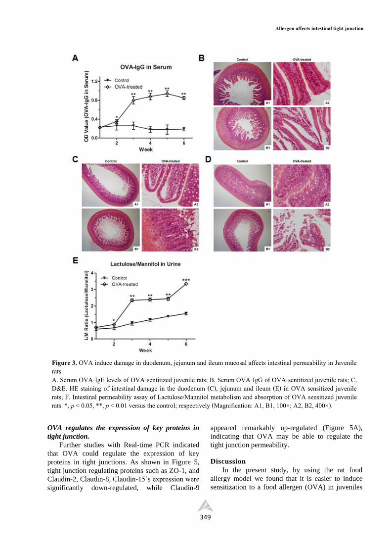
OVA induces damage in duodenum, jejunum and
ileum mucosal to affect intestinal permeability
Our results further suggested that in juvenile
rats OVA induced damage exists not just in
duodenum (Figure 3B), but also in the villi of
jejunum (Figure 3C) and ileum (Figure 3D) in
juvenile rats. Compared with the control group,
damage and erosion could be found more frequently
in the intestinal villi and mucosa of OVA-treated
rats, with a certain amount leakage of mucus and
fibrous protein (Figure 3B, 3C and 3D).
We then used the Lactulose/Mannitol assay to
assess the intestinal permeability change. As shown
in Figure 3E, the intestinal permeability was greatly
increased, with the L/M value rising significantly
from the 2nd week, indicating OVA induced
damage in the duodenal, jejunal and ileal mucosa.
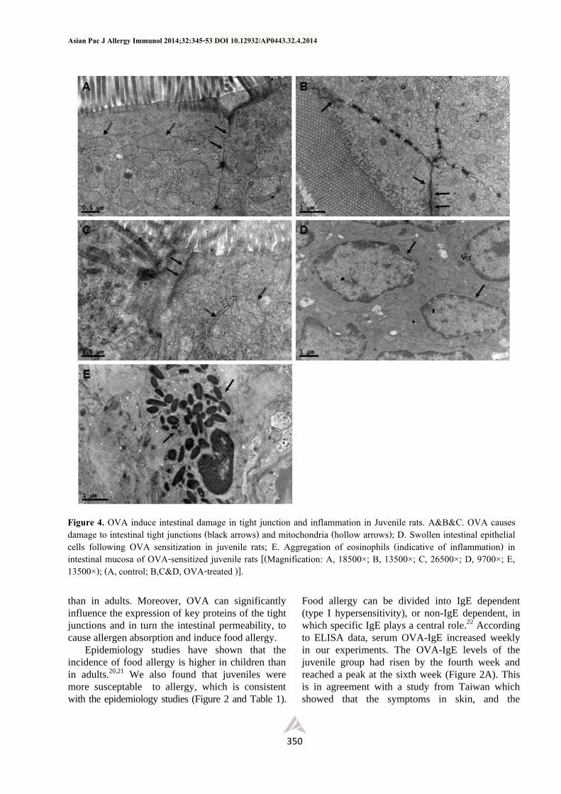
OVA causes damage to tight junctions of the
intestinal villi epithelial cells
Compared with the control group, conspicuous
damage can be seen in ileum villus in the OVA-
treated group (Figure 4A, 4B and 4C). Moreover,
we could see the gap expansion as well as tight
junction damage in intestinal villus epithelial cells
(marked with two black arrows) and swelling of
mitochondria (Figure 4B and 4C). It’s worth
mentioning that the epithelial cells also swelled
(Figure 4D) and we could see the increase and
aggregation of eosinophils (Figure 4F), indicating
that the basement membrane might be damaged
causing osmosis, inflammation and aggregation of
immune cells.
Figure 2. Juvenile rats were easier to get allergy under OVA induction compared with the adults. A. Serum OVA-IgE levels rose in both juvenile and adult groups; B. Serum OVA-IgG level increased significantly, both in the juvenile and adult rats; C&D. HE staining of intestinal damage in the upper (C) or lower (D) jejunal villi of OVA sensitized rats. *, p < 0.05, **, p < 0.01 versus the control; respectively (Magnification: A1, A2, 400×; B1, B2, 400×).
Allergen affects intestinal tight junction
349
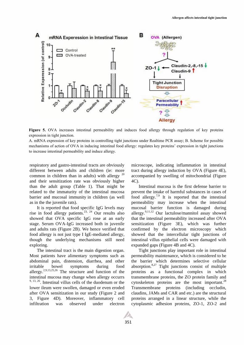
OVA regulates the expression of key proteins in
tight junction.
Further studies with Real-time PCR indicated
that OVA could regulate the expression of key
proteins in tight junctions. As shown in Figure 5,
tight junction regulating proteins such as ZO-1, and
Claudin-2, Claudin-8, Claudin-15’s expression were
significantly down-regulated, while Claudin-9
appeared remarkably up-regulated (Figure 5A),
indicating that OVA may be able to regulate the
tight junction permeability.
Discussion
In the present study, by using the rat food
allergy model we found that it is easier to induce
sensitization to a food allergen (OVA) in juveniles
Figure 3. OVA induce damage in duodenum, jejunum and ileum mucosal affects intestinal permeability in Juvenile rats. A. Serum OVA-IgE levels of OVA-sentitized juvenile rats; B. Serum OVA-IgG of OVA-sentitized juvenile rats; C, D&E. HE staining of intestinal damage in the duodenum (C), jejunum and ileum (E) in OVA sensitized juvenile rats; F. Intestinal permeability assay of Lactulose/Mannitol metabolism and absorption of OVA sensitized juvenile rats. *, p < 0.05, **, p < 0.01 versus the control; respectively (Magnification: A1, B1, 100×; A2, B2, 400×).
Asian Pac J Allergy Immunol 2014;32:345-53 DOI 10.12932/AP0443.32.4.2014
350
than in adults. Moreover, OVA can significantly
influence the expression of key proteins of the tight
junctions and in turn the intestinal permeability, to
cause allergen absorption and induce food allergy.
Epidemiology studies have shown that the
incidence of food allergy is higher in children than
in adults.20,21 We also found that juveniles were
more susceptable to allergy, which is consistent
with the epidemiology studies (Figure 2 and Table 1).
Food allergy can be divided into IgE dependent
(type I hypersensitivity), or non-IgE dependent, in
which specific IgE plays a central role.22 According
to ELISA data, serum OVA-IgE increased weekly
in our experiments. The OVA-IgE levels of the
juvenile group had risen by the fourth week and
reached a peak at the sixth week (Figure 2A). This
is in agreement with a study from Taiwan which
showed that the symptoms in skin, and the
Figure 4. OVA induce intestinal damage in tight junction and inflammation in Juvenile rats. A&B&C. OVA causes damage to intestinal tight junctions (black arrows) and mitochondria (hollow arrows); D. Swollen intestinal epithelial cells following OVA sensitization in juvenile rats; E. Aggregation of eosinophils (indicative of inflammation) in intestinal mucosa of OVA-sensitized juvenile rats [(Magnification: A, 18500×; B, 13500×; C, 26500×; D, 9700×; E, 13500×); (A, control; B,C&D, OVA-treated )].
Allergen affects intestinal tight junction
351
respiratory and gastro-intestinal tracts are obviously
different between adults and children (ie: more
common in children than in adults) with allergy 20
and their sensitization rate was obviously higher
than the adult group (Table 1). That might be
related to the immaturity of the intestinal mucosa
barrier and mucosal immunity in children (as well
as in the the juvenile rats).
It is reported that food specific IgG levels may
rise in food allergy patients.23, 24 Our results also
showed that OVA specific IgG rose at an early
stage. Serum OVA-IgG increased both in juvenile
and adults rats (Figure 2B). We hence verified that
food allergy is not just type I IgE-mediated allergy,
though the underlying mechanisms still need
exploring.
The intestinal tract is the main digestion organ.
Most patients have alimentary symptoms such as
abdominal pain, distension, diarrhea, and other
irritable bowel symptoms during food
allergy.2,9,13,25,26 The structure and function of the
intestinal mucosa may change when allergy occurrs 9, 13, 26. Intestinal villus cells of the duodenum or the
lower ileum were swollen, damaged or even eroded
after OVA sensitization in our study (Figure 2 and
3, Figure 4D). Moreover, inflammatory cell
infiltration was observed under electron
microscope, indicating inflammation in intestinal
tract during allergy induction by OVA (Figure 4E),
accompanied by swelling of mitochondrial (Figure
4C).
Intestinal mucosa is the first defense barrier to
prevent the intake of harmful substances in cases of
food allergy.7,9 It is reported that the intestinal
permeability may increase when the intestinal
mucosal barrier function is damaged during
allergy.9,11,12 Our lactulose/mannitol assay showed
that the intestinal permeability increased after OVA
sensitization (Figure 3E), which was further
confirmed by the electron microscopy which
showed that the intercellular tight junctions of
intestinal villus epithelial cells were damaged with
expanded gaps (Figure 4B and 4C).
Tight junctions play important role in intestinal
permeability maintenance, which is considered to be
the barrier which determines selective cellular
absorption.8,27 Tight junctions consist of multiple
proteins as a functional complex in which
transmembrane proteins, the ZO protein family and
cytoskeleton proteins are the most important.28
Transmembrane proteins (including occludin,
claudins, JAMs and CAR and etc.) are the structural
proteins arranged in a linear structure, while the
cytoplasmic adhesion proteins, ZO-1, ZO-2 and
Figure 5. OVA increases intestinal permeability and induces food allergy through regulation of key proteins expression in tight junction. A. mRNA expression of key proteins in controlling tight junctions under Realtime PCR assay; B. Scheme for possible mechanisms of action of OVA in inducing intestinal food allergy: regulates key proteins’ expression in tight junctions to increase intestinal permeability and induce allergy.
Asian Pac J Allergy Immunol 2014;32:345-53 DOI 10.12932/AP0443.32.4.2014
352
ZO-3, are the foundations or supporting structure
and are responsible for the connection of
transmembrane proteins to the cytoskeleton.28-30
We further explored the underlying mechanisms
that are responsible for intestinal barrier damage
and tight junction permeability induced by OVA.
Our Real-time PCR study showed that OVA could
down-regulate the expression of ZO-1, Claudin-2,
Claudin-8 and Claudin-15 (Figure 5A). ZO-1 plays
an important role in the regulation of tight junction
permeability.27,31 Studies have shown that the
inflammatory bowel disease, chylous diarrhea, is
closely related to ZO-1 expression.32 Celiac patients
have been found to have reduced ZO-1 expression
in the duodenal mucosa.27,31 Claudins also play an
important role in intestinal barrier function
regulation.32-35 Abnormal expression of Claudins
(e.g., claudins-3, -5, and -8) can lead to Crohn’s
disease.33 Transgenic Claudin-1 knockout mice may
die rapidly because of a serious lack of transdermal
water loss.33 Our results suggested that OVA
induces damage to the intestinal barrier and tight
junction permeability was related to the expression
and regulation of ZO-1, Claudins and other key
tight junction proteins (Figure 5B). However,
Claudin-9 appeared to be significantly up-regulated,
which may be due to its differential expression style
between the adults and juveniles35 and there were no
significant changes in Occluding expression (Figure
5A).
In summary, our study has demonstrated that
juvenile rats were more easily sensitized by OVA
induction; OVA could modulate the intestinal
permeability through tight junction (key proteins
expressions) regulation, which might cause
intestinal mucosal leakage and induce intestinal
food allergy (Figure 5B). Though the exact
underlying molecular mechanism remains to be
clarified, these findings to some extent add new
knowledge to our understandings of intestinal food
allergy and provide new insights for food allergy
treatment or prevention.
Conflict of interest
The authors confirm that there are no conflicts
of interest.
Acknowledgement
This study was supported by Grants from
Natural Science Foundation of China (31101280,
81271950, 81300292, 81373176), Grants from
International/HMT (Hong Kong, Macau, and
Taiwan) Cooperation and Innovation Platform in
Science and Technology of Guangdong Higher
Education Institutions (2012gjhz0009), Grants of
Science and Technology plan of Shenzhen
(JCYJ20120613172559904, GJHZ20130408174112021),
Key Laboratory Construction Program of Shenzhen
(SW201110010), to Liu ZG; and by Grants from the
China Postdoctoral Science Foundation
(2012M521585, 2013T60791), to Chen T.
References
1. Boyce JA, Assa'ad A, Burks AW, Jones SM, Sampson HA, Wood
RA, et al. Guidelines for the Diagnosis and Management of Food
Allergy in the United States: Summary of the NIAID-Sponsored
Expert Panel Report. J Allergy Clin Immunol 2010;126:1105-18.
2. Boyce JA, Assa'ad A, Burks AW, Jones SM, Sampson HA, Wood
RA, et al. Guidelines for the diagnosis and management of food
allergy in the United States: summary of the NIAID-sponsored
expert panel report. Nutr Res 2011;31:61-75.
3. Macdougall CF, Cant AJ, Colver AF. How dangerous is food
allergy in childhood? The incidence of severe and fatal allergic
reactions across the UK and Ireland. Arch Dis Child 2002;86:236-
9.
4. Sakaguchi M, Nakayama T, Inouye S. Food allergy to gelatin in
children with systemic immediate-type reactions, including
anaphylaxis, to vaccines. J Allergy Clin Immunol 1996;98:1058-
61.
5. Sicherer SH, Leung DY. Advances in allergic skin disease,
anaphylaxis, and hypersensitivity reactions to foods, drugs, and
insects in 2008. J Allergy Clin Immunol 2009;123:319-27.
6. Farhadi A, Banan A, Fields J, Keshavarzian A. Intestinal barrier:
an interface between health and disease. J Gastroenterol Hepatol
2003;18:479-97.
7. Groschwitz KR, Hogan SP. Intestinal barrier function: molecular
regulation and disease pathogenesis. J Allergy Clin Immunol
2009;124:3-20; quiz 1-2.
8. Arrieta MC, Bistritz L, Meddings JB. Alterations in intestinal
permeability. Gut 2006;55:1512-20.
9. Secondulfo M, Iafusco D, Carratu R, deMagistris L, Sapone A,
Generoso M, et al. Ultrastructural mucosal alterations and
increased intestinal permeability in non-celiac, type I diabetic
patients. Dig Liver Dis 2004;36:35-45.
10. Majamaa H, Isolauri E. Evaluation of the gut mucosal barrier:
evidence for increased antigen transfer in children with atopic
eczema. J Allergy Clin Immunol 1996;97:985-90.
11. Caffarelli C, Cavagni G, Menzies IS, Bertolini P, Atherton DJ.
Elimination diet and intestinal permeability in atopic eczema: a
preliminary study. Clin Exp Allergy 1993;23:28-31.
12. Laudat A, Arnaud P, Napoly A, Brion F. The intestinal
permeability test applied to the diagnosis of food allergy in
paediatrics. West Indian Med J 1994;43:87-8.
Allergen affects intestinal tight junction
353
13. Perrier C, Corthesy B. Gut permeability and food allergies. Clin
Exp Allergy 2011;41:20-8.
14. Iwata K, Yamada Y, Nakata A, Oghiso Y, Tani S, Doi K, et al.
Co-operative effects of thoracic X-ray irradiation and N-
nitrosobis(2-hydroxypropyl) amine administration on lung
tumorigenesis in neonatal, juvenile and adult Wistar rats. Toxicol
Appl Pharmacol 2013;267:266-75.
15. Knippels LM, Houben GF, Spanhaak S, Penninks AH. An oral
sensitization model in Brown Norway rats to screen for potential
allergenicity of food proteins. Methods 1999;19:78-82.
16. Pilegaard K, Madsen C. An oral Brown Norway rat model for
food allergy: comparison of age, sex, dosing volume, and allergen
preparation. Toxicology 2004;196:247-57.
17. Jarvinen KM, Konstantinou GN, Pilapil M, Arrieta MC, Noone S,
Sampson HA, et al. Intestinal permeability in children with food
allergy on specific elimination diets. Pediatr Allergy Immunol
2013;24:589-95.
18. Quadro L, Gamble MV, Vogel S, Lima AA, Piantedosi R, Moore
SR, et al. Retinol and retinol-binding protein: gut integrity and
circulating immunoglobulins. J Infect Dis 2000;182 Suppl 1:S97-
S102.
19. Li F, Wang L, Jin XM, Yan CH, Jiang S, Shen XM. The
immunologic effect of TGF-beta1 chitosan nanoparticle plasmids
on ovalbumin-induced allergic BALB/c mice. Immunobiology
2009;214:87-99.
20. Hsin YC, Huang JL, Yeh KW. Clinical features of adult and
pediatric anaphylaxis in Taiwan. Asian Pac J Allergy Immunol
2011;29:307-12.
21. Lyons AC, Forde EM. Food allergy in young adults: perceptions
and psychological effects. J Health Psychol 2004;9:497-504.
22. Sicherer SH. Food allergy. Lancet 2002;360:701-10.
23. Dannaeus A, Inganas M. A follow-up study of children with food
allergy. Clinical course in relation to serum IgE- and IgG-antibody
levels to milk, egg and fish. Clin Allergy 1981;11:533-9.
24. Sicherer SH, Sampson HA. Food allergy. J Allergy Clin Immunol
2010;125:S116-25.
25. Mansueto P, Montalto G, Pacor ML, Esposito-Pellitteri M, Ditta
V, Lo Bianco C, et al. Food allergy in gastroenterologic diseases:
Review of literature. World J Gastroenterol 2006;12:7744-52.
26. Piche T, Barbara G, Aubert P, Bruley des Varannes S, Dainese R,
Nano JL, et al. Impaired intestinal barrier integrity in the colon of
patients with irritable bowel syndrome: involvement of soluble
mediators. Gut 2009;58:196-201.
27. Pizzuti D, Bortolami M, Mazzon E, Buda A, Guariso G,
D'Odorico A, et al. Transcriptional downregulation of tight
junction protein ZO-1 in active coeliac disease is reversed after a
gluten-free diet. Dig Liver Dis 2004;36:337-41.
28. Gonzalez-Mariscal L, Betanzos A, Avila-Flores A. MAGUK
proteins: structure and role in the tight junction. Semin Cell Dev
Biol 2000;11:315-24.
29. Gonzalez-Mariscal L, Betanzos A, Nava P, Jaramillo BE. Tight
junction proteins. Prog Biophys Mol Biol 2003;81:1-44.
30. Coyne CB, Bergelson JM. CAR: a virus receptor within the tight
junction. Adv Drug Deliv Rev 2005;57(6):869-82.
31. Montalto M, Cuoco L, Ricci R, Maggiano N, Vecchio FM,
Gasbarrini G. Immunohistochemical analysis of ZO-1 in the
duodenal mucosa of patients with untreated and treated celiac
disease. Digestion 2002;65:227-33.
32. Gassler N, Rohr C, Schneider A, Kartenbeck J, Bach A,
Obermuller N, et al. Inflammatory bowel disease is associated
with changes of enterocytic junctions. Am J Physiol Gastrointest
Liver Physiol 2001;281:G216-28.
33. Furuse M, Hata M, Furuse K, Yoshida Y, Haratake A, Sugitani Y,
et al. Claudin-based tight junctions are crucial for the mammalian
epidermal barrier: a lesson from claudin-1-deficient mice. J Cell
Biol 2002;156:1099-111.
34. Furuse M, Fujita K, Hiiragi T, Fujimoto K, Tsukita S. Claudin-1
and -2: novel integral membrane proteins localizing at tight
junctions with no sequence similarity to occludin. J Cell Biol
1998;141:1539-50.
35. Abuazza G, Becker A, Williams SS, Chakravarty S, Truong HT,
Lin F, et al. Claudins 6, 9, and 13 are developmentally expressed
renal tight junction proteins. Am J Physiol Renal Physiol
2006;291:F1132-41.