fluoroscopy credentialing for non-radiologist physiciansfluoroscopy credentialing for...
TRANSCRIPT
Fluoroscopy Credentialing for non-Radiologist PhysiciansRevised April 2019
Tom LaVoy, Radiation Safety OfficerSUNY Upstate Medical UniversityRadiation Safety Office(315) 464-6510
Credentialing Requirements
• The hospital requires appropriate training before granting fluoroscopic privileges.
• In order to become credentialed, a physician must do the following:
- View this PowerPoint presentation (or equivalent)- Complete the quiz- Upon completing the training for the first time only, each physician will need to schedule time with Sue Baran in radiology for the hands–on orientation
• Every two year re-qualification with online training and test
• Occupational Radiation Protection• Personnel Monitoring• Patient Radiation Exposure• Mobile C-arms• Basic Radiation Biology• Radiation and Pregnancy
Topics
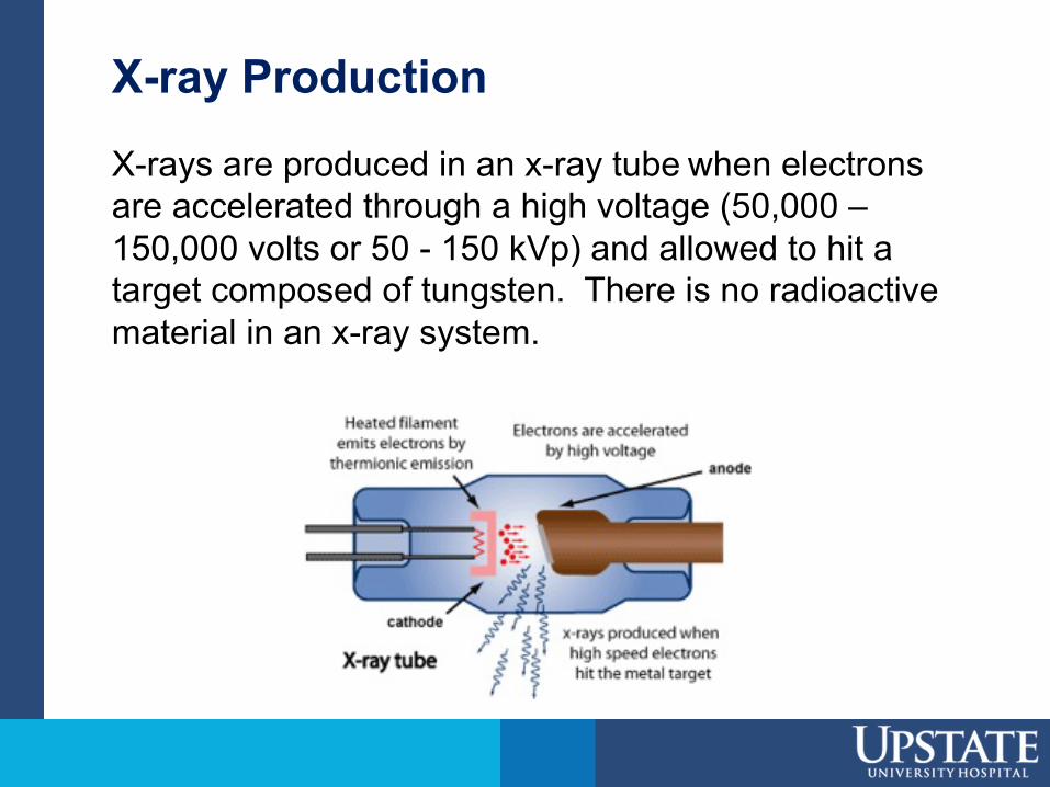
X-ray ProductionX-rays are produced in an x-ray tube when electrons are accelerated through a high voltage (50,000 –150,000 volts or 50 - 150 kVp) and allowed to hit a target composed of tungsten. There is no radioactive material in an x-ray system.
Mobile C-arms
• Can be used in approved areas of the hospital only.
• A mobile C-arm can put out just as much radiation as a full size, fixed fluoroscopy unit.
• A C-arm contains an x-ray tube at one end and an image receptor (called an Image Intensifier) at the other.
• The display monitors and computer are mounted on a separate cart.

Factors Influencing Fluoroscopy Exposure Rate
A fluoroscope automatically adjusts the amount of radiation output to compensate for the size of the body part.
Note: Exposure to a thicker or more dense body part will be greater than to a thin or less dense body part. The abdomen will require a greater exposure than a chest, a hip will require more exposure than a wrist.
Exposure During Angiography
• A C-arm fluoroscope can be used for angiography
• Many still images are acquired during the injection of the contrast media
• The Exposure Rate to the patient is ten times higher during angiography
• That means exposure to everyone in the room is ten times higher – So please stand back during Angiography!
Scattered Radiation
• Scatter radiation comes from the portion of the patient that we are imaging
• Scatter radiation goes in all directions • As we move away from the patient the
exposure is reduced as an inverse square function
• The Primary Beam is about 1,000 times more intense than the scatter radiation level at three feet from the patient
Factors which increase our exposure from scatter radiation:
• Larger body parts will cause the automatic brightness control (ABC) to adjust the kVp and mA to higher values which also causes greater amounts of scatter radiation.
• A larger x-ray field (a result of not restricting field size) will increase scatter radiation.
• The length of time the fluoroscopy unit is on. Complex examinations will require greater procedure time, increasing dose to both the patient and operator.
Scattered Radiation
• The more radiation exposure the patient gets, the more scatter radiation emitted.
• When there is more scattered radiation, everyone in the area receives more exposure.
• The difference in scattered radiation between the examination of an ankle and the abdomen of a large patient is greater than a factor of 100.
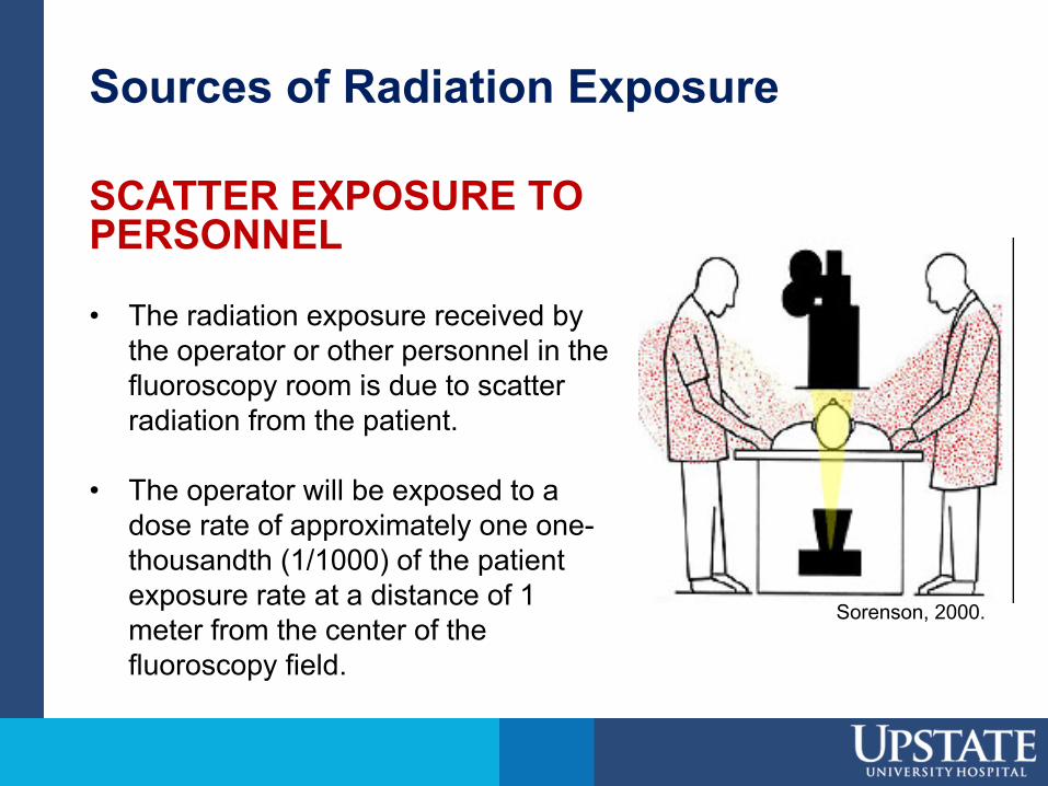
Sources of Radiation Exposure
SCATTER EXPOSURE TO PERSONNEL
• The radiation exposure received by the operator or other personnel in the fluoroscopy room is due to scatter radiation from the patient.
• The operator will be exposed to a dose rate of approximately one one-thousandth (1/1000) of the patient exposure rate at a distance of 1 meter from the center of the fluoroscopy field.
Sorenson, 2000.
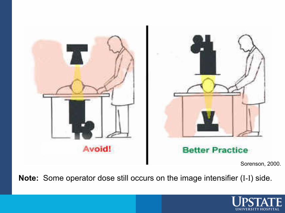
Note: Some operator dose still occurs on the image intensifier (I-I) side.
Sorenson, 2000.
Radiation ProtectionThe three most productive means of reducingradiation exposure are:
• Time: Minimize time spent in the radiation field.Use of “last-image-hold” and pulse fluoro featuresare technical advantages in reducing the total timex-rays are produced.
• Distance: Radiation dose rates increase or decreaseaccording to the inverse square law
Ex: Double your distance from the source and decrease your exposure by a factor of 4
• Shielding: Use of lead garments, lead gloves, thyroidshields, leaded eyeglasses, lead drapes andclear leaded glass barriers between the patientand operator
Effective Dose Equivalent• Lead garments (aprons, vest/skirts, thyroid collars)
are designed to shield the radiation sensitive portions of your body. The shielding reduces exposure to the covered areas by about 95%.
• The other portions of the body that are not shielded are much less sensitive to radiation.
• Lead garments must be worn during fluoroscopy, they are not optional.
• The risk of potential health effects is significantly less with the proper use of shielding.
• The EDE is a calculation of the effective risk to an individual from this non-uniform irradiation.
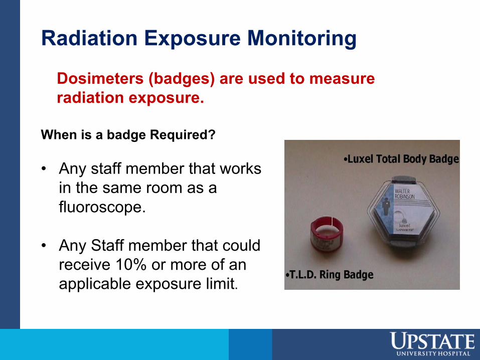
Radiation Exposure Monitoring
When is a badge Required?
• Any staff member that works in the same room as a fluoroscope.
• Any Staff member that could receive 10% or more of an applicable exposure limit.
Dosimeters (badges) are used to measure radiation exposure.
Personnel Monitoring
• Even when radiation protection techniques and engineering controls are in place to reduce personnel exposure, individual dose monitoring is required. (commonly called a “film badge”)
• Badges are assigned to one individual and must never be shared.
• A badge designed to measure your whole body exposure should be worn at the collar/chest area –OUTSIDE the lead garments.
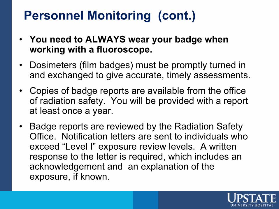
Personnel Monitoring (cont.)
• You need to ALWAYS wear your badge when working with a fluoroscope.
• Dosimeters (film badges) must be promptly turned in and exchanged to give accurate, timely assessments.
• Copies of badge reports are available from the office of radiation safety. You will be provided with a report at least once a year.
• Badge reports are reviewed by the Radiation Safety Office. Notification letters are sent to individuals who exceed “Level I” exposure review levels. A written response to the letter is required, which includes an acknowledgement and an explanation of the exposure, if known.
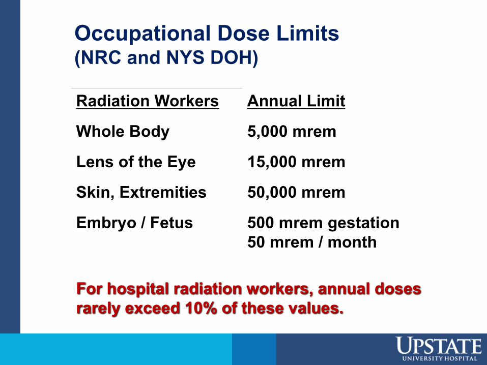
Occupational Dose Limits(NRC and NYS DOH)
For hospital radiation workers, annual dosesrarely exceed 10% of these values.
Radiation Workers Annual Limit
Whole Body 5,000 mrem
Lens of the Eye 15,000 mrem
Skin, Extremities 50,000 mrem
Embryo / Fetus 500 mrem gestation50 mrem / month
Pregnancy and Radiation
• The decision to perform a radiological procedure on a patient who may be pregnant is a medical decision and shall be made by a physician in consultation with the patient. If the procedure is to be performed, the physician must explain the risks to the patient, provide informed consent and the appropriate consent form shall be signed.
• Shielding shall be used to shield the abdomen from radiation provided it does not interfere with the procedure.
• Every attempt must be made to minimize direct exposure to the fetus according to the principles described in this presentation.
• Medical emergency radiological procedures however take precedence over pregnancy status.
Factors Influencing Fluoroscopy Exposure Rate
• The following slides detail patient exposure reduction methods.
• The Radiologic Technologist (RT) is familiar with these methods, but they require the physicians input and cooperation.
Any time we reduce patient exposure, we reduce the exposure of everyone else from scattered radiation.
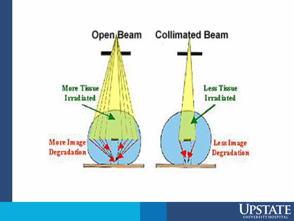
Recommendation #1: Use Collimation
• Restrict the x-ray field as much as possible (by using collimation). This will decreasethe volume of patient tissue exposed to radiation.
• Restricting the field size improves image quality by reducing scatter.
• Restricting the x-ray field reduces the amount of scatter everyone in the area is exposed to.
Factors Influencing Fluoroscopy Exposure Rate
Collimation
• Reducing the field size also makes it less likely that you will get your hands in the primary x-ray beam.
• REMEMBER – The primary beam is hundreds of times more intense than scattered radiation.
• Collimation is the single most effective means of exposure reduction for the patient and everyone else in the room.
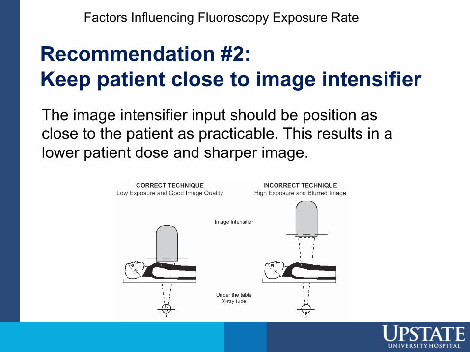
Recommendation #2: Keep patient close to image intensifierThe image intensifier input should be position as close to the patient as practicable. This results in a lower patient dose and sharper image.
Factors Influencing Fluoroscopy Exposure Rate
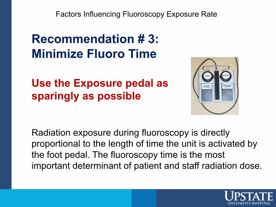
Recommendation # 3: Minimize Fluoro Time
Use the Exposure pedal as sparingly as possible
Factors Influencing Fluoroscopy Exposure Rate
Radiation exposure during fluoroscopy is directly proportional to the length of time the unit is activated by the foot pedal. The fluoroscopy time is the most important determinant of patient and staff radiation dose.
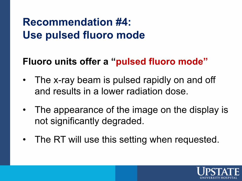
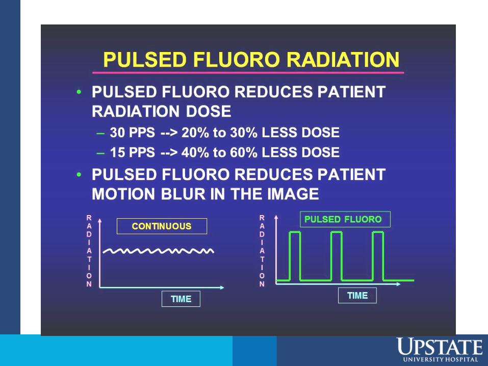
Recommendation #4: Use pulsed fluoro mode
Fluoro units offer a “pulsed fluoro mode”
• The x-ray beam is pulsed rapidly on and off and results in a lower radiation dose.
• The appearance of the image on the display is not significantly degraded.
• The RT will use this setting when requested.
Basic Radiation Biology• X-rays from fluoroscopy interact with biological
materials by transferring their energy to an electron which subsequently interacts with the target molecule to produce an ion or a free radical.
• Indirect action is the creation of free radicals from interactions with water molecules. Free radicals may then chemically interact with biologically sensitive molecules (DNA, RNA, proteins) causing damage.
• Direct action is the interaction of ionizing radiation with biologically sensitive molecules such as DNA causing direct destruction or mutation.
• Since water molecules are much more numerous than biologically sensitive molecules, indirect action is the most common form of biological damage.
Basic Radiation Biology (cont.)
• Cells can sustain a variable amount of radiation and still repair themselves from sub-lethal damage.
• Continuous high intensity radiation will produce greater damage than an equivalent fractionated (multiple smaller) dose since fractionation allows for cell repair.
A given organ’s response to radiation depends on:- Total dose- Dose rate- Fractionated scheme- Volume of irradiated tissue- Inherent tissue radiation sensitivity
• A large total dose, high dose rate and small fractionated schedule (which are all possible in fluoroscopy) will cause a greater degree of damage.
• Major concern in fluoroscopy is the possibility of acute, direct or deterministic, radiation damage which manifests as a skin injury. The severity of skin injury is dose-dependent; more dose means more severe symptoms.
Basic Radiation Biology (cont.)
As Low As Reasonable Achievable(ALARA)
• The core concept of radiation safety.
• ALARA is also a main component in regulations.
• Always take steps to keep your exposure to a minimum and minimize the risk.
• Applies to patients, staff, visitors, EVERYONE
Principles for Fluoroscopy
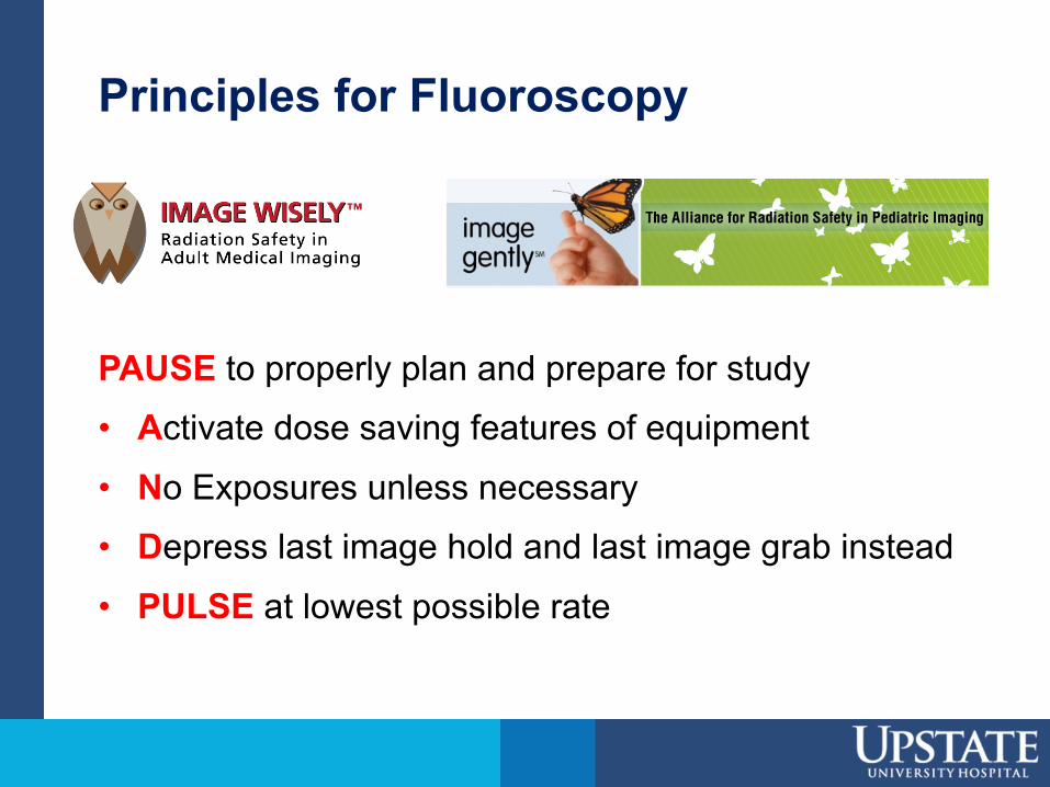
PAUSE to properly plan and prepare for study• Activate dose saving features of equipment
• No Exposures unless necessary• Depress last image hold and last image grab instead
• PULSE at lowest possible rate
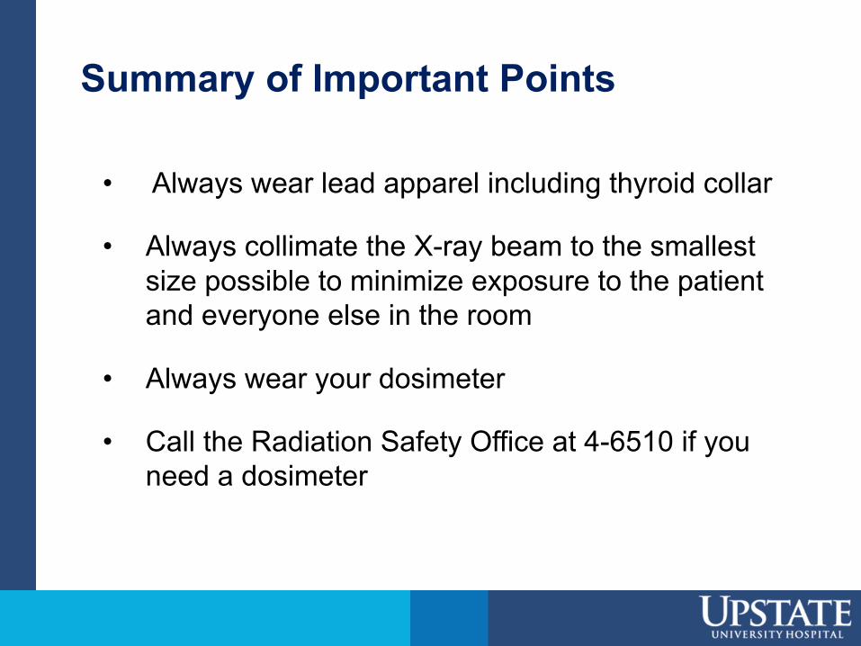
Summary of Important Points
• Always wear lead apparel including thyroid collar
• Always collimate the X-ray beam to the smallest size possible to minimize exposure to the patient and everyone else in the room
• Always wear your dosimeter
• Call the Radiation Safety Office at 4-6510 if you need a dosimeter
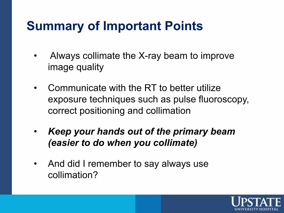
Summary of Important Points
• Always collimate the X-ray beam to improve image quality
• Communicate with the RT to better utilize exposure techniques such as pulse fluoroscopy, correct positioning and collimation
• Keep your hands out of the primary beam (easier to do when you collimate)
• And did I remember to say always use collimation?