fluorescence detection of protein s-nitrosation influenced … · fluorescence detection of protein...
TRANSCRIPT
Micah Doty and Paul DomanskiBiochemistry Department Cayman Chemical Company Ann Arbor MI
Fluorescence detection of proteinS-nitrosation influenced by pH and NObull donor
Figure 1 ndash Thiosulfonate Switch Technique (TST) sample treatments
ABSTRACTProtein S-nitrosothiol formation depends upon several factors including thiol accessibility protein conformation and concentration of nitric oxide The recently developed Thiosulfonate Switch Technique (TST)12 compliments the traditional Biotin Switch Technique (BST)3 but can be completed in half the time TST reactions are performed at pH 40 convergent with the application of a unique class of NOmiddot donors NOmiddot is released from the aryloxathiazolyliumolate class of NOmiddot donors in a pH-dependent manner4 Parallel treatment of proteins with S-nitroso-L-glutathione (GSNO) and aryloxathiazolyliumolate (CAY10562) at two different pH levels generated differential protein S-nitrosation as detected by the TST Further evaluation of pH and donor dependent exclusive protein S-nitrosothiols by LC-MSMS is planned with the development of heavy stable and light biotin probes The TST may prove useful for the characterization of S-nitrosation denitrosation and protein signaling within unique cells and sub-cellular compartments
1 Reeves BD Joshi N et al Org Biomol Chem 2014 12(40) 7942-7956 2 Reeves BD Hilmer JK et al Tetrahedron Lett 2013 54(42) 5707-5710 3 Jaffrey SR and Snyder SH Sci STKE 2001 86 pl1 4 D Lu J Nadas et al J AM CHEM SOC 2007 129 5503-5514
BACKGROUNDThe Thiosulfonate Switch Technique (TST) was developed by Drrsquos Ed Dratz Paul Grieco David Singel and Benjamin Reeves at the Department of Chemistry amp Biochemistry of Montana State University Their fast method blocks free thiols at pH 40 avoids the use of ascorbate high temperatures and denaturing reagents to visualize protein S-nitrosothiols as rhodamine mixed disulfides
HYPOTHESISCAY10562 may reveal unique protein S-nitrosothiols at pH 40 by the TST
METHODS1 Protein samples isolated from mouse muscle tissue by Precellysreg homogenization from
either HENS pH 77 or TST Assay Buffer pH 40
2 Treated 200-500 μg of clarified proteins with and without NO donors according to the scheme in Figure 2 1 hour at ambient temperature
3 Desalted all samples and exchanged buffers to pH 40 with ZebaTM 05 mL desalting columns (Thermo)
4 Added TST reagents sequentially to each sample for the times listed in Figure 1
5 Diluted MMTS treated samples with non-reducing PAGE buffer
6 SDS-PAGE 12 acrylamide tris-tricine gels (CBS Scientific) 100V
7 Rhodamine fluorescence imaging (excitation 532 nm emission 580 nm)
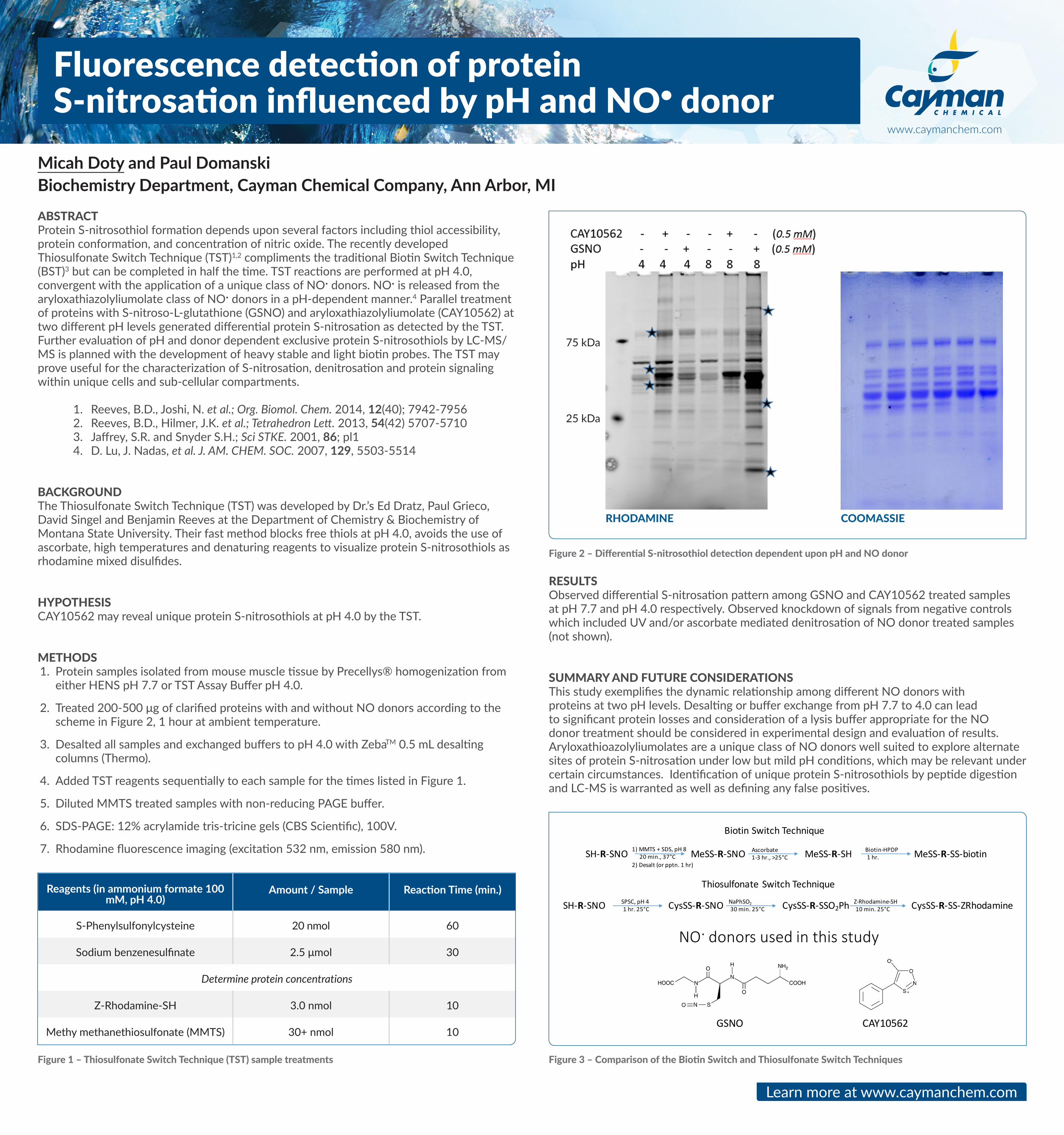
RESULTSObserved differential S-nitrosation pattern among GSNO and CAY10562 treated samples at pH 77 and pH 40 respectively Observed knockdown of signals from negative controls which included UV andor ascorbate mediated denitrosation of NO donor treated samples (not shown)
SUMMARY AND FUTURE CONSIDERATIONS This study exemplifies the dynamic relationship among different NO donors with proteins at two pH levels Desalting or buffer exchange from pH 77 to 40 can lead to significant protein losses and consideration of a lysis buffer appropriate for the NO donor treatment should be considered in experimental design and evaluation of results Aryloxathioazolyliumolates are a unique class of NO donors well suited to explore alternate sites of protein S-nitrosation under low but mild pH conditions which may be relevant under certain circumstances Identification of unique protein S-nitrosothiols by peptide digestion and LC-MS is warranted as well as defining any false positives
Learn more at wwwcaymanchemcom
wwwcaymanchemcom
Reagents (in ammonium formate 100 mM pH 40)
Amount Sample Reaction Time (min)
S-Phenylsulfonylcysteine 20 nmol 60
Sodium benzenesulfinate 25 μmol 30
Determine protein concentrations
Z-Rhodamine-SH 30 nmol 10
Methy methanethiosulfonate (MMTS) 30+ nmol 10
Figure 3 ndash Comparison of the Biotin Switch and Thiosulfonate Switch Techniques
Figure 2 ndash Differential S-nitrosothiol detection dependent upon pH and NO donor
RHODAMINE COOMASSIE
75 kDa
25 kDa
SH-R-SNO MeSS-R-SNO MeSS-R-SH MeSS-R-SS-biotin
BiotinSwitchTechnique
ThiosulfonateSwitchTechnique
SH-R-SNO CysSS-R-SNO CysSS-R-SSO2PhCysSS-R-SS-ZRhodamine
1)MMTS+SDSpH820min37degC
2)Desalt(orpptn1hr)
Ascorbate1-3hrgt25degC
Biotin-HPDP1hr
SPSCpH41hr25degC
NaPhSO230min25degC
Z-Rhodamine-SH10min25degC
SH-R-SNO MeSS-R-SNO MeSS-R-SH MeSS-R-SS-biotin
BiotinSwitchTechnique
ThiosulfonateSwitchTechnique
SH-R-SNO CysSS-R-SNO CysSS-R-SSO2PhCysSS-R-SS-ZRhodamine
1)MMTS+SDSpH820min37degC
2)Desalt(orpptn1hr)
Ascorbate1-3hrgt25degC
Biotin-HPDP1hr
SPSCpH41hr25degC
NaPhSO230min25degC
Z-Rhodamine-SH10min25degC
NOdonorsusedinthisstudy
GSNO CAY10562