fk228analoguesinducefetalhemoglobinin ... · hindawi publishing corporation anemia volume 2012,...
TRANSCRIPT
Hindawi Publishing CorporationAnemiaVolume 2012, Article ID 428137, 13 pagesdoi:10.1155/2012/428137
Research Article
FK228 Analogues Induce Fetal Hemoglobin inHuman Erythroid Progenitors
Levi Makala,1 Salvatore Di Maro,2, 3 Tzu-Fang Lou,4 Sharanya Sivanand,5
Jung-Mo Ahn,2 and Betty S. Pace1
1 Department of Pediatrics, Georgia Health Sciences University, Augusta, GA 30912, USA2 Department of Chemistry, University of Texas at Dallas, Richardson, TX 75083, USA3 Department of Pharmacological and Toxicological Chemistry, University of Naples Federico II, 80100 Naples, Italy4 Department of Molecular and Cell Biology, University of Texas at Dallas, TX 75080, USA5 Department of Developmental Biology, University of Texas Southwestern Medical Center, Dallas, TX 75390, USA
Correspondence should be addressed to Betty S. Pace, [email protected]
Received 16 December 2011; Accepted 7 March 2012
Academic Editor: Solomon F. Ofori-Acquah
Copyright © 2012 Levi Makala et al. This is an open access article distributed under the Creative Commons Attribution License,which permits unrestricted use, distribution, and reproduction in any medium, provided the original work is properly cited.
Fetal hemoglobin (HbF) improves the clinical severity of sickle cell disease (SCD), therefore, research to identify HbF-inducingagents for treatment purposes is desirable. The focus of our study is to investigate the ability of FK228 analogues to induce HbFusing a novel KU812 dual-luciferase reporter system. Molecular modeling studies showed that the structure of twenty FK228analogues with isosteric substitutions did not disturb the global structure of the molecule. Using the dual-luciferase system, asubgroup of FK228 analogues was shown to be inducers of HbF at nanomolar concentrations. To determine the physiologicalrelevance of these compounds, studies in primary erythroid progenitors confirmed that JMA26 and JMA33 activated HbF synthesisat levels comparable to FK228 with low cellular toxicity. These data support our lead compounds as potential therapeutic agentsfor further development in the treatment of SCD.
1. Introduction
Several classes of pharmacological compounds that reactivateγ-globin gene transcription have been identified. They in-clude cytotoxic agents, DNA methyl transferase, and his-tone deacetylase (HDAC) inhibitors. Cytotoxic compoundsterminate actively cycling progenitors and perturb cellulargrowth to trigger rapid erythroid regeneration and γ-globingene activation. S-stage cytotoxic drugs, such as cytosinearabinoside [1], myleran [2], vinblastine [3], and hydrox-yurea [4, 5], induce HbF production in primates and humans[4, 6, 7]. The Multicenter Study of Hydroxyurea establishedthis agent as the first FDA-approved treatment for SCD [7].Hydroxyurea was shown to reduce vaso-occlusive episodes inthe majority of sickle-cell patients treated. However, limita-tions to using hydroxyurea such as bone marrow suppression
[8], concerns over long-term carcinogenic complications,and a 30% non-response rate [7, 9], make the developmentof alternative therapies desirable.
The HDAC inhibitors have also been shown to bepotent HbF inducers. These agents target HDACs, whichplay a dynamic role in regulating cell cycle progression andchromatin conformation by changes in histone acetylationstatus. Aberrant transcriptional repression mediated by ClassI and II HDACs has been demonstrated in many cancers [10].Thus, HDAC inhibitors have been developed as promisinganticancer therapeutics [11]. Structurally diverse classes ofnatural and synthetic HDAC inhibitors bind target HDACsto block histone deacetylation [12] and produce an openchromatin confirmation and gene activation [13].
There has been great interest in HDAC inhibitors asHbF inducers to treat SCD. They include (1) short-chain
2 Anemia
fatty acids such as sodium butyrate (NaB), the first HDACinhibitor reported [14, 15]; (2) the benzamides (i.e., MS-275); (3) non cyclic and cyclic hydroxamates, like SAHA(suberoylanilide hydroxamic acid) and TSA (TrichostatinA); (4) cyclic peptides including FK228 (depsipeptide).NaB induces differentiation in mouse erythroleukemia cellsvia Stat5 phosphorylation and HbF synthesis through p38mitogen-activated protein kinase signaling [16–18]. Otherfatty acids including phenylacetate and propionate [19–21], induce HbF in erythroid progenitors, however, theseagents are rapidly metabolized and oral preparations arenot available. These published studies serve as the basis forresearch efforts to develop HDAC inhibitors as therapeuticagents for SCD.
Of the hydroxamic acid derivatives, the prototype TSAis a potent HDAC inhibitor [22, 23]. It interacts with adivalent zinc-binding motif in the binding pocket of ClassI and II HDACs [24]. Other HDAC inhibitors in the hydrox-amic acid class include the second-generation analogues ofTSA, identified from a library screen of 600 synthesizedcompounds [25]. The most widely studied TSA analoguesare SAHA and Scriptaid. SAHA targets HDAC1, 3, and 4and inhibits prostate cancer cell growth in vitro and invivo [26, 27]. Recently, it was demonstrated by Pace andcolleagues that SAHA and Scriptaid induce HbF synthesiscomparable to NaB and TSA in erythroid cells and β-YACtransgenic mice respectively [28]. However, limitations tothe further development of these agents included toxicity inprimary cells.
Another potent HDAC inhibitor is FK228, also knownas depsipeptide, isolated from Chromobacterium violaceum[29]. This compound has a unique bicyclic structure and isa stable pro-drug activated by the reduction of the disulfidebond by glutathione to produce an active form (redFK) afteruptake into cells [30]. The reduced sulfhydryl group interactsstrongly with the zinc ion at the active site of the enzyme andhas been shown to inhibit tumor proliferation in vitro andin vivo at nanomolar concentrations [27, 31, 32]. Recently,FK228 was tested in the μLCRβprRluc
AγprFluc GM979 stablecell line and erythroid progenitors grown in methylcellulosecolonies produced from peripheral blood mononuclear cells[33]. FK228 was shown to induce HbF in both systems.The level of γ-globin and β-globin promoter activity wasquantified indirectly using firefly (γ) and renilla (β) luciferaseactivity.
Drug-mediated HbF induction remains the best ap-proach to ameliorate the symptoms and complications ofSCD. Among many compounds, FK228 showed efficacy ininducing γ-globin transcription at low concentrations, how-ever, cell toxicity was observed and the drug is difficult tosynthesize. It is a bicyclic depsipeptide almost exclusivelycomprised of unnatural amino acids, D-valine, D-cysteineand, (Z)-dehydrobutyrine (Dhb) as well as a (3S, 4E)-3-hydroxy-7-mercapto-4-heptenoic acid, which is a keycomponent to form the highly constrained bicyclic structure.The high content of the unnatural amino acids and theconstrained bicyclic structure make it extremely stable inphysiological condition. Simon and coworkers first reportedits total synthesis in 1996 [34], and suggested a laborious
synthetic route with moderate yield (18% overall yield withover 16 steps).
Despite its exceptionally high in vitro and in vivoactivity, FK228 has not been explored due to its non-trivialand challenging synthesis, which hampered its productionand the design of analogues. The latter would aide ourunderstanding of the molecular mechanism of FK228 and toachieve higher potency and selectivity for HbF induction. Infact, only a few FK228 analogues have been created to dateeven after intensive synthetic efforts were made [34–36]. Tocircumvent this problem, we used in silico structure analysisand molecular modeling to design twenty FK228 structuralanalogues that can be easily synthesized [37]. Furthermore,two isosteric substitutions were made without altering itsglobal conformation.
The objective of our study was to investigate the abilityof the newly synthesized FK228 analogues to induce γ-globin gene transcription using a dual luciferase-based assaysystem. We identified two lead compounds, JMA26 andJMA33, which induce HbF expression in primary erythroidprogenitors. The potential of HDAC enzymes as druggabletargets in the treatment of SCD is discussed.
2. Materials and Methods
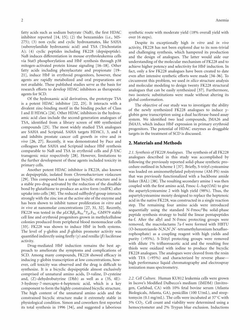
2.1. Synthesis of FK228 Analogues. The synthesis of all FK228analogues described in this study was accomplished byfollowing the previously reported solid-phase synthetic pro-cedure outlined in Scheme 1 [37]. Briefly, S-trityl cysteaminewas loaded on aminomethylated polystyrene (AM-PS) resinthat was previously functionalized with a backbone amidelinker (BAL) [38]. The resulting secondary amine 1 was thencoupled with the first amino acid, Fmoc-L-Asp(OAl) to givethe aspartylcysteamine 2 with high yield (98%). Thus, theaspartylcysteamine moiety designed to replace the heptenoicacid in the native FK228, was constructed in a single reactionstep. The remaining four amino acids were introducedsequentially using the standard N-Fmoc/tBu solid-phasepeptide synthesis strategy to build the linear pentapeptides6a-f. After the allyl and N-Fmoc protecting groups wereremoved, the macrolactams 7a-f were formed with HBTU(O-benzotriazole-N,N,N′,N′-tetramethyluronium hexafluo-rophosphate) as a coupling reagent with high yields andpurity (>95%). S-Trityl protecting groups were removedwith dilute 1% trifluoroacetic acid and the resulting freethiols were oxidized with iodine to produce the bicyclicFK228 analogues. The analogues were cleaved from the resinwith TFA (>95%) and characterized by reverse phase—high performance liquid chromatography and electrosprayionization mass spectrometry.
2.2. Cell Culture. Human KU812 leukemia cells were grownin Iscove’s Modified Dulbecco’s medium (IMDM) (Invitro-gen, Carlsbad, CA) with 10% fetal bovine serum (AtlantaBiologicals, Atlanta, GA), penicillin (100 U/mL), and strep-tomycin (0.1 mg/mL). The cells were incubated at 37◦C with5% CO2. Cell count and viability were determined using ahemocytometer and 2% Trypan blue exclusion. Inductions
Anemia 3
a
h, c, i
b
NH
NH
NH
SS
O
OO
OO
O
NH
NH
N
OO
O
OO
O
N
OO
OAl
N
OO
O
HN
NH
NH
NTrtS
OO
O
OO
O H
NH
N
OO
OO
NH
NTrtS
OO
O
OO
ONH
12
H2N STrtNH
TrtS TrtS
TrtS
TrtS
TrtS
TrtS
TrtS
TrtS
NHFmoc
NHFmoc
NHFmoc
NFmoc
Fmoc HN
R2
R2 R2
R2R2
R1
R1 R1
R1R1
R1
HNHN
HN
HNHN
3a–f
5a–f
4a–f
1. c2. d
1. c2. f
1. c2. e
1. c2. g
OAI
OAI OAI
OAI
NH
NHNH
NH
NH
j, k, l
JMA 1 R1 = L-Val,JMA 2 R1 = L-Val,JMA 12 R1 = L-Phe,JMA 26 R1 = L-PheJMA 33 R1 = L-(2)-NalJMA 112 R1 = L-Phe
R2 = L-AlaR2 = L-PheR2 = L-AlaR2 = L-PheR2 =L-(2)-NalR2 =L-Lys (FITC)
7
7a–f
6a–f
Scheme 1: Synthesis of FK228 analogues. Shown in the schematic are the steps, reagents, and conditions used for FK228 analogue synthesis.Compounds 1–7 are the intermediates during the synthesis. For steps 7a–f, the different R1 and R2 group substitutions were made to generatethe various JMA analogues shown. Symbols: (a) NaBH3CN; (b) Fmoc-Asp(OAl), DIC; (c) Piperidine; (d) Fmoc-AA1, HBTU; (e) Fmoc-AA2,HBTU; (f) Fmoc-D-Cys(Trt), HBTU; (g) Fmoc-D-Val, HBTU; (h) Pd(PPh3)4, DMBA; (i) HBTU; (j) 1% TFA; (k) I2; (l) TFA (>95%).
were performed with one million cells treated for 48 hr withthe following drugs purchased from Sigma (St Louis, MO):50 μM Hem (hemin), 2 mM NaB (sodium buytrate), 0.5 μMTSA, 10 mM Cys (cysteine), 1.5 nM FK228, and 100 μM HU(hydroxyurea). We also tested 5 μM SAHA, a gift from Merck& Co. Inc. (Whitehouse Station, NJ).
2.3. KU812 Stable Lines. KU812 stable cell lines were createdby co-transfecting wild-type KU812 cells with pEGFP-NI(G418 selectable marker) and the μLCRβprRluc
AγprFluc dual-reporter a kind gifts from Dr. George Stamatoyannopoulos
(University of Washington). Briefly, the 315-bp human β-globin gene promoter sequence was inserted upstream ofthe Renilla along with a polyadenylation signal downstreamto create PβprRluc. Likewise, 1.4 kb of human Aγ-globinpromoter was inserted upstream of firefly luciferase tocreate AγprFluc. The μLCR (locus control region), PβprRluc,and AγprFluc fragments were subsequently cloned into themammalian vector, pRL-null [39].
The dual-luciferase reporter lines were produced using10 μg each of linearized μLCRβprRluc
AγprFluc and pEGFP-NIplasmids co-transfected into KU812 cells by electroporation
4 Anemia
(S) (S)
(S)
(R) (R)
(S)
(S)
(S)
(S) (S)
(S)
(S)
O ONH
O OHO
O
NHNH
NH
S
S
O
O
O
OO
ONH
O
NH
SS
O
O
OO
O
HO
OH OH
SH SHNH
NH
NH
NHNH
SHHO
NH2
L-Asp Cysteamine
FK228 FK228 analogues
HN
R2R1
(3S,4E)-3-hydroxy-7-mercapto-4-heptenoic acid
(a)
D-val
(3S,4E)-3-hydroxy-7-mercapto-4-heptenoic acid
D-Cys
Dehydro-butyrine
L-Val
(b)
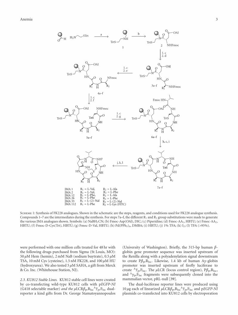
Figure 1: Structures of FK228 analogues. (a) The parent compound FK228 was transformed into novel structural analogues by two isostericsubstitutions. The modification of the trans-double bond and ester linkage in the native FK228 with two isosteric amide functional groupsallows facile synthesis of analogues as well as retention of the same backbone structure. Various amino acids such as Val, Ala, Phe, 2-Nal, andLys were introduced to investigate potency of the analogues. (b) Superimposed structures of FK228 (green) and a modified FK228 analogue(orange).
at 260 V, 975 μF (Bio-Rad, Hercules CA). After 72 hr, G418was added at a concentration of 900 μg/μl for 3 daysthen maintained under selection pressure indefinitely at aconcentration of 400 μg/μl. KU812 stable lines were treatedwith the various drugs at the same concentrations describedabove. FK228 and analogues were screened at concentrationsbetween 1–1000 nM for 48 hr and cell toxicity was monitoredby 2% Trypan blue exclusion. The effect of drug treatmentson γ-globin and β-globin promoter activity was monitoredby luciferase assay.
2.4. Dual Luciferase Assay. Luciferase activity was monitoredunder the different experimental conditions using the DualLuciferase Assay Reporter System (Promega, Madison, WI).The activity of firefly luciferase represents γ-globin promoter
activity (γF), while the renilla luciferase is the read-out forβ-globin promoter activity (βR). The β-globin promoter wasstrategically cloned between the LCR and γ-globin promoterto increase β expression, while simultaneously increasing thesensitivity of detection of γ-globin gene inducers [40].
After drug treatments, KU812 stable cells were washedwith 1X phosphate buffered saline and lysed in 1X PassiveLysis Buffer for 15 min, then protein extracts were added tothe Luciferase Assay Reagent II and firefly luciferase activityquantified in a Turner Designs TD-20/20 luminometer (Sun-nyvale, CA). To measure βR activity, Stop & Glo Reagents wasadded to measure the renilla luciferase activity. Total proteinwas determined by Bradford assay on a Beckman DU 640spectrophotometer (Chaska, MN) and luciferase activity wascorrected for total protein.
Anemia 5
SAH
A20
00 n
M
FK22
81.
5 n
M
HU
100 μ
M
KU812 cellsFo
ld in
crea
se
UT
50 μ
MH
emin
Cys
10 m
M
NaB
2 m
M
γ/β
glob
in m
RN
A ∗∗
∗ ∗∗
∗
0
2
4
6
8
10
12
14
16
(a)
S N H A
Renilla FireflyAγ-Proβ-ProμLCR
(b)
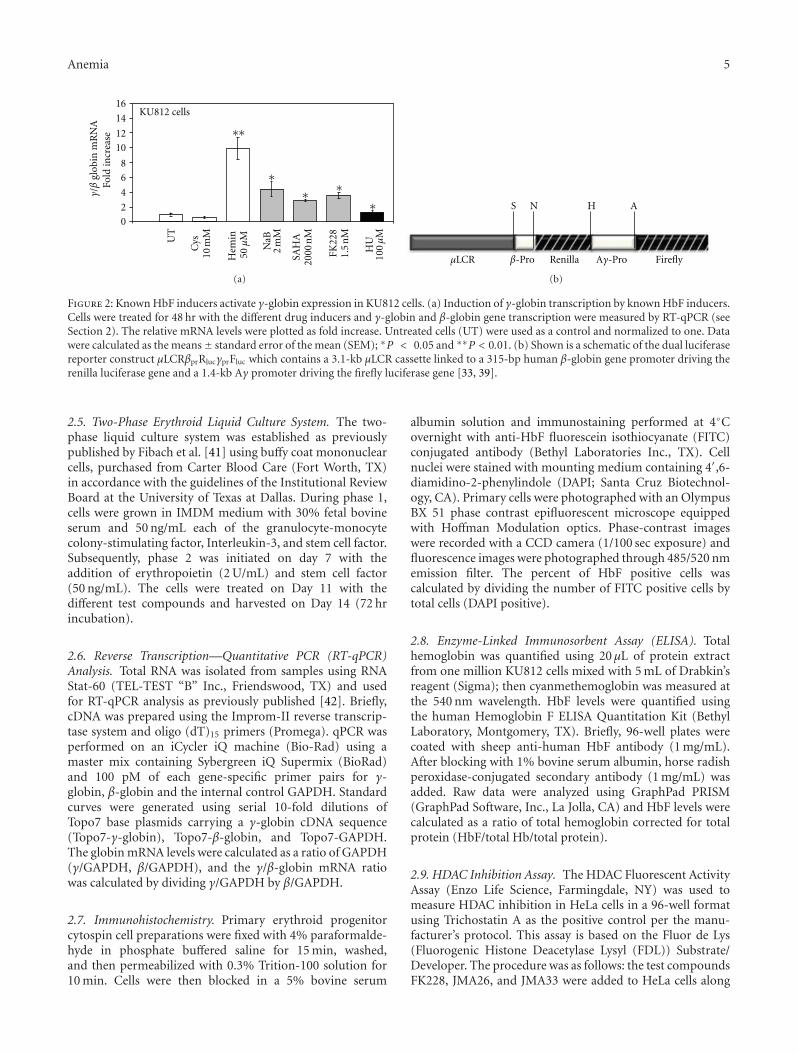
Figure 2: Known HbF inducers activate γ-globin expression in KU812 cells. (a) Induction of γ-globin transcription by known HbF inducers.Cells were treated for 48 hr with the different drug inducers and γ-globin and β-globin gene transcription were measured by RT-qPCR (seeSection 2). The relative mRNA levels were plotted as fold increase. Untreated cells (UT) were used as a control and normalized to one. Datawere calculated as the means± standard error of the mean (SEM); ∗P < 0.05 and ∗∗P < 0.01. (b) Shown is a schematic of the dual luciferasereporter construct μLCRβprRlucγprFluc which contains a 3.1-kb μLCR cassette linked to a 315-bp human β-globin gene promoter driving therenilla luciferase gene and a 1.4-kb Aγ promoter driving the firefly luciferase gene [33, 39].
2.5. Two-Phase Erythroid Liquid Culture System. The two-phase liquid culture system was established as previouslypublished by Fibach et al. [41] using buffy coat mononuclearcells, purchased from Carter Blood Care (Fort Worth, TX)in accordance with the guidelines of the Institutional ReviewBoard at the University of Texas at Dallas. During phase 1,cells were grown in IMDM medium with 30% fetal bovineserum and 50 ng/mL each of the granulocyte-monocytecolony-stimulating factor, Interleukin-3, and stem cell factor.Subsequently, phase 2 was initiated on day 7 with theaddition of erythropoietin (2 U/mL) and stem cell factor(50 ng/mL). The cells were treated on Day 11 with thedifferent test compounds and harvested on Day 14 (72 hrincubation).
2.6. Reverse Transcription—Quantitative PCR (RT-qPCR)Analysis. Total RNA was isolated from samples using RNAStat-60 (TEL-TEST “B” Inc., Friendswood, TX) and usedfor RT-qPCR analysis as previously published [42]. Briefly,cDNA was prepared using the Improm-II reverse transcrip-tase system and oligo (dT)15 primers (Promega). qPCR wasperformed on an iCycler iQ machine (Bio-Rad) using amaster mix containing Sybergreen iQ Supermix (BioRad)and 100 pM of each gene-specific primer pairs for γ-globin, β-globin and the internal control GAPDH. Standardcurves were generated using serial 10-fold dilutions ofTopo7 base plasmids carrying a γ-globin cDNA sequence(Topo7-γ-globin), Topo7-β-globin, and Topo7-GAPDH.The globin mRNA levels were calculated as a ratio of GAPDH(γ/GAPDH, β/GAPDH), and the γ/β-globin mRNA ratiowas calculated by dividing γ/GAPDH by β/GAPDH.
2.7. Immunohistochemistry. Primary erythroid progenitorcytospin cell preparations were fixed with 4% paraformalde-hyde in phosphate buffered saline for 15 min, washed,and then permeabilized with 0.3% Trition-100 solution for10 min. Cells were then blocked in a 5% bovine serum
albumin solution and immunostaining performed at 4◦Covernight with anti-HbF fluorescein isothiocyanate (FITC)conjugated antibody (Bethyl Laboratories Inc., TX). Cellnuclei were stained with mounting medium containing 4′,6-diamidino-2-phenylindole (DAPI; Santa Cruz Biotechnol-ogy, CA). Primary cells were photographed with an OlympusBX 51 phase contrast epifluorescent microscope equippedwith Hoffman Modulation optics. Phase-contrast imageswere recorded with a CCD camera (1/100 sec exposure) andfluorescence images were photographed through 485/520 nmemission filter. The percent of HbF positive cells wascalculated by dividing the number of FITC positive cells bytotal cells (DAPI positive).
2.8. Enzyme-Linked Immunosorbent Assay (ELISA). Totalhemoglobin was quantified using 20 μL of protein extractfrom one million KU812 cells mixed with 5 mL of Drabkin’sreagent (Sigma); then cyanmethemoglobin was measured atthe 540 nm wavelength. HbF levels were quantified usingthe human Hemoglobin F ELISA Quantitation Kit (BethylLaboratory, Montgomery, TX). Briefly, 96-well plates werecoated with sheep anti-human HbF antibody (1 mg/mL).After blocking with 1% bovine serum albumin, horse radishperoxidase-conjugated secondary antibody (1 mg/mL) wasadded. Raw data were analyzed using GraphPad PRISM(GraphPad Software, Inc., La Jolla, CA) and HbF levels werecalculated as a ratio of total hemoglobin corrected for totalprotein (HbF/total Hb/total protein).
2.9. HDAC Inhibition Assay. The HDAC Fluorescent ActivityAssay (Enzo Life Science, Farmingdale, NY) was used tomeasure HDAC inhibition in HeLa cells in a 96-well formatusing Trichostatin A as the positive control per the manu-facturer’s protocol. This assay is based on the Fluor de Lys(Fluorogenic Histone Deacetylase Lysyl (FDL)) Substrate/Developer. The procedure was as follows: the test compoundsFK228, JMA26, and JMA33 were added to HeLa cells along
6 Anemia
Primary cells
∗∗ ∗∗ ∗∗
∗∗
SAH
A20
00 n
M
FK22
81.
5 n
M
Fold
incr
ease
UT
50 μ
MH
emin
Cys
10 m
M
JMA
11.
5 n
MJM
A2
1 n
M
JMA
1210
00 n
M
JMA
2650
0 n
M
JMA
3310
00 n
M
γ/β
glob
in m
RN
A
0
1
2
3
4
5
(a)
FITC
DAPI
FITC
DAPI
Untreated Cysteine Hemin SAHA FK228
JMA1 JMA2 JMA12 JMA26 JMA33
(b)
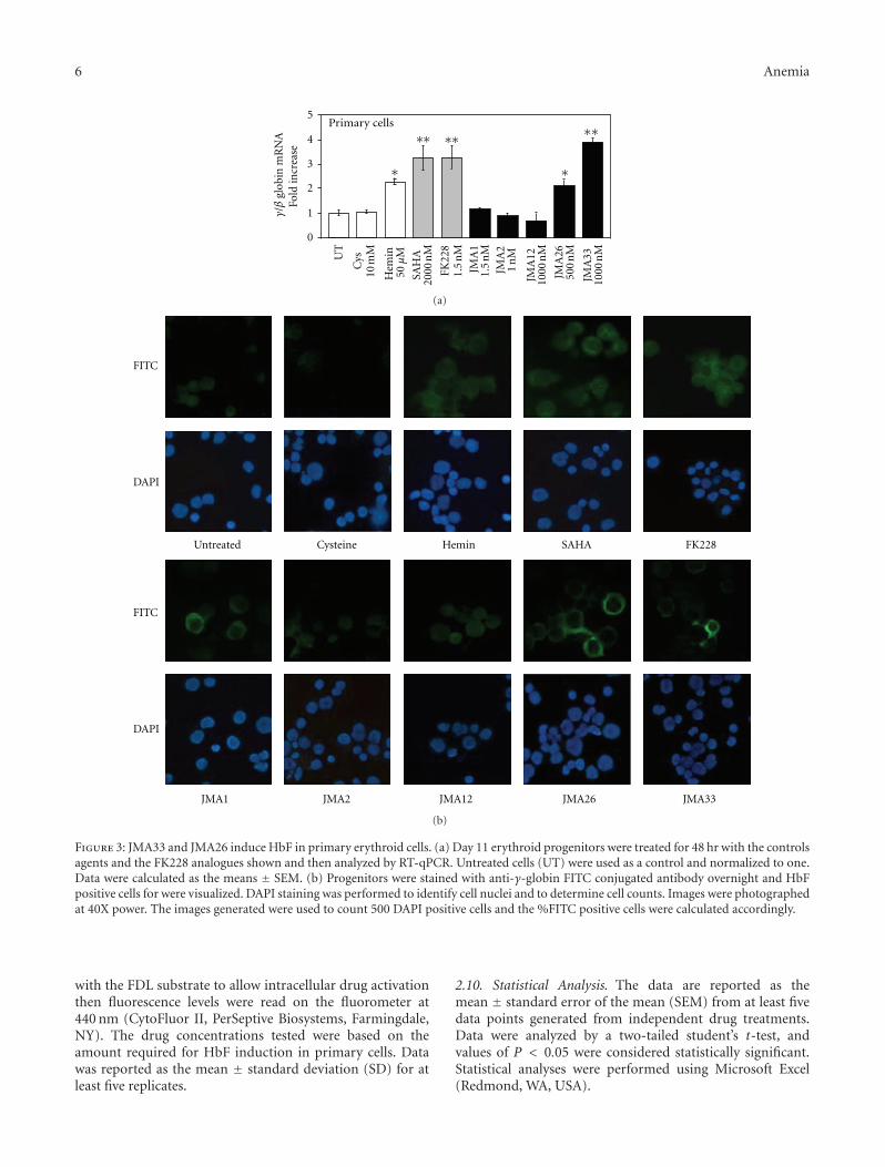
Figure 3: JMA33 and JMA26 induce HbF in primary erythroid cells. (a) Day 11 erythroid progenitors were treated for 48 hr with the controlsagents and the FK228 analogues shown and then analyzed by RT-qPCR. Untreated cells (UT) were used as a control and normalized to one.Data were calculated as the means ± SEM. (b) Progenitors were stained with anti-γ-globin FITC conjugated antibody overnight and HbFpositive cells for were visualized. DAPI staining was performed to identify cell nuclei and to determine cell counts. Images were photographedat 40X power. The images generated were used to count 500 DAPI positive cells and the %FITC positive cells were calculated accordingly.
with the FDL substrate to allow intracellular drug activationthen fluorescence levels were read on the fluorometer at440 nm (CytoFluor II, PerSeptive Biosystems, Farmingdale,NY). The drug concentrations tested were based on theamount required for HbF induction in primary cells. Datawas reported as the mean ± standard deviation (SD) for atleast five replicates.
2.10. Statistical Analysis. The data are reported as themean ± standard error of the mean (SEM) from at least fivedata points generated from independent drug treatments.Data were analyzed by a two-tailed student’s t-test, andvalues of P < 0.05 were considered statistically significant.Statistical analyses were performed using Microsoft Excel(Redmond, WA, USA).
Anemia 7
0
1000
2000
3000
4000
5000
6000
7000
8000
9000
HD
AC
act
ivit
y (%
)
TSA (nM)
0 0.5 5 50 500 1000
∗
∗∗
∗∗
(a)
0
1000
2000
3000
4000
5000
6000
7000
8000
9000
FK228 (nM)
HD
AC
act
ivit
y (%
)
0 0.15 1.5 15 150 300
∗∗
∗∗ ∗∗
∗
(b)
0
1000
2000
3000
4000
5000
6000
7000
8000
9000
HD
AC
act
ivit
y (%
)
0 0.1 1 10 100 200 0.1 1 10 100 200
JMA33 (μM)JMA26 (μM)
∗ ∗∗ ∗
∗
(c)
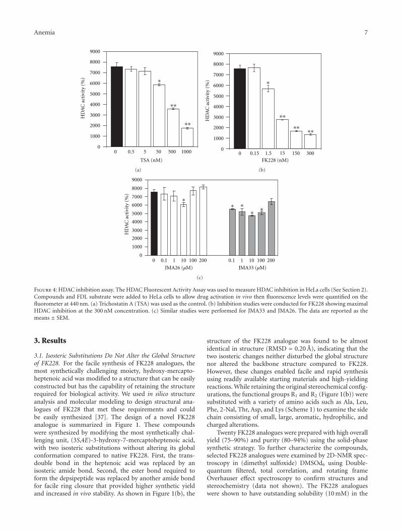
Figure 4: HDAC inhibition assay. The HDAC Fluorescent Activity Assay was used to measure HDAC inhibition in HeLa cells (See Section 2).Compounds and FDL substrate were added to HeLa cells to allow drug activation in vivo then fluorescence levels were quantified on thefluorometer at 440 nm. (a) Trichostatin A (TSA) was used as the control. (b) Inhibition studies were conducted for FK228 showing maximalHDAC inhibition at the 300 nM concentration. (c) Similar studies were performed for JMA33 and JMA26. The data are reported as themeans ± SEM.
3. Results
3.1. Isosteric Substitutions Do Not Alter the Global Structureof FK228. For the facile synthesis of FK228 analogues, themost synthetically challenging moiety, hydroxy-mercapto-heptenoic acid was modified to a structure that can be easilyconstructed but has the capability of retaining the structurerequired for biological activity. We used in silico structureanalysis and molecular modeling to design structural ana-logues of FK228 that met these requirements and couldbe easily synthesized [37]. The design of a novel FK228analogue is summarized in Figure 1. These compoundswere synthesized by modifying the most synthetically chal-lenging unit, (3S,4E)-3-hydroxy-7-mercaptoheptenoic acid,with two isosteric substitutions without altering its globalconformation compared to native FK228. First, the trans-double bond in the heptenoic acid was replaced by anisosteric amide bond. Second, the ester bond required toform the depsipeptide was replaced by another amide bondfor facile ring closure that provided higher synthetic yieldand increased in vivo stability. As shown in Figure 1(b), the
structure of the FK228 analogue was found to be almostidentical in structure (RMSD = 0.20 A), indicating that thetwo isosteric changes neither disturbed the global structurenor altered the backbone structure compared to FK228.However, these changes enabled facile and rapid synthesisusing readily available starting materials and high-yieldingreactions. While retaining the original stereochemical config-urations, the functional groups R1 and R2 (Figure 1(b)) weresubstituted with a variety of amino acids such as Ala, Leu,Phe, 2-Nal, Thr, Asp, and Lys (Scheme 1) to examine the sidechain consisting of small, large, aromatic, hydrophilic, andcharged alterations.
Twenty FK228 analogues were prepared with high overallyield (75–90%) and purity (80–94%) using the solid-phasesynthetic strategy. To further characterize the compounds,selected FK228 analogues were examined by 2D-NMR spec-troscopy in (dimethyl sulfoxide) DMSOd6 using Double-quantum filtered, total correlation, and rotating frameOverhauser effect spectroscopy to confirm structures andstereochemistry (data not shown). The FK228 analogueswere shown to have outstanding solubility (10 mM) in the
8 Anemia
Table 1: γ-globin induction in KU812-γF/βR stable lines by FK228 derivatives1.
Line 1Drug
concentrationγ/γ + 2β
MeanSEM P value
Foldchange
Untreated none 0.0402 0.0091 n/a 1
Cys 10 mM 0.0388 0.0092 0.8528 0.950
Hemin 50 μM 0.1732 0.0399 0.0050 4.325
SAHA 2000 nM 0.2157 0.0142 0.0001 5.400
FK228 1.5 nM 0.1197 0.0325 0.0150 2.975
1127Ox 1000 nM 0.0363 0.0022 0.8167 0.900
JMA1 1.5 nM 0.0473 0.0149 0.6720 1.175
JMA2 1.0 nM 0.0157 0.0003 0.1629 0.400
JMA12 1000 nM 0.0187 0.0009 0.2156 0.475
JMA26 500 nM 0.1460 0.0234 0.0004 3.650
JMA33 1000 nM 0.1373 0.0117 0.0002 3.425
JMA112 100 nM 0.0187 0.0007 0.2156 2.453
Line 2Drug
concentrationγ/γ + 2β
MeanSEM P value
Foldchange
Untreated none 0.1573 0.0150 n/a 1
Cys 10 mM 0.1200 0.0056 0.1977 0.764
Hemin 50 μM 0.6593 0.0839 0.0001 4.261
SAHA 2000 nM 0.6500 0.0208 0.0001 4.140
FK228 1.5 nM 0.6167 0.0338 0.0001 3.929
1127Ox 1000 nM 0.6133 0.0371 0.0001 3.904
JMA1 1.5 nM 0.1260 0.0152 0.2896 0.803
JMA2 1.0 nM 0.1183 0.0071 0.1814 0.752
JMA12 1000 nM 0.1470 0.0027 0.7093 0.936
JMA26 500 nM 0.2487 0.0308 0.0153 1.579
JMA33 1000 nM 0.3313 0.0256 0.0002 2.108
JMA112 100 nM 0.1070 0.0076 0.0938 0.682
Line 3Drug
concentrationγ/γ + 2β
MeanSEM P value
Foldchange
Untreated none 0.0721 0.0052 n/a 1
Cys 10 mM 0.0860 0.0050 0.1814 1.194
Hemin 50 μM 0.2508 0.0153 0.0001 3.486
SAHA 2000 nM 0.2520 0.0066 0.0001 3.500
FK228 1.5 nM 0.1583 0.0198 0.0007 2.194
1127Ox 1000 nM 0.0370 0.0095 0.0076 0.514
JMA1 1.5 nM 0.0770 0.0036 0.6179 1.069
JMA2 1.0 nM 0.0740 0.0036 0.8463 1.027
JMA12 1000 nM 0.0537 0.0023 0.0779 0.750
JMA26 500 nM 0.2560 0.0192 0.0001 3.555
JMA33 1000 nM 0.5120 0.1765 0.0007 7.111
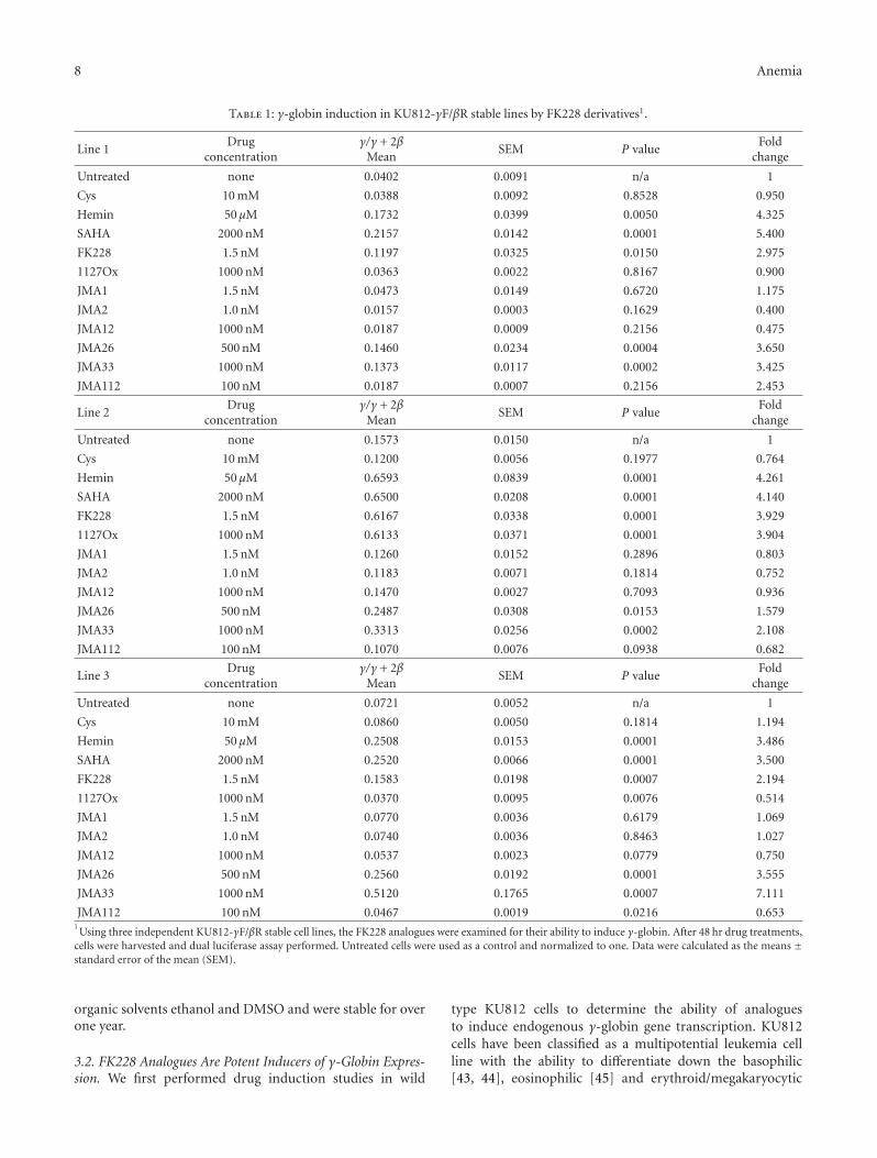
JMA112 100 nM 0.0467 0.0019 0.0216 0.6531Using three independent KU812-γF/βR stable cell lines, the FK228 analogues were examined for their ability to induce γ-globin. After 48 hr drug treatments,
cells were harvested and dual luciferase assay performed. Untreated cells were used as a control and normalized to one. Data were calculated as the means ±standard error of the mean (SEM).
organic solvents ethanol and DMSO and were stable for overone year.
3.2. FK228 Analogues Are Potent Inducers of γ-Globin Expres-sion. We first performed drug induction studies in wild
type KU812 cells to determine the ability of analoguesto induce endogenous γ-globin gene transcription. KU812cells have been classified as a multipotential leukemia cellline with the ability to differentiate down the basophilic[43, 44], eosinophilic [45] and erythroid/megakaryocytic
Anemia 9
lineages [46]. Previous studies from our laboratory demon-strated that KU812 cells express γ-globin, β-globin andthe erythroid markers CD36, and erythropoietin receptor[47]. Therefore, we used these cells to perform initial drugscreens to determine the suitability of KU812 cells for ourdual luciferase reporter stable lines. We observed a 1.5- to-10-fold increase in the γ/β-globin mRNA levels after Hem(50 μM), NaB (2 mM), SAHA (2 μM)), and FK228 (1.5 nM)treatment (Figure 2(a)). In the untreated and negativecontrols, cysteine-treated cells, γ-globin gene expression wasnot induced. These data demonstrated that the intracellularenvironment in KU812 is conducive to identifying γ-globingene activators in our FK228 analogue drug screen.
Subsequently, three independent dual-luciferase reporterKU812 stable cell lines were established to analyze theability FK228 analogues to induce γ-globin promoter activitywithout an effect on β-globin transcription. The stable celllines were created with the μLCRβprRlucγprFluc construct(Figure 2(b)) containing a 3.1-kb μLCR cassette linked toa 315-bp human β-globin promoter driving the renilla (R)and a 1.4-kb Aγ-globin promoter driving the firefly (F)luciferase genes [33, 39]. Since the firefly luciferase gene(γF) has approximately 50% greater luminescence than therenilla gene (βR), the renilla activity was multiplied by twoto adjust for the difference in luminescence [33] yieldingthe γ/γ+2β final measurement. The FK228 analogues wereexamined at concentrations ranging from 1–1000 nM inthe three stable lines. After 48-hour treatments, cells wereharvested and protein isolated for luciferase activity using theDual Luciferase Reporter Assay. Of the twenty compoundstested, five induced γ-promoter activity. The remainingagents were either toxic at the concentrations tested or didnot induce γ-globin (data not shown). Table 1 summarizesthe γ-promoter activity for FK228 analogues that were testedfurther in primary erythroid cells. Cell viability by Trypanblue exclusion remained at 90–95% for the concentrationsshown. Of note are the FK228 analogues, JMA26 and JMA33(Table 2) containing aromatic side chains in the functionalR1 and R2 groups which produced statistically significantγ-promoter activation comparable to FK228. Additionalanalogues can be designed based on these observations toincrease potency, while sparing toxicity.
3.3. FK228 Analogues Activate HbF Synthesis in PrimaryErythroid Progenitors. Next, we examined the ability ofthe lead FK228 analogues to induce HbF expression inprimary erythroid progenitors grown from peripheral bloodmononuclear cells in the two-phase liquid culture system.As shown in Figure 3(a), JMA26 and JMA33 induced γ-globin transcription at the mRNA level 2.1-fold and 3.9-fold,respectively, compared to a maximal 3.2-fold, induction bySAHA and FK228. However, FK228 derivatives induce γ-promoter activity at significantly lower drug concentrationscompared to SAHA and Hem. At the concentrations tested,greater than 90% cell viability was observed in primary cellsat all concentrations tested for the synthesized compounds.The similarity of these results to those acquired with theKU812 dual-luciferase reporter cell lines also validates thesystem for drug screening.
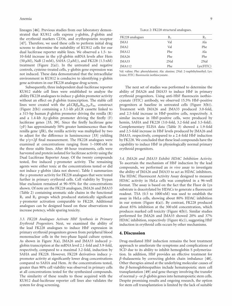
Table 2: FK228 structural analogues.
FK228 analogues R1 R2
JMA1 Val Ala
JMA2 Val Phe
JMA12 Phe Ala
JMA26 Phe Phe
JMA33 2Nal 2Nal
JMA112 Phe Lys(FITC)
Val: valine; Phe: phenylalanini; Ala: alanine; 2Nal: 2-naphthylmethyl; Lys:lysine; FITC: fluorescein isothiocyanate.
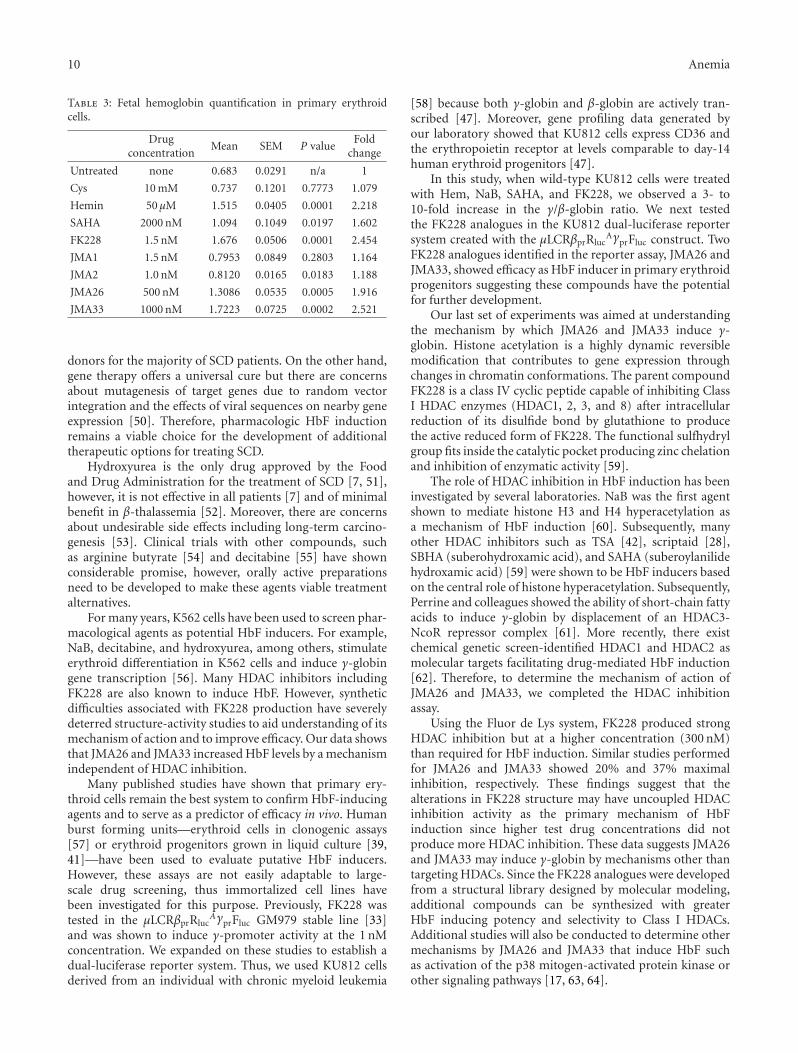
The next set of studies was performed to determine theability of JMA26 and JMA33 to induce HbF in primaryerythroid progenitors. Using anti-HbF fluorescein isothio-cyanate (FITC) antibody, we observed 15.5% HbF-positiveprogenitors at baseline in untreated cells (Figure 3(b)).Treatment with JMA26 and JMA33 produced 3.0-fold,and 2.5-fold increase in HbF-positive cells, respectively. Asimilar increase in HbF-positive cells, were produced byhemin, SAHA and FK228 (3.0-fold, 3.2-fold and 3.5-fold).Complementary ELISA data (Table 3) showed a 1.9-foldand 2.5-fold increase in HbF levels produced by JMA26 andJMA33, respectively, compared to a 2.4-fold HbF inductionby FK228. We concluded that these lead compounds have thecapability to induce HbF in physiologically normal primaryerythroid progenitors.
3.4. JMA26 and JMA33 Exhibit HDAC Inhibition Activity.To ascertain the mechanism of HbF induction by the leadcompounds, we performed an in vivo assay to investigatethe ability of JMA26 and JMA33 to act as HDAC inhibitors.The HDAC Fluorescent Activity Assay designed to measureHDAC activity in HeLa cells was completed in a 96-wellformat. The assay is based on the fact that the Fluor de Lyssubstrate is deacetylated by HDACs to generate a fluorescentreadout. TSA (0.5 to 1000 nM) was used to establish theassay in HeLa cells, showing about 80% HDAC inhibitionin our system (Figure 4(a)). By contrast, FK228 producedabout 85% inhibition at the 300 nM concentration, whichproduces marked cell toxicity (Figure 4(b)). Similar studiesperformed for JMA26 and JMA33 showed 20% and 37%HDAC inhibition, respectively (Figure 4(c)), suggesting HbFinduction in erythroid cells occurs by other mechanisms.
4. Discussion
Drug-mediated HbF induction remains the best treatmentapproach to ameliorate the symptoms and complications ofSCD due to its ability to inhibit hemoglobin S polymeriza-tion. In addition, HbF provides an effective treatment forβ-thalassemia by correcting globin chain imbalance [48].Other therapies aimed at the underlying molecular causes ofthe β-hemoglobinopathies include hematopoietic stem celltransplantation [49] and gene therapy involving the transferof normal γ- or β-globin genes into hematopoietic stem cells.Despite promising results and ongoing research, the optionfor stem cell transplantation is limited by the lack of suitable
10 Anemia
Table 3: Fetal hemoglobin quantification in primary erythroidcells.
Drugconcentration
Mean SEM P valueFold
change
Untreated none 0.683 0.0291 n/a 1
Cys 10 mM 0.737 0.1201 0.7773 1.079
Hemin 50 μM 1.515 0.0405 0.0001 2.218
SAHA 2000 nM 1.094 0.1049 0.0197 1.602
FK228 1.5 nM 1.676 0.0506 0.0001 2.454
JMA1 1.5 nM 0.7953 0.0849 0.2803 1.164
JMA2 1.0 nM 0.8120 0.0165 0.0183 1.188
JMA26 500 nM 1.3086 0.0535 0.0005 1.916
JMA33 1000 nM 1.7223 0.0725 0.0002 2.521
donors for the majority of SCD patients. On the other hand,gene therapy offers a universal cure but there are concernsabout mutagenesis of target genes due to random vectorintegration and the effects of viral sequences on nearby geneexpression [50]. Therefore, pharmacologic HbF inductionremains a viable choice for the development of additionaltherapeutic options for treating SCD.
Hydroxyurea is the only drug approved by the Foodand Drug Administration for the treatment of SCD [7, 51],however, it is not effective in all patients [7] and of minimalbenefit in β-thalassemia [52]. Moreover, there are concernsabout undesirable side effects including long-term carcino-genesis [53]. Clinical trials with other compounds, suchas arginine butyrate [54] and decitabine [55] have shownconsiderable promise, however, orally active preparationsneed to be developed to make these agents viable treatmentalternatives.
For many years, K562 cells have been used to screen phar-macological agents as potential HbF inducers. For example,NaB, decitabine, and hydroxyurea, among others, stimulateerythroid differentiation in K562 cells and induce γ-globingene transcription [56]. Many HDAC inhibitors includingFK228 are also known to induce HbF. However, syntheticdifficulties associated with FK228 production have severelydeterred structure-activity studies to aid understanding of itsmechanism of action and to improve efficacy. Our data showsthat JMA26 and JMA33 increased HbF levels by a mechanismindependent of HDAC inhibition.
Many published studies have shown that primary ery-throid cells remain the best system to confirm HbF-inducingagents and to serve as a predictor of efficacy in vivo. Humanburst forming units—erythroid cells in clonogenic assays[57] or erythroid progenitors grown in liquid culture [39,41]—have been used to evaluate putative HbF inducers.However, these assays are not easily adaptable to large-scale drug screening, thus immortalized cell lines havebeen investigated for this purpose. Previously, FK228 wastested in the μLCRβprRluc
AγprFluc GM979 stable line [33]and was shown to induce γ-promoter activity at the 1 nMconcentration. We expanded on these studies to establish adual-luciferase reporter system. Thus, we used KU812 cellsderived from an individual with chronic myeloid leukemia
[58] because both γ-globin and β-globin are actively tran-scribed [47]. Moreover, gene profiling data generated byour laboratory showed that KU812 cells express CD36 andthe erythropoietin receptor at levels comparable to day-14human erythroid progenitors [47].
In this study, when wild-type KU812 cells were treatedwith Hem, NaB, SAHA, and FK228, we observed a 3- to10-fold increase in the γ/β-globin ratio. We next testedthe FK228 analogues in the KU812 dual-luciferase reportersystem created with the μLCRβprRluc
AγprFluc construct. TwoFK228 analogues identified in the reporter assay, JMA26 andJMA33, showed efficacy as HbF inducer in primary erythroidprogenitors suggesting these compounds have the potentialfor further development.
Our last set of experiments was aimed at understandingthe mechanism by which JMA26 and JMA33 induce γ-globin. Histone acetylation is a highly dynamic reversiblemodification that contributes to gene expression throughchanges in chromatin conformations. The parent compoundFK228 is a class IV cyclic peptide capable of inhibiting ClassI HDAC enzymes (HDAC1, 2, 3, and 8) after intracellularreduction of its disulfide bond by glutathione to producethe active reduced form of FK228. The functional sulfhydrylgroup fits inside the catalytic pocket producing zinc chelationand inhibition of enzymatic activity [59].
The role of HDAC inhibition in HbF induction has beeninvestigated by several laboratories. NaB was the first agentshown to mediate histone H3 and H4 hyperacetylation asa mechanism of HbF induction [60]. Subsequently, manyother HDAC inhibitors such as TSA [42], scriptaid [28],SBHA (suberohydroxamic acid), and SAHA (suberoylanilidehydroxamic acid) [59] were shown to be HbF inducers basedon the central role of histone hyperacetylation. Subsequently,Perrine and colleagues showed the ability of short-chain fattyacids to induce γ-globin by displacement of an HDAC3-NcoR repressor complex [61]. More recently, there existchemical genetic screen-identified HDAC1 and HDAC2 asmolecular targets facilitating drug-mediated HbF induction[62]. Therefore, to determine the mechanism of action ofJMA26 and JMA33, we completed the HDAC inhibitionassay.
Using the Fluor de Lys system, FK228 produced strongHDAC inhibition but at a higher concentration (300 nM)than required for HbF induction. Similar studies performedfor JMA26 and JMA33 showed 20% and 37% maximalinhibition, respectively. These findings suggest that thealterations in FK228 structure may have uncoupled HDACinhibition activity as the primary mechanism of HbFinduction since higher test drug concentrations did notproduce more HDAC inhibition. These data suggests JMA26and JMA33 may induce γ-globin by mechanisms other thantargeting HDACs. Since the FK228 analogues were developedfrom a structural library designed by molecular modeling,additional compounds can be synthesized with greaterHbF inducing potency and selectivity to Class I HDACs.Additional studies will also be conducted to determine othermechanisms by JMA26 and JMA33 that induce HbF suchas activation of the p38 mitogen-activated protein kinase orother signaling pathways [17, 63, 64].
Anemia 11
5. Conclusions
The current drug treatment options for SCD are limitedwith hydroxyurea being the only FDA-approved drug. Thekey finding of this study is the high-efficiency synthesis ofFK228 analogues with structural modifications which didnot disturb the global chemical structure of the parentcompound. The analogues exhibited HbF induction atnanomolar concentrations in primary erythroid progenitorsdemonstrating physiological relevance. These data supportthe FK228 analogues as potential therapeutic agents andalso validates the KU812 dual-luciferase stable cell linesas an efficacious screening system to identify γ-globinactivators. Long-term our goal is to establish a group of HbFinducers that selectively inhibit Class I HDACs to expand ourunderstanding of epigenetic mechanisms of γ-globin generegulation and to facilitate the development of drug therapyfor SCD.
Acknowledgment
This work was supported by a Grant from the NationalHeart Lung and Blood Institute (R01HL069234; BSP), theFrancis J. Tedesco Distinguished Chair in Pediatric Hematol-ogy/Oncology (BSP), and the Robert A. Welch Foundation(AT-1595; JMA).
References
[1] T. Papayannopoulou, A. Torrealba De Ron, and R. Veith,“Arabinosylcytosine induces fetal hemoglobin in baboons byperturbing erythroid cell differentiation kinetics,” Science, vol.224, no. 4649, pp. 617–619, 1984.
[2] D. P. Liu, C. C. Liang, Z. H. Ao et al., “Treatment of severeβ-thalassemia (patients) with myleran,” American Journal ofHematology, vol. 33, no. 1, pp. 50–55, 1990.
[3] R. Veith, T. Papayannopoulou, S. Kurachi, and G. Stamatoy-annopoulos, “Treatment of baboon with vinblastine: insightsinto the mechanisms of pharmacologic stimulation of hb f inthe adult,” Blood, vol. 66, no. 2, pp. 456–459, 1985.
[4] R. Galanello, G. Stamatoyannopoulos, and PapayannopoulouTh., “Mechanism of hb f stimulation by s-stage compounds. invitro studies with bone marrow cells exposed to 5-azacytidine,ara-c, or hydroxyurea,” Journal of Clinical Investigation, vol. 81,no. 4, pp. 1209–1216, 1988.
[5] N. L. Letvin, D. C. Linch, and G. P. Beardsley, “Augmenta-tion of fetal-hemoglobin production in anemic monkeys byhydroxyurea,” New England Journal of Medicine, vol. 310, no.14, pp. 869–873, 1984.
[6] M. H. Steinberg, F. Barton, O. Castro et al., “Effect ofhydroxyurea on mortality and morbidity in adult sickle cellanemia: risks and benefits up to 9 years of treatment,” Journalof the American Medical Association, vol. 289, no. 13, pp. 1645–1651, 2003.
[7] S. Charache, M. L. Terrin, R. D. Moore et al., “Effect ofhydroxyurea on the frequency of painful crises in sickle cellanemia. Investigators of the Multicenter Study of Hydroxyureain Sickle Cell Anemia,” The New England Journal of Medicine,vol. 332, pp. 1317–1322, 1995.
[8] M. H. Steinberg, Z. H. Lu, F. B. Barton, M. L. Terrin, S.Charache, and G. J. Dover, “Fetal hemoglobin in sickle cell
anemia: determinants of response to hydroxyurea,” Blood, vol.89, no. 3, pp. 1078–1088, 1997.
[9] X. De La Cruz, S. Lois, S. Sanchez-Molina, and M. A.Martınez-Balbas, “Do protein motifs read the histone code?”Bioessays, vol. 27, no. 2, pp. 164–175, 2005.
[10] P. A. Marks and X. Jiang, “Histone deacetylase inhibitors inprogrammed cell death and cancer therapy,” Cell Cycle, vol. 4,no. 4, pp. 549–551, 2005.
[11] C. Monneret, “Histone deacetylase inhibitors,” European Jour-nal of Medicinal Chemistry, vol. 40, no. 1, pp. 1–13, 2005.
[12] K. N. Prasad, “Butyric acid: a small fatty acid with diversebiological functions,” Life Sciences, vol. 27, no. 15, pp. 1351–1358, 1980.
[13] A. E. Smith, P. J. Hurd, A. J. Bannister, T. Kouzarides, andK. G. Ford, “Heritable gene repression through the action ofa directed dna methyltransferase at a chromosomal locus,”Journal of Biological Chemistry, vol. 283, no. 15, pp. 9878–9885, 2008.
[14] S. D. Gore and M. A. Carducci, “Modifying histones totame cancer: clinical development of sodium phenylbutyrateand other histone deacetylase inhibitors,” Expert Opinion onInvestigational Drugs, vol. 9, no. 12, pp. 2923–2934, 2000.
[15] T. Yamashita, H. Wakao, A. Miyajima, and S. Asano, “Differ-entiation inducers modulate cytokine signaling pathways in amurine erythroleukemia cell line,” Cancer Research, vol. 58, no.3, pp. 556–561, 1998.
[16] O. Witt, K. Sand, and A. Pekrun, “Butyrate-induced erythroiddifferentiation of human k562 leukemia cells involves inhibi-tion of erk and activation of p38 map kinase pathways,” Blood,vol. 95, no. 7, pp. 2391–2396, 2000.
[17] B. S. Pace, X. H. Qian, J. Sangerman et al., “P38 map kinaseactivation mediates γ-globin gene induction in erythroidprogenitors,” Experimental Hematology, vol. 31, no. 11, pp.1089–1096, 2003.
[18] S. Torkelson, B. White, D. V. Faller, K. Phipps, C. Pantazis,and S. P. Perrine, “Erythroid progenitor proliferation isstimulated by phenoxyacetic and phenylalkyl acids,” BloodCells, Molecules, and Diseases, vol. 22, no. 2, pp. 150–158, 1996.
[19] G. J. Dover, S. Brusilow, and S. Charache, “Induction of fetalhemoglobin production in subjects with sickle cell anemia byoral sodium phenylbutyrate,” Blood, vol. 84, no. 1, pp. 339–343, 1994.
[20] E. Liakopoulou, C. A. Blau, Q. Li et al., “Stimulation of fetalhemoglobin production by short chain fatty acids,” Blood, vol.86, no. 8, pp. 3227–3235, 1995.
[21] N. Tsuji, M. Kobayashi, and K. Nagashima, “A new antifungalantibiotic, trichostatin,” Journal of Antibiotics, vol. 29, no. 1,pp. 1–6, 1976.
[22] M. Yoshida, M. Kijima, M. Akita, and T. Beppu, “Potent andspecific inhibition of mammalian histone deacetylase bothin vivo and in vitro by trichostatin a,” Journal of BiologicalChemistry, vol. 265, no. 28, pp. 17174–17179, 1990.
[23] M. S. Finnin, J. R. Donigian, A. Cohen et al., “Structures ofa histone deacetylase homologue bound to the tsa and sahainhibitors,” Nature, vol. 401, no. 6749, pp. 188–193, 1999.
[24] V. M. Richon, S. Emiliani, E. Verdin et al., “A class of hybridpolar inducers of transformed cell differentiation inhibitshistone deacetylases,” Proceedings of the National Academy ofSciences of the United States of America, vol. 95, no. 6, pp. 3003–3007, 1998.
[25] L. M. Butler, D. B. Agus, H. I. Scher et al., “Suberoylanilidehydroxamic acid, an inhibitor of histone deacetylase, sup-presses the growth of prostate cancer cells in vitro and in vivo,”Cancer Research, vol. 60, no. 18, pp. 5165–5170, 2000.
12 Anemia
[26] P. A. Marks, V. M. Richon, R. Breslow, and R. A. Rifkind,“Histone deacetylase inhibitors as new cancer drugs,” CurrentOpinion in Oncology, vol. 13, no. 6, pp. 477–483, 2001.
[27] H. Ueda, H. Nakajima, Y. Hori et al., “Fr901228, a novel anti-tumor bicyclic depsipeptide produced by chromobacteriumviolaceum no. 968. i. taxonomy, fermentation, isolation,physico-chemical and biological properties, and antitumoractivity,” Journal of Antibiotics, vol. 47, no. 3, pp. 301–310,1994.
[28] J. Johnson, R. Hunter, R. McElveen, X. H. Qian, B. S. Baliga,and B. S. Pace, “Fetal hemoglobin induction by the histonedeacetylase inhibitor, scriptaid,” Cellular and Molecular Biol-ogy, vol. 51, no. 2, pp. 229–238, 2005.
[29] R. Furumai, A. Matsuyama, N. Kobashi et al., “Fk228 (dep-sipeptide) as a natural prodrug that inhibits class i histonedeacetylases,” Cancer Research, vol. 62, no. 17, pp. 4916–4921,2002.
[30] J. J. Xiao, J. Byrd, G. Marcucci, M. Grever, and K. K. Chan,“Identification of thiols and glutathione conjugates of dep-sipeptide fk228 (fr901228), a novel histone protein deacetylaseinhibitor, in the blood,” Rapid Communications in MassSpectrometry, vol. 17, no. 8, pp. 757–766, 2003.
[31] H. Kosugi, M. Ito, Y. Yamamoto et al., “In vivo effects of ahistone deacetylase inhibitor, fk228, on human acute promye-locytic leukemia in nod/shi-scid/scid mice,” Japanese Journalof Cancer Research, vol. 92, no. 5, pp. 529–536, 2001.
[32] K. A. Fecteau, M. E. I. Jianxun, and H. C. Wang, “Differentialmodulation of signaling pathways and apoptosis of ras-transformed 10t1/2 cells by the depsipeptide fr901228,” Jour-nal of Pharmacology and Experimental Therapeutics, vol. 300,no. 3, pp. 890–899, 2002.
[33] H. Cao and G. Stamatoyannopoulos, “Histone deacetylaseinhibitor fk228 is a potent inducer of human fetal hemo-globin,” American Journal of Hematology, vol. 81, no. 12, pp.981–983, 2006.
[34] K. W. Li, J. Wu, W. Xing, and J. A. Simon, “Total synthesis ofthe antitumor depsipeptide fr-901,228,” Journal of the Ameri-can Chemical Society, vol. 118, no. 30, pp. 7237–7238, 1996.
[35] T. J. Greshock, D. M. Johns, Y. Noguchi, and R. M. Williams,“Improved total synthesis of the potent hdac inhibitor FK228(FK-901228),” Organic Letters, vol. 10, no. 4, pp. 613–616,2008.
[36] A. A. Bowers, T. J. Greshook, N. West et al., “Synthesis andconformation-activity relationships of the peptide isosteres offk228 and largazole,” Journal of the American Chemical Society,vol. 131, no. 8, pp. 2900–2905, 2009.
[37] S. Di Maro, R. C. Pong, J. T. Hsieh, and J. M. Ahn, “Efficientsolid-phase synthesis of fk228 analogues as potent antitumoralagents,” Journal of Medicinal Chemistry, vol. 51, no. 21, pp.6639–6641, 2008.
[38] K. J. Jensen, J. Alsina, M. F. Songster, J. Vagner, F. Albericio,and G. Barany, “Backbone amide linker (BAL) strategyfor solid-phase synthesis of c-terminal-modified and cyclicpeptides,” Journal of the American Chemical Society, vol. 120,no. 22, pp. 5441–5452, 1998.
[39] E. Skarpidi, G. Vassilopoulos, Q. Li, and G. Stamatoyannopou-los, “Novel in vitro assay for the detection of pharmacologicinducers of fetal hemoglobin,” Blood, vol. 96, no. 1, pp. 321–326, 2000.
[40] C. Y. Gui and A. Dean, “Acetylation of a specific promoternucleosome accompanies activation of the ε-globin gene byβ-globin locus control region HS2,” Molecular and CellularBiology, vol. 21, no. 4, pp. 1155–1163, 2001.
[41] E. Fibach, L. P. Burke, A. N. Schechter, C. T. Noguchi, and G. P.Rodgers, “Hydroxyurea increases fetal hemoglobin in culturederythroid cells derived from normal individuals and patientswith sickle cell anemia or β- thalassemia,” Blood, vol. 81, no. 6,pp. 1630–1635, 1993.
[42] J. Sangerman, S. L. Moo, X. Yao et al., “Mechanism forfetal hemoglobin induction by histone deacetylase inhibitorsinvolves γ-globin activation by creb1 and atf-2,” Blood, vol.108, no. 10, pp. 3590–3599, 2006.
[43] T. Fukuda, K. Kishi, Y. Ohnishi, and A. Shibata, “Bipotentialcell differentiation of ku-812: evidence of a hybrid cell line thatdifferentiates into basophils and macrophage-like cells,” Blood,vol. 70, no. 3, pp. 612–619, 1987.
[44] K. Kishi, “A new leukemia cell line with Philadelphia chro-mosome characterized as basophil precursors,” LeukemiaResearch, vol. 9, no. 3, pp. 381–390, 1985.
[45] M. Yamashita, A. Ichikawa, Y. Katakura et al., “Inductionof basophilic and eosinophilic differentiation in the humanleukemic cell line ku812,” Cytotechnology, vol. 36, no. 1–3, pp.179–186, 2001.
[46] M. Nakazawa, M. T. Mitjavila, N. Debili et al., “Ku 812: apluripotent human cell line with spontaneous erythroid ter-minal maturation,” Blood, vol. 73, no. 7, pp. 2003–2013, 1989.
[47] S. Zein, W. Li, V. Ramakrishnan et al., “Identification offetal hemoglobin-inducing agents using the human leukemiaku812 cell line,” Experimental Biology and Medicine, vol. 235,no. 11, pp. 1385–1394, 2010.
[48] D. G. Nathan and R. B. Gunn, “Thalassemia: the consequencesof unbalanced hemoglobin synthesis,” the American Journal ofMedicine, vol. 41, no. 5, pp. 815–830, 1966.
[49] M. Bhatia and M. C. Walters, “Hematopoietic cell transplan-tation for thalassemia and sickle cell disease: past, present andfuture,” Bone Marrow Transplantation, vol. 41, no. 2, pp. 109–117, 2008.
[50] C. E. Dunbar, “The yin and yang of stem cell gene therapy:insights into hematopoiesis, leukemogenesis, and gene therapysafety,” Hematology/the Education Program of the AmericanSociety of Hematology. American Society of Hematology. Edu-cation Program, pp. 460–465, 2007.
[51] M. H. Steinberg, Z. H. Lu, F. B. Barton, M. L. Terrin, S.Charache, and G. J. Dover, “Fetal hemoglobin in sickle cellanemia: determinants of response to hydroxyurea,” Blood, vol.89, no. 3, pp. 1078–1088, 1997.
[52] H. Fathallah, M. Sutton, and G. F. Atweh, “Pharmacologicalinduction of fetal hemoglobin: why haven’t we been moresuccessful in thalassemia?” Annals of the New York Academy ofSciences, vol. 1054, pp. 228–237, 2005.
[53] S. C. Davies and A. Gilmore, “The role of hydroxyurea in themanagement of sickle cell disease,” Blood Reviews, vol. 17, no.2, pp. 99–109, 2003.
[54] G. F. Atweh, M. Sutton, I. Nassif et al., “Sustained inductionof fetal hemoglobin by pulse butyrate therapy in sickle celldisease,” Blood, vol. 93, no. 6, pp. 1790–1797, 1999.
[55] Y. Saunthararajah, R. Molokie, S. Saraf et al., “Clinicaleffectiveness of decitabine in severe sickle cell disease,” BritishJournal of Haematology, vol. 141, no. 1, pp. 126–129, 2008.
[56] R. Mabaera, R. J. West, S. J. Conine et al., “A cell stresssignaling model of fetal hemoglobin induction: what doesn’tkill red blood cells may make them stronger,” ExperimentalHematology, vol. 36, no. 9, pp. 1057–1072, 2008.
[57] P. Constantoulakis, G. Knitter, and G. Stamatoyannopoulos,“On the induction of fetal hemoglobin by butyrates: in vivoand in vitro studies with sodium butyrate and comparison of
Anemia 13
combination treatments with 5-AZAC and ARAC,” Blood, vol.74, no. 6, pp. 1963–1971, 1989.
[58] A. Goga, J. McLaughlin, D. E. H. Afar, D. C. Saffran, andO. N. Witte, “Alternative signals to ras for hematopoietictransformation by the bcr- abl oncogene,” Cell, vol. 82, no. 6,pp. 981–988, 1995.
[59] R. Furumai, A. Matsuyama, N. Kobashi et al., “FK228 (dep-sipeptide) as a natural prodrug that inhibits class i histonedeacetylases,” Cancer Research, vol. 62, no. 17, pp. 4916–4921,2002.
[60] H. Fathallah, R. S. Weinberg, Y. Galperin, M. Sutton, and G.F. Atweh, “Role of epigenetic modifications in normal globingene regulation and butyrate-mediated induction of fetalhemoglobin,” Blood, vol. 110, no. 9, pp. 3391–3397, 2007.
[61] R. Mankidy, D. V. Faller, R. Mabaera et al., “Short-chain fattyacids induce γ-globin gene expression by displacement of ahdac3-ncor repressor complex,” Blood, vol. 108, no. 9, pp.3179–3186, 2006.
[62] J. E. Bradner, R. Mak, S. K. Tanguturi et al., “Chemical geneticstrategy identifies histone deacetylase 1 (HDAC1) and HDAC2as therapeutic targets in sickle cell disease,” Proceedings of theNational Academy of Sciences of the United States of America,vol. 107, no. 28, pp. 12617–12622, 2010.
[63] O. Witt, K. Sand, and A. Pekrun, “Butyrate-induced erythroiddifferentiation of human k562 leukemia cells involves inhi-bition of ERK and activation of p38 map kinase pathways,”Blood, vol. 95, no. 7, pp. 2391–2396, 2000.
[64] O. Witt, S. Monkemeyer, K. Kanbach, and A. Pekrun, “Induc-tion of fetal hemoglobin synthesis by valproate: modulationof mapkinase pathways,” American Journal of Hematology, vol.71, no. 1, pp. 45–46, 2002.

Submit your manuscripts athttp://www.hindawi.com
Stem CellsInternational
Hindawi Publishing Corporationhttp://www.hindawi.com Volume 2014
Hindawi Publishing Corporationhttp://www.hindawi.com Volume 2014
MEDIATORSINFLAMMATION
of
Hindawi Publishing Corporationhttp://www.hindawi.com Volume 2014
Behavioural Neurology
EndocrinologyInternational Journal of
Hindawi Publishing Corporationhttp://www.hindawi.com Volume 2014
Hindawi Publishing Corporationhttp://www.hindawi.com Volume 2014
Disease Markers
Hindawi Publishing Corporationhttp://www.hindawi.com Volume 2014
BioMed Research International
OncologyJournal of
Hindawi Publishing Corporationhttp://www.hindawi.com Volume 2014
Hindawi Publishing Corporationhttp://www.hindawi.com Volume 2014
Oxidative Medicine and Cellular Longevity
Hindawi Publishing Corporationhttp://www.hindawi.com Volume 2014
PPAR Research
The Scientific World JournalHindawi Publishing Corporation http://www.hindawi.com Volume 2014
Immunology ResearchHindawi Publishing Corporationhttp://www.hindawi.com Volume 2014
Journal of
ObesityJournal of
Hindawi Publishing Corporationhttp://www.hindawi.com Volume 2014
Hindawi Publishing Corporationhttp://www.hindawi.com Volume 2014
Computational and Mathematical Methods in Medicine
OphthalmologyJournal of
Hindawi Publishing Corporationhttp://www.hindawi.com Volume 2014
Diabetes ResearchJournal of
Hindawi Publishing Corporationhttp://www.hindawi.com Volume 2014
Hindawi Publishing Corporationhttp://www.hindawi.com Volume 2014
Research and TreatmentAIDS
Hindawi Publishing Corporationhttp://www.hindawi.com Volume 2014
Gastroenterology Research and Practice
Hindawi Publishing Corporationhttp://www.hindawi.com Volume 2014
Parkinson’s Disease
Evidence-Based Complementary and Alternative Medicine
Volume 2014Hindawi Publishing Corporationhttp://www.hindawi.com