fixed-drug eruption: a retrospective study in a single referral center
TRANSCRIPT
at SciVerse ScienceDirect
DERMATOLOGICA SINICA 30 (2012) 11e15
Contents lists available
Dermatologica Sinica
journal homepage: http: / /www.derm-sinica.com
ORIGINAL ARTICLE
Fixed-drug eruption: A retrospective study in a single referral centerin northern Taiwan
Cheng-Han Lee 1, Yi-Chun Chen 2, Yung-Tsu Cho 1, Chia-Ying Chang 1, Chia-Yu Chu 1,*
1Department of Dermatology, National Taiwan University Hospital and National Taiwan University College of Medicine, Taipei, Taiwan2Department of Dermatology, Cathay General Hospital, Taipei, Taiwan
a r t i c l e i n f o
Article history:Received: Jul 19, 2011Revised: Oct 4, 2011Accepted: Feb 9, 2012
Keywords:generalized bullous fixed drug eruptionfixed drug eruptionStevens-Johnson syndrometoxic epidermal necrolysis
* Corresponding author. Chia-Yu Chu, DepartmenTaiwan University Hospital, Number 7, Chung-Shan S
E-mail address: [email protected] (C.-Y. Chu).
1027-8117/$ e see front matter Copyright � 2012, Tadoi:10.1016/j.dsi.2012.02.002
a b s t r a c t
Background/Objective: Fixed drug eruption (FDE) is a dermatosis characterized by recurrent patches orplaques at exactly the same sites with each administration of the causative drug. Vesicles or bullae maysometimes be found, and generalized bullous fixed drug eruption (GBFDE) may be confused withStevens-Johnson syndrome (SJS) or toxic epidermal necrolysis (TEN). This study aimed to investigate theclinical and pathologic features of FDE in Taiwan.Methods: A retrospective analysis evaluated patients with FDE in a referral center in Taiwan coveringa period of 11 years. Clinical data, suspected etiologies, and pathology/patch test results were collected.We also compared the GBFDE cases with SJS/TEN overlap or TEN cases to find differentiating clues.Results: There were 39 FDE patients, including nine GBFDE cases. The most frequent causative drugs werenon-steroidal anti-inflammatory drugs (five cases, 12.8%) and antibiotics (four cases, 10.3%). Extremitiesother than the hands (71.8%) were the most frequently affected sites, followed by the trunk (51.3%),mucosa (38.5%), and hands (33.3%). The average age of FDE patients was 52.2 years (median, 56 years;range, 4e86 years). Patients with GBFDE were significantly older than non-GBFDE patients (69.1� 19.7vs. 47.2� 23.6, p¼ 0.0124) and the trunk was more likely to be involved in GBFDE cases (88.9% vs. 40.0%,p¼ 0.0197). GBFDE cases also showed tendency to have more mucosal involvement (66.7% vs. 30.0%,p¼ 0.0631). Although similar to SJS/TEN, GBFDE cases had fewer constitutional symptoms, less mucosalinvolvement but had previous episodes. Histopathologically, the presence of more than two aggregateddyskeratotic keratinocytes (fire flag sign) in the epidermis was more frequently observed in SJS/TEN,whereas GBFDE had superficial and deep dermal infiltration of eosinophils and melanophages.Conclusion: FDE is one of the specialized cutaneous drug reactions and GBFDE should be kept in mind anddifferentiated from SJS/TEN.
Copyright � 2012, Taiwanese Dermatological Association.Published by Elsevier Taiwan LLC. All rights reserved.
Introduction
Fixed drug eruptions (FDEs) are defined as recurrent lesions atthe same skin or mucosal sites after repeated intake of the causa-tive agent.1 FDEs usually present as itching or burning, well-circumscribed, erythematous macules, patches, or plaques thatleave hyperpigmentation after resolving. Vesicles or bullae mayoccasionally be seen. There are many causative agents and theincidence of FDE for a particular drug depends on the frequency ofits use. Therefore, the list of etiologic drugs varies from one place toanother and from time to time.2
t of Dermatology, Nationalouth Road, Taipei, Taiwan.
iwanese Dermatological Associatio
Generalized bullous fixed drug eruption (GBFDE) may beconfused with toxic epidermal necrolysis (TEN) or Stevens-Johnsonsyndrome (SJS). The present study aimed to investigate the clinicaland pathologic features of FDE in Taiwan and identify severaldifferentiating features between GBFDE and non-GBFDE, as well asbetween GBFDE and SJS/TEN.
Methods
Patients
From January 2000 to February 2011, cases with suspected diagnosisof FDE recorded in the patch-testing database or skin pathologydatabase of the Department of Dermatology of the National TaiwanUniversity Hospital in Taipei, Taiwan, were recruited. FDE wasdiagnosed according to the typical clinical features: erythematous,
n. Published by Elsevier Taiwan LLC. All rights reserved.
C.-H. Lee et al. / Dermatologica Sinica 30 (2012) 11e1512
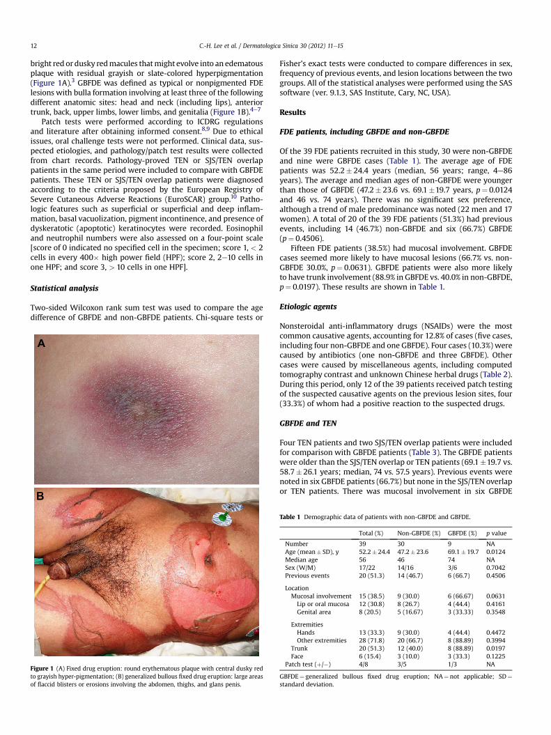
bright red ordusky redmacules thatmight evolve into an edematousplaque with residual grayish or slate-colored hyperpigmentation(Figure 1A).3 GBFDE was defined as typical or nonpigmented FDElesions with bulla formation involving at least three of the followingdifferent anatomic sites: head and neck (including lips), anteriortrunk, back, upper limbs, lower limbs, and genitalia (Figure 1B).4e7
Patch tests were performed according to ICDRG regulationsand literature after obtaining informed consent.8,9 Due to ethicalissues, oral challenge tests were not performed. Clinical data, sus-pected etiologies, and pathology/patch test results were collectedfrom chart records. Pathology-proved TEN or SJS/TEN overlappatients in the same period were included to compare with GBFDEpatients. These TEN or SJS/TEN overlap patients were diagnosedaccording to the criteria proposed by the European Registry ofSevere Cutaneous Adverse Reactions (EuroSCAR) group.10 Patho-logic features such as superficial or superficial and deep inflam-mation, basal vacuolization, pigment incontinence, and presence ofdyskeratotic (apoptotic) keratinocytes were recorded. Eosinophiland neutrophil numbers were also assessed on a four-point scale[score of 0 indicated no specified cell in the specimen; score 1, < 2cells in every 400� high power field (HPF); score 2, 2e10 cells inone HPF; and score 3, > 10 cells in one HPF].
Statistical analysis
Two-sided Wilcoxon rank sum test was used to compare the agedifference of GBFDE and non-GBFDE patients. Chi-square tests or
Figure 1 (A) Fixed drug eruption: round erythematous plaque with central dusky redto grayish hyper-pigmentation; (B) generalized bullous fixed drug eruption: large areasof flaccid blisters or erosions involving the abdomen, thighs, and glans penis.
Fisher’s exact tests were conducted to compare differences in sex,frequency of previous events, and lesion locations between the twogroups. All of the statistical analyses were performed using the SASsoftware (ver. 9.1.3, SAS Institute, Cary, NC, USA).
Results
FDE patients, including GBFDE and non-GBFDE
Of the 39 FDE patients recruited in this study, 30 were non-GBFDEand nine were GBFDE cases (Table 1). The average age of FDEpatients was 52.2� 24.4 years (median, 56 years; range, 4e86years). The average and median ages of non-GBFDE were youngerthan those of GBFDE (47.2� 23.6 vs. 69.1�19.7 years, p¼ 0.0124and 46 vs. 74 years). There was no significant sex preference,although a trend of male predominance was noted (22 men and 17women). A total of 20 of the 39 FDE patients (51.3%) had previousevents, including 14 (46.7%) non-GBFDE and six (66.7%) GBFDE(p¼ 0.4506).
Fifteen FDE patients (38.5%) had mucosal involvement. GBFDEcases seemed more likely to have mucosal lesions (66.7% vs. non-GBFDE 30.0%, p¼ 0.0631). GBFDE patients were also more likelyto have trunk involvement (88.9% in GBFDE vs. 40.0% in non-GBFDE,p¼ 0.0197). These results are shown in Table 1.
Etiologic agents
Nonsteroidal anti-inflammatory drugs (NSAIDs) were the mostcommon causative agents, accounting for 12.8% of cases (five cases,including four non-GBFDE and one GBFDE). Four cases (10.3%) werecaused by antibiotics (one non-GBFDE and three GBFDE). Othercases were caused by miscellaneous agents, including computedtomography contrast and unknown Chinese herbal drugs (Table 2).During this period, only 12 of the 39 patients received patch testingof the suspected causative agents on the previous lesion sites, four(33.3%) of whom had a positive reaction to the suspected drugs.
GBFDE and TEN
Four TEN patients and two SJS/TEN overlap patients were includedfor comparison with GBFDE patients (Table 3). The GBFDE patientswere older than the SJS/TEN overlap or TEN patients (69.1�19.7 vs.58.7� 26.1 years; median, 74 vs. 57.5 years). Previous events werenoted in six GBFDE patients (66.7%) but none in the SJS/TEN overlapor TEN patients. There was mucosal involvement in six GBFDE
Table 1 Demographic data of patients with non-GBFDE and GBFDE.
Total (%) Non-GBFDE (%) GBFDE (%) p value
Number 39 30 9 NAAge (mean� SD), y 52.2� 24.4 47.2� 23.6 69.1� 19.7 0.0124Median age 56 46 74 NASex (W/M) 17/22 14/16 3/6 0.7042Previous events 20 (51.3) 14 (46.7) 6 (66.7) 0.4506
LocationMucosal involvement 15 (38.5) 9 (30.0) 6 (66.67) 0.0631Lip or oral mucosa 12 (30.8) 8 (26.7) 4 (44.4) 0.4161Genital area 8 (20.5) 5 (16.67) 3 (33.33) 0.3548
ExtremitiesHands 13 (33.3) 9 (30.0) 4 (44.4) 0.4472Other extremities 28 (71.8) 20 (66.7) 8 (88.89) 0.3994
Trunk 20 (51.3) 12 (40.0) 8 (88.89) 0.0197Face 6 (15.4) 3 (10.0) 3 (33.3) 0.1225
Patch test (þ/�) 4/8 3/5 1/3 NA
GBFDE¼ generalized bullous fixed drug eruption; NA¼ not applicable; SD¼standard deviation.
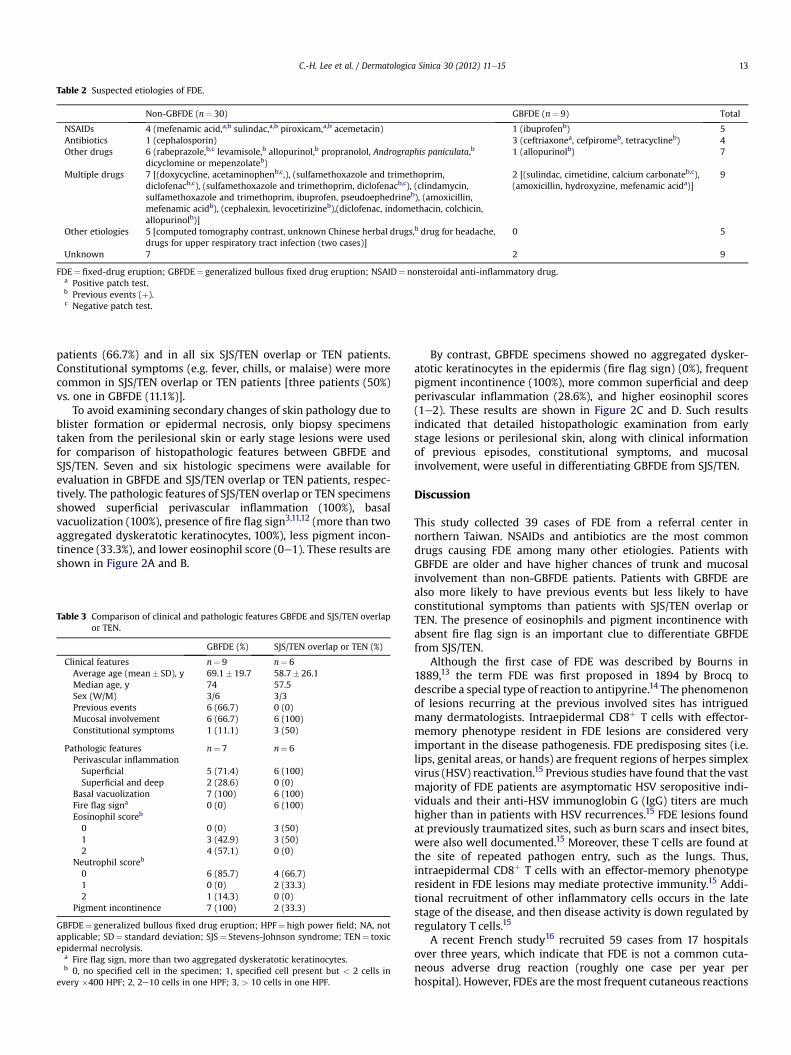
Table 2 Suspected etiologies of FDE.
Non-GBFDE (n¼ 30) GBFDE (n¼ 9) Total
NSAIDs 4 (mefenamic acid,a,b sulindac,a,b piroxicam,a,b acemetacin) 1 (ibuprofenb) 5Antibiotics 1 (cephalosporin) 3 (ceftriaxonea, cefpiromeb, tetracyclineb) 4Other drugs 6 (rabeprazole,b,c levamisole,b allopurinol,b propranolol, Andrographis paniculata,b
dicyclomine or mepenzolateb)1 (allopurinolb) 7
Multiple drugs 7 [(doxycycline, acetaminophenb,c,), (sulfamethoxazole and trimethoprim,diclofenacb,c), (sulfamethoxazole and trimethoprim, diclofenacb,c), (clindamycin,sulfamethoxazole and trimethoprim, ibuprofen, pseudoephedrineb), (amoxicillin,mefenamic acidb), (cephalexin, levocetirizineb),(diclofenac, indomethacin, colchicin,allopurinolb)]
2 [(sulindac, cimetidine, calcium carbonateb,c),(amoxicillin, hydroxyzine, mefenamic acida)]
9
Other etiologies 5 [computed tomography contrast, unknown Chinese herbal drugs,b drug for headache,drugs for upper respiratory tract infection (two cases)]
0 5
Unknown 7 2 9
FDE¼ fixed-drug eruption; GBFDE¼ generalized bullous fixed drug eruption; NSAID¼ nonsteroidal anti-inflammatory drug.a Positive patch test.b Previous events (þ).c Negative patch test.
C.-H. Lee et al. / Dermatologica Sinica 30 (2012) 11e15 13
patients (66.7%) and in all six SJS/TEN overlap or TEN patients.Constitutional symptoms (e.g. fever, chills, or malaise) were morecommon in SJS/TEN overlap or TEN patients [three patients (50%)vs. one in GBFDE (11.1%)].
To avoid examining secondary changes of skin pathology due toblister formation or epidermal necrosis, only biopsy specimenstaken from the perilesional skin or early stage lesions were usedfor comparison of histopathologic features between GBFDE andSJS/TEN. Seven and six histologic specimens were available forevaluation in GBFDE and SJS/TEN overlap or TEN patients, respec-tively. The pathologic features of SJS/TEN overlap or TEN specimensshowed superficial perivascular inflammation (100%), basalvacuolization (100%), presence of fire flag sign3,11,12 (more than twoaggregated dyskeratotic keratinocytes, 100%), less pigment incon-tinence (33.3%), and lower eosinophil score (0e1). These results areshown in Figure 2A and B.
Table 3 Comparison of clinical and pathologic features GBFDE and SJS/TEN overlapor TEN.
GBFDE (%) SJS/TEN overlap or TEN (%)
Clinical features n¼ 9 n¼ 6Average age (mean� SD), y 69.1� 19.7 58.7� 26.1Median age, y 74 57.5Sex (W/M) 3/6 3/3Previous events 6 (66.7) 0 (0)Mucosal involvement 6 (66.7) 6 (100)Constitutional symptoms 1 (11.1) 3 (50)
Pathologic features n¼ 7 n¼ 6Perivascular inflammationSuperficial 5 (71.4) 6 (100)Superficial and deep 2 (28.6) 0 (0)
Basal vacuolization 7 (100) 6 (100)Fire flag signa 0 (0) 6 (100)Eosinophil scoreb
0 0 (0) 3 (50)1 3 (42.9) 3 (50)2 4 (57.1) 0 (0)
Neutrophil scoreb
0 6 (85.7) 4 (66.7)1 0 (0) 2 (33.3)2 1 (14.3) 0 (0)
Pigment incontinence 7 (100) 2 (33.3)
GBFDE¼ generalized bullous fixed drug eruption; HPF¼ high power field; NA, notapplicable; SD¼ standard deviation; SJS¼ Stevens-Johnson syndrome; TEN¼ toxicepidermal necrolysis.
a Fire flag sign, more than two aggregated dyskeratotic keratinocytes.b 0, no specified cell in the specimen; 1, specified cell present but < 2 cells in
every �400 HPF; 2, 2e10 cells in one HPF; 3, > 10 cells in one HPF.
By contrast, GBFDE specimens showed no aggregated dysker-atotic keratinocytes in the epidermis (fire flag sign) (0%), frequentpigment incontinence (100%), more common superficial and deepperivascular inflammation (28.6%), and higher eosinophil scores(1e2). These results are shown in Figure 2C and D. Such resultsindicated that detailed histopathologic examination from earlystage lesions or perilesional skin, along with clinical informationof previous episodes, constitutional symptoms, and mucosalinvolvement, were useful in differentiating GBFDE from SJS/TEN.
Discussion
This study collected 39 cases of FDE from a referral center innorthern Taiwan. NSAIDs and antibiotics are the most commondrugs causing FDE among many other etiologies. Patients withGBFDE are older and have higher chances of trunk and mucosalinvolvement than non-GBFDE patients. Patients with GBFDE arealso more likely to have previous events but less likely to haveconstitutional symptoms than patients with SJS/TEN overlap orTEN. The presence of eosinophils and pigment incontinence withabsent fire flag sign is an important clue to differentiate GBFDEfrom SJS/TEN.
Although the first case of FDE was described by Bourns in1889,13 the term FDE was first proposed in 1894 by Brocq todescribe a special type of reaction to antipyrine.14 The phenomenonof lesions recurring at the previous involved sites has intriguedmany dermatologists. Intraepidermal CD8þ T cells with effector-memory phenotype resident in FDE lesions are considered veryimportant in the disease pathogenesis. FDE predisposing sites (i.e.lips, genital areas, or hands) are frequent regions of herpes simplexvirus (HSV) reactivation.15 Previous studies have found that the vastmajority of FDE patients are asymptomatic HSV seropositive indi-viduals and their anti-HSV immunoglobin G (IgG) titers are muchhigher than in patients with HSV recurrences.15 FDE lesions foundat previously traumatized sites, such as burn scars and insect bites,were also well documented.15 Moreover, these T cells are found atthe site of repeated pathogen entry, such as the lungs. Thus,intraepidermal CD8þ T cells with an effector-memory phenotyperesident in FDE lesions may mediate protective immunity.15 Addi-tional recruitment of other inflammatory cells occurs in the latestage of the disease, and then disease activity is down regulated byregulatory T cells.15
A recent French study16 recruited 59 cases from 17 hospitalsover three years, which indicate that FDE is not a common cuta-neous adverse drug reaction (roughly one case per year perhospital). However, FDEs are themost frequent cutaneous reactions

Figure 2 Pathologic features of toxic epidermal necrolysis (TEN) and generalized bullous fixed drug eruption (GBFDE). (A, B) TEN: superficial perivascular lymphocytic inflam-mation, aggregation of more than two dyskeratotic keratinocytes (fire flag sign, arrows), and basal vacuolization (hematoxylin-eosin, �40 and �100); (C) GBFDE: discretedyskeratosis, basal vacuolization, and superficial perivascular lymphocytic inflammation (hematoxylin-eosin, �100; (D) GBFDE: pigment incontinence and eosinophils in the upperdermis (hematoxylin-eosin, �200).
C.-H. Lee et al. / Dermatologica Sinica 30 (2012) 11e1514
in one report from India, accounting for 30% of all cutaneousadverse drug reactions (61 cases over a 10-year study).17 Therewere a total of 39 cases over an 11-year period in one referral centerfor drug eruption and patch testing in northern Taiwan. It seemsthat the incidence rate of FDE in Taiwan is similar to that of India.There may be some evidence of genetic predisposition to FDEs.1
The average age of the patients in the current study is 52.2 years,which is older than those of previous reported series (around thefourth decade).18e20 However, the median age of one recent study(58 years) is close to the present findings.16 The average age ofGBFDE patients is higher than those of non-GBFDE patients. Thismay be explained by the fact that GBFDE patients have moreprevious episodes than non-GBFDE patients, even though thedifference is not statistically significant. It is possible that somepatients may overlook or forget prior episodes.
Some studies, including the present one, show a trend towarda male predominance,2 although female predominance has alsobeen reported.16 In the present study, extremities (not includinghands)(71.8%) were the most frequently affected sites, followed bytrunk (51.3%), mucosa (38.5%), and hands (33.3%). In a study of siteinvolvement in FDE from 105 patients from Turkey, genital mucosa(50.5%) was the most frequently involved site, followed by thetrunk (38.1%), lips (37.1%), and hands (32.4%).20 However, somestudies show that the lips are the most frequently affectedsite.18,19,21
The etiology of FDE varies depending on the drugs in vogue atthe time in different regions.2 The list of causative drugs is long,including non-narcotic analgesics, antibacterial agents, antifungalagents, antipsychotics, other miscellaneous drugs, and even ultra-violet radiation, emotional and psychiatric factors, heat, menstrualabnormalities, pregnancy, fatigue, cold, and undue effort.2 There iseven one report describing a male patient with postcoital FDE afterhis wife took the causative drug trimethoprim/sulfamethoxazole.22
In the present study, the etiologies are diverse. It is very commonthat patients take several drugs at one time and, in this study, ninepatients took multiple possible drugs. Therefore, it is difficult toidentify the true etiology. Furthermore, it is difficult to pinpointa culprit drug in subsequent episodes considering cross- andpolysensitivity.2 Verbov23 reported a case of FDE caused byparacetamol-chlormezanone combination, but not by either drugalone. Drug interactions may lead to formation of chemicalscausing FDE. Although oral challenge with subtherapeutic doseremains the most reliable method for defining the etiology, it maycause severe flare-up reactions. Thus, only patch tests were done onthe previously involved sites in this study.
Because there is no clear definition of GBFDE in the literature,we proposed that it should fulfill the description of typical FDE ornonpigmented lesions and have at least bullae involving at leastthree of the following different anatomic sites: head and neck(including the lips), anterior trunk, back, upper limbs, lowerlimbs, and genitalia.4e7 GBFDE is sometimes confused with SJS/TEN.12 There have been reports of patients surviving from recur-rent TEN episodes but such cases are rare. Some authors evensuspect that some of these cases are actually GBFDE rather thanTEN.12
The present study shows that GBFDE patients are older than SJS/TEN patients and may have previous episodes. Mucosal involve-ment and constitutional symptoms are less frequent. Commonlyincriminated drugs for bullous FDE are rifampicin, metronidazole,paracetamol, paclitaxel, vinburnine, erythromycin, and ibuprofen.3
This study showed antibiotics (three cases) are the most frequentcausative drugs in GBFDE. Histopathologically, FDE and SJS/TENboth showed basal vacuolization and dyskeratosis. However, thepresence of eosinophils, neutrophils, or melanophages in thesuperficial and deep infiltrates favors GBFDE over SJS/TEN.12
The absence of fire flag sign and higher eosinophil scores further

C.-H. Lee et al. / Dermatologica Sinica 30 (2012) 11e15 15
differentiate FDE from SJS/TEN.3,11,12 Unfortunately, because of thelimited number of cases, further investigations are warranted.
In conclusion, NSAIDs and antibiotics are the most frequentcausative drugs of FDEs in Taiwan. Patients with GBFDE are olderthan non-GBFDE patients, have more involvement of the trunk,and are easily misdiagnosed as TEN. They are less likely to haveconstitutional symptoms and mucosal involvement, and may haveprevious episodes. Skin biopsy is important because the absence offire flag sign and eosinophils and melanophages in the superficialand deep infiltration favors GBFDE. Detailed histopathologicexamination from early stage lesions or perilesional skin, alongwith clinical information of previous episode, constitutionalsymptoms and mucosal involvement, will be useful in differenti-ating GBFDE from SJS/TEN.
Acknowledgments
The authors thank Yu-Hsian Tseng for her assistance in the statis-tical computations.
References
1. Ozkaya E. Fixed drug eruption: state of the art. J Dtsch Dermatol Ges 2008;6:181e8.
2. Sehgal VN, Srivastava G. Fixed drug eruption (FDE): changing scenario ofincriminating drugs. Int J Dermatol 2006;45:897e908.
3. Wolff KGL, Katz SI, Gilchrest BA, Paller AS, Leffell DJ. Fitzpatrick’s dermatology ingeneral medicine. 7th ed. New York: McGraw-Hill; 2008. 359e360.
4. Rai R, Jain R, Kaur I, Kumar B. Multifocal bullous fixed drug eruptionmimicking Stevens-Johnson syndrome. Indian J Dermatol Venereol Leprol 2002;68:175e6.
5. Shiohara T, Mizukawa Y. Fixed drug eruption: a disease mediated by self-inflicted responses of intraepidermal T cells. Eur J Dermatol 2007;17:201e8.
6. Mizukawa Y, Shiohara T. Nonpigmenting fixed drug eruption as a possibleabortive variant of toxic epidermal necrolysis: immunohistochemical andserum cytokine analyses. Clin Exp Dermatol 2010;35:493e7.
7. Mockenhaupt M. Severe drug-induced skin reactions: clinical pattern, diag-nostics and therapy. J Dtsch Dermatol Ges 2009;7:142e60.
8. Long CC, Finlay AY, Marks R. Fixed drug eruption to mefenamic acid: a report ofthree cases. Br J Dermatol 1992;126:409e11.
9. Tanaka S. Fixed drug eruption from piroxicam with positive lesional patch test.Contact Dermatitis 2002;46:174.
10. Auquier-Dunant A, Mockenhaupt M, Naldi L, Correia O, Schroder W, Roujeau JC.Correlations between clinical patterns and causes of erythema multiformemajus, Stevens-Johnson syndrome, and toxic epidermal necrolysis: results ofan international prospective study. Arch Dermatol 2002;138:1019e24.
11. Eduardo Calonje TB, Lazar A, McKee PH. McKee’s pathology of the skin: expertconsult. 4th ed. 2012.
12. Lin TK, Hsu MM, Lee JY. Clinical resemblance of widespread bullous fixed drugeruption to Stevens-Johnson syndrome or toxic epidermal necrolysis: report oftwo cases. J Formos Med Assoc 2002;101:572e6.
13. Bourns D. Unusual effects of antipyrine. BMJ 1889;2:818e20.14. Brocq L. Eruption erythemato-pigmentee fixe due a I’antipyrine. Ann Dermatol
Venereol 1894;5:308e13.15. Shiohara T. Fixed drug eruption: pathogenesis and diagnostic tests. Curr Opin
Allergy Clin Immunol 2009;9:316e21.16. Brahimi N, Routier E, Raison-Peyron N, et al. A three-year-analysis of fixed drug
eruptions in hospital settings in France. Eur J Dermatol 2010;20:461e4.17. Patel RM, Marfatia YS. Clinical study of cutaneous drug eruptions in 200
patients. Indian J Dermatol Venereol Leprol 2008;74:430.18. Chan HL. Fixed drug eruptions. A study of 20 occurrences in Singapore. Int J
Dermatol 1984;23:607e9.19. Mahboob A, Haroon TS. Drugs causing fixed eruptions: a study of 450 cases. Int
J Dermatol 1998;37:833e8.20. Ozkaya-Bayazit E. Specific site involvement in fixed drug eruption. J Am Acad
Dermatol 2003;49:1003e7.21. Gupta R. Drugs causing fixed drug eruptions: confirmed by provocation tests.
Indian J Dermatol Venereol Leprol 2003;69:120e1.22. Gruber F, Stasic A, Lenkovic M, Brajac I. Postcoital fixed drug eruption in a man
sensitive to trimethoprim-sulphamethoxazole. Clin Exp Dermatol 1997;22:144e5.
23. Verbov J. Fixed drug eruption due to a drug combination but not to itsconstituents. Dermatologica 1985;171:60e1.