first annual meeting of the royal london hospital...
TRANSCRIPT
First Annual Meeting Of The
Royal London Hospital Orthopaedic & Trauma Society
29th February 2008
Kensington Roof Gardens
London

The Royal London Hospital Orthopaedic and Trauma Society
Dear Friends and Colleagues
Welcome to the first annual academic meeting of the Royal London Hospital Orthopaedic and Trauma Society. We would like to extend a special welcome to our guest surgeons from Graz.
The year 2007 was the 250th anniversary of The Royal London Hospital’s first patient receiving treatment at the Whitechapel site. In honour of this occasion, we decided to form the Royal London Hospital Orthopaedic and Trauma Society. The efforts of the society in its first year have been towards organising the academic meeting and the website that will become an educational source for all of the Royal London trainees. We would also like to thank Johnson & Johnson for sponsoring our first meeting.
A long tradition of orthopaedic surgery has existed at The London. In 1760 the first archives relating to surgery appeared, including the setting of fractures and drainage of abscess. In 1791, William Blizard, a general surgeon, performed the first amputation. Blizard’s manual became the standard text for the ship’s surgeon on Nelson’s fleet. William John Little, an ex-student of The London Hospital Medical College, was elected Assistant Physician to the Royal London in 1839. He went on to raise funds and founded the Institute of Orthopaedics in Bloomsbury Square, known today as the Royal National Orthopaedic Hospital.
Orthopaedic Surgery was not separated from General Surgery until 1943, with the establishment of the Orthopaedic and Accident department by Sir Reginald Watson-Jones and Sir Henry Osmond-Clarke. Based on his experiences in pre-war Liverpool, Watson-Jones’ famous book “Fractures and Joint Injuries” became a major work of reference and influenced orthopaedic departments worldwide. With the addition of Scotty Law and Oliver Vaughan-Jackson, a formidable orthopaedic department was established at The Royal London.
Michael Freeman joined The Royal London Hospital Orthopaedic department in 1968, and began and co-directed the Biomechanics Unit of Imperial College with Dr S Swanson. He collaborated in the design and implantation techniques of the knee, developing what would turn out to be the first cemented condylar total knee replacement. In 1980, with help from Dr K Samuelson, the implant evolved into the Freeman-Samuelson knee prosthesis.
For generations, the trainees of the Royal London Hospital Orthopaedic Department have produced the best calibre of Orthopaedic surgeons. The Royal London Hospital Orthopaedic and Trauma Society has been re-established with the specific aim of maintaining the standards of the past and setting standards for the future. We will provide an educational forum for trainees and trainers and an opportunity to meet professionally and socially.
The future success of Royal London Hospital Orthopaedic and Trauma Society depends on your support and commitment.
Mr JMH Paterson Chairman Royal London Hospital Orthopaedic and Trauma Society [email protected] Ali Noorani Nic Wardle Wai Yoon Nima Heidari Treasurer Webmaster Social Secretary Academic Secretary [email protected] [email protected] [email protected] [email protected]
Johnson & Johnson are the world’s largest and most diverse healthcare provider. We are proud and honoured to be supporting the Royal London Hospital Trauma &
Orthopaedics Society. Please take the time to visit our exhibition stands throughout the day.
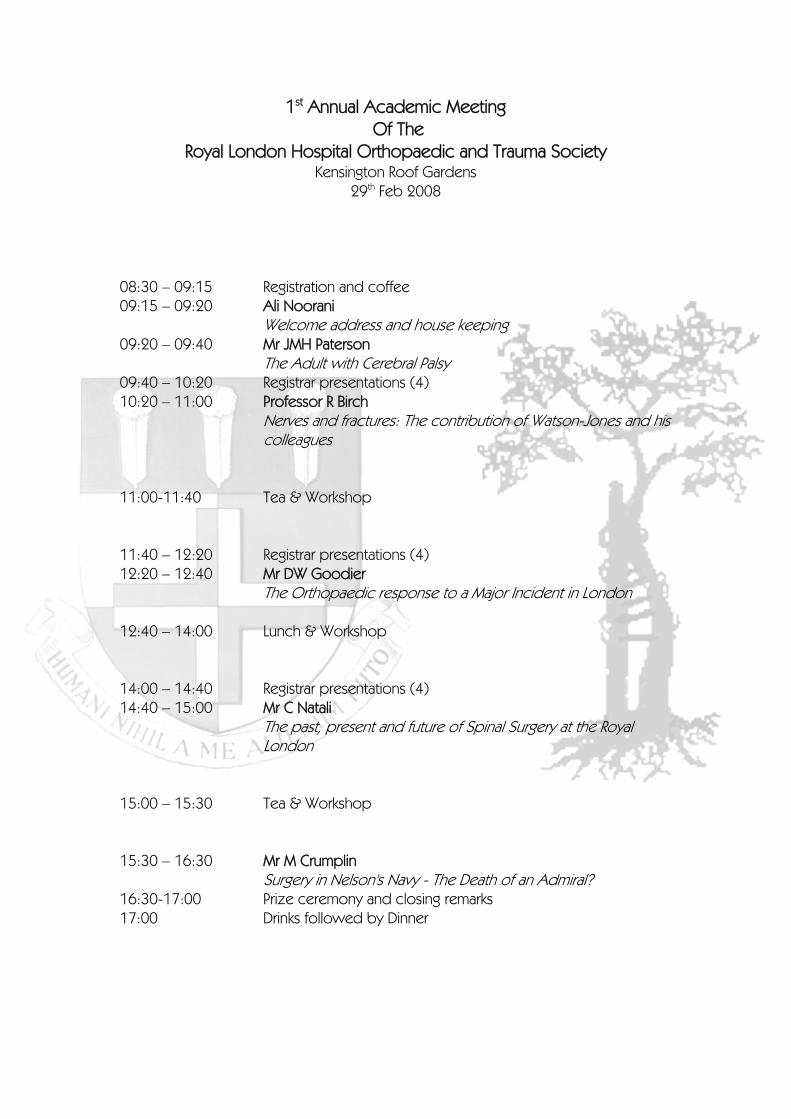
1st Annual Academic Meeting
Of The Royal London Hospital Orthopaedic and Trauma Society
Kensington Roof Gardens 29th Feb 2008
08:30 – 09:15 Registration and coffee 09:15 – 09:20 Ali Noorani
Welcome address and house keeping 09:20 – 09:40 Mr JMH Paterson The Adult with Cerebral Palsy 09:40 – 10:20 Registrar presentations (4) 10:20 – 11:00 Professor R Birch
Nerves and fractures: The contribution of Watson-Jones and his colleagues
11:00-11:40 Tea & Workshop 11:40 – 12:20 Registrar presentations (4) 12:20 – 12:40 Mr DW Goodier
The Orthopaedic response to a Major Incident in London 12:40 – 14:00 Lunch & Workshop 14:00 – 14:40 Registrar presentations (4) 14:40 – 15:00 Mr C Natali
The past, present and future of Spinal Surgery at the Royal London
15:00 – 15:30 Tea & Workshop 15:30 – 16:30 Mr M Crumplin
Surgery in Nelson's Navy - The Death of an Admiral? 16:30-17:00 Prize ceremony and closing remarks 17:00 Drinks followed by Dinner

Registrar Podium Presentations
Group 1 9:40- 10:20 Medialisation of the tibial insertion of the patella tendon in patients with open growth plates: a retrospective study C. Maizen, F. Schneider, W. Linhart Altered physeal gene-expression due to diaphyseal fracture of a growing bone E.E. Fischerauer, C. Gülly, G. Janezic, M.E. Höllwarth, A.M. Weinberg A retrospective study of Galeazzi fractures in the paediatric population R. Eberl Diagnostic features of septic arthritis of the knee in patients with renal failure: a retrospective study A. Berridge , R. Tahmassebi
Group 2 11:40- 12:20 The medial rotation knee: a ten-year follow up study K. Mannan, G. Scott Bilateral cementless total knee replacement following previous unilateral high tibial osteotomy: functional results at an average of 8 years P.J.H. Sloper, M.J. Cross Arthroscopic ankle fusion Z. Dannawi, A. Patel, D.J. Moore A stereophotographic study of ankle joint contact area S. Millington, M. Grabner, R. Wozelka, S. Hurwitz, J. Crandall
Group 3 14:00- 14:40 Pelvic trauma: how do we manage it? C. Jowett, S. McMahon Development of a clinical device for measuring rotational laxity of the knee M. Alam, A.M.J. Bull, R.W. Thomas, A.A. Amis Correct implant placement in proximal femur fractures K. Tanzer, W. Hartwagner, F.J. Seibert, W. Seggl The Role of Judet decortication in the 21st century C. Jayadev, M. Mullins, P. Piriou, T. Judet
Registrar Poster Presentations Therapeutic Shoulder Arthroscopy procedures in patients with clotting disorders: A Case Series. J.M.C. Thomas, E.G. Tuddenham, P.M. Ahrens Operative reconstruction of the radio-humeral joint in cases of neglected dislocation of the radial head in children. T. Kraus, A.K. Hell, C.C. Hasler, W.E. Linhart
Invited Keynote Speakers J Mark H Paterson FRCS(Eng) Chairman of the Royal London Hospital Orthopaedics & Trauma Society Consultant Orthopaedic Surgeon, Royal London Hospital Rolfe Birch MChir FRCS, FRCS Eng by election
)
Professor in Neurological Orthopaedic Surgery, RNOH David Goodier FRCS(Orth) Consultant Orthopaedic Surgeon, Royal London Hospital Colin Natali FRCS(Orth Program Director of the Royal London RotationConsultant Orthopaedic Surgeon, Royal London Hospital Michael Crumplin FRCS Retired General Surgeon Curator & Archivist at the Royal College Of Surgeons Of England
ABSTRACTS Medialisation of the tibial insertion of the patella tendon in patients with open growth plates. A retrospective study C. Maizen, F. Schneider and W. Linhart Department of Paediatric Orthopaedics, University Hospital for Paediatric Surgery Graz, Austria Introduction From 1997 to 2001 we performed 98 medialisations of the tibial insertion of the patella tendon in patients between 10 and 18 years of age. We want to describe a solely soft tissue procedure for medialisation of the tibial insertion of the patella tendon after recurrent patella dislocation in patients with open growth plates. We start with the incision of the periost at the anterior margin of the tibia in a thought line of continuation of the patella tendon, starting from the medial side of the tibial tuberosity extending to about 10 cm distally and slightly medially. Then the insertion of the patella tendon and the periosteum are lifted off the tibia so that the attachment is now shifted further distally and medially. As the procedure is combined with a lateral release it allows the patella to centralise naturally in the femoral groove and improves or even restores mal-alignement. The tendon is not fixed to the tibia in any way. Postoperative treatment consists of physiotherapy and mobilisation with crutches non weight bearing for 4 weeks. Methods Retrospective analysis of postoperative results in 36 knees after a mean follow up period of 4 years. We evaluated the efficiency of the described operation in terms of recurrence of dislocation, over all patient satisfaction, occurrence of growth disturbance and clinical outcome with functional scores. Results 36 knees in 27 patients were evaluated postoperatively. The mean follow up period was 47 months. 15 patients had a flake fracture of the patella and/or an osteochondral flake of the lateral femoral condyle. There were no peri- or post-operative complications. In 4 cases (12%) a recurrence of dislocation occurred. In 3 cases we had to perform a second arthroscopy due to the extensive damage of the cartilage. Most of the patients had a mean decrease of 1,5 points in the Tegner Score for sporting activities at the time of follow up Discussion In our opinion the medialisation of the patella tendon as a soft tissue procedure is a simple and effective method for treatment of recurrent dislocation of the patella in children and adolescents.
Altered physeal gene-expression due to diaphyseal fracture of a growing bone E.E. Fischerauer1, C. Gülly2, G. Janezic1, M.E. Höllwarth1, A-M Weinberg1
1Department of pediatric surgery, Medical University of Graz, Austria; 2 Core Facility Molecular Biology, Center for Medical Research, Medical University of Graz, Austria. Background and Aims Children bones are unique in responding to a fracture by overgrowth or growth arrest. Accelerated bone growth is hypothesised to be the result of an enhanced chondrocyte proliferation rate at the growth plate due to the interaction of different cytokines, morphogens, agiogenics, and growth factors. By the performance of a microarray study and qRT-PCR analysis, an anticipated change of physeal gene-expression after fracture was investigated in an animal model. Material and Methods Male Sprague-Dawley rats at the age of 4 weeks were used as a living fracture-animal-model. In general anaesthesia, experimental animals sustained a unilateral fracture with a Bonnarens and Einhorn device. After euthanasia at scheduled time-points, tissue from the proximal tibial growth plate was harvested and analysed by Microarray and qRT-PCR for growth factors such as bone morphogenetic proteins (BMP), fibroblast growth factor (FGF), or vascular endothelial growth factor (VEGF). Results Microarray analysis revealed a strong systemic immune response 3 days post-trauma. 13301 physeal genes were differentially expressed between the fractured, contra-lateral and control bone. PANTHER analysis showed that the Wnt/beta-catenin signalling pathway which is involved in osteogenesis, is one of the most significantly down-regulated pathways between the experimental and control group on day 3 post-fracture. Furthermore, significant differences in regenerative (cell division/mitosis and motility) and developmental processes could be seen between the experimental and the control group. Quantitative gene-expression analysis of growth factors which are known to be involved in bone development, displayed lower physeal expression levels of the fractured tibial bone compared to contra-laterals and controls 3 days post-fracture. Growth factors such as Parathyroid hormone related protein (PTHrP), BMPs, FGFs and their receptors show increasing expression from day 3, with overall highest levels on day 14 or 29 post-fracture in the fractured bone. Towards day 29, gene-expression levels of BMP-2,-6,-7, -receptor 1a, VEGF, FGF receptor 3 of the fractured and contra-lateral intact bones are converging and eventually, display higher expression levels than in control samples. Conclusions The strong physeal immune response to a fracture evaluated by microarray analysis indicates that inflammation is prominent at the growth plate initially after fracture, probably influencing chondrocytes proliferation and thus contributing to overgrowth. Quantitative RT-PCR data revealed an initial physeal down-regulation of many growth factors such as BMPs which are known potent growth factors that promote chondrogenesis and osteogenesis, and thus indicate that these growth factors can not account for the high physeal turn-over seen 3 days post-fracture in the fractured bone, and rather promote bone growth during later stages of fracture healing. Furthermore, microarray and qRT-PCR results displayed that there are similar physeal gene-expression changes in both tibiae of the experimental animal, suggesting a cross-talk between the affected and the contra-lateral intact bone, which might minimise deregulated limb length growth.
A retrospective study of Galeazzi fractures in the paediatric population Robert Eberl Introduction A Galeazzi fracture is defined as a fracture of the radius associated with dislocation of the ulna in the distal radioulnar joint (DRUJ). Treatment in children and adolescents differs from that in adults and is usually possible with conservative therapy using plaster casting. The objective of this retrospectively designed study was to describe all Galeazzi fractures treated at our department over a period of three years. Patients and Methods Data of 198 patients with dislocated fractures of the radius alone or both bones of the forearm were reviewed. Conservative treatment was done with plaster casting and operative intervention by intramedullary nailing or dorsal plate fixation. Results In 26 (13%) cases we found a Galeazzi fracture and the patients were included in a follow up. The outcome was measured using the Gartland-Werley-Score. 8 of 26 (31%) fractures were recognized initially and classified as a Galeazzi lesion. Conservative treatment following fracture reposition was possible in 22 patients. 13 patients were treated with immobilization in a below-elbow cast, 9 with an above elbow cast. 4 patients were treated operatively. The results using the Gartland-Werley-Score were excellent in 23 cases and good in 3 cases. Discussion In cases of distal forearm fractures a possible Galeazzi-lesion should be considered. However, proper reduction of the radius with subsequent reduction of the ulna in the DRUJ and cast-immobilization provides good to excellent outcome even if the Galeazzi-lesion is primarily under-diagnosed. Long-term instability of the DRUJ following Galeazzi fractures was not observed in our series of pediatric patients.
Diagnostic features of septic arthritis of the knee in patients with renal failure: a retrospective study A. Berridge, R. Tammessebi Department of Trauma and Orthopaedics, The Royal London Hospital Background Patients with end stage renal failure receiving renal replacement therapy are at an increased risk of developing septic arthritis due to immunosuppressive disease, medications and active infections at distant sites (including intravenous lines). However the clinical picture is different to that seen in patients with normal renal function, making the diagnosis more challenging. In addition, the risks associated with surgical washout in such patients who often have multiple co-morbidities are frequently high. Our aim was to review the diagnostic features and microbiological findings in this patient group, and produce evidence that would help guide our practice in the future. Hypothesis We considered that the classical clinical findings of a painful hot swollen joint with minimal range of movement would be less marked, and that the laboratory findings of a neutrophillia, raised CRP and ESR would not necessarily be present. We expected the organisms found at microscopy and culture to be similar to those found in patients without renal disease-predominantly staphylococcal and streptococcal species. Method A retrospective study of patients undergoing treatment for ESRF at the Royal London Hospital who were suspected of having septic arthritis of the knee and who had either synovial fluid aspirated or washout of the knee. Patients were identified through the Renal Department database and data was collected from the medical notes. Risk factors were identified (for example immunosuppressive medication), clinical signs and symptoms, inflammatory markers, findings at aspiration and surgical washout and response to treatment. Additional information was gathered from the Electronic Patient Record System on gram stain and culture results from aspirate and washout specimens. Results We found that the presenting symptoms and signs of a hot swollen joint in association with fever were common, although the inability to weight bear was less frequently found. A mild raise in inflammatory markers was present, although the ESR was more likely to be raised. Concurrent infection, for example of the dialysis access site, was present in a significant number of patients. Microscopic findings of pus cells but no organisms were found in a surprisingly high number of patients. Conclusions The assessment of patients with renal failure who are suspected of having septic arthritis is difficult. They often do not display the classical symptoms and signs of the disease, and haematological investigations, in particular the CRP, can be misleading. A high index of suspicion is needed, and close liaison with both the Renal and Anaesthetic team is vital.
Bilateral Cementless Total Knee Replacement Following Previous Unilateral High Tibial Osteotomy: Functional Results at an Average of 8 Years P. J. H. Sloper FRCS (T&O)1 M. J. Cross, OAM, MD, FRACS2
1Specialist Registrar, Princess Alexandra Hospital, Harlow. 2Orthopaedic Knee Surgeon, Australian Institute of Musculo-Skeletal Research, Sydney, Australia. Background High tibial osteotomy (HTO) has been used as a surgical treatment for isolated medial compartment osteoarthritis in younger patients for many years. Whilst the results of HTO are good in the short term, they tend to deteriorate with time and total knee replacement (TKR) is often required at a later date. There is considerable debate regarding whether previous HTO compromises the outcome of TKR when compared to primary TKR. Some studies have demonstrated similar functional outcomes, whilst others have shown inferior results and/or higher rates of complications. A limitation of many of these studies is that they have to use matched groups, thus introducing an additional layer of variability into the result. One study avoided this by comparing the results of TKR after HTO with the results of contralateral primary TKR in patients with bilateral TKR, but the TKRs in this study were performed by a number of different surgeons. We report the results of bilateral cementless TKR performed by a single surgeon in patients with previous unilateral HTO, thus allowing a direct comparison. Hypothesis Our hypothesis was that the results of TKR after HTO are no different to those of TKR without prior HTO. Patients and methods Between 1993 and 2002, 23 patients underwent bilateral primary TKR using an unstemmed, cementless, HA-coated posterior-cruciate-retaining prosthesis following a previous unilateral proximal tibial valgus osteotomy. All of the TKR procedures were performed by the senior author using a standard technique. Eleven of the osteotomies were performed by the senior author, with the rest being performed by multiple other surgeons. Patients were reviewed at an average of 8 years and were assessed using the Knee Society knee and functional scores. Pre-operative demographic and intra-operative surgical data were also compared. Statistical analysis was performed with the Fisher exact test and a 2-tailed Student t-test, paired for repeat observations on the same knee and unpaired for comparisons between knees. Results There was a significant difference between the two groups in the number of other procedures, such as arthroscopy (but excluding the HTO, see Table 1), which were performed prior to TKR (p<0.02), but there were no other significant differences in demographic data. There were no significant differences between the groups in Knee Society scores or range of movement pre-operatively, at 5 years or at final review. The polyethylene tibial inserts were significantly thicker in the osteotomised group (p<0.01), but there were no significant differences in the rate of lateral retinacular release or patella resurfacing. There was a higher rate of complications in the osteotomy group and 2 patients required revision for infection (1 early, 1 late). No patients required revision in the primary TKR group. Conclusions The results of this study do not show a significant difference between the functional results of TKR after previous HTO and those of primary TKR on the contralateral knee in a group of 23 patients. However, the possibility of a higher rate of complications and revisions following TKR in the previously osteotomised knees deserves further examination.
Arthroscopic ankle fusion Z Dannawi, A Patel, DJ Moore Background The aim of this study was to evaluate the outcome of arthroscopic ankle fusion carried out at our institution Patients and Methods Between 1999 and 2007, 54 patients underwent an arthroscopic ankle arthrodesis for end stage arthritis. Arthroscopic ankle arthrodesis was accomplished with a consistent technique using 2 cannulated screws. The average follow up was 52 months (range 11 to 108 months). Clinical records and preoperative and postoperative radiographs were reviewed. Patients were evaluated subjectively and objectively using the Mazur grading system. Results Four patients died before final review. 3 patients were lost to follow-up, leaving us with 47 patients. The overall fusion rate was 91% (43/47). The preoperative tibio-talar angle in the coronal plane was between 21° valgus and 23° varus. The mean time to union was 10.5 weeks. There were 81% good to excellent results according to the Mazur grading system. Non union occurred in 4 cases (9%) necessitating an open procedure. There were 2 superficial infections, two deep vein thromboses. Three patients required removal of screws for prominence. Conclusion Arthroscopic ankle fusion achieves high union rates, short time to union and low complication rates. We also feel that the indications can be broadened to include patients with significant angulation provided the foot can be squared.
A stereophotographic study of ankle joint contact area Millington S1, Grabner M2, Wozelka R2, Hurwitz S3, Crandall J3. 1. Royal London Hospital, London, UK; 2. Technical University of Graz, Austria; 3 University of Virginia, USA Introduction There is considerable variation in the size and location of the contact area in the ankle joint reported in the literature. However, knowledge of the contact area under physiological load is essential for understanding the biomechanics of the ankle joint, is beneficial for understanding the pathogenesis of joint degeneration, as well as improving prosthetic design and ligament reconstruction surgery. Therefore this study was performed to measure the ankle joint contact area and describe the contact area distribution under physiological load using a stereophotography technique that allows accurate analysis of the entire joint surface without disrupting the joint during loading. Methods 10 cadaveric foot and ankle specimens were loaded to 1000 N in neutral, and 20°dorsiflexion, inversion, eversion, and plantarflexion. Photo targets rigidly fixed to each of the bones were imaged in the loaded joint position using a high resolution stereophotography system (ATOS™). After testing each ankle was disarticulated and the joint surfaces imaged relative to the photo targets. The photo targets were then used to spatially register the joint surfaces into the loaded joint position, the overlap of the surfaces was used to determine the joint contact area. Results The mean talo-tibia contact area was greatest in dorsiflexion 7.34 cm2 ± 1.69 cm2 and was significantly larger than in plantar flexion (P < 0.05) which showed the smallest joint contact area 4.39 cm2 ± 1.41 cm2. Considering talo-fibula contact area, the maximum contact occurred in dorsiflexion, 2.02 cm2 ± 0.78 cm2, and the minimum contact area occurred in eversion, 0.77 cm2 ± 0.49 cm2, respectively (P<0.05). Conclusions The reported stereophotography technique allows measurement of the joint contact area without disrupting the joint during loading. The contact area is larger than previously reported, as the entire joint surface was analysed. Joint contact was largely distributed over the talar shoulders where the cartilage is thickest, subchondral bone is most dense and osteochondritis dissecans lesions commonly occur, as opposed to the central talar dome as previously reported.
PELVIC TRAUMA: how do we manage it? Jowett, C., McMahon, S. Department of Trauma and Orthopaedics, The Royal London Hospital Background The Royal London Hospital receives a volume of major trauma from London and the surrounding regions. 236 patients with pelvic fractures were admitted between January 2005 and November 2007, the majority of whom were multiply injured. Hypothesis This study asks four questions related to the management of pelvic trauma: What are we dealing with? How do we manage it? Is our management of these injuries systematic? Is there room for improvement? Methods We reviewed the notes and initial imaging of patients admitted with a pelvic fracture during the last twelve months (70 patients). Using a proforma, we collated data including the age and sex of the patient; co-morbidities; mechanism of injury; type of pelvic fracture; other injuries; GCS on scene; haemodynamic parameters on admission; fluid and transfusion requirements; initial examination, interventions and management; emergency management of pelvic fracture; use of angiography; definitive management of pelvic fracture; documented nursing instructions; use of thromboprophylaxis; ITU stay; and outcome ( death, transfer to specialist unit, discharge) Results Of seventy patients admitted with pelvic fractures, 21 died ( 30%) and 18 were transferred out (25.7%). The remaining 39 patients (55.7%) were eventually discharged home. The number of patients undergoing formal internal examination to establish whether the fracture was open or closed was negligible, as was the rate of anterior external fixation. No patients had a C clamp applied, and no patients had internal fixation of their pelvic fracture at the Royal London Hospital. Conclusions The Royal London Hospital received a significant volume of pelvic trauma. More than a third (36.7%) of patients who survived for 24 hours required operative management by pelvic specialists, and had to wait for beds to become available at the receiving unit. The pelvic service could be streamlined and improved by clear guidance and leadership, as well as the operative ability, of a surgeon specialised in pelvic trauma surgery.
Development of a Clinical Device for Measuring Rotational Laxity of the Knee Alam M, Bull AMJ, Thomas RW, Amis AA Introduction A clinical tool to assess rotational knee laxity would aid diagnosis, monitoring of treatment progress and comparisons between surgical interventions. Such a device is not currently available. Aim To design and test a clinical tool for measuring rotational laxity of the knee. Methods The device comprises a boot and tibial mould with inclinometers. The boot provides constraint via pneumatic pressure cuffs at the foot and ankle and allows a defined torque to be applied across the knee. The tibial mould is attached to the tibia via velcro straps and the inclinometers measure induced rotation. Simultaneous measurements of tibio-femoral rotation using an electromagnetic ‘Nest of Birds (NOB)’ device and the inclinometer device were taken. The NOB were attached to the femur using a custom splint to compensate for soft tissue motion due to femoral rotation and to the tibia using the mould. These measures were validated using intra-operative pin-fixation to bone in a separate study. Bilateral measurements were taken of 25 normal subjects at 90° and 30° flexion in a supine position by a single examiner, repeated at 24hrs. Knee flexion was maintained by positioning the lower leg on the adjustable ´head end´ of an examination couch. The thighs were immobilised by a large velcro strap. Measurements were taken using 4, 6, 8Nm torque, followed by a clinical hard endpoint, mimicking the ‘dial test’. Results All subjects reported the testing as comfortable. RMS errors of the boot inclinometer measurements for the internal-external rotation envelope were greater at 30° and were less than 5° at 90° knee flexion, when comparing the inclinometer readings with the NOB measurements. At 30°, these errors increased with increasing torque for the inclinometer device, measuring less than 4° at 4Nm and increasing to 8° at 8Nm. The errors were maximal with the endpoint method, measuring at 8.5°. Internal and external rotations increased with increasing torque, and were maximal using the clinical endpoint. Knee internal and external rotation was greater in the 30° flexed position compared to the 90° position for all torques, and this difference increased with increasing torques. This difference was approximately 5° for both internal and external rotation at 8Nm. Side to side differences, the method used to differentiate between normal and abnormal in the clinical setting, were less than 2° at 90° knee flexion. This was consistent throughout all torque levels and using the clinical endpoint method. Side to side differences were greater for 30° knee flexion. Differences were approximately 2° at 4Nm, but increased to 4° at 8Nm. They were above 8° for the hard endpoint method. Conclusions Studies reporting on clinical devices for measuring rotatory laxity of the knee have had problems with inadequate control of ipsilateral joints, soft tissue artefact and defining neutral rotation position. Initial data from our study show side to side differences of less than 2° using the devices at 90° flexion. This may allow differentiation between normal and abnormal laxities at 90°. At 30° flexion the side to side differences were 2° at low torque, but increased with greater torque levels. RMS error data suggests the measuring device is representative of the true knee rotations to 5° for 90° knee flexion. The errors are greater at lower knee flexion levels. The measuring device allows a greater appreciation of the total internal and external rotations at different knee flexions in a clinical setting. We are continuing our work by increasing our sample size, analysing intra- and inter-examiner repeatability and carrying out measurements on abnormal knees.
Correct implant placement in proximal femur fractures K. Tanzer, W. Hartwagner, F.J. Seibert, W. Seggl Department of Traumatology, University Hospital Graz Introduction A new proximal femur nail with one single femoral neck stabilisation element was introduced in our institution 04/03 and over 250 patients were prospectively monitored. According to Levy, screw support in the head-neck fragment should be in the posterior inferior part of the head to have enough hold. We tried to analyse if we are able to achieve this position with our implant and if the outcome correlates with the placement of our device. Patients and Methods 300 patients had their unstable trochanteric femoral fracture stabilized by this femur nail until now. The age of the female patients was between 70-93. Male patients age was between 19-90 years. All patients had been mobilized under full weight bearing. Preoperative, intra-/postoperative and the last available x-rays were analysed concerning to the tip-apex-Distance and the inferior-superior- and the posterior-anterior ratio. Results Special attention was drawn to implant migration in consideration to implant placement. Our hypothesis is the following now: the antirotational blade should be placed as inferior as possible in ap view and as central as possible in the axial view. The tip-apex distance should be less than 1 cm. Reduction in both planes in mandatory and axial alignment is crucial. Netherless the nail with its antirotational blade is more forgiving than thought. Conclusion Low rate of migration if blade was placed correctly. Postoperative mobilization with full weight bearing was possible without a higher amount of complications. This intramedullary device is easy to handle and is a real advantage in fixing all types of proximal femur fractures.
The Role of Judet Decortication in the 21st Century C. Jayadev, M. Mullins, P. Piriou, T. Judet Background The treatment of failure of fracture union is a complex affair. Initial management must address the underlying cause such as soft tissue interposition, infection, ischaemia and excessive movement at the fracture site. There are a number of interventions available to encourage bone consolidation. These include internal and external fixation, bone grafting, bone transport and distraction osteo-modelling by the Illizarov method and decortication techniques. Despite the historical background, scientific basis and versatility of decortication in the treatment of non-union the technique has fallen out of vogue with bone grafting and distraction osteo-modelling being the favoured options. Hypothesis We aim to show that Judet’s method of osteoperiosteal decortication still has a strong role to play in the management of failure of union in the 21st century. Methods Prospective analysis of 297 cases of osteoperiosteal decortication for failure fracture union. Concurrent stabilisation was with either internal or external fixation as appropriate to fracture location and configuration, and bone grafting for major structural defects was used in 34% (n=102) of cases. Results Union was successfully achieved in 294 of the 297 patients (99%) after a mean delay of 8 months (range 6 weeks to 46 months, SD 6.5 weeks). Two hundred and twenty patients (74%) achieved union following the decortication procedure without subsequent operations. There was no significant difference in union time with respect to initial fracture location, open vs. closed fractures or atrophic vs. hypertrophic non-union (p<0.05). Union was successful in all but one case of infected non-union (98%). Patients with infected non-union took 60% longer for union to be achieved than those with no initial infection. Conclusions Osteoperiosteal decortication remains a highly effective solution in the management of failed fracture union regardless of location, type of non-union and presence of infection.
Therapeutic Shoulder Arthroscopy procedures in patients with clotting disorders: A Case Series. JMCThomas1, EG Tuddenham2, PM Ahrens1
Department of Orthopaedic Surgery1 and Haemophilia Centre2
The Royal Free Hospital, London, UK Background Orthopaedic surgery in patients with clotting disorders represents a challenge to surgeons and haematologists alike. Storti1 performed the first documented open knee synovectomy in 1966, since then series of elective orthopaedic procedures performed on patients with clotting disorders have been documented2.Arthroscopy of the shoulder has been used to perform a synovectomy, but a review of the literature shows no previous documentation of therapeutic arthroscopic procedures of the shoulder in patients with clotting disorders. Hypothesis Ho – Patients with clotting disorders are not contraindicated for undergoing therapeutic shoulder arthroscopic procedures. Methods We report a consecutive case series of eight arthroscopic procedures in seven patients with clotting disorders of varying severity and who had undergone therapeutic shoulder arthroscopic procedures for pathology that was unrelated to their haematology. Close liaison with the haematology centre at our institute was maintained and these patients were managed in a multidisciplinary team. Results All patients showed significant improvement following the procedure. One post operative portal haematoma had a two day delay to discharge, however no significant morbidity occurred. Conclusion Clotting disorders do not preclude the use of reconstructive arthroscopic techniques in the shoulder as long as the appropriate specialist multidisciplinary team manages these patients. References 1) Storti E Traldi A, Tosatti E, Davoli P. Synovectomy a New Approach to Haemophiliac Arthropathy.
Acta Haematol 1969;41:193 2) ScharrerI, Bray GL, Neutzling O. Incidence of Inhibitors in Haemophilia A Patients – a Review of
Recent Studies of Recombinant and Plasma-derived Factor VIII Concentrates. Haemophilia 1999;5;145-154
Operative reconstruction of the radio-humeral joint in cases of neglected dislocation of the radial head in children T. Kraus, A.K. Hell, C.C. Hasler, W. E.Linhart Introduction Neglected dislocation of the radial head in children can present as a congenital anomaly, improperly managed monteggia fracture-dislocations, or missed isolated dislocations. Clinical findings include instability at the elbow and restriction in flexion and extension. A valgus deformity of the elbow is often seen as a late secondary change, which may produce a tardy unlar paresis. Principles of operative correction include either a corrective osteotomy combined if necessary with ulnar lengthening, and open reduction of the dislocated radius with reconstruction of the radio-humeral joint. In this report we present our results of surgical management in cases of neglected radial head dislocation in children. Materials and Methods The collective consisted of 21 children with neglected radial head dislocation managed by the Pediatric Orthopedics Units at the University Hospitals in Basel (Switzerland), and Graz (Austria). Corrective osteotomy with open reduction of the radial head was performed in 18 cases, and ulnar lengthening followed by open reduction of the radial head in 3 cases. The annular ligament was not reconstructed in this series. Results All the children (13 from Basel, 8 from Graz) were evaluated retrospectively using subjective, clinical, and radiological criteria. The average follow-up period was 8.2 years. There were 11 girls and 10 boys with average 5.5 years age at diagnosis, and 8 years at begin of surgical management. Primary reconstruction of the radial head could be achieved in 86% (n=18) cases. Recurrence of dislocation in the postoperative period was observed in the remaining 3 cases. Two of these could be stabilised with repeated open reduction, in one child the dislocation persisted inspite of revision surgery. Realignment of the elbow axis could be achieved in all cases. About half of the cases had restriction of joint movements preoperatively, approximately 50% of these showed an improvement in the joint movements postoperatively. About 25% cases showed decrease in the elbow movements after surgery. Discussion The management of neglected dislocations of radial head is complicated and needs to be planned carefuly. Good results in producing a stable joint and improving the elbow motion depends on the extent of the anatomy and congruence of the radio-capitular joint after open reduction.

Delegate List P Achan M Alam W Al-Hakim S Al-Nammari F Altaf A Amin S Ang M Barry A Berridge H Bhinda R Birch H Bosman J Bradley T Briggs T Bucknill P Calder R Carew C Critchley D Crone M Crumplin S Curry Z Dannawi L Di Mascio R Eberl M El-Zebdeh E Fischerauer M Freeman I Garnham A Ghassemi D Goodier B Grange T Greer N Heidari S Ilyas C J Spivey C Jayadev C Jowett V Khanduja M Lee M Loeffler
H Maan A Macdowell J Mahaluxmivala C Maizen O Malaga Shaw J Mangwani K Mannan S Matthews T McAuliffe S McMahon C Middleton S Millington D Moore C Natali D Nawabi A Noorani H Nordeen S Owen-Johnstone J Parker M Paterson T Peckham A Sanghrajka G Scott F Seibert M Shafafy A Sivaraman P Sloper M Taba R Tahmassebi K Tanzer J Targett M Taylor A Thomas J Thomas N Wardle A Watson A Way A Weinberg G Wright W Yoon