fine structure of meiotic prophase chromosomes...
TRANSCRIPT

J. Cell Sci. 37, 69-84 (i979) 69Printed in Great Brtitain © Company of Biologists Limited 1979
FINE STRUCTURE OF MEIOTIC PROPHASE
CHROMOSOMES AND MODIFIED
SYNAPTONEMAL COMPLEXES IN DIPLOID
AND TRIPLOID RHOEO SPATHACEA
YUE J. LIN
Department of Genetics, Ohio State University, Columbus Ohio 43210,U.S.A. Present address: Department of Biological Sciences, St John'sUniversity ,Jamaica, New York 11439, U.S.A.
SUMMARY
The synaptonemal complex (SC) in the diploid Rhoeo consists of 2 amorphous lateral elements,each about 46-0 nm thick, and one amorphous central element about 300 nra thick. The centralregion is about 1150 nm wide. SC in the triploid have essentially the same dimensions as thoseof the diploid; both lateral (460 nm) and central (30-0 nm) elements are amorphous, and thecentral region is about 1175 nm wide. The coil, observed in both diploid and triploid, is amodified short segment of SC with several twists at the end of a synapsed bivalent that is at-tached to the nuclear membrane. Serial sections in a diploid cell reveal that a coil extends in-wards about 3-5 /4m from the nuclear membrane and makes a complete turn at a distance ofevery 0-5 (tm. There is a correlation between the modified ends of SC and terminal chiasmatain Rhoeo. The coils might have a positive role in the process of crossing over, or alternativelymight be involved in ring formation by holding chromosome ends together while chiasmata arenot involved. SC are present in chromocentres of both diploid and triploid. Chromocentres indiploid and triploid are indistinguishable, and appear to be formed from the aggregation ofpericentromeric heterochromatin as a result of translocations which occurred close to the centro-meres. 3-dimensional hypothetical pachytene configuration of the diploid is presented.
INTRODUCTION
Diploid Rhoeo spathacea (Swartz) Stern {zn = 12) is characterized by chromosomalstructural hybridity to such an extent that no 2 chromosomes in the diploid plant aregenetically identical. At first meiotic division, the 12 chromosomes are usually arrangedinto a ring. One, two or three chains are often observed (Lin & Paddock, 19736). Thering has been interpreted to be a result of extensive reciprocal translocation, so thatthe 2 arms of each chromosome synapse respectively with an arm of each of 2 otherchromosomes. Rhoeo has not been satisfactory for light-microscope studies of synapsisbecause of the unfavourable and elongated state of chromosomes at zygonema andpachynema (Figs. 5-8). Electron microscopy allows the study of synapsis at an ultra-structural level.
Synaptonemal complexes (SC hereafter) have been shown to be associated withbivalents at meiotic prophase in many species of eukaryotes and have become regardedto be involved in synapsis and perhaps subsequent crossing over and chiasma forma-tion although the exact function is yet not clear (see Moses, 1968; Westergaard &Wettstein, 1972; Gillies, 1975, for review).

70 Y.J.Lin
SC among different species have a remarkable morphological consistency despitegreat phylogenetic diversity. An SC usually consists of a set of 3 parallel strands, lyingin a single plane that curves and twists along the axis of the synapsed chromosomes inwhich it lies. Two dense lateral elements are held in register by the central regionwhich contains the central element. The central element is generally less dense and isjoined to the lateral elements by thin transverse filaments.
Modified lateral elements of SC have been reported. In an allotriploid lily, Moens(1968) observed unusual tube-like structures over a distance of a few micrometres inone or both of the lateral elements in nearly every section of a pachytene nucleus. Hespeculated that this deformed structure might be associated with the chromosomeheterozygosity because this species is an allotriploid derived from hybridization of twoor more closely related species. Further studies of SC in an autotetraploid lily (Moens,1970) revealed no such abnormality. Later a very similar deformity was reported inpollen mother cells of both diploid and triploid Phaedranassa viridiflora (LaCour &Wells, 19736). LaCour & Wells (1973 a) also studied the SC in a lily hybrid, whichhad almost complete bivalent formation at metaphase I, and observed rather bizarreirregularities in lateral elements and in whole SC.
In his studies of the SC in diploid Rhoeo, Moens (1972) found coiled SC attachingto the inner membrane of the double nuclear membrane. Therefore, diploid Rhoeopresents another example of a modified SC. The coiled SC, hereafter simply the coil,extends approximately 4 /tm from the nuclear membrane and as many as 10 suchattachments (of 12 possible attachments) were observed in a small region of the nu-clear membrane near the nucleolus (Moens, 1972). Moens concluded that perhaps allthe ends of SC in diploid Rhoeo are coiled and attached to the nuclear membrane.This type of coil has not been reported except in a grasshopper, Chloealtis conspersa(Moens, 1974). Later McQuade & Wells (1975) confirmed Moens' finding of coils indiploid Rhoeo.
Studies on SC have been concentrated mostly in diploid organisms. This studywas aimed at investigating the SC and its possible structural modification in triploidRhoeo, and comparing the SC in the triploid with that in the diploid.
MATERIALS AND METHODS
The process of microsporogenesis and maturation is often synchronous within an anther butis less so among the anthers in a flower of Rhoeo. For accurate identification of the meiotic stagesin pollen mother cells (PMC hereafter), an anther in proper size was first cut into 2 halves whichwere fixed immediately: one half was placed in fixative for squash preparation, and the other wasfixed in glutaraldehyde for electron microscopy. If the PMC in squash preparation were found tobe in the proper stages, the other half was further prepared for electron-microscope studies.Further stage identification was done with thick sections (o-8 fim) prior to ultrathin sectioning.
The method of making slides for light microscopy has been described (Lin & Paddock,1973a)-
For electron microscopy, excised anthers were fixed immediately for 1 h in 2 % glutaralde-hyde buffered in o-2M Sorensen's phosphate buffer at pH 7-2, rinsed in buffer at pH 7-2 for30 min (2 changes), and postfixed for 1 h in phosphate-buffered 1 % osmium tetroxide. Antherswere then dehydrated in a graded ethanol series at 4 °C, and embedded in hard Spurr's lowviscosity resin (Spurr, 1969) at room temperature. Thick sections (o-8 /im) were obtained with
Synaptonemal complexes in Rhoeo 71
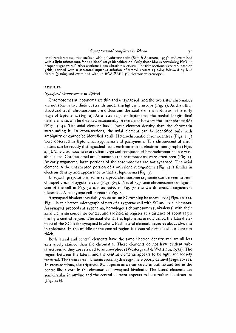
an ultramicrotome, then stained with polychrome stain (Sato & Shamato, 1973), and examinedwith a light microscope for additional stage identification. Only those blocks containing PMC inproper stages were further sectioned into ultrathin sections. The thin sections were mounted ongrids, stained with a saturated aqueous solution of uranyl acetate (3 min) followed by leadcitrate (3 min) and examined with an RCA-EMU 3G electron microscope.
RESULTS
Synapsed chromosomes in diploid
Chromosomes at leptonema are thin and unsynapsed, and the two sister chromatidsare not 9een as two distinct strands under the light microscope (Fig. 1). At the ultra-Structural level, chromosomes are diffuse and the axial element is elusive in the earlystage of leptonema (Fig. 2). At a later stage of leptonema, the medial longitudinalaxial elements can be detected occasionally in the space between the sister chromatids(Figs. 3, 4). The axial element has a lower electron density than the chromatinsurrounding it. In cross-sections, the axial element can be identified only withambiguity or cannot be identified at all. Heterochromatic chromocentres (Figs. 2, 3)were observed in leptonema, zygonema and pachynema. The chromocentral chro-matins can be readily distinguished from euchromatin in electron micrographs (Figs.2, 3). The chromocentres are often large and composed of heterochromatins in 2 vari-able states. Chromosomal attachments to the chromocentre were often seen (Fig. 2).At early zygonema, large portions of the chromosomes are not synapsed. The axialelement in the unsynapsed portion of a univalent at zygonema (Fig. 4) is similar inelectron density and appearance to that at leptonema (Fig. 3).
In squash preparations, some synapsed chromosome segments can be seen in less-clumped areas of zygotene cells (Figs. 5-7). Part of zygotene chromosome configura-tion of the cell in Fig. 7A is interpreted in Fig. 7B-F and a differential segment isidentified. A pachytene cell is seen in Fig. 8.
A synapsed bivalent invariably possesses an SC running its central axis (Figs. 10-12).Fig. 4 is an electron micrograph of part of a zygotene cell with SC and axial elements.As synapsis proceeds at zygonema, homologous chromosomes (univalents) with theiraxial elements come into contact and are held in register at a distance of about 115-0nm by a central region. The axial element at leptonema is now called the lateral ele-ment of the SC in the synapsed bivalent. Each lateral element measures about 46-0 nmin thickness. In the middle of the central region is a central element about 30-0 nmthick.
Both lateral and central elements have the same electron density and are all lessextensively stained than the chromatin. These elements do not have evident sub-structures so they are referred to as amorphous (Westergaard & Wettstein, 1972). Theregion between the lateral and the central elements appears to be light and looselytextured. The transverse filaments crossing this region are poorly defined (Figs. 10-12).In cross-sections, the tripartite SC appears as a near-circle in outline and lies in thecentre like a core in the chromatin of synapsed bivalents. The lateral elements aresemicircular in outline and the central element appears to be a rather flat structure(Fig. I2B).


Synaptonemal complexes in Rhoeo 73
The twists and turns of SC, revealed by serial section reconstruction or whole mountpreparations, are generally very gentle (Gillies, 1972; Counce & Meyer, 1973). In thediploid, coiling was observed at the ends of synapsed bivalents (Figs. 9, 13). The coilswere always near the nuclear membrane on which the synapsed chromosome endswere attached. The coil in Fig. 13 extends inwards about 3-5 /tm from the nuclearmembrane and makes one complete turn at a distance of every 0-5 /tm. The dimensionsof the lateral and the central elements in the coil are the same as those where the com-plexes are not coiled.
SC were also observed in chromocentres (Fig. 14). The SC in chromocentres andthe role of the coil are considered in the Discussion.
Measurements of SC in the diploid are compared with the measurements made byMcQuade & Wells (1975) in Table 1: the methods of fixation appear to alter thedimensions of SC. The authors' measurements of SC in Rhoeo are very close to theaverage dimensions reported from 1968 to 1971 in various organisms as shown intable I of Westergaard & Wettstein (1972). McQuade & Wells' measurements of thelateral and central elements in the diploid are apparently much smaller than the average.
Synapsed chromosomes in triploid
Leptotene chromosomes in triploid (Fig. 15) have an appearance similar to thoseof diploid (Fig. 1). At zygonema, the homologous chromosomes synapse (Fig. 16). Apachytene cell is in Fig. 18. Two-by-two synapsis leaving the third unsynapsed hasbeen observed in a squash preparation (Lin & Paddock, 1978). With the electronmicroscope, SC were also found to be present in synapsed bivalents in triploid andthey were not distinguishable from those in diploid. In Fig. 17 a portion of a zygotenecell with SC distributed over the section is shown. Figs. 19-21 are electron micrographsof SC in various cells. The 2 lateral elements appear as 2 ribbons each about 46-0 nmthick and separated by a region in the centre of which lies a strip of central elementabout 30-0 nm thick. Fine filaments traversing the central region between the lateralelements are more obvious in Fig. 21. The distance between the lateral elements isabout 117-5 n m (Table 1). A cross-sectional view of an SC is in Fig. 20B.
Chromocentres were also observed at prophase I in triploid (Figs. 22, 23). Diffusechromosomes and chromocentres of a leptotene cell can be seen in Fig. 22; in somesections, some chromocentres did have a suggestion of SC (Fig. 23).
Figs. 1-3. Diploid leptonema.Fig. 1. Diploid leptotene cell from a squash preparation, x 1850.Fig. 2. Section through a diploid early leptotene cell. Chromatin (ch) in electron
micrograph appears quite diffuse and the axial element is hardly seen. Arrow marksthe chromosome attachment with chromocentre (cc). x 20000.
Fig. 3. Diploid leptotene chromosomes with axial elements (ae) in the space betweenthe sister chromatids prior to synapsis. Note the 2 variable states of heterochromatinin chromocentre (cc). x 15000.
Fig. 4. Synapsed chromosomes with an SC and unsynapsed chromosomes with axialelements (ae) in a diploid zygotene cell, x 15000.

74 Y.J.Lin
5 B

Synaptonemal complexes in Rhoeo 75
Coiling at the ends of synapsed bivalents was also observed in triploid (Fig. 24). InFig. 24, the end of the chromosome is associated with a nucleolus and the nuclearmembrane. It appears then that a nucleolar organizer region is located at the tip ofthis chromosome.
No SC were found inside nucleoli in this study although central region materialshave been found inside nucleoli of a fungus, Neottiella (Westergaard & Wettstein,1970). Triple chromosome synapsis as reported in triploid chickens (Comings &Okada, 1971) was not observed in triploid Rhoeo.
No abnormal tubular lateral elements such as those described in triploid lily (Moens,1968) and triploid Phaedranassa (LaCour & Wells, 19736) were observed either intriploid or in diploid Rhoeo in this study. Therefore the deformed lateral elements inthese 2 instances may not be due to the triploidy.
DISCUSSION
Coils and chiasmata in Rhoeo
The association of chromosome ends with the nuclear membrane at prophase I hasbeen found consistently in many animals (insects: Moens, 1969; Church, 1976;Wettstein & Sotelo, 1967; mammals: Baker & Franchi, 1967; birds: Ford & Woolam,1964; snail: Gall, 1961; and rat: Esponda& Gime"nez-Martin, 1972). Occasionally, theassociation was observed in higher plants (Figs. 9, 13, 24 in Rhoeo; Gillies, 1973, inmaize). In the ascomycete fungi Neottiella (Westergaard & Wettstein, 1970) andNeurospora crassa (Gillies, 1972) the ends of chromosomes are also connected withthe nuclear membrane. Both ends of the same chromosomes attaching to the nuclearmembrane were observed in some organisms (Ford & Woollam, 1964; Wettstein &Sotelo, 1967; Moens, 1969V
The attachment points may be polarized to a small region near the centriolesmaking a bouquet configuration in Locusta migratoria and synapsis was found to beinitiated near the attachments and proceeded away from the nuclear membrane inthis organism (Moens, 1969).
Fig. 5. A, diploid zygotenc cell, B, an interpretation of A. Chromosome 11' is synapsingwith 2 chromosomes J 2 and 1' 2'. x 1850.
Fig. 6. Synapsed segments (arrows) of a bivalent are shown in this diploid zygotenecell. Synapsis appears to be initiated at the same time in various segments of homo-logous chromosomes, x 1850.
Fig. 7. A, squash preparation of a diploid zygotene cell. Arrows indicate synapsedchromosome ends, B-F, an interpretation of A. The added dotted lines are not seenin the photo-micrograph. Each chromosome is synapsed with 2 or more differentchromosomes, e.g. chromosome 13 is synapsed with 3 chromosomes: 1' 2'', 1 2 and 3 3'.Chromosome 1' 3' is synapsed with 2 chromosomes: 1' 2' and 3 3'. The chromo-centre is not associated with the 2 translocation points. The segment between 2translocation points is called a differential segment (ds). x 1850.
Fig. 8. Diploid pachytene cell, x 1850.
Fig. 9. Section through a diploid zygotene cell with 3 coils at the ends of 3 synapsedbivalents. x 12000.

Y.J.Lin
DFigs. 10-12. Synaptonemal complexes in diploid. ce, central element; le, lateral ele-ment, x 40000. Fig. i2B. Cross-sectional view of 2 SC in diploid. Both LE and CEare less dense than the chromatin. x 40000.Fig. 13. A-D, 4 consecutive sections of zygotene bivalent in diploid at its point ofattachment to the nuclear membrane. The coil structure is evident, E, reconstructionof lateral elements. F, reconstruction of central element; the lateral elements are shownby dotted lines, x 20000.

Synaptonemal complexes in Rhoeo 77
Fig. 14. Chromocentre with SC (arrow) in diploid. x 20000.Fig. 15. A triploid leptotene cell, x 1850.Fig. I6A, B. A triploid zygotene cell and diagrammatic representation. The arrow indi-cates a point where the synaptic partners change which may be a translocation point,x 1850.
Fig. 17. Section through a triploid zygotene cell. Many chromosomes contain SC.x12 000.Fig. 18. Triploid pachytene cell in a squash preparation, x 1850.Fig. 19. Synaptonemal complexes in triploid. ce, central element; le, lateral element,x 40000.
fi CEL 37

mlyEDTAethanolic PTA°OsO4
OsO4
ions"
76-48678 9 293-897-6
i iS-o
"7-5io6-2105-0
1 7 82 2 4
24-41 6 646-0
46-040-045O
1 5 0
I9-314-2I2'2
I3-O3O-O
3O-O
3O-O
as -o
78 Y. J. Lin
The polarization of chromosome ends at prophase I is probably important to theprocess of synapsis. It may provide a firm association of chromosome ends with eachother in the beginning of synapsis and promote the chance of the synaptic partnersmeeting.
Table 1. Dimensions (in nm) of synaptonemal complexes related tomethods of fixation
Central Lateral CentralFixation region element element
Diploid RhoeoMcQuade & Wells (1975)0
Formalin; OsO4
GAP"; OsO4
Glutaraldehyde onlyGlutaraldehyde;Glutaraldehyde;
Lin Glutaraldehyde;Triploid Rhoeo
Lin Glutaraldehyde;Average monocot.''Average of reported dimensions"
a. Their Table I.b. GAP = glutaraldehyde-acrolein-paraformaldehyde technique.c. PTA = phosphotungstic acid.d. See table I of Westergaard & Wettstein (1972).e. This is the average of all reported dimensions in various organisms from 1968 to 1971 as
reviewed by Westergaard & Wettstein (1972) in their table I.
The attachment of chromosome ends with nuclear membrane has been noted onlyrarely in higher plants (Westergaard & Wettstein, 1972). In my study, both diploidand triploid Rhoeo were found to have not only the attachments but also coils (Figs.9, 13, 24). From a large number of serial sections, as many as 10 such attachments wereobserved in a cell by Moens (1972) and 11 by McQuade & Wells (1975). The 3 nucleiexamined with serial sections by Moens had all the attachments polarized on a smallarea of the nuclear membrane. Three coils together in a small region of nucleus canbe seen clearly in a single thin section (my Fig. 9). Similarly to Moens' (1969) observa-tion, McQuade & Wells' (1975) data indicate that the initiation of synapsis in diploidRhoeo probably begins near the nuclear membrane. Synapsis in triploid Rhoeo perhapsproceeds in a similar fashion.
The existence of coils has been reported only in Rhoeo and a grasshopper, Chloealtisconspersa (Moens, 1974). The coils in C. conspersa are located in the terminal portion ofsome of the bivalents.
There is a striking correlation between such modified ends of SC and terminalchiasmata. In diploid Rhoeo, the 12 meiotic chromosomes at diakinesis are joined byterminal chiasmata and form a ring. In C. conspersa, there are 3 pairs of large chro-mosomes with terminal chiasmata which always form rings at meiosis (Moens, 1974).Another species of grasshopper, Chorthippus longicornis, has a karyotype similar to

Synaptonemal complexes in Rhoeo
ce
M X ^ &. .->
24Figs. 20, 21. SC in triploid. c«, central element; le, lateral element. Fig. 20B is a cross-sectional view. Transverse filaments (//) are more evident in Fig. 21. x 40000.Fig. 22. Electron micrograph of a triploid leptotene cell with 2 chromocentres (cc).Chromosomes (ch) are diffuse and the axial element is hardly discernible, x 10000.Fig. 23. A chromocentre in triploid with a suggestion of SC. x 20000.Fig. 24. A coil in triploid. The nucleolus («) is attached to this chromosome which isassociated with the nuclear membrane (rnn). x 25000.

80 Y. J. Lin
C. conspersa but there are no terminal chiasmata, and coils have not been found in thisspecies (Moens, 1974). Such correlations suggest that the modified SC are probablyinvolved in terminal chiasma formation.
X
X
y
y
~ y
x'
y'
Fig. 25. A. Reciprocal translocation between 2 pairs of non-homologous chromosomesmay result in a chromocentre at pachynema. The translocation points are near thecentromeres. B. An additional ring-enlarging translocation may result in a larger chro-mocentre.
The indication from light-microscope studies that synapsis in diploid Rhoeo is limi-ted to short terminal regions of chromosomes (Lin & Paddock, 19736) is in conflictwith the observation that synapsis (SC formation) occurs all over in the interior of thenucleus (McQuade & Wells, 1975). This can be explained, however, by the fact thatSC does not always lead to chiasma formation. The role of the modified SC, coils,perhaps is in enhancing the chance of crossing-over at terminal regions. Light-micro-scope studies showed the frequency of terminal chiasmata tends to be high in diploidRhoeo meiotic cells (Lin & Paddock, 19736). Only a very small percentage of cellshad less than 9 terminal chiasmata. On the other hand, prophase I cells seem to havea high number of coils in each cell because the 2 zygotene and 1 pachytene nucleistudied had 8, 10 and 11 attachments (coils), respectively (McQuade & Wells, 1975).Thus, the number of terminal chiasmata probably corresponds to the number of coils.The chiasma failure might be due simply to the failure in coil formation at the attach-ment during synapsis. An alternative hypothesis is that the chromosome ends are heldtogether by nothing more than the coils themselves, i.e. chiasmata are not involvedin the ring formation at all.
Synaptonemal complexes in Rhoeo 81
Chromocentre and synopsis
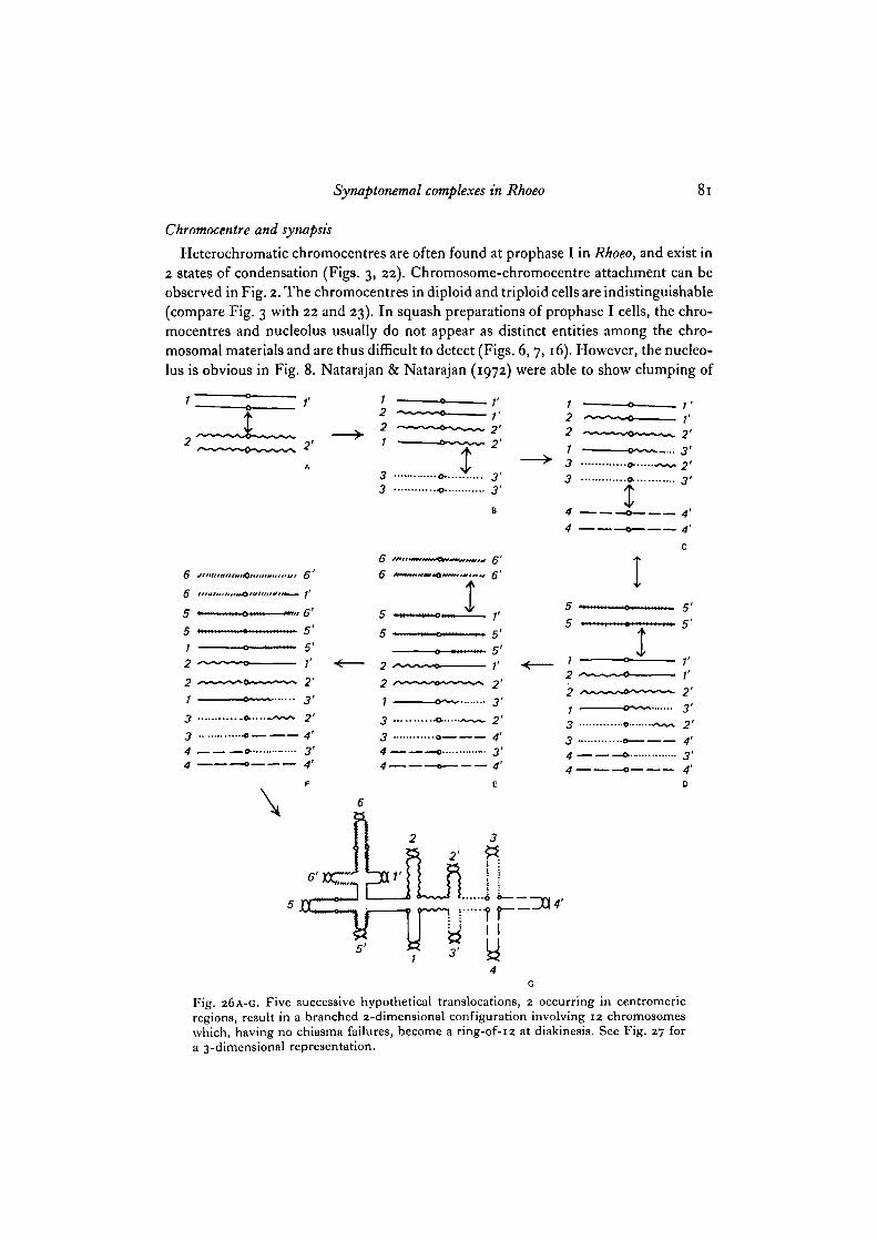
Heterochromatic chromocentres are often found at prophase I in Rhoeo, and exist in2 states of condensation (Figs. 3, 22). Chromosome-chromocentre attachment can beobserved in Fig. 2. The chromocentres in diploid and triploid cells are indistinguishable(compare Fig. 3 with 22 and 23). In squash preparations of prophase I cells, the chro-mocentres and nucleolus usually do not appear as distinct entities among the chro-mosomal materials and are thus difficult to detect (Figs. 6, 7,16). However, the nucleo-lus is obvious in Fig. 8. Natarajan & Natarajan (1972) were able to show clumping of
6 J
6 '
5
221
6'1'
5'r2'3'2'3 « • • • • -
3 -o 4'4 o. -.... 3'4 0 4'
\
1221
33
66
55
221334-4-
X
v1'2'2'
3'3'
I— 5'— 5'— ;'-~ 2'
3'
4'
3'4'
55
12213J **
44
5'5'
2'3'2'4'3'4'D
Fig. 26A-G. Five successive hypothetical translocations, 2 occurring in centromericregions, result in a branched 2-dimensional configuration involving 12 chromosomeswhich, having no chiasma failures, become a ring-of-12 at diakinesis. See Fig. 27 fora 3-dimensional representation.

82 Y. J. Lin
chromocentres at pachynema in diploid. The heterochromatins in diploid are situatedaround the centromeres (pericentromeric) in all the 12 chromosomes and are latereplicating (Natarajan & Natarajan, 1972). They are constitutive heterochromatins andmay be considered to contain highly repetitive DNA sequences (Yunis & Yasmineh,1971). Aggregation of heterochromatin has been shown in many organisms (Yunis &Yasmineh, 1971). In a recent report (Godin & Stack, 1975), association of telomericheterochromatins was shown in rye, Secale cereale. In Rhoeo, the chromocentre form-ation thus appears to be from the aggregation of at least part of the pericentromericheterochromatins. It can be conceived as a result of extensive reciprocal translocation
Fig. 27. Hypothetical pachytene configuration in diploid, based on Fig. 26c Onlylateral elements are shown. All chromosome ends are polarized to a small region in thenucleus. Coiled SC are located at the ends of synapsed bivalents. Two adjacent trans-location points neither of which is involved in a chromocentre are at left, cc, chromo-centre.
in the following way. It is generally accepted that the structural hybridity in Rhoeo hasresulted from a series of translocations. As shown in Fig. 25 A if the break points wereat or near the centromeres, then heterochromatins in the 4 chromosomes could bebrought together at pachynema and a large mass of heterochromatic chromocentreformed. A ring-enlarging translocation, if at centromeric vicinities, will give rise to aneven larger chromocentre by clumping together of centromeric heterochromatins in6 chromosomes (Fig. 25 B).
The indefinite shapes of chromocentres may reflect that the various reciprocal trans-locations involved in building the ring did not occur equally distant from their res-pective centromeres.
Short stretches of synaptonemal complexes were occasionally found in chromo-centres in both diploid and triploid. Those in Figs. 14 and 23 and in McQuade & Wells'(1975) figs. 11 and 12 are all in the less-condensed area which has a similar appearanceto that of chromosome. It is possible but doubtful that SC in Rhoeo do not occur in thehighly condensed region of the chromocentres.

Synaptonemal complexes in Rhoeo 83
Electron-microscope studies of meiosis in Fritillaria lanceolata (LaCour & Wells,1970) also showed the presence of SC in the chromocentres.
The view that heterochromatin as such may aid in initial alignment prior to synapsiswas criticized by Maguire (1972) who considers that a direct functional role of hetero-chromatin in synapsis still lacks sound evidence. In Rhoeo, chromocentres are probablya result of synapsis causing association of pericentromeric heterochromatins.
Based on considerations in the foregoing discussion and some observations in thestudy, theoretical configurations of diploid pachytene cells are presented in Figs. 26and 27. Only lateral elements of SC are presented. As shown in Fig. 26 G, 5 successivetranslocations among non-homologous chromosomes occurring 2 in centromericregions and 3 in non-centromeric regions result in a branched 2-dimensional pachyteneconfiguration containing 12 chromosomes that can become a ring at diakinesis. Acorresponding 3-dimensional pachytene configuration is in Fig. 27. A chromocentreforms wherever more than 2 centromeric regions aggregate. Where translocation hasnot occurred at a centromeric region, such as in Fig. 26B, D and E, no chromocentre isexpected to be present at that point. The speculation that break points are not neces-sarily localized at centromeric regions is based on the interpretation in Fig. 7B-Fin which neither of 2 adjacent translocation points is associated with the chromocentres(see also Fig. 27).
This paper represents part of a dissertation submitted for a Ph.D. degree at The Ohio StateUniversity, Columbus, Ohio, U.S.A. I am thankful to Dr Elton F. Paddock for his guidance,patience and encouragement during the course of this study.
REFERENCES
BAKER, T. G. & FRANCHI, L. L. (1967). The fine structure of oogonia and oocytes in humanovaries. J. Cell Sci. 2, 213-224.
CHURCH, K. (1976). Arrangement of chromosome ends and axial core formation during earlymeiotic prophase in the male grasshopper Brachystola magna by 3D, E.M. reconstruction.Chromosoma 58, 365-376.
COMINGS, D. E. & OKADA, T. A. (1971). Triple chromosome pairing in triploid chickens.Nature, Lond. 231, 119-121.
COUNCE, S. J. & MEYER, G. F. (1973). Differentiation of the synaptonemal complex and thekinetochore in Locusta spermatocytes studied by whole mount electron microscopy. Chromo-soma 44, 231-253.
ESPONDA, P. & GIMENEZ-MART/N, G. (1972). The attachment of the synaptonemal complex tothe nuclear envelope. An ultrastructural and cytochemical analysis. Chromosoma 38, 405-417.
FORD, E. H. R. & WOOLLAM, D. H. M. (1964). Testicular chromosomes of Gallus domesticus.Chromosoma 15, 568-578.
GALL, J. G. (1961). Centriole replication. J. biophys. biochtm. Cytol. io, 163-193.GILLIES, C. B. (1972). Reconstruction of the Neiirospora crassa pachytene karyotype from
serial sections of synaptonemal complexes. Chromosoma 36, 119-130.GILLIES, C. B. (1973). Ultrastructural analysis of maize pachytene karyotypes by three dimen-
sional reconstruction of the synaptonemal complexes. Chromosoma 43, 145-176.GILLIES, C. B. (1975). Synaptonemal complex and chromosome structure. A. Rev. Genet. 9,
91-109.GODIN, D. E. & STACK, S. M. (1975). Heterochromatic connectives between the chromosomes
of Secale cereale. Can. J. Genet. Cytol. 17, 269-273.LACOUR, L. F. & WELLS, B. (1970). Chromocentres and the synaptinemal complex. J. Cell Sci.
6, 655-667.

84 Y. J. Lin
LACOUR, L. F. & WELLS, B. (1973 a). Abnormalities in synaptonemal complexes in pollen mothercells of a lily hybrid. Chromosoma 42, 137-144.
LACOUR, L. F. & WELLS, B. (19736). Deformed lateral elements in synaptonemal complexes ofPhaedranassa viridiflora. Chromosoma 41, 289-296.
LIN, Y. J. & PADDOCK, E. F. (1973a). Ring-position and frequency of adjacent distribution ofmeiotic chromosomes in Rhoeo spathacea. Am. J. Bot. 60, 685-690.
LIN, Y. J. & PADDOCK, E. F. (19736). Ring-position and frequency of chiasma failure in Rhoeospathacea. Am.J. Bot. 60, 1023-1027.
LIN, Y. J. & PADDOCK, E. F. (1978). Identification of complexes in a spontaneous triploid R/ioeospathacea. Chromosoma 67, 97-1C8.
MAGUIRE, M. P. (1972). Role of heterochromatin in homologous chromosome pairing: Evalu-ation of evidence. Science, N. Y. 176, 543-544.
MCQUADE, H. A. & WELLS, B. (1975). The synaptonemal complex \r\ Rhoeo spatliacea. J. Cell Sci.
MOENS, P. B. (1968). Synaptinemal complex of Lilium tigrinwn (triploid) sporocytes. Can. J.Genet. Cytol. io, 799-807.
MOENS, P. B. (1969). The fine structure of meiotic chromosome polarization and pairing inLocusta migratoria spermatocytes. Chromosoma 28, 1-25.
MOENS, P. B. (1970). The fine structure of meitoic chromosome pairing in natural and artificialLilium polyploids. J. Cell Sci. 7, 55-64.
MOENS, P. B. (1972). Fine structure of chromosome coiling at meiotic prophase in Rhoeodiscolor. Can. J. Genet. Cytol. 14, 801-808.
MOENS, P. B. (1974). Coincidence of modified crossover distribution with modified synapto-nemal complexes. In Mechanisms in Recombination, Proc. 27th Res. Conf., Biol. Division,Oak Ridge Nat. Lib. (ed. R. F. Grell), pp. 377-383. New York: Plenum Press.
MOSES, M. J. (1968). Synaptinemal complex. A. Rev. Genet. 2, 363-412.NATARAJAN, A. T. & NATARAJAN, S. (1972). The heterochromatin of Rhoeo discolor. Hereditas 72,
323-33O-SATO, T. & SHAMATO, M. (1973). A simple rapid polychrome stain for epoxy-embedded tissue.
Stain Technol. 48, 223-227.SPURR, A. R. (1969). A low-viscosity epoxy resin embedding medium for electron microscopy.
J. Ultrastruct. Res. 26, 31-43.WESTERGAARD, M. & WETTSTEIN, D. von. (1970). Studies on the mechanism of crossing over IV.
The molecular organization of the synaptinemal complex in Neottiella (Cooke) saccardo(Ascomycetes). C. r. Trav. Lab. Carlsberg 37, 239-268.
WESTERGAARD, M. & WETTSTEIN, D. von. (1972). The synaptinemal complex. A. Rev. Genet. 6,71-110.
WETTSTEIN, R. & SOTELO, J. R. (1976). Electron microscope serial reconstruction of the sper-matocyte I nuclei at pachytene. J. Microscopie 6, 557-576.
YuNlS, J. J. & YASMINEH, W. G. (1971). Heterochromatin, satellite DNA, and cell function.Science, N.Y. 174, 1201-1209.
(Received 25 October 1978)