figure 1 - key endocrine glands - anatomy and physiology · the endocrine system figure 1 - key...
TRANSCRIPT
The Endocrine System
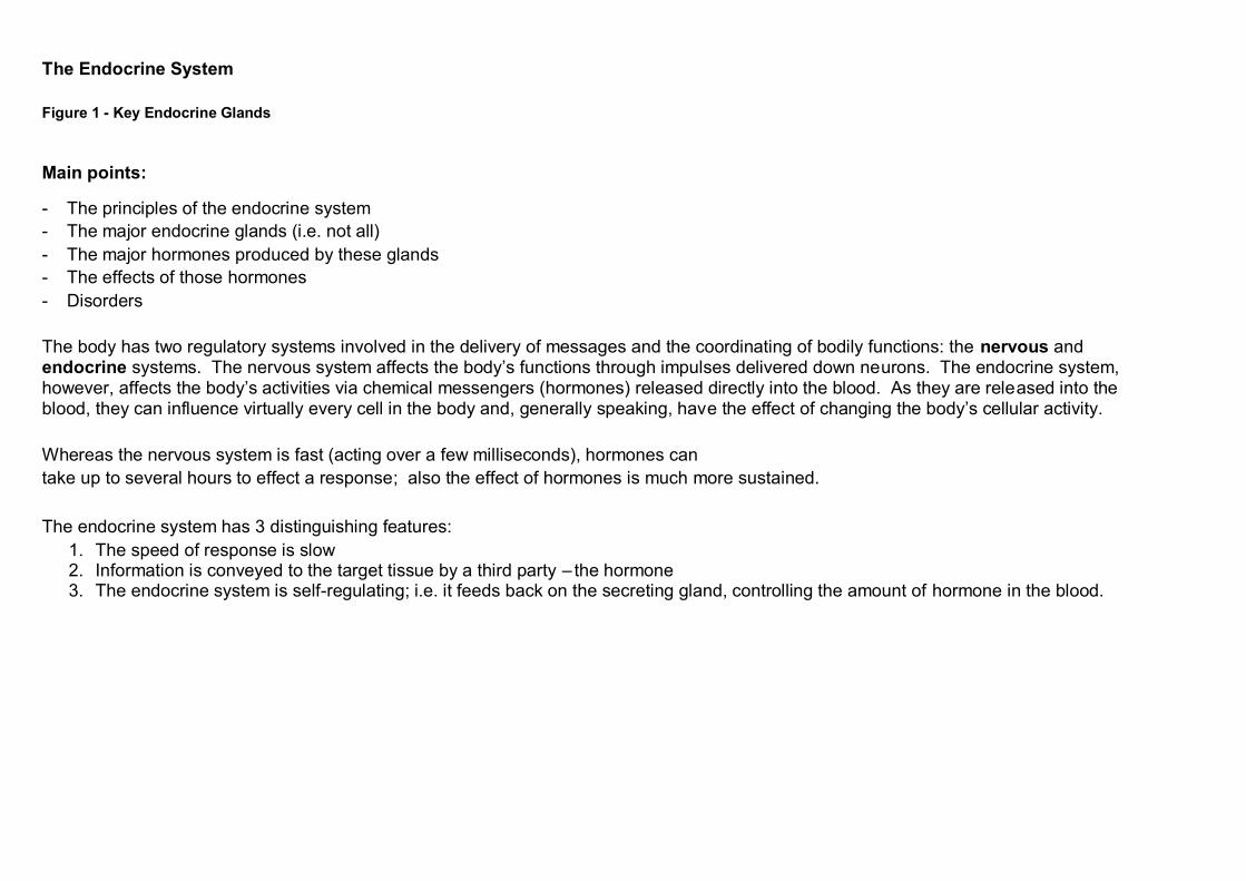
Figure 1 - Key Endocrine Glands Main points:
- The principles of the endocrine system
- The major endocrine glands (i.e. not all)
- The major hormones produced by these glands
- The effects of those hormones
- Disorders
The body has two regulatory systems involved in the delivery of messages and the coordinating of bodily functions: the nervous and endocrine systems. The nervous system affects the body’s functions through impulses delivered down neurons. The endocrine system, however, affects the body’s activities via chemical messengers (hormones) released directly into the blood. As they are released into the blood, they can influence virtually every cell in the body and, generally speaking, have the effect of changing the body’s cellular activity.
Whereas the nervous system is fast (acting over a few milliseconds), hormones can
take up to several hours to effect a response; also the effect of hormones is much more sustained.
The endocrine system has 3 distinguishing features:
1. The speed of response is slow 2. Information is conveyed to the target tissue by a third party – the hormone 3. The endocrine system is self-regulating; i.e. it feeds back on the secreting gland, controlling the amount of hormone in the blood.
The endocrine glands are:
1) Pituitary
2) Thyroid
3) Parathyroid
4) Suprarenal
5) Pancreas
6) Ovaries and testes
7) Pineal
8) Thymus
9) Placenta
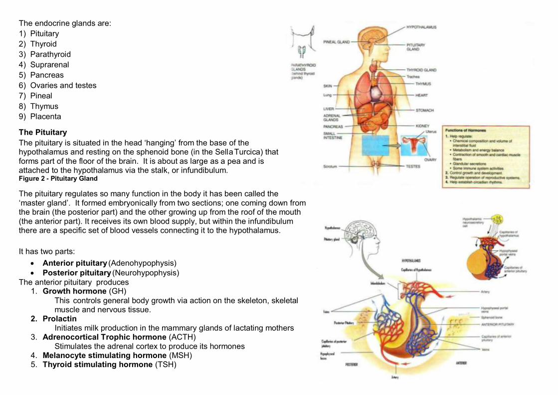
The Pituitary
The pituitary is situated in the head ‘hanging’ from the base of the hypothalamus and resting on the sphenoid bone (in the SellaTurcica) that forms part of the floor of the brain. It is about as large as a pea and is attached to the hypothalamus via the stalk, or infundibulum. Figure 2 - Pituitary Gland
The pituitary regulates so many function in the body it has been called the ‘master gland’. It formed embryonically from two sections; one coming down from the brain (the posterior part) and the other growing up from the roof of the mouth (the anterior part). It receives its own blood supply, but within the infundibulum there are a specific set of blood vessels connecting it to the hypothalamus.
It has two parts:
Anterior pituitary (Adenohypophysis)
Posterior pituitary (Neurohypophysis)
The anterior pituitary produces 1. Growth hormone (GH)
This controls general body growth via action on the skeleton, skeletal muscle and nervous tissue.
2. Prolactin Initiates milk production in the mammary glands of lactating mothers
3. Adrenocortical Trophic hormone (ACTH) Stimulates the adrenal cortex to produce its hormones
4. Melanocyte stimulating hormone (MSH) 5. Thyroid stimulating hormone (TSH)
Stimulates the thyroid to produce hormones 6. Follicle stimulating hormone (FSH)
a. Stimulates the ovaries to produce ova (eggs) b. Stimulates the testes to produce sperm
7. Luteinizing hormone (LH)
a. In the female, it stimulates the ovaries to release the ova and prepares the endometrium of the uterus for implantation; it also stimulates the corpus luteum in the ovary. (The corpus luteum produces progesterone and prepares the mammary glands for milk production)
b. In the male it stimulates the production of testosterone in the testes. The posterior pituitary produces:
Oxytocin – this stimulates the muscle in the wall of the uterus in childbirth (therefore produced initially at this time). It also stimulates
milk ejection from the mammary glands during breastfeeding.
Anti-diuretic hormone (ADH aka vasopressin) – this reduces the volume of urine produced by stimulating water resorption in the
distal convoluted tubule and collecting duct in the kidney. It also acts to increase blood pressure directly by causing constriction of the walls of arterioles.
In the anterior pituitary, the hormones are produced and stored in situ, but are only released when ‘told to’ by the hypothalamus, via releasing factors coming down the connecting vessels. In the Neurohypophysis, the hormones are synthesized in the hypothalamus and are stored in the posterior pituitary and released when required.
Disorders:
GH – Reduced secretion of this causes pituitary dwarfism, as opposed to genetic dwarfism, caused by
undersecretion of growth hormone (officially as dwarf is anyone shorter than 4ft 10ins). Pituitary dwarfism is different to achondroplasia (circus’ dwarfism):
In pituitary dwarfism all the body parts are in proportion to each other.
In achondroplasia is genetic with the head and body are relatively normal, but the limbs are short
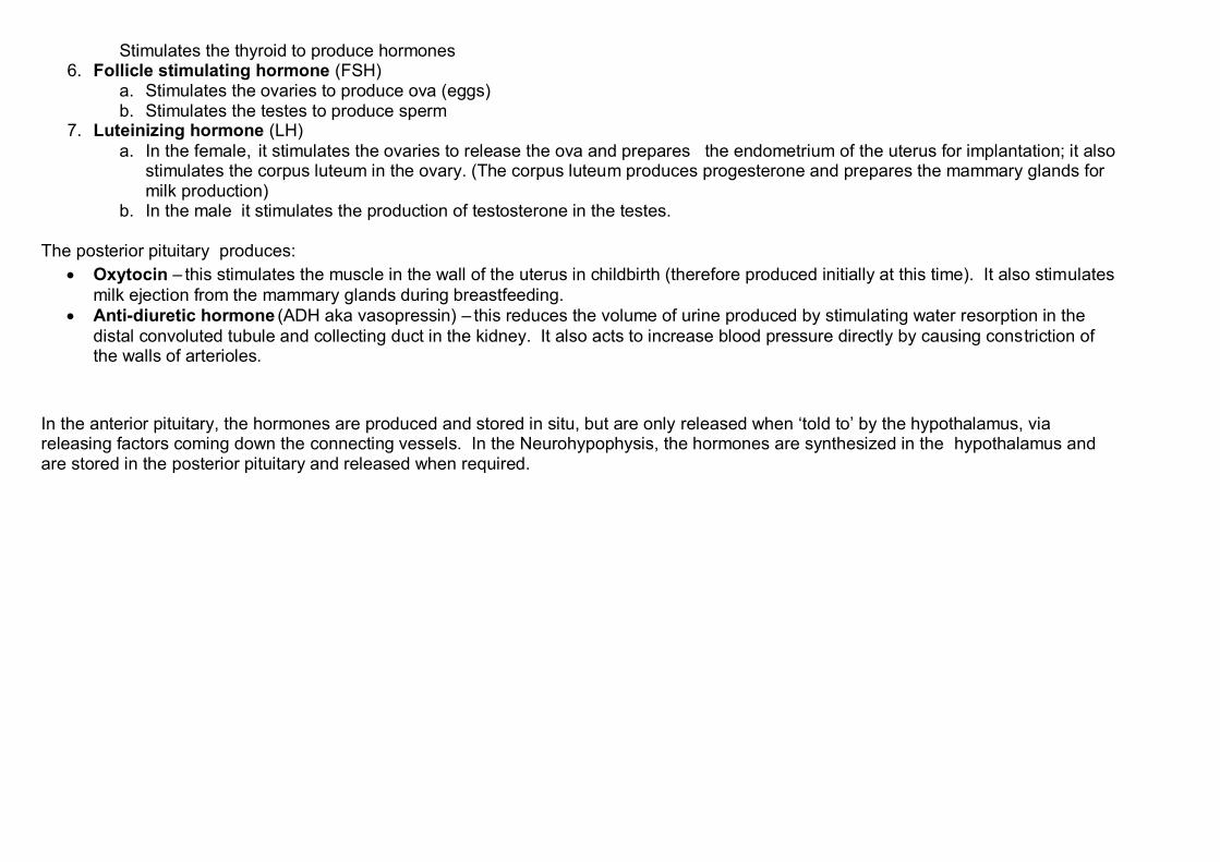
GH – over secretion causes giantism. Giantism is anyone over 6ft 4in
Figure 3 - Giantism
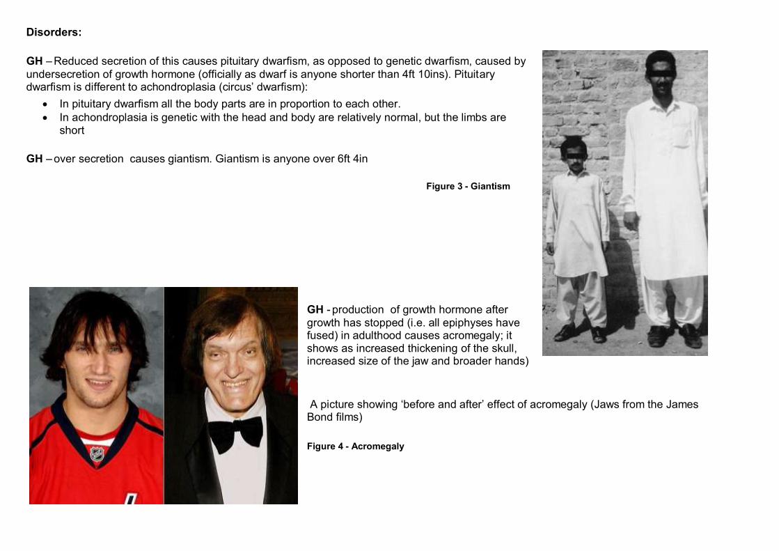
GH - production of growth hormone after
growth has stopped (i.e. all epiphyses have fused) in adulthood causes acromegaly; it shows as increased thickening of the skull, increased size of the jaw and broader hands)
A picture showing ‘before and after’ effect of acromegaly (Jaws from the James Bond films)
Figure 4 - Acromegaly
TSH – increased production, with a resultant increased release of thyroid hormones
and increase in metabolic rate causes Graves Disease. This will increase the rate of all metabolic functions in the body. Symptoms include: increased heart rate, breathing, tremor, possible swelling of the gland and exophthalmos (bulging eyes)
FSH– reduced production of this can cause sterility via an inability to produce eggs or sperm
LH – lack of this also causes sterility, via an inability to produce eggs or
sperm
Prolactin– increased production causes gallactorrhea (inappropriate milk production) and amenorrhea (absence of menstrual cycles) in women, and impotence (inability to attain an erection) and infertility in males
Pineal Gland
This is situated at the roof of the third ventricle of the brain; its function is still unclear, though. It produces melatonin; more of this at night, and less in strong sunlight. The presence of increased amounts of melatonin causes sleepiness, and may contribute to the body’s biological clock.
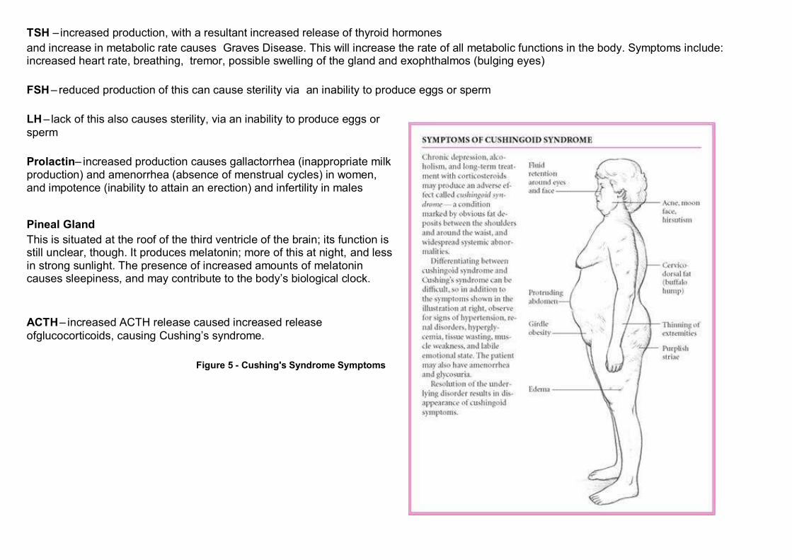
ACTH– increased ACTH release caused increased release
ofglucocorticoids, causing Cushing’s syndrome.
Figure 5 - Cushing's Syndrome Symptoms
ADH– Reduced ADH output causes diabetes insipidus
Disorders:
Seasonal Affective Disorder (SAD) – a type of depression arising during the winter months, because there is less light. Some broad
spectrum lights are thought to help this.
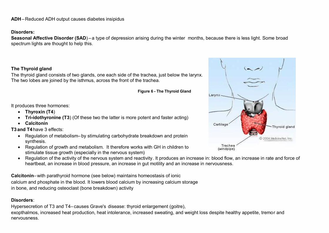
The Thyroid gland
The thyroid gland consists of two glands, one each side of the trachea, just below the larynx. The two lobes are joined by the isthmus, across the front of the trachea.
Figure 6 - The Thyroid Gland
It produces three hormones:
Thyroxin (T4)
Tri-idothyronine (T3) (Of these two the latter is more potent and faster acting)
Calcitonin
T3 and T4 have 3 effects:
Regulation of metabolism–by stimulating carbohydrate breakdown and protein synthesis.
Regulation of growth and metabolism. It therefore works with GH in children to stimulate tissue growth (especially in the nervous system)
Regulation of the activity of the nervous system and reactivity. It produces an increase in: blood flow, an increase in rate and force of heartbeat, an increase in blood pressure, an increase in gut motility and an increase in nervousness.
Calcitonin– with parathyroid hormone (see below) maintains homeostasis of ionic
calcium and phosphate in the blood. It lowers blood calcium by increasing calcium storage
in bone, and reducing osteoclast (bone breakdown) activity
Disorders:
Hypersecretion of T3 and T4– causes Grave's disease: thyroid enlargement (goitre),
exopthalmos, increased heat production, heat intolerance, increased sweating, and weight loss despite healthy appetite, tremor and nervousness.
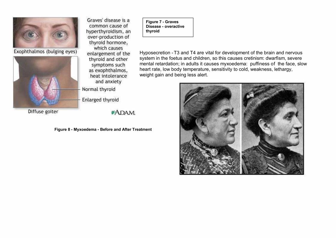
Hyposecretion -T3 and T4 are vital for development of the brain and nervous system in the foetus and children, so this causes cretinism: dwarfism, severe mental retardation; in adults it causes myxoedema: puffiness of the face, slow heart rate, low body temperature, sensitivity to cold, weakness, lethargy, weight gain and being less alert.
Figure 8 - Myxoedema - Before and After Treatment
Figure 7 - Graves Disease - overactive thyroid

Parathyroid glands
There are 4 parathyroids, 2 on each side on the posterior surface of the thyroid
gland.
They produce parathyroid hormone (aka PTH or parathormone). This increases the levels of calcium and phosphate in the blood by:
Increasing their absorption from the gut
Releasing them from bone (i.e. from storage)
Stimulating the reabsorption of calcium and phosphate in the kidney
Disorders:
Hyposecretion – causes muscles twitches and spasms, leading to tetany.
Hypersecretion – results in osteitis fibrosa cystica, from demineralisation and causes them to become brittle.
Thymus Gland
This is a bi-lobed lymphatic organ situated in the mediastinum (behind the sternum,
between the lungs). It is particularly large in infants; it reduces significantly in size by adulthood:
Strictly speaking it is not an endocrine gland, it is involved in the immune system, but releases hormones as well; these hormones are mainly to proliferate and help mature T cells, but evidence exists that they also help retard the aging process.
Suprarenal (adrenal) glands
There are two suprarenal glands, one top of each kidney. Each gland has two
parts:
Adrenal cortex (outer)
Adrenal medulla (inner)
Figure 10 - Adrenal (Suprarenal) Gland
Figure 9 - Position of Parathyroid
Adrenal cortex
This produces:
Aldosterone – this acts via the kidney to increase the amount of sodium in the blood by reabsorbing it from the urine (increasing the salt
content of the blood causes a consequent reabsorption of water [to dilute it], hence increasing blood volume and blood pressure).
Glucocorticoids (steroids)
These stimulate the conversion of fat and protein to glucose (gluconeogenesis)
They help the body cope with stress; via gluconeogenesis and making us more alert, thus helping us cope with stressful situations
They are anti-inflammatory – but at the same time they slow healing and reduce the body’s ability to fight disease.
Adrenal medulla
This produces adrenalin (mainly), which is a sympathomimetic, i.e. it has the same actions as the sympathetic nervous system:
Increases blood pressure (via increased heart rate and vasoconstriction)
Increased rate of respiration
Dilates the bronchioles
Increases the efficiency of muscle contraction
Increases blood glucose level
Increases cell metabolism
Increases sphincter tone
Decreases the rate of digestion
It can be seen from these that the general effect is to prepare the body for the ‘fight or flight’ response.
Disorders:
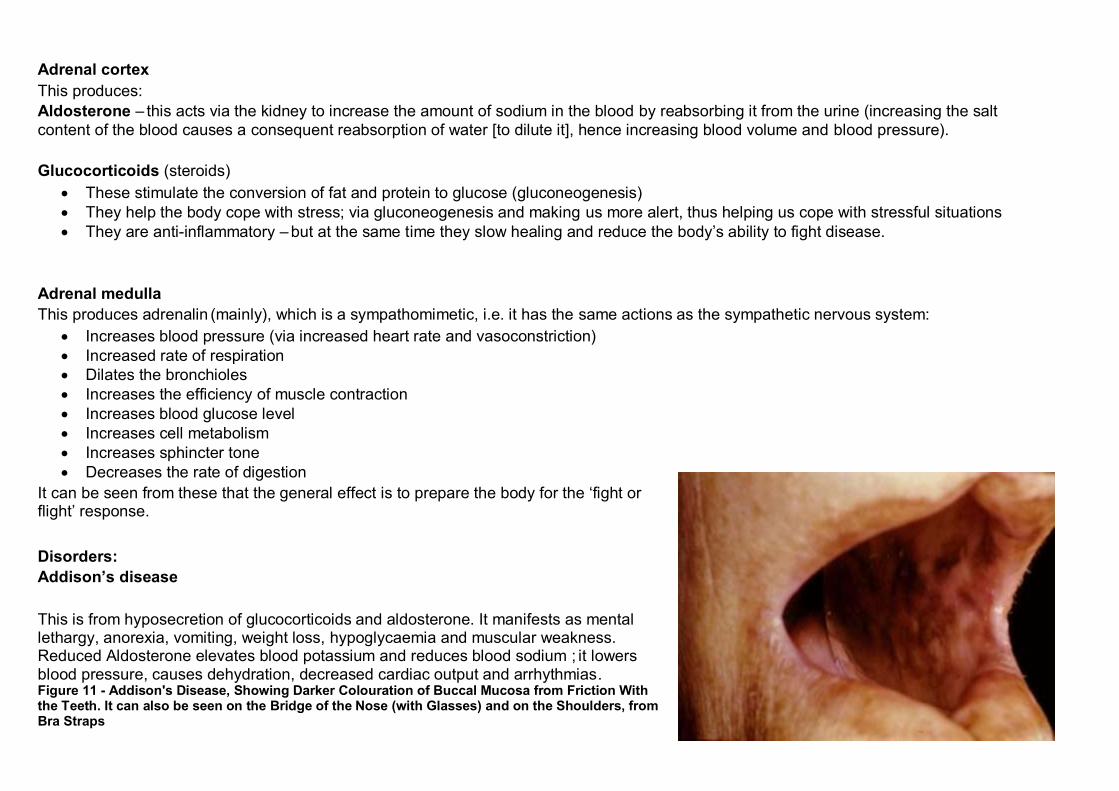
Addison’s disease
This is from hyposecretion of glucocorticoids and aldosterone. It manifests as mental lethargy, anorexia, vomiting, weight loss, hypoglycaemia and muscular weakness. Reduced Aldosterone elevates blood potassium and reduces blood sodium ; it lowers blood pressure, causes dehydration, decreased cardiac output and arrhythmias. Figure 11 - Addison's Disease, Showing Darker Colouration of Buccal Mucosa from Friction With the Teeth. It can also be seen on the Bridge of the Nose (with Glasses) and on the Shoulders, from Bra Straps
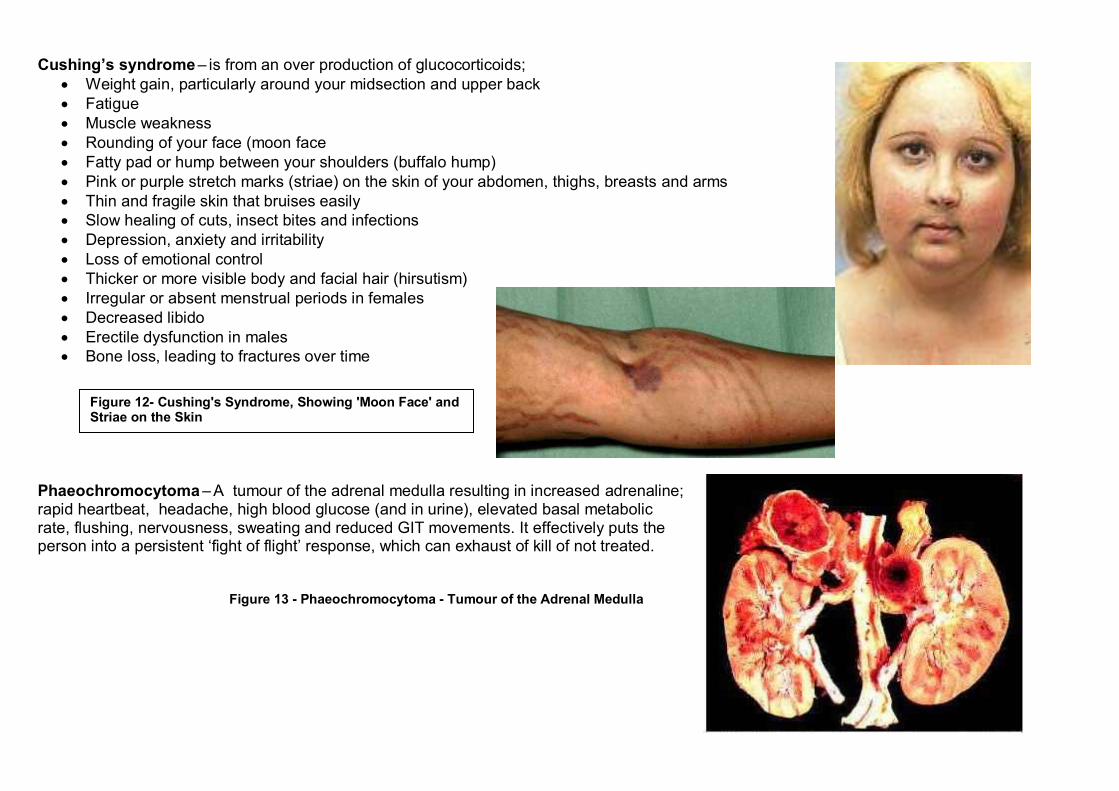
Cushing’s syndrome – is from an over production of glucocorticoids;
Weight gain, particularly around your midsection and upper back
Fatigue
Muscle weakness
Rounding of your face (moon face
Fatty pad or hump between your shoulders (buffalo hump)
Pink or purple stretch marks (striae) on the skin of your abdomen, thighs, breasts and arms
Thin and fragile skin that bruises easily
Slow healing of cuts, insect bites and infections
Depression, anxiety and irritability
Loss of emotional control
Thicker or more visible body and facial hair (hirsutism)
Irregular or absent menstrual periods in females
Decreased libido
Erectile dysfunction in males
Bone loss, leading to fractures over time
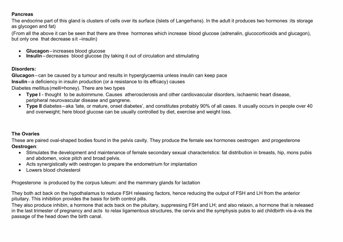
Phaeochromocytoma – A tumour of the adrenal medulla resulting in increased adrenaline; rapid heartbeat, headache, high blood glucose (and in urine), elevated basal metabolic rate, flushing, nervousness, sweating and reduced GIT movements. It effectively puts the person into a persistent ‘fight of flight’ response, which can exhaust of kill of not treated. Figure 13 - Phaeochromocytoma - Tumour of the Adrenal Medulla
Figure 12- Cushing's Syndrome, Showing 'Moon Face' and Striae on the Skin
Pancreas
The endocrine part of this gland is clusters of cells over its surface (Islets of Langerhans). In the adult it produces two hormones :its storage as glycogen and fat)
(From all the above it can be seen that there are three hormones which increase blood glucose (adrenalin, glucocorticoids and glucagon), but only one that decrease s it –insulin)
Glucagon– increases blood glucose Insulin– decreases blood glucose (by taking it out of circulation and stimulating
Disorders:
Glucagon– can be caused by a tumour and results in hyperglycaemia unless insulin can keep pace
Insulin– a deficiency in insulin production (or a resistance to its efficacy) causes
Diabetes mellitus (melli=honey). There are two types
Type I - thought to be autoimmune. Causes atherosclerosis and other cardiovascular disorders, ischaemic heart disease,
peripheral neurovascular disease and gangrene.
Type II diabetes –aka ‘late, or mature, onset diabetes’, and constitutes probably 90% of all cases. It usually occurs in people over 40
and overweight; here blood glucose can be usually controlled by diet, exercise and weight loss.
The Ovaries
These are paired oval-shaped bodies found in the pelvis cavity. They produce the female sex hormones oestrogen and progesterone
Oestrogen:
Stimulates the development and maintenance of female secondary sexual characteristics: fat distribution in breasts, hip, mons pubis and abdomen, voice pitch and broad pelvis.
Acts synergistically with oestrogen to prepare the endometrium for implantation
Lowers blood cholesterol
Progesterone is produced by the corpus luteum: and the mammary glands for lactation They both act back on the hypothalamus to reduce FSH releasing factors, hence reducing the output of FSH and LH from the anterior pituitary. This inhibition provides the basis for birth control pills.
They also produce inhibin, a hormone that acts back on the pituitary, suppressing FSH and LH; and also relaxin, a hormone that is released in the last trimester of pregnancy and acts to relax ligamentous structures, the cervix and the symphysis pubis to aid childbirth vis-à-vis the passage of the head down the birth canal.
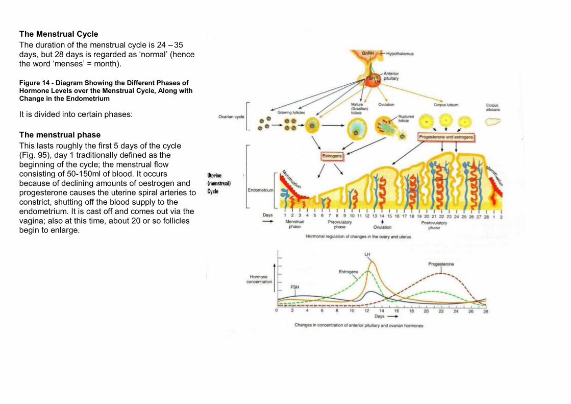
The Menstrual Cycle
The duration of the menstrual cycle is 24 – 35 days, but 28 days is regarded as ‘normal’ (hence the word ‘menses’ = month).
Figure 14 - Diagram Showing the Different Phases of Hormone Levels over the Menstrual Cycle, Along with Change in the Endometrium
It is divided into certain phases:
The menstrual phase
This lasts roughly the first 5 days of the cycle (Fig. 95), day 1 traditionally defined as the beginning of the cycle; the menstrual flow consisting of 50-150ml of blood. It occurs because of declining amounts of oestrogen and progesterone causes the uterine spiral arteries to constrict, shutting off the blood supply to the endometrium. It is cast off and comes out via the vagina; also at this time, about 20 or so follicles begin to enlarge.
Preovulatory phase
This occurs between menstruation and ovulation; it lasts between days 6 – 13. The 20 or so follicles continue to grow and begin to secrete oestrogen and inhibin. By about day 6 one follicle has outgrown all the others and becomes the dominant one; the oestrogen and inhibin suppress the release of FSH, so the other follicles stop developing and undergo atresia. The developing follicle now becomes mature (Graafian follicle) and increases its oestrogen production under the influence of LH, and at maturity is about 2cm in diameter.
Oestrogens are the primary ovarian hormones before ovulation; the menstrual and
Preovulatory phases are called the follicular phase.
The oestrogens produced by the follicle pass into the blood and stimulate the repair of the endometrium; its thickness doubling to 4 - 6mm, hence is called the proliferative phase of the uterus.
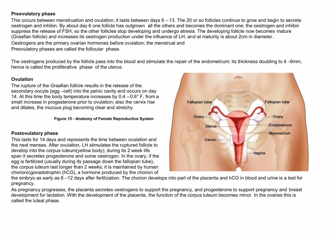
Ovulation
The rupture of the Graafian follicle results in the release of the secondary oocyte (egg –cell) into the pelvic cavity and occurs on day 14. At this time the body temperature increases by 0.4 –0.6° F, from a small increase in progesterone prior to ovulation; also the cervix rise and dilates, the mucous plug becoming clear and stretchy.
Figure 15 - Anatomy of Female Reproductive System
Postovulatory phase
This lasts for 14 days and represents the time between ovulation and the next menses. After ovulation, LH stimulates the ruptured follicle to develop into the corpus luteum(yellow body); during its 2 week life span it secretes progesterone and some oestrogen. In the ovary, if the egg is fertilized (usually during its passage down the fallopian tube), the corpus luteum last longer than 2 weeks; it is maintained by human chorionicgonadotrophin (hCG), a hormone produced by the chorion of the embryo as early as 8 –12 days after fertilization. The chorion develops into part of the placenta and hCG in blood and urine is a test for pregnancy.
As pregnancy progresses, the placenta secretes oestrogens to support the pregnancy, and progesterone to support pregnancy and breast development for lactation. With the development of the placenta, the function of the corpus luteum becomes minor. In the ovaries this is called the luteal phase.
If hCG does not rescue the corpus luteum, it degenerates into a scar called the corpus albicans (white body); the consequent lack of oestrogen and progesterone then causes menstruation. The reduction of oestrogen and progesterone and inhibin promote the release of releasing factors for FSH and the cycle starts all over again.
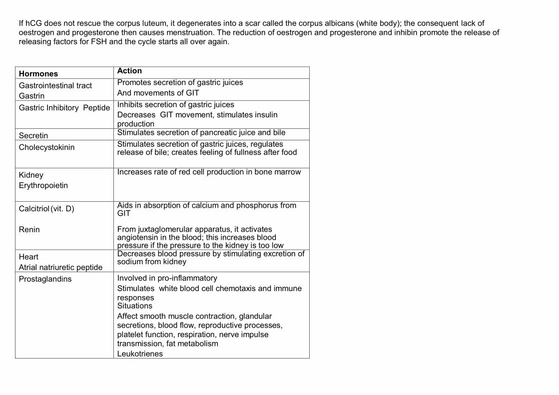
Hormones Action
Gastrointestinal tract
Gastrin
Promotes secretion of gastric juices
And movements of GIT
Gastric Inhibitory Peptide Inhibits secretion of gastric juices
Decreases GIT movement, stimulates insulin production
Secretin Stimulates secretion of pancreatic juice and bile
Cholecystokinin Stimulates secretion of gastric juices, regulates release of bile; creates feeling of fullness after food
Kidney
Erythropoietin
Increases rate of red cell production in bone marrow
Calcitriol (vit. D)
Renin
Aids in absorption of calcium and phosphorus from GIT From juxtaglomerular apparatus, it activates angiotensin in the blood; this increases blood pressure if the pressure to the kidney is too low
Heart
Atrial natriuretic peptide
Decreases blood pressure by stimulating excretion of sodium from kidney
Prostaglandins Involved in pro-inflammatory
Stimulates white blood cell chemotaxis and immune responses Situations
Affect smooth muscle contraction, glandular secretions, blood flow, reproductive processes, platelet function, respiration, nerve impulse transmission, fat metabolism
Leukotrienes
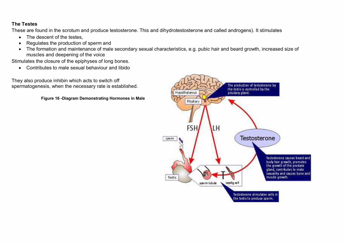
The Testes
These are found in the scrotum and produce testosterone. This and dihydrotestosterone and called androgens). It stimulates
The descent of the testes,
Regulates the production of sperm and
The formation and maintenance of male secondary sexual characteristics, e.g. pubic hair and beard growth, increased size of muscles and deepening of the voice
Stimulates the closure of the epiphyses of long bones.
Contributes to male sexual behaviour and libido
They also produce inhibin which acts to switch off spermatogenesis, when the necessary rate is established.
Figure 16 -Diagram Demonstrating Hormones in Male