fidelity of uracil-initiated base excision dna repair in escherichia
TRANSCRIPT
Fidelity of Uracil-Initiated Base Excision DNA Repair in Escherichia coli
Cell Extracts*
Jung-Suk Sung‡, Samuel E. Bennett‡, and Dale W. Mosbaugh‡§⁄⁄¶
‡Departments of Environmental and Molecular Toxicology, and §Biochemistry and Biophysics, and
the ⁄⁄Environmental Health Science Center, Oregon State University, Corvallis, Oregon 97331-7301
Running title: Fidelity of Uracil-initiated Base Excision DNA Repair
Copyright 2000 by The American Society for Biochemistry and Molecular Biology, Inc.
JBC Papers in Press. Published on October 16, 2000 as Manuscript M008147200 by guest on M
arch 15, 2018http://w
ww
.jbc.org/D
ownloaded from
2
SUMMARY
The error frequency and mutational specificity associated with Escherichia coli uracil-initiated base
excision repair were measured using an M13mp2 lacZα DNA-based reversion assay. Repair was
detected in cell-free extracts utilizing a form I DNA substrate containing a site-specific uracil residue.
The rate and extent of complete uracil-DNA repair were measured using uracil-DNA glycosylase
(Ung) or double-strand uracil-DNA glycosylase (Dug) proficient and deficient isogenic E. coli cells.
In reactions utilizing E. coli NR8051 (ung+ dug+), approximately 80 % of the uracil-DNA was
repaired, whereas about 20 % repair was observed using NR8052 (ung- dug+) cells. The Ung-deficient
reaction was insensitive to inhibition by the PBS2 uracil-DNA glycosylase inhibitor protein, implying
the involvement of Dug activity. Under both conditions, repaired form I DNA accumulated in
conjunction with limited DNA synthesis associated with a repair patch size of 1-20 nucleotides.
Reactions conducted with E. coli BH156 (ung- dug+), BH157 (ung+ dug-), and BH158 (ung- dug-) cells
provided direct evidence for the involvement of Dug in uracil-DNA repair. The rate of repair was 5-
fold greater in the Ung-proficient than in the Ung-deficient reactions, while repair was not detected in
reactions deficient in both Ung and Dug. The base substitution reversion frequency associated with
uracil-DNA repair was determined to be ~5.5 x 10⁻⁴ with transversion mutations dominating the
mutational spectrum. In the presence of Dug, inactivation of Ung resulted in up to a 7.3-fold increase
in mutation frequency without a dramatic change in mutational specificity.
by guest on March 15, 2018
http://ww
w.jbc.org/
Dow
nloaded from
3
INTRODUCTION
Uracil-mediated base excision DNA repair serves as a strategic cellular defense mechanism to
maintain the genetic stability of the Escherichia coli genome (1). This BER1 process protects the DNA
from premutagenic U•G2 mispairs formed by cytosine deamination and U•A base pairs produced by
incorporation of dUMP during DNA synthesis (2, 3). BER is initiated when uracil-DNA glycosylase
recognizes a uracil residue in DNA and catalyzes the cleavage of the N-glycosylic bond that links the
uracil base to the deoxyribose phosphate DNA backbone (4). This hydrolytic reaction results in the
release of free uracil and creates an abasic site in the DNA (4). Incision by a class II AP endonuclease,
either the endonuclease IV (Nfo) (5) or exonuclease III (Xth) (6), cleaves the phosphodiester bond on
the 5'-side of the AP-site to generate a terminal 3'-hydroxyl-containing nucleotide and a deoxyribose
5'-phosphate residue (7). Approximately 90% of the AP endonuclease activity detected in E. coli is
accounted for by the Xth protein (8). Removal of the dRP moiety prior to gap-filling DNA synthesis
can occur by the deoxyribophosphodiesterase (dRPase) activity of RecJ, which also possesses a 5' to 3'
single-stranded exonuclease (9, 10). In addition, the product of the mutM gene (Fpg) has been
reported to catalyze the 5' to 3' removal of incised dRP residues by a β-elimination mechanism (11).
Furthermore, exonuclease I (SbcB) has been shown to release dRP from an AP-site incised by Nfo and
also to remove 4-hydroxy-2-pentenal-5-phosphate from an abasic site incised by AP lyase on the 3’-
side of the lesion (12). Collectively, RecJ, Fpg, and SbcB may be responsible for dRP removal in E.
coli, since inactivation of all three dRP excision activities produced a lethal phenotype (12). In
association with the dRP excision step, one or more nucleotides may be removed from the uracil-
by guest on March 15, 2018
http://ww
w.jbc.org/
Dow
nloaded from
4
containing DNA strand by a 5’ to 3’ exonuclease activity. However, appreciable degradation of the
incision site in the 3’ to 5’ direction does not seem to occur (13). Gap-filling DNA synthesis replaces
the excised nucleotide(s) by the action of a DNA polymerase (Pol) and DNA ligase (Lig) re-establishes
the continuity of the phosphodiester backbone (10).
In E. coli, two genetically distinct forms of uracil-DNA glycosylase have been purified to apparent
homogeneity, characterized, and demonstrated to initiate uracil-mediated BER (4, 14-17). E. coli
uracil-DNA glycosylase (Ung) was the first DNA glycosylase identified and consists of a
monofunctional single polypeptide with a molecular weight of 25,558 daltons (18, 19). Ung prefers to
act on uracil residues located in single-stranded DNA but also recognizes uracil in duplex DNA with
moderately reduced efficiency (20). A second protein, referred to as double-stranded uracil-DNA
glycosylase (Dug, also termed Mug), was recently purified as a 18,672 dalton polypeptide and
characterized (14, 16). Based on amino acid sequence alignment, Dug lacks strong sequence identity
(~10 %) with Ung but shares remarkable similarity in tertiary structure (17). Dug preferentially
removes uracil from DNA containing a U•G or U•T mispair but inefficiently recognizes a U•A
basepair target, and lacks detectable activity on single-stranded uracil-containing DNA (14, 16).
The activity of Ung can also be differentiated from that of Dug based on several other biochemical
properties: (i) Ung activity is inhibited by the PBS1 and 2 uracil-DNA glycosylase inhibitor (Ugi)
protein which forms an essentially irreversible Ung•Ugi complex (21), whereas Dug activity is
insensitive to Ugi (14, 17, 22); (ii) purified Dug is a relatively inefficient enzyme that exhibits a
significantly lower uracil-excision turnover number than Ung (4, 14, 17); (iii) Dug is strongly inhibited
by guest on March 15, 2018
http://ww
w.jbc.org/
Dow
nloaded from
5
by apyrimidinic sites but not by free uracil, whereas Ung exhibits modest inhibition by both reaction
end products (14); and (iv) Dug efficiently cleaves 3,N4-ethenocytosine from duplex DNA, while this
exocyclic DNA adduct remains refractory to removal by Ung (14, 16). The involvement of Ung in
uracil-DNA repair has been well-documented (1, 13, 23); however, the role of Dug remains to be fully
elucidated. Sung and Mosbaugh (14) recently demonstrated the involvement of Dug in BER using an
E. coli NR8052 (ung) cell-free extract and an M13mp2 form I DNA substrate containing a site-specific
U•G mispair. Uracil-initiated BER conducted in the absence of Ung was shown to be both insensitive
to Ugi and stimulated by the addition of exogenous Dug (14). Thus, while it has been established that
E. coli possesses two classes of uracil-DNA glycosylase capable of initiating BER, the relative
contribution of Ung and Dug in mediating repair remains to be determined.
Studies of the repair patch size associated with gap-filling DNA synthesis in conjunction with
uracil-initiated BER have been conducted using both E. coli cell-free extracts and reconstituted
enzyme systems (13, 23, 24). Two types of repair patch size have been described involving either the
incorporation of a single nucleotide (short patch) or 2-19 nucleotides (long patch). Dianov et al. (13)
reported that BER initiated at a U•G target site contained in an oligonucleotide (30-mer) DNA
substrate was mainly repaired by a short patch process in wild-type E. coli NH5033 cell extracts.
Similar results were obtained using the same DNA substrate and a reconstituted enzyme system
composed of Ung, Nfo, RecJ, Pol I, and Lig (24). In these experiments, short patch BER was largely
dependent on the presence of RecJ (24). In sharp contrast, Sandigursky et al. (23) reported that E. coli
AB1157 cell extracts did not efficiently support short patch repair when a closed circular DNA
by guest on March 15, 2018
http://ww
w.jbc.org/
Dow
nloaded from
6
containing a U•G mispair was used as substrate. In this case, long patch BER was observed that was
not dependent on either SbcB and RecJ or Fpg and RecJ (23). Given the apparent disparity between
these two reports, further investigation into the BER patch size is warranted.
The biochemical steps involved in E. coli uracil-initiated BER have been elucidated in substantial
detail. However, an assessment of the fidelity of DNA repair synthesis associated with the completed
BER process initiated by either E. coli Ung or Dug has not been investigated. In the present study we
have (i) used a site-specific uracil-containing circular duplex DNA substrate to monitor the relative
rate of Ung- and Dug-initiated BER in E. coli cell extracts; (ii) determined the repair patch size
associated with BER initiated by the two uracil-DNA glycosylases; (iii) measured the base substitution
error frequency produced during BER; and (iv) assessed the error specificity associated with Ung- and
Dug-mediated BER.
EXPERIMENTAL PROCEDURES
Materials−PCR primers, GTGTGGAATTGTGAGCGG (FP-18-mer) and
CGTGCATCTGCCAGTTTG (RP-18-mer), and sequencing primer,
GCACTCCAGCCAGCTTTCCGG (S-21-mer) were obtained from Research Genetics, Inc. Two
oligodeoxynucleotides, CCCAGTCACGTCUTTGTAAAACG (U-23-mer) and
CCCAGTCACGTCATTGTAAAACG (A-23-mer) were synthesized and purified by Oligos Etc; 5'-
end-phosphorylated oligodeoxynucleotides were prepared as described previously (25).
by guest on March 15, 2018
http://ww
w.jbc.org/
Dow
nloaded from
7
E. coli strains NR8051 and NR8052, NR9162, and CSH50 were provided by T. A. Kunkel
(NIEHS, National Institute of Health), and E. coli BH156, BH157, and BH158 were obtained from A.
S. Bhagwat (Wayne State University). E. coli JM109 was procured from New England Biolabs. P1
lysate containing mutS::Tn-10 was obtained from J. Hays (Oregon State University) and E. coli mutS-
strains, NR80511 and NR80521 were constructed by the P1 transduction of mutS::Tn-10 into E. coli
NR8051 and NR8052, respectively.
E. coli uracil-DNA glycosylase (fraction V) and Ugi (fraction IV) were purified as described by
Sanderson and Mosbaugh (26). Double-strand uracil-DNA glycosylase (fraction VI) was isolated as
described by Sung and Mosbaugh (14). E. coli endonuclease IV (fraction V) was provided by B.
Demple (Harvard University) and T4 DNA polymerase was purified as previously described (25). T4
polynucleotide kinase, restriction endonuclease HinfI, and T4 DNA ligase were purchased from New
England Biolabs. Proteinase K and creatine phosphokinase (type I, rabbit) were from Sigma, and
ribonuclease A was from Worthington Biochemicals.
Preparation of Base Excision Repair DNA Substrates−M13mp2op14 DNA that contained an
opal codon located at nucleotide position 78-80 of lacZα gene was constructed and isolated as
described previously by Sanderson and Mosbaugh (25), with minor modifications. Briefly,
M13mp2op14 phage were propagated in E. coli JM109 cells, and single-stranded M13mp2op14 DNA
was purified using the CTAB DNA precipitation method (27). Single-stranded M13mp2op14 DNA
was then annealed to 5'-end phosphorylated oligodeoxynucleotides U-23-mer or A-23-mer, and the
primed template DNA was subjected to a primer extension reaction. Primer extension reaction
by guest on March 15, 2018
http://ww
w.jbc.org/
Dow
nloaded from
8
mixtures (3030 µl) contained 20 mM Hepes-KOH (pH 7.4), 2 mM dithiothreitol, 10 mM MgCl2, 1 mM
ATP, 500 µM each of dATP, dTTP, dGTP, and dCTP, 400 units of T4 DNA polymerase, 40,000 units
of T4 DNA ligase, and 200 pmol of primed template DNA containing either U•T mispair or A•T base
pair were incubated for 4 h at 37 °C. Covalently closed circular duplex DNA reaction products were
isolated by ethidium bromide cesium chloride gradient centrifugation, as previously described (25).
Form I DNA was isolated, extracted four times with an equal volume of 1-butanol saturated with 5 M
NaCl, concentrated using a Centricon-30 concentrator (Amicon), and buffer exchanged into TE buffer
(10 mM Tris-HCl pH 8.0, 1 mM EDTA). The isolated DNA3 was > 95% form I as determined by
0.8% agarose gel electrophoresis.
Preparation of E. coli Cell Free Extracts−E. coli cells grown at 37 °C in 500 ml of YT medium
(0.5% yeast extract, 0.8% tryptone and 0.5% NaCl) to mid-log phase were harvested by centrifugation
at 7,000 x g for 15 min at 4 °C. The cell pellet was resuspended in 10 ml of sonification buffer
containing 50 mM Tris-HCl (pH 8.0), 1 mM EDTA, and 0.1 mM dithiothreitol, and cells were lysed on
ice by sonification. After cell debris was removed by centrifugation at 20,000 x g for 20 min at 4 °C,
protein was precipitated from the supernatant by addition of 0.35 g of powdered ammonium sulfate per
ml and the precipitate recovered by centrifugation 20,000 x g for 20 min at 4 °C. The pellet was
resuspended in 5 ml of R-buffer containing 50 mM Tris-HCl (pH 7.5), 1 mM EDTA, 1 mM
dithiothreitol, 10% (w/v) glycerol, dialyzed against the same buffer, and the protein concentration of
the cell free extract was determined by the Bradford reaction (28) using Bio-Rad Protein Assay.
by guest on March 15, 2018
http://ww
w.jbc.org/
Dow
nloaded from
9
Base Excision DNA Repair Reaction−Standard BER reaction mixtures (100 µl) containing 100
mM Tris-HCl (pH 7.5), 5 mM MgCl2, 1 mM dithiothreitol, 0.1 mM EDTA, 2 mM ATP, 0.5 mM β-
NAD, 20 µM each of dATP, dTTP, dGTP, and dCTP, 5 mM phosphocreatine di-Tris salt, 200 units/ml
of phosphocreatine kinase, 10 µg/ml of M13mp2op14 (U•T) heteroduplex or (A•T) homoduplex DNA
(form I), and 1 mg/ml of E. coli cell free extract protein were prepared on ice. In some experiments,
400 µCi/ml of [α-32P]dATP (6000 Ci/mmol) was also included in the reaction mixture. Following
incubation at 30 °C in the presence or absence of 1000 units of Ugi, the reactions were terminated after
various times by adjustment to 20 mM EDTA and heated at 70 °C for 3 min. RNase A was then added
to 80 µg/ml, and the reaction mixtures were incubated at 37 °C for 10 min. Each reaction was then
adjusted to 0.5% SDS, proteinase K was added to 190 µg/ml, and incubated for 30 min at 37 °C. The
M13mp2op14 DNA was then isolated and resuspended in 20 µl of TE buffer as described previously
(25).
Analysis of Base Excision DNA Repair Reaction Product−DNA samples (5 µl), isolated as
described above, were removed and treated with 100 units of Ung for 30 min at 37 °C. After
quenching the reaction by adding 1000 units of Ugi, samples were further incubated with 1 unit of Nfo
for 30 min at 37 °C. Nfo was inactivated by heating at 70 °C for 3 min, and form I DNA that was
resistant to Ung/Nfo cleavage was resolved from form II DNA by 0.8% agarose gel electrophoresis
(25). The amount of form I and II DNA was determined by comparing the fluorescence at 300 nm of
the ethidium bromide-stained DNA reaction products with that of co-electrophoresced form I and II
DNA (6.25-100 ng) standards. The fluorescence intensity data was captured using a gel
by guest on March 15, 2018
http://ww
w.jbc.org/
Dow
nloaded from
10
documentation system (Ultra Violet Products Ltd.) and quantified with the ImageQuant computer
program (Molecular Dynamics). After correcting for ~0.7-fold reduced staining intensity of form I
DNA relative to form II DNA, the percentage of form I DNA was calculated by dividing the amount of
form I (ng) by that of form I plus form II DNA and multiplying by 100.
Analysis of Base Excision Repair DNA Synthesis−Standard BER reactions were conducted in
the presence of 400 µCi/ml of [α-32P]dATP, and DNA reaction products were isolated, treated with
Ung/Nfo, and resolved by 0.8% agarose gel electrophoresis as described above. The [32P]DNA was
then transferred from the agarose gel to a Gene Screen Plus (NEN Life Science Products) membrane as
described by Koetsier et al. (29). The membrane was dried and used to expose a phosphor screen; 32P-
labeled DNA bands were visualized by PhosphorImager using the Scanner Control program
(Molecular Dynamics). Form I [32P]DNA (~100 ng) isolated from the BER reactions was treated with
excess HinfI restriction endonuclease, and restriction fragments were resolved by 5% nondenaturing
polyacrylamide gel electrophoresis (25). After drying the gel, DNA bands were visualized by
PhosphorImager, and the relative intensity of various [32P]DNA bands was quantified using the
ImageQuant computer program.
Isolation of Repaired Form I DNA−Form I DNA bands was isolated from agarose gel slices by
electroelution using an Elutrap apparatus (Schleicher and Schuell). Electroelution was conducted for 3
h at 150 V in TAE buffer (40 mM Tris acetate, 1 mM EDTA (pH 8.0)), the recovered form I DNA was
concentrated using a Centricon 30 concentrator (Amicon) and buffer-exchanged into distilled water.
by guest on March 15, 2018
http://ww
w.jbc.org/
Dow
nloaded from
11
Determination of Repair Patch Size−Standard BER reaction mixtures were prepared as
described above except that a 2'-deoxyribonucleoside α-thioltriphosphate was used in place of each of
the four 2-deoxyribonucleoside triphosphates and 32P labeled M13mp2op14 (U•T) form I DNA was
used as the BER substrate. The [32P]DNA substrate was constructed as previously described (30, 31)
and contained a 32P radiolabel located 13 nucleotides upstream of the target uracil and 7 nucleotides
downstream of the SmaI restriction site on the transcribed strand of the lacZα gene sequence.
Following the BER reactions, DNA products were isolated and resuspended in 20 µl of TE buffer as
described above. Samples (4 µl, ~200 ng) were removed for digestion with 10 units of EcoRI for 1 h
at 25°C. The reaction was terminated at 70°C for 10 min and samples were incubated in the absence or
the presence of various amounts of E. coli exonuclease III (Xth) for 1 h at 37°C. After incubation,
each sample was heated at 70°C for 10 min and the [32P]DNA was then cleaved with 10 units of SmaI
for 1 h at 25°C. An equal volume of denaturing formamide dye buffer was added and [32P]DNA
products were resolved by 12% polyacrylamide/8.3 M urea gel electrophoresis (25). After drying the
gel under vacuum, autoradiography was performed and the amount of 32P radioactivity associated with
each band was quantified using a PhosphorImager.
Transfection of E. coli and Determination of Reversion Frequencies−E. coli NR9162 (mutS)
cells were transfected with repaired M13mp2op14 DNA (form I) as previously described (25). Briefly,
the transfected cells were diluted into SOB medium, combined with E. coli CSH50 (indicator strain)
and top agar containing 0.4 mM β-D-thiogalactopyranoside and 1 mg/ml of 5-bromo-4-chloro-3-
indolyl β-D-galactopyranoside, and the mixture was plated on M9 plates. After scoring the phage
by guest on March 15, 2018
http://ww
w.jbc.org/
Dow
nloaded from

12
plaques as either colorless or blue, the reversion frequency was calculated as the ratio of the number of
blue plaques to total plaques detected. Secondary screening of each blue plaque was then conducted to
purify phage in preparation for nucleotide sequence analysis (25).
Determination of Mutational Spectrum−An individual blue plaque was picked by touching a
P10 pipet tip (Pipetman) to the surface of the plaque so as to extract a mini-agarose plug approximately
0.3 mm long. This material was then injected into an Eppendorf microcentrifuge tube containing 100
µl of PCR reaction mixture (New England Biolabs) comprising 1 unit of Deep Vent DNA polymerase,
1 x Thermopol buffer including 2 mM MgCl2, 200 µM dNTPs (Pharmacia, Ultrapure), and 100 pmol
each of the lacZα forward primer (FP-18-mer) and reverse primer (RP-18-mer). PCR was carried out
in a Hybaid PCR Express Gradient thermocycler under active temperature control (150 µl of mineral
oil in the temperature reference tube) using the following cycling parameters: 94 °C (1 min), 30 cycles
of 94 °C (1 min), 45 °C (1 min), and 72 °C (3 min); and 72 °C (10 min). PCR products were purified
with a StrataPrep PCR Purification Kit (Stratagene). Approximately 70 µl of 30-70 µg/ml of double-
stranded DNA was obtained from a single purified M13 plaque. An aliquot of the purified DNA
product (~30 ng) together with 12 pmol of sequencing primer (S-21-mer) complimentary to the (+)
strand of M13mp2op14 lacZα gene was used for DNA sequence analysis conducted by the Center for
Gene Research and Biotechnology (Oregon State University).
by guest on March 15, 2018
http://ww
w.jbc.org/
Dow
nloaded from
13
RESULTS
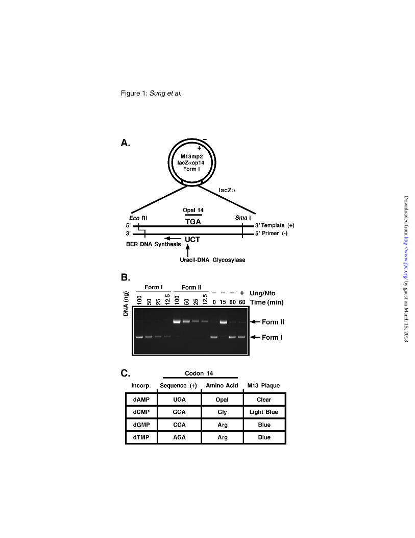
Uracil-initiated base excision DNA repair assay– An M13mp2 lacZα DNA-based reversion
assay was utilized to detect uracil-initiated DNA base excision repair and to determine the base
substitution error frequency associated with the completed repair reaction (25). Briefly, the arginine
codon 14 (CGT) of the lacZα gene, was replaced with an opal codon (TGA) by site-directed
mutagenesis. Heteroduplex DNA (form I) substrate was synthesized containing a U•T base mispair at
the first position of the opal codon 14 that served as the uracil target for repair; excision of the uracil
residue by uracil-DNA glycosylase initiated the BER pathway (Fig. 1A). Following BER in cell
extracts, the M13mp2op14 DNA was recovered and treated in vitro with excess E. coli Ung and Nfo to
convert uracil- or AP-site-containing form I DNA to form II molecules. This step effectively removed
unreacted and incompletely repaired substrates from the pool of fully repaired form I DNA molecules
which were resistant to the Ung/Nfo treatment. The progress of the uracil-initiated BER reaction was
monitored by product analysis using agarose gel electrophoresis and ethidium bromide staining (Fig.
1B). As expected, the M13mp2op14 DNA substrate was rapidly converted from form I to form II
DNA following uracil-excision and incision of the resulting apyrimidinic site. As the DNA repair
progressed, form II DNA was reconverted to form I DNA, which was resistant to the Ung/Nfo
treatment, in accordance with completed BER.
The uracil residue in the M13mp2op14 (U•T) DNA substrate was strategically located such that
faithful and unfaithful uracil-initiated DNA repair synthesis could be distinguished by the lacZα
complementation phenotype of the M13mp2 phage genome. If faithful DNA synthesis occurs during
by guest on March 15, 2018
http://ww
w.jbc.org/
Dow
nloaded from
14
BER opposite the template thymine residue at position 78, a dAMP nucleotide will be incorporated
into the (-) strand, and the opal codon will be re-established in both DNA strands. As the (-) strand
DNA serves as the template for production of single-stranded M13 DNA in E. coli, the resulting phage
will be defective in α-complementation, and the plaques they produce on the indicator strain will be
colorless when grown on medium containing IPTG and X-gal (Fig. 1C.). However, if BER DNA
synthesis is unfaithful, dCMP, dGMP, or dTMP may be misincorporated. Each of these base
substitutions restores (reverts to wild-type) the α-complementation phenotype and produces blue-
colored plaques.
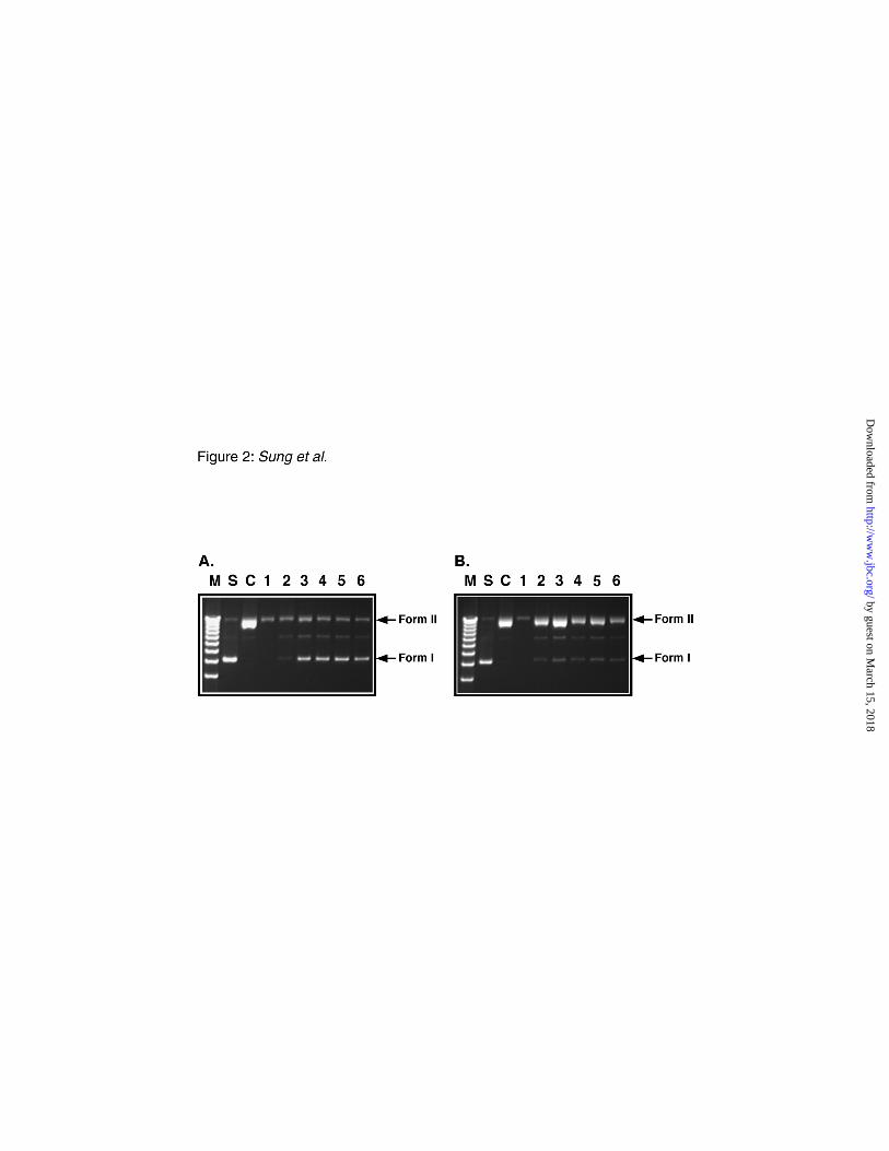
Detection of uracil-initiated BER in E. coli NR8051 (ung+) and NR8052 (ung-) cell
extracts–Initial experiments were conducted to detect uracil-initiated BER in E. coli extracts of Ung-
proficient (NR8051) and Ung-deficient (NR8052) cells. M13mp2op14 (U•T) DNA (form I) was
incubated with each cell-free extract for various times and then the reactions products were resolved by
agarose gel electrophoresis (Fig. 2). In both the Ung-proficient and Ung-deficient cell extracts, a time-
dependence appearance of repaired form I DNA was observed that indicated completed BER.
However, the rate of DNA repair, as determined from the percentage of Ung/Nfo-resistant form I
DNA, was much faster with the Ung-proficient strain (Fig. 2A) compared to the Ung-deficient strain
(Fig. 2B).
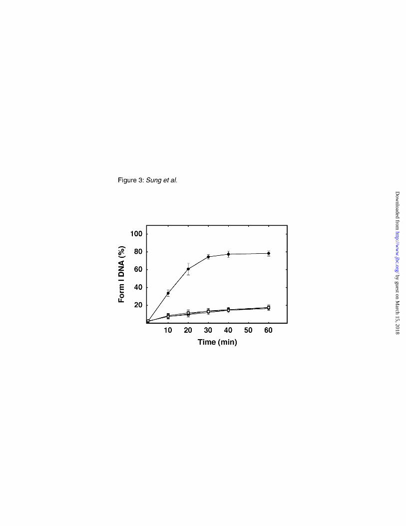
Impact of Ugi on uracil-initiated BER–To further examine the observation that uracil-initiated
BER occurred in Ung-deficient cell extracts, we utilized the Ugi protein to inactivate E. coli uracil-
DNA glycosylase. BER reactions were performed in extracts of E. coli NR8051 and NR8052 cells in
by guest on March 15, 2018
http://ww
w.jbc.org/
Dow
nloaded from
15
the absence or presence of excess Ugi. A time course of repair was conducted, and the amount of
repaired DNA (form I) detected relative to form II DNA was ascertained (Fig. 3). The results showed
quantitatively that during the first 20 min of the reaction, the initial rate of repair was ~5.5-fold slower
in E. coli NR8052 (ung-1) compared to the NR8051 cell extract. As expected, the addition of Ugi did
not appear to affect the rate or extent of repair in extracts of NR8052 (ung-1). However, uracil-
initiated BER was substantially diminished in extracts of the Ung-proficient strain NR8051 when
supplemented with Ugi. The level of repaired DNA, detected after 60 min, in the Ugi-supplemented
Ung-proficient cell extract was comparable to that observed in both the Ung-deficient and Ugi-
supplemented Ung-deficient cell extracts.
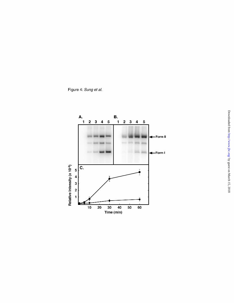
Evidence for uracil-mediated BER DNA synthesis–To determine if uracil-initiated BER DNA
synthesis was involved in the production of Ung/Nfo-resistant form I DNA, standard BER reactions
were conducted in the presence of [α-32P]dATP. After agarose gel electrophoresis of the BER reaction
products, examination of the PhosphoImages showed the incorporation of [32P]dAMP into the
Ung/Nfo-resistant form I DNA recovered from reactions containing cell extracts of E. coli NR8051
(ung+) (Fig. 4A) as well as NR8052 (ung-) (Fig. 4B). Notably, the intensity of form I [32P]DNA
generated in the Ung-proficient cell extract was considerably greater than that produced in the Ung-
deficient extract, although the amount of 32P radioactivity incorporated into the total DNA (form I +
form II) in each reaction was similar. Quantification of the 32P radioactivity associated with the form I
DNA band revealed that the amount of DNA synthesis generated in Ung-deficient cell extracts was
~10-fold less than that produced in Ung-proficient extracts after 60 min (Fig. 4C). When taken
by guest on March 15, 2018
http://ww
w.jbc.org/
Dow
nloaded from
16
together with the findings in Figure 3, the results suggest that the reduced level of DNA synthesis was
the likely result of an overall reduction in the level of BER.
Specificity of uracil-mediated DNA repair synthesis in E. coli Ung-proficient cell extracts– To
determine the distribution of [32P]dAMP incorporation associated with the repaired DNA molecules,
[32P]DNA isolated from the BER reactions described in Figure 4 was subjected to HinfI restriction
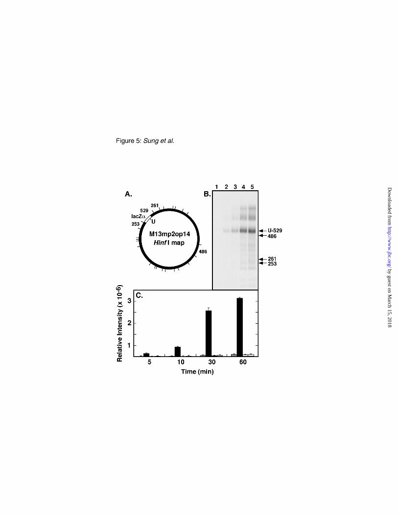
endonuclease digestion and resolved by non-denaturing polyacrylamide gel electrophoresis (Fig. 5).
While there are 26 HinfI recognition sites in the M13mp2op14 DNA sequence, HinfI digestion
produces but 16 DNA fragments in excess of 200 bp. Among these, the 529 bp fragment containing
the uracil site is bordered by 262 bp and 253 bp fragments positioned 5' and 3', respectively (Fig. 5A).
Following electrophoresis, inspection of the PhosphoImage showed that [32P]dAMP incorporation
occurred preferentially in the 529 bp fragment in a time-dependent manner (Fig. 5B and C). The minor
amount of incorporation detected on the neighboring fragments (262- and 253-bp) was assumed to
correspond to non-specific background, since a similar level of incorporation was observed associated
with a 486 bp fragment located on the opposite side of the M13mp2op14 DNA molecule (Fig. 5C).
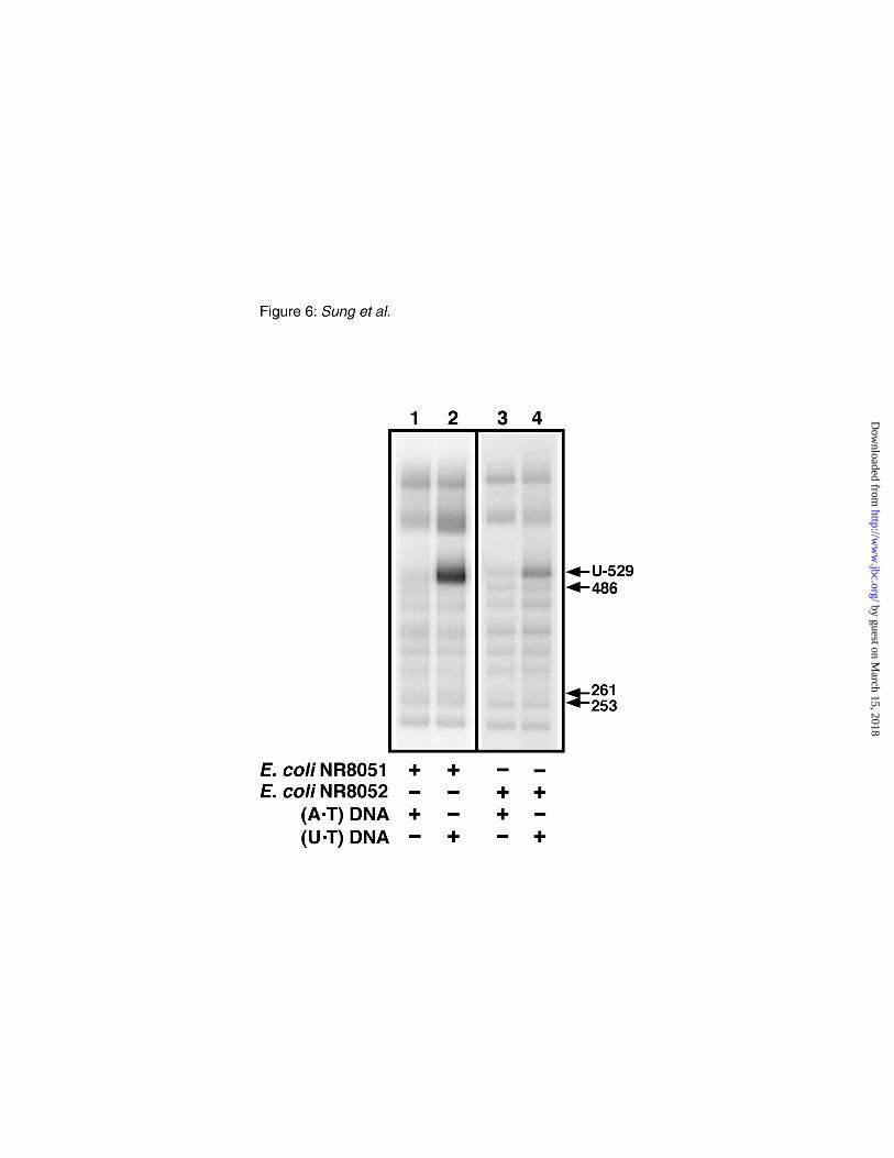
Uracil-dependent BER DNA synthesis–To determine whether the preferential incorporation of
[32P]dAMP into the 529 bp fragment observed in Figure 5 was the result of BER DNA repair synthesis
instigated by uracil excision, standard BER reactions were conducted as before, but the M13mp2op14
DNA substrate contained either a U•T or A•T base pair at the first position of the lacZα opal codon 14.
After incubation for 1 h the reaction products were recovered, and HinfI [32P]DNA fragments were
resolved by polyacrylamide gel electrophoresis (Fig. 6). Inspection of the PhosphoImage showed that
by guest on March 15, 2018
http://ww
w.jbc.org/
Dow
nloaded from
17
preferential incorporation of [32P]dAMP into the 529 bp fragment of the (U•T) DNA occurred in both
the E. coli NR8051 and NR8052 cell extracts relative to [32P]dAMP incorporation into the same 529 bp
fragment of the (A•T) DNA. The results indicated that a uracil residue located in the target DNA
fragment stimulated DNA synthesis by 17.2- and 5.8-fold above background for the reactions
containing E. coli NR8051 and NR8052 cell extracts, respectively.
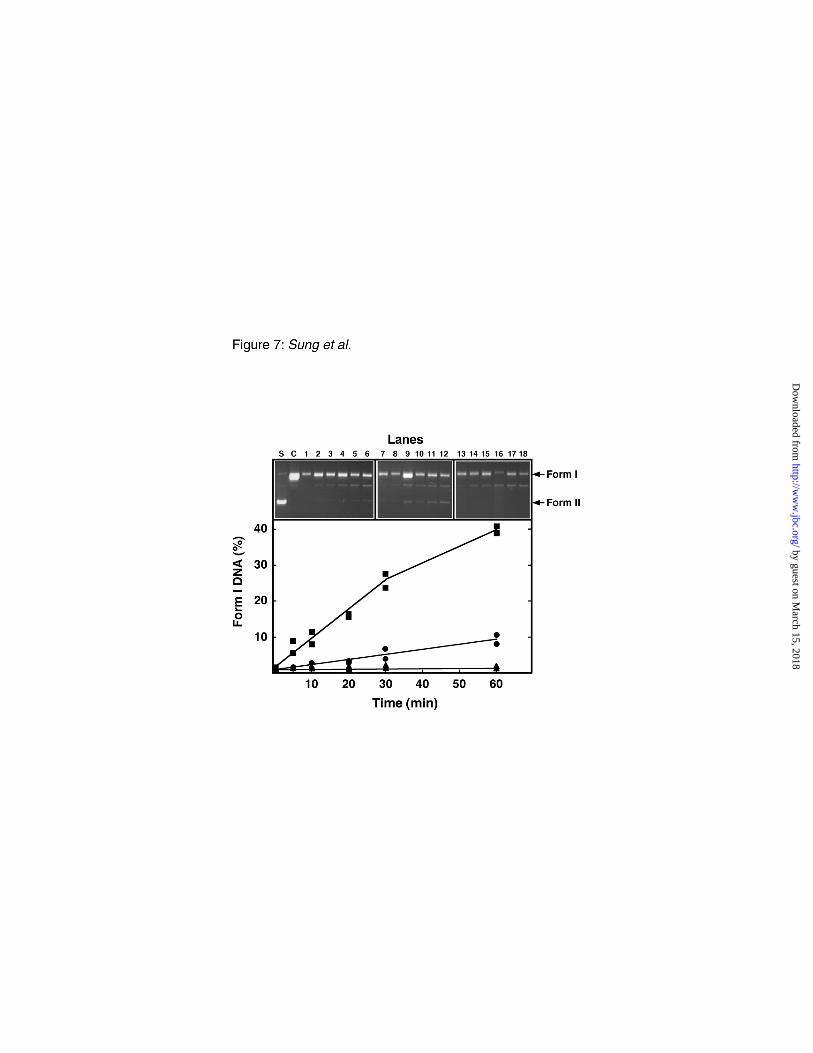
Uracil-initiated BER in cell extracts of E. coli defective in ung and dug – To further assess the
role of the dug gene product in uracil-initiated BER, repair reactions were conducted using the three E.
coli isogenic strains BH156 (ung- dug+), BH157 (ung+ dug-), and BH158 (ung- dug-). Examination of
the uracil-initiated BER reaction time course by agarose gel electrophoresis revealed a time-dependent
accumulation of Ung/Nfo-resistant form I DNA in both the E. coli BH156 and BH157 cell extracts
(Fig. 7). However, the rate of repair appeared to be 5-fold greater in the reactions containing E. coli
BH157 cell extract. In contrast, Ung/Nfo-resistant form I DNA was not observed in BER reactions
with BH158 cell extracts (Fig. 7, lanes 13-18). This experiment indicated that uracil-mediated BER
occurred in the absence of either Ung or Dug but at different rates.
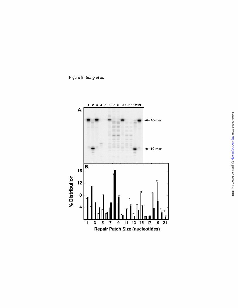
Uracil-initiated BER patch size– We utilized the approach developed by Huang et al. (32) and
Gish and Eckstein (33), and modified as previously described (30, 31), to determine the patch size of
DNA repair synthesis associated with uracil-mediated BER using the M13mp2op14 DNA substrate
(Fig. 8). This approach relies on the incorporation of 2'-deoxyribonucleoside α-thiolmonophosphates
during the BER reaction to render the repaired DNA strand resistant to in vitro digestion with E. coli
exonuclease III. As illustrated in Figure 1A, the uracil-target site in M13mp2op14 DNA was located
by guest on March 15, 2018
http://ww
w.jbc.org/
Dow
nloaded from

18
on the (-) strand, 20 nucleotides upstream (5'-side) of a unique EcoRI site, and 19 nucleotides
downstream (3'-side) of a unique SmaI site. For the purpose of measuring the repair patch size, a site-
specific [32P]dCMP residue was introduced between the uracil-target and the SmaI site, at nucleotide
position 90 in the (-) strand DNA. Two reference standards were created by treating the M13mp2op14
[32P]DNA substrate with EcoRI and SmaI alone (Fig. 8A, lanes 1 and 13) or in conjunction with Ung
and Nfo (Fig. 8A, lanes 2 and 12) which produced the expected 32P-labeled fragments of 40 and 19
nucleotides, respectively. The 19-mer corresponded to the BER intermediate formed immediately
prior to DNA synthesis and defined the 5’-boundary of the repair patch (30, 31). In order to locate the
3’-boundary of DNA synthesis produced during ung-proficient and ung-deficient BER, recovered
M13mp2op14 [32P]DNA was examined from BER reactions conducted with E. coli NR8051, NR8052,
and mock treatments. In each case, the [32P]DNA was linearized with EcoRI and then digested in the
3’-5’ direction with excess exonuclease III. Exonuclease III digestion was expected to terminate upon
encountering the first phosphorothiol-linkage (34); accordingly, this ultimate dNMP[αS] residue
defined the 3’-boundary of the repair patch. Subsequent cleavage with SmaI produced a [32P]DNA
fragment, the length of which indicated the BER patch size (i.e., 20-, 21-, and 22-mers corresponded to
a repair tract of 1, 2, and 3 nucleotides, respectively). As anticipated, exonuclease III digestion of the
mock treated [32P]DNA (Fig. 8A, lanes 4 and 5) did not produce detectable fragments smaller than the
40-mer control (Fig. 8A, lane 3) since BER had not occurred and dNMP[αS]s were not incorporated
into the [32P]DNA substrate. In contrast, discrete fragments were generated following exonuclease III
digestion of repaired M13mp2op14 [32P]DNA produced in BER reactions containing E. coli NR8051
by guest on March 15, 2018
http://ww
w.jbc.org/
Dow
nloaded from
19
cell extracts (Fig. 8A, lanes 6-8) and NR8052 (Fig. 8A, lanes 9-11). The amount of each 32P-labeled
DNA fragment was quantitatively determined and the distribution of the repair patch size was plotted
in Figure 8B. In both cell extracts, the vast majority of BER occurred via a long patch mechanism,
whereas short patch (1 nucleotide) repair accounted for ~7 % of the BER events. While the repair
patch size distribution was similar in each reaction, the ung-proficient BER reaction was biased toward
longer repair patches.
Error frequency associated with uracil-initiated BER– The M13mp2op14 lacZα DNA-based
reversion assay was utilized to ascertain the frequency of mutations produced during uracil-initiated
BER (Table I). Initially, the background reversion frequency of the M13mp2op14 (A•T) DNA was
determined in extracts of E.coli NR8051 and NR8052 cells. In each case, a similar reversion
frequency, 0.14 x 10⁻⁴ (NR8051) and 0.21 x 10⁻⁴ (NR8052), was observed. Next, the reversion
frequency associated with uracil-mediated BER of M13mp2op14 (U•T) DNA was determined to be 5.5
x 10-4 and 19.7 x 10⁻⁴ for reactions conducted with E. coli NR8051 and NR8052 cell extracts,
respectively (Table I). Thus, the absence of ung promoted an increase (~3.6-fold) in the reversion
frequency. Similar results were obtained when BER of M13mp2op14 (U•T) DNA was performed
using the analogous cell extracts (NR80511 and NR80521) that contained a mutS mutation. Therefore,
methyl-directed mismatch repair did not seem to influence the fidelity of the BER reaction.
In an effort to clarify the role of Dug as a potential mediator of the elevated error frequency
associated Ung-deficient uracil-initiated BER, repair reactions containing cell extracts of E. coli
NR8051 were supplemented with Ugi or Dug protein, and reactions containing NR8052 cell extracts
by guest on March 15, 2018
http://ww
w.jbc.org/
Dow
nloaded from
20
were supplemented with Ugi or Ung protein. Following the recovery of repaired DNA, the reversion
frequency of the M13mp2op14 (U•T) DNA substrate was determined (Table II). Supplementation of
NR8051 extracts with Ugi gave rise to a reversion frequency (40.3 x 10⁻⁴) elevated ~7-fold relative to
that measured for NR8051 extracts alone (5.5 x 10⁻⁴). Conversely, when extracts of NR8052 were
supplemented with Ung, the reversion frequency was reduced ~8-fold relative to that obtained for
NR8052 extracts alone. Taken together, these results suggest that uracil-mediated BER conducted in
the absence of Ung was more mutagenic than Ung-initiated BER. Consistent with this interpretation,
addition of Dug to NR8051 extracts resulted in an increase (~2-fold) in the frequency of opal codon 14
reversion, whereas the addition of Ugi to NR8052 extracts did not appreciably augment the elevated
mutation frequency.
The reversion frequency of uracil-initiated BER at opal codon 14 was also determined for E. coli of
a different genetic background (Table II). Experiments conducted with E. coli BH156 (ung- dug+)
exhibited a reversion frequency of 25.9 x 10⁻⁴. This value was similar to those obtained for E. coli
NR8052 (19.7 x 10⁻⁴), NR8052 supplemented with Ugi (39.4 x 10⁻⁴), and NR8051 supplemented with
Ugi (40.3 x 10⁻⁴). In the complementary experiment, uracil-initiated BER in extracts of E. coli BH157
(ung+ dug-) resulted in a relatively low reversion frequency of 5.7 x 10⁻⁴ which compared favorably to
that obtained for extracts of NR8051 (5.5 x 10⁻⁴). Determination of the reversion frequency associated
with the E. coli BH158 (ung- dug-) was not possible, as the production of Ung/Nfo-resistant Form I
DNA was not detected (Fig. 7, lanes 13-18).
by guest on March 15, 2018
http://ww
w.jbc.org/
Dow
nloaded from
21
Mutational spectra–DNA from individual revertant M13mp2op14 phage plaques was amplified
and the nucleotide sequence of the lacZα gene encompassing the opal codon 14 reversion target was
determined to define the nature and the distribution of base substitutions introduced during the process
of BER. First, the spontaneous mutational spectrum was determined for M13mp2op14 (A•T) DNA
obtained from the BER reactions containing E. coli NR8051 cell extracts. Inspection of the mutational
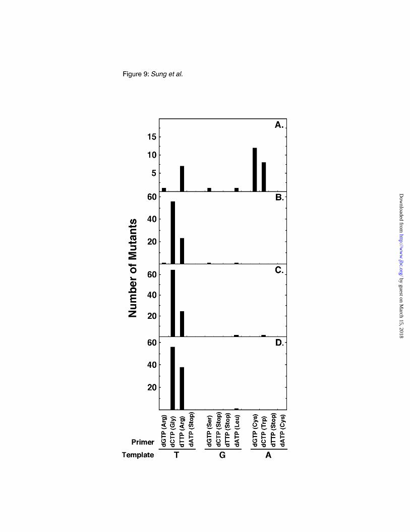
distribution showed that of the 30 mutants sequenced, 20 (67 %) reverted by base substitutions in the
third nucleotide position of the opal codon (Fig. 9A). Of these 20, 12 were A to C transversions, and 8
were A to G transition mutations. A relatively minor class of mutations occurred in the first position, 8
mutations were detected; 7 of these were T to A transversions. A similar bias for mutations at the third
position was also observed for M13mp2op14 (A•T) DNA incubated in a BER reaction containing
extracts of NR8052 (data not shown). Second, the mutational spectrum of uracil-initiated BER,
obtained using the M13mp2op14 (U•T) DNA substrate and E. coli NR8051 cell extract, revealed that
80 of the 82 mutants sequenced reverted at the first nucleotide position of the opal codon (Fig. 9B);
thus, these mutations occurred almost exclusively at the uracil target site. The large majority of these
uracil-initiated BER mutations were T to G transversions (70 %) and T to A transversions (29 %).
Third, the mutational spectrum of uracil-initiated BER in E. coli NR8051 and NR8052 cell extracts
supplemented with Ugi was compared using the M13mp2op14 (U•T) DNA substrate. From the BER
reaction containing NR8051 cell extracts, 90 revertants were analyzed and 98 % (88 of 90) of the base
substitutions occurred in the first nucleotide position (Fig 9C). The majority of these mutations (73 %)
were determined to be T to G transversions (64 of 88); T to A transversions accounted for the
by guest on March 15, 2018
http://ww
w.jbc.org/
Dow
nloaded from
22
remainder. The spectra of mutations obtained from the BER reaction containing NR8052 extracts also
consisted almost exclusively of first nucleotide base substitution mutations (94 out of 95) (Fig. 9D).
Of these, 60 % (56 of 94) were T to G transversions, while 40 % were T to A transversions.
DISCUSSION
We have examined the ability of various E. coli cell extracts to carry out uracil-initiated BER of a
covalently closed circular M13mp2 lacZα DNA substrate for the purpose of determining the fidelity
associated with complete BER reactions. To our knowledge, this report is the first to present fidelity
measurements of BER associated with Ung-proficient or Ung-deficient E. coli cells. Several previous
studies have implicated Dug as participating in uracil-DNA repair in Ung-deficient cells (14, 16, 22);
however, a recent report concluded that Dug plays no role in uracil-mediated BER (35). The results
presented here support the proposition that Dug can participate in uracil-DNA repair, and provide
additional evidence that Dug is primarily responsible for uracil-mediated BER in the absence of Ung.
These findings reinforce the observations of Gallinari and Jiricny (22), who originally identified the
double-strand uracil-DNA glycosylase activity in cell-free extracts of E. coli NR8052 that carried the
ung-1 mutation; however, they do not exclude the possibility that Dug may also play a role in the
repair of εC residues, as has been suggested by other investigators (16, 35).
Several lines of evidence support the interpretation that the uracil-mediated repair observed in this
study occurred via a BER pathway. Firstly, we observed that upon conclusion of the BER reaction,
form I DNA was generated that was resistant to cleavage by the combined treatment of Ung and Nfo.
by guest on March 15, 2018
http://ww
w.jbc.org/
Dow
nloaded from
23
This finding indicated that all steps of uracil-DNA repair had been completed. Secondly, we
demonstrated that DNA synthesis occurred preferentially in the HinfI DNA fragment (529 bp)
encompassing the uracil target. Furthermore, DNA synthesis within the 529 bp fragment was almost
exclusively dependent on the presence of a uracil residue. Thirdly, the addition of Ugi to cell extracts
of E. coli NR8051 substantially inhibited the formation of Ung/Nfo resistant form I DNA. However,
complete inhibition was not observed, as E. coli NR8051 is proficient for Dug activity, which is
insensitive to inhibition by Ugi (14). Fourthly, DNA synthesis was associated with a repair patch
involving ≤20 nucleotides and was oriented 3' to the uracil target. These results were not characteristic
of the E. coli methyl-directed DNA mismatch repair pathway, where DNA repair synthesis tracts of 1
kb or more can occur (36). Lastly, formation of Ung/Nfo-resistant form I DNA was not observed in
BER reactions containing extracts of E. coli BH158, in which both ung and dug are inactivated. This
observation strongly suggests that Ung and Dug are the predominant, if not exclusive, uracil excision
activities in wild type E. coli, and that BER was initiated by one or the other uracil-DNA glycosylase.
Examination of the kinetics of BER in extracts of NR8051 (ung+) cells showed that 60 % of the
M13mp2op14 (U•T) DNA substrate was repaired after a 20 min reaction. In contrast, the rate of BER
in extracts of NR8052 (ung-) cells was ~5.5-fold lower. One interpretation of these data is that Ung
rapidly turns over during BER, whereas Dug has a low rate of turnover. Consistent with this
interpretation is the observation of Sung and Mosbaugh (14, 17) that the addition of purified Dug to the
Ung-deficient reaction led to an increase in the rate of repair early in the reaction time course. Since
strong binding by Dug to its reaction product AP-site•G DNA has been demonstrated (14), it is
by guest on March 15, 2018
http://ww
w.jbc.org/
Dow
nloaded from
24
tempting to speculate that Dug binding hinders efficient processing of the abasic site, impeding
completion of the BER pathway. While E. coli endonuclease IV was shown to stimulate the catalytic
turnover of Dug, Dug-mediated BER remained significantly less than Ung-initiated BER, even under
the stimulated condition (14, 17). Thus, the role of Dug in uracil-DNA repair conducted via the BER
pathway may be to provide a auxiliary repair system that serves as a secondary line of defense against
uracil-provoked mutagenesis.
What influence does the fidelity of BER have on uracil-initiated mutagenesis in E. coli? Based on
the results reported in Table I, the error frequency associated with Ung-mediated BER in cell-free
extracts was determined to be 5.5 x 10⁻⁴ per repaired uracil residue. Under normal conditions, the vast
majority of uracil in E. coli DNA results from dUMP incorporation during replication (1, 3). Tye et al.
(3) reported that one uracil residue was introduced per 1200 nucleotides polymerized. Accordingly,
one would anticipate that ~4000 uracil residues are incorporated per round of chromosomal DNA
replication (1). Based on these reports, we extrapolate that E. coli Ung-proficient BER could generate
~2 mutations per cycle of semi-conservative DNA replication, providing that error correction did not
occur prior to mutation fixation. Addition of Ugi to the Ung-proficient BER reactions resulted in a ~7-
fold increase in reversion frequency without an accompanying change in the mutational specificity.
Thus, Ugi produced an ung phenotype that reflected the elevated reversion frequency and mutational
specificity associated with Dug-mediated BER. The ung mutator phenotype was reproduced in strains
sharing the E. coli GM31 genetic background, namely, BH156 and BH157. Taken together, these
results indicate that Ung-mediated BER occurs with higher fidelity than that initiated by Dug.
by guest on March 15, 2018
http://ww
w.jbc.org/
Dow
nloaded from
25
The mutational specificity of E. coli uracil-initiated BER repair observed in the opal codon 14
TGA reversion assay appears distinct from that observed in other fidelity assays conducted with
purified E. coli DNA polymerase I (large fragment). In extracts of Ung-proficient E. coli cells
(NR8051), our mutational analysis revealed that T to G transversions, resulting presumably from T•C
mispairs, were dominant (56 of 79), while T to A transversions, the likely result of T•T mispairs,
comprised the remainder (23 of 79). In contrast, Minnick et al. (37), using a 361-base gap-filling TGA
reversion assay and 3' to 5' exonuclease-deficient DNA polymerase I (large fragment), observed that
the error rate of dGTP incorporation opposite T was ~49-fold greater than that of dCTP incorporation
opposite T, and ~10-fold greater than dTTP incorporation opposite T. Perhaps it is not surprising that
our results differ from those reported by Minnick et al. (37), since we have utilized a system in which
all steps of the BER pathway are represented. The mutational specificity of uracil-mediated BER is
the end result of DNA repair synthesis, which includes misincorporation, proofreading, and/or
misextension, and must be followed by ligation. On the other hand, the gap-filling assay is restricted
to measurement of the accuracy of the polymerization step in the absence of competing reactions, and
does not require ligation.
We examined the patch size of BER DNA synthesis associated with Ung- versus Dug-mediated
repair. In both cases, the patch size was heterogeneous, ranging from 1 to ~20 nucleotides in length,
although the size of the repair patch produced in Dug-mediated BER reactions was consistently
somewhat shorter. Quantification of the distribution of the repair patches showed that the mean patch
size was 11 nucleotides in Ung-mediated BER compared to 7 nucleotides in Dug-mediated BER. A
by guest on March 15, 2018
http://ww
w.jbc.org/
Dow
nloaded from
26
small amount (~7 %) of one nucleotide replacement synthesis was observed in both systems; however,
the predominant type of DNA repair synthesis was long-patch. The latter observation is consistent
with the results of Sandigursky et al.(23), who found that repair of a U•G base pair in a closed circular
plasmid involved replacement of ~15 nucleotides downstream of the uracil target. The first
experiments conducted to elucidate the repair patch size associated with BER in E. coli cell extracts
utilized a duplex oligodeoxynucleotide 30-mer DNA with a single U•G base pair located
approximately in the middle of the substrate (13). Interestingly, under these conditions, more than 70
% of DNA repair synthesis involved incorporation of a single nucleotide (13). As previously pointed
out, the size of the repair patch may be influenced by the nature of the DNA repair substrate (23, 38).
Thus, short oligodeoxynucleotide substrates may not provide a platform sufficient for interaction with
DNA polymerase and accessory repair proteins.
It is generally accepted that DNA polymerase I occupies the primary role in uracil-mediated DNA
repair synthesis in E. coli (39, 40). Our analysis of the mutational spectra derived from Ung-proficient
and Ung-deficient extracts suggests that the specificity of misinsertion remains essentially the same
regardless of the uracil-DNA glycosylase involved; accordingly, one might infer that the same DNA
polymerase is involved in Ugi-resistant as well as in Ugi-sensitive uracil-DNA repair. Given the
relatively high mutation frequencies we observed in this study, a role for the newly discovered Pol IV
and/or Pol V DNA polymerases in BER cannot be formally excluded. These DNA polymerases have
been described as low fidelity enzymes that exhibit error rates of ~10-3 to 5 x 10⁻⁴ when copying
undamaged DNA in vitro (41); however, experiments to assess the contribution of these enzymes to
by guest on March 15, 2018
http://ww
w.jbc.org/
Dow
nloaded from
27
BER have not yet been carried out. The molecular mechanisms underlying the mutational specificity
of uracil-initiated BER in E. coli and the increased reversion frequency associated with the Dug-
mediated BER pathway await further elucidation.
Acknowledgments–We thank Nanci Adair for conducting the DNA sequence analysis at the Center for
Gene Research and Biotechnology.
by guest on March 15, 2018
http://ww
w.jbc.org/
Dow
nloaded from

28
REFERENCES
1. Mosbaugh, D. W., and Bennett, S.E. (1994) Prog. Nucleic Acid Res. Mol. Biol. 48, 315-370
2. Duncan, B. K., and Miller, J. H. (1980) Nature 287, 560-561
3. Tye, B.-K., Chien, J., Lehman, I. R., Duncan, B. K., and Warner, H. R. (1978) Proc. Natl. Acad.
Sci. U. S. A. 75, 233-237
4. Lindahl, T., Ljungquist, S., Siegert, W., Nyberg, B., and Sperens, B. (1977) J. Biol. Chem. 252,
3286-3294
5. Chan, E., and Weiss, B. (1987) Proc. Natl. Acad. Sci. U. S. A. 84, 3189-3193
6. Gossard, F., and Verly, W. G. (1978) Eur. J. Biochem. 82, 321-332
7. Levin, J. D., and Demple, B. (1990) Nucleic Acids Res. 18, 5069-5075
8. Doetsch, P. W., and Cunningham, R. P. (1990) Mutation Res. 236, 173-201
9. Franklin, W. A., and Lindahl, T. (1988) EMBO J. 7, 3617-3622
10. Dianov, G., Sedgwick, B., Daly, G., Olsson, M., Lovett, S., and Lindahl, T. (1994) Nucleic Acids
Res. 22, 993-998
11. Graves, R. J., Felzenszwalb, I., Laval, J., and O'Connor, T. R. (1992) J. Biol. Chem. 267, 14429-
14435
12. Sandigursky, M., and Franklin, W. A. (1992) Nucleic Acids Res. 20, 4699-4703
13. Dianov, G., Price, A., and Lindahl, T. (1992) Mol. Cell. Biol. 12, 1605-1612
14. Sung, J.-S., and Mosbaugh, D. W. (2000) Biochemistry 39, 10224-10235
15. Varshney, U., Hutcheon, T., and van de Sande, J. H. (1988) J. Biol. Chem. 263, 7776-7784
16. Saparbaev, M., and Laval, J. (1998) Proc. Natl. Acad. Sci. U. S. A. 95, 8508-8513
17. Barrett, T. E., Savva, R., Panayotou, G., Barlow, T., Brown, T., Jiricny, J., and Pearl, L. H. (1998)
Cell 92, 117-129
18. Roy, S., Purnapatre, K., Handa, P., Boyanapalli, M., and Varshney, U. (1998) Protein Expr. Purif.
13, 155-162
19. Bennett, S. E., Jensen, O. N., Barofsky, D. F., and Mosbaugh, D. W. (1994) J. Biol. Chem. 269,
21870-21879
by guest on March 15, 2018
http://ww
w.jbc.org/
Dow
nloaded from
29
20. Bennett, S. E., Sanderson, R. J., and Mosbaugh, D. W. (1995) Biochemistry 34, 6109-6119
21. Bennett, S. E., and Mosbaugh, D. W. (1992) J. Biol. Chem. 267, 22512-22521
22. Gallinari, P., and Jiricny, J. (1996) Nature 383, 735-738
23. Sandigursky, M., Freyer, G.A., and Franklin, W.A. (1998) Nucleic Acids Res. 26, 1282-1287
24. Dianov, G., and Lindahl, T. (1994) Curr. Biol. 4, 1069-1076
25. Sanderson, R. J., and Mosbaugh, D. W. (1998) J. Biol. Chem. 273, 24822-24831
26. Sanderson, R. J., and Mosbaugh, D. W. (1996) J. Biol. Chem. 271, 29170-29181
27. Del Sal, G., Manfioletti, G., and Schneider, C. (1989) Biotechniques 7, 514-520
28. Bradford, M. M. (1976) Anal. Biochem. 72, 248-254
29. Koetsier, P. A., Schorr, J., and Doerfler, W. (1993) BioTechniques 15, 260-262
30. Sanderson, R. J., Bennett, S. E., Sung, J.-S., and Mosbaugh, D. W. (2000) Prog. Nucleic Acid Res.
Mol. Bio. (in press)
31. Sanderson, R. J. (1998) Uracil-DNA Glycosylase Inhibitor Protein: Role of Carboxylic Acid
Residues and Use for Measuring the Fidelity of Uracil-Excision DNA Repair in Human Cell
Extracts. , Ph.D. thesis, Oregon State University
32. Huang, J. C., Svoboda, J. T., Reardon, J. T., and Sancar, A. (1992) Proc. Natl. Acad. Sci. U. S. A.
89, 3664-3668
33. Gish, G., and Eckstein, F. (1988) Science 240, 1520-1522
34. Putney, S. D., Benkovic, S. J., and Schimmel, P. R. (1981) Proc. Natl. Acad. Sci. U. S. A. 78, 7350-
7354
35. Lutsenko, E., and Bhagwat, A.S. (1999) J. Biol. Chem. 274, 31034-31038
36. Lahue, R. S., Au, K. G., and Modrich, P. (1989) Science 245, 160-164
37. Minnick, D. T., Bebenek, K., Osheroff, W. P., Turner, R. M. J., Astatke, M., Liu, L., Kunkel, T. A.,
and Joyce, C. M. (1999) J. Biol. Chem 274, 3067-3075
38. Biade, S., Sobol, R. W., Wilson, S. H., and Matsumoto, Y. (1998) J. Biol. Chem. 273, 898-902
39. Tye, B.-K., Nyman, P.-O., Lehman, I. R., Hochhauser, S., and Weiss, B. (1977) Proc. Natl. Acad.
Sci. U. S. A. 74, 154-157
by guest on March 15, 2018
http://ww
w.jbc.org/
Dow
nloaded from
30
40. Lindahl, T. (1979) Prog. Nucleic Acid Res. Mol. Biol. 22, 135-192
41. Tang, M., Pham, P., Shen, X., Taylor, J. S., O'Donnell, M., Woodgate, R., and Goodman, M. F.
(2000) Nature 404, 1014-1018
by guest on March 15, 2018
http://ww
w.jbc.org/
Dow
nloaded from
31
FOOTNOTES
*This work was supported by National Institutes of Health Grants GM32823 and ES00210. This is
Technical Report 11708 from the Oregon Agricultural Experiment Station. The cost for publication of
this article are defrayed in part by the payment of page charges. This article must by hereby marked
“advertisement” in accordance with 18 U.S.C. Section 1734 solely to indicate this fact.
¶To whom correspondence should be addressed. Tel: (541) 737-1797; FAX; (541) 737-0497;
email [email protected].
1 The abbreviations used are: BER, base excision repair; AP, apurinic/apyrimidinic; dRP,
deoxyribose 5'-phosphate; PCR, polymerase chain reaction; EDTA, ethylenediaminetetraacetic; SDS,
sodium dodecyl sulfate; IPTG, β-D-thiogalactopyranoside; X-gal, 5-bromo-4-chloro-3-indolyl β-D-
galactopyranoside; bp, base pair(s); kb, kilo-base pairs; dNMP[αS], 2'-deoxyribonucleoside α-
thiotriphosphate; εC, 3,N4-ethenocytosine.
2 Base pairs and mispairs are described by listing the base in the repaired strand first and then the
base in the template strand.
3 No conversion of form I M13mp2op14 (A•T) DNA to form II following Ung/Nfo treatment was
detected, indicating that this DNA preparation did not support uracil-initiated BER.
by guest on March 15, 2018
http://ww
w.jbc.org/
Dow
nloaded from
32
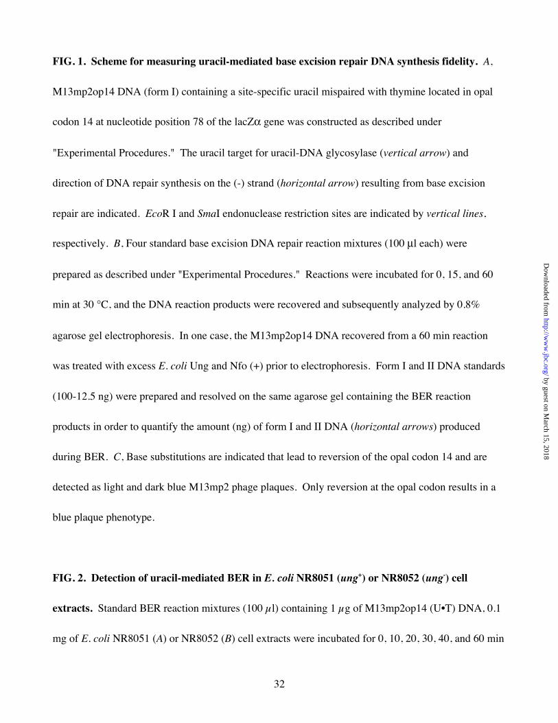
FIG. 1. Scheme for measuring uracil-mediated base excision repair DNA synthesis fidelity. A,
M13mp2op14 DNA (form I) containing a site-specific uracil mispaired with thymine located in opal
codon 14 at nucleotide position 78 of the lacZα gene was constructed as described under
"Experimental Procedures." The uracil target for uracil-DNA glycosylase (vertical arrow) and
direction of DNA repair synthesis on the (-) strand (horizontal arrow) resulting from base excision
repair are indicated. EcoR I and SmaI endonuclease restriction sites are indicated by vertical lines,
respectively. B, Four standard base excision DNA repair reaction mixtures (100 µl each) were
prepared as described under "Experimental Procedures." Reactions were incubated for 0, 15, and 60
min at 30 °C, and the DNA reaction products were recovered and subsequently analyzed by 0.8%
agarose gel electrophoresis. In one case, the M13mp2op14 DNA recovered from a 60 min reaction
was treated with excess E. coli Ung and Nfo (+) prior to electrophoresis. Form I and II DNA standards
(100-12.5 ng) were prepared and resolved on the same agarose gel containing the BER reaction
products in order to quantify the amount (ng) of form I and II DNA (horizontal arrows) produced
during BER. C, Base substitutions are indicated that lead to reversion of the opal codon 14 and are
detected as light and dark blue M13mp2 phage plaques. Only reversion at the opal codon results in a
blue plaque phenotype.
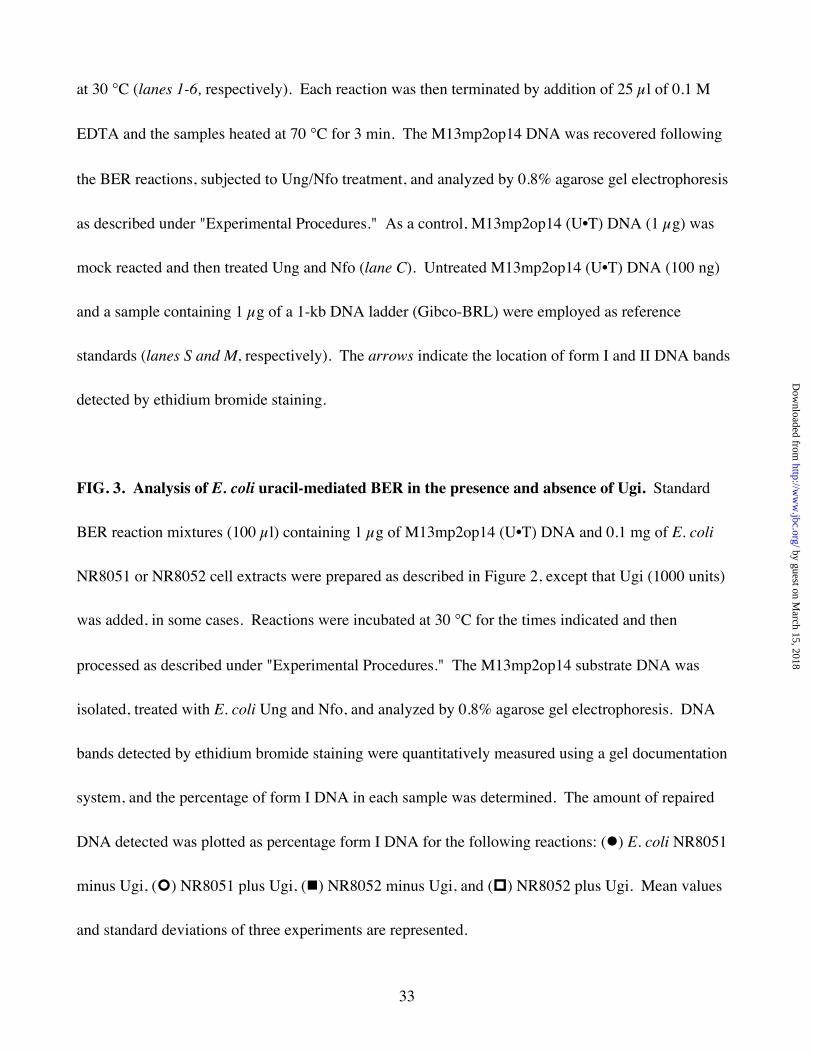
FIG. 2. Detection of uracil-mediated BER in E. coli NR8051 (ung+) or NR8052 (ung-) cell
extracts. Standard BER reaction mixtures (100 µl) containing 1 µg of M13mp2op14 (U•T) DNA, 0.1
mg of E. coli NR8051 (A) or NR8052 (B) cell extracts were incubated for 0, 10, 20, 30, 40, and 60 min
by guest on March 15, 2018
http://ww
w.jbc.org/
Dow
nloaded from
33
at 30 °C (lanes 1-6, respectively). Each reaction was then terminated by addition of 25 µl of 0.1 M
EDTA and the samples heated at 70 °C for 3 min. The M13mp2op14 DNA was recovered following
the BER reactions, subjected to Ung/Nfo treatment, and analyzed by 0.8% agarose gel electrophoresis
as described under "Experimental Procedures." As a control, M13mp2op14 (U•T) DNA (1 µg) was
mock reacted and then treated Ung and Nfo (lane C). Untreated M13mp2op14 (U•T) DNA (100 ng)
and a sample containing 1 µg of a 1-kb DNA ladder (Gibco-BRL) were employed as reference
standards (lanes S and M, respectively). The arrows indicate the location of form I and II DNA bands
detected by ethidium bromide staining.
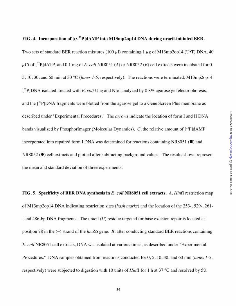
FIG. 3. Analysis of E. coli uracil-mediated BER in the presence and absence of Ugi. Standard
BER reaction mixtures (100 µl) containing 1 µg of M13mp2op14 (U•T) DNA and 0.1 mg of E. coli
NR8051 or NR8052 cell extracts were prepared as described in Figure 2, except that Ugi (1000 units)
was added, in some cases. Reactions were incubated at 30 °C for the times indicated and then
processed as described under "Experimental Procedures." The M13mp2op14 substrate DNA was
isolated, treated with E. coli Ung and Nfo, and analyzed by 0.8% agarose gel electrophoresis. DNA
bands detected by ethidium bromide staining were quantitatively measured using a gel documentation
system, and the percentage of form I DNA in each sample was determined. The amount of repaired
DNA detected was plotted as percentage form I DNA for the following reactions: ( ) E. coli NR8051
minus Ugi, ( ) NR8051 plus Ugi, ( ) NR8052 minus Ugi, and ( ) NR8052 plus Ugi. Mean values
and standard deviations of three experiments are represented.
by guest on March 15, 2018
http://ww
w.jbc.org/
Dow
nloaded from
34
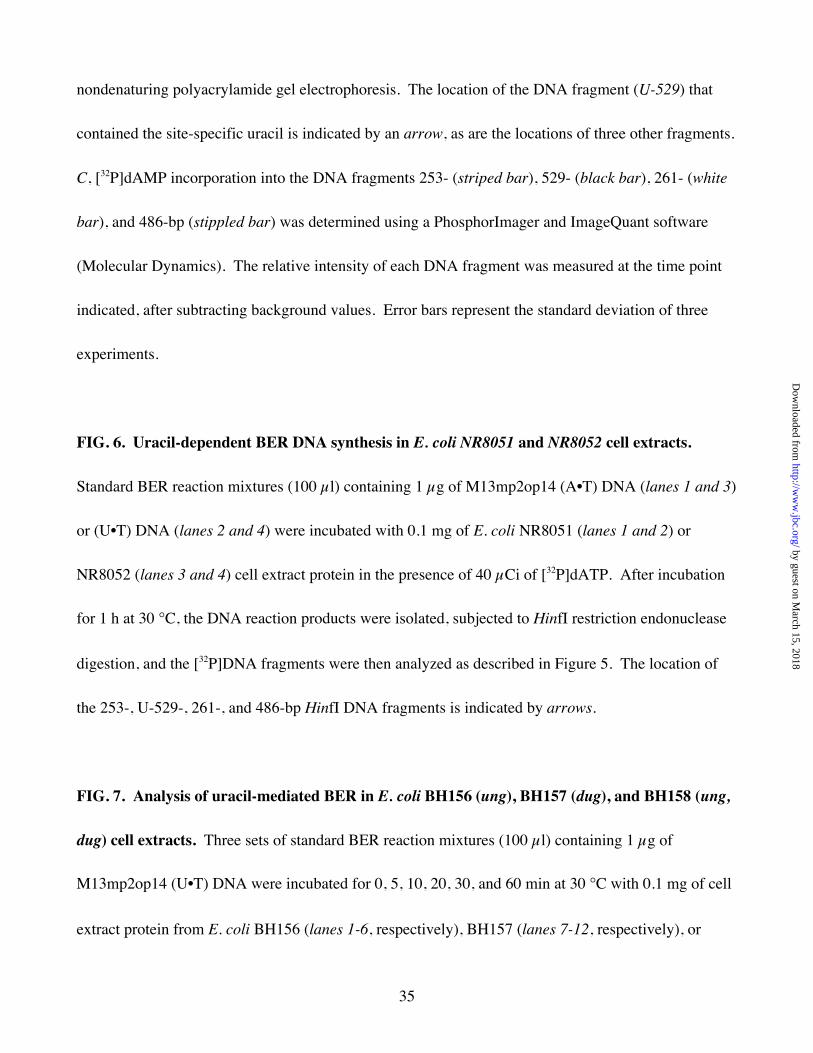
FIG. 4. Incorporation of [α-32P]dAMP into M13mp2op14 DNA during uracil-initiated BER.
Two sets of standard BER reaction mixtures (100 µl) containing 1 µg of M13mp2op14 (U•T) DNA, 40
µCi of [32P]dATP, and 0.1 mg of E. coli NR8051 (A) or NR8052 (B) cell extracts were incubated for 0,
5, 10, 30, and 60 min at 30 °C (lanes 1-5, respectively). The reactions were terminated, M13mp2op14
[32P]DNA isolated, treated with E. coli Ung and Nfo, analyzed by 0.8% agarose gel electrophoresis,
and the [32P]DNA fragments were blotted from the agarose gel to a Gene Screen Plus membrane as
described under "Experimental Procedures." The arrows indicate the location of form I and II DNA
bands visualized by PhosphorImager (Molecular Dynamics). C, the relative amount of [32P]dAMP
incorporated into repaired form I DNA was determined for reactions containing NR8051 ( ) and
NR8052 ( ) cell extracts and plotted after subtracting background values. The results shown represent
the mean and standard deviation of three experiments.
FIG. 5. Specificity of BER DNA synthesis in E. coli NR8051 cell extracts. A, HinfI restriction map
of M13mp2op14 DNA indicating restriction sites (hash marks) and the location of the 253-, 529-, 261-
, and 486-bp DNA fragments. The uracil (U) residue targeted for base excision repair is located at
position 78 in the (−) strand of the lacZα gene. B, after conducting standard BER reactions containing
E. coli NR8051 cell extracts, DNA was isolated at various times, as described under "Experimental
Procedures." DNA samples obtained from reactions conducted for 0, 5, 10, 30, and 60 min (lanes 1-5,
respectively) were subjected to digestion with 10 units of HinfI for 1 h at 37 °C and resolved by 5%
by guest on March 15, 2018
http://ww
w.jbc.org/
Dow
nloaded from
35
nondenaturing polyacrylamide gel electrophoresis. The location of the DNA fragment (U-529) that
contained the site-specific uracil is indicated by an arrow, as are the locations of three other fragments.
C, [32P]dAMP incorporation into the DNA fragments 253- (striped bar), 529- (black bar), 261- (white
bar), and 486-bp (stippled bar) was determined using a PhosphorImager and ImageQuant software
(Molecular Dynamics). The relative intensity of each DNA fragment was measured at the time point
indicated, after subtracting background values. Error bars represent the standard deviation of three
experiments.
FIG. 6. Uracil-dependent BER DNA synthesis in E. coli NR8051 and NR8052 cell extracts.
Standard BER reaction mixtures (100 µl) containing 1 µg of M13mp2op14 (A•T) DNA (lanes 1 and 3)
or (U•T) DNA (lanes 2 and 4) were incubated with 0.1 mg of E. coli NR8051 (lanes 1 and 2) or
NR8052 (lanes 3 and 4) cell extract protein in the presence of 40 µCi of [32P]dATP. After incubation
for 1 h at 30 °C, the DNA reaction products were isolated, subjected to HinfI restriction endonuclease
digestion, and the [32P]DNA fragments were then analyzed as described in Figure 5. The location of
the 253-, U-529-, 261-, and 486-bp HinfI DNA fragments is indicated by arrows.
FIG. 7. Analysis of uracil-mediated BER in E. coli BH156 (ung), BH157 (dug), and BH158 (ung,
dug) cell extracts. Three sets of standard BER reaction mixtures (100 µl) containing 1 µg of
M13mp2op14 (U•T) DNA were incubated for 0, 5, 10, 20, 30, and 60 min at 30 °C with 0.1 mg of cell
extract protein from E. coli BH156 (lanes 1-6, respectively), BH157 (lanes 7-12, respectively), or
by guest on March 15, 2018
http://ww
w.jbc.org/
Dow
nloaded from
36
BH158 (lanes 13-18, respectively). Form I DNA reaction products were isolated, treated with E. coli
Ung and Nfo, and resolved by 0.8% agarose gel electrophoresis (inset) as described under
"Experimental Procedures." Untreated M13mp2op14 (U•T) DNA (100 ng) was used as a reference
standard (lanes S). As a control, M13mp2op14 (U•T) DNA (1 µg) was mock-reacted, isolated, and
then subjected to Ung/Nfo treatment (lane C). The location of form I and II DNA bands is indicated
by arrows. The amount of form I and II DNA detected by ethidium bromide staining was
quantitatively measured with a gel documentation system and the percentage of form I DNA was
determined. The results of two independent experiments are plotted for E. coli BH156 ( ), BH157
( ), and BH158 ( ).
Fig. 8. Analysis of DNA repair patch size associated with uracil-mediated BER reactions. A,
Standard BER reaction mixtures (100 µl) containing 1 µg of M13mp2op14 (U•T) [32P]DNA, 20 µM
each of dATP[αS], dTTP[αS], dGTP[αS], and dCTP[αS] and 0.1 mg of cell extract protein of E. coli
NR8051 (lanes 6-8) and NR8052 (lanes 9-11) were incubated for 60 min at 30 °C. As a control,
M13mp2op14 (U•T) [32P]DNA (1 µg) was mock-reacted in the absence of cell extract protein (lanes 3-
5). DNA products were isolated, samples (~200 ng) were digested with EcoRI, and then incubated
with 0 (lanes 3, 6, and 9), 2 (lanes 4, 7, and 10), and 20 (lanes 5, 8, and 11) units of E. coli exonuclease
III. Following exonuclease III digestion, the DNA was cleaved with SmaI, and then resolved by 12%
polyacrylamide/8.3 M urea gel electrophoresis as described under "Experimental Procedures." The
DNA size markers, 40-mer (lanes 1 and 13) generated by digesting 200 ng of M13mp2op14 (U•T)
by guest on March 15, 2018
http://ww
w.jbc.org/
Dow
nloaded from
37
[32P]DNA with EcoRI and SmaI, and the 19-mer (lanes 2 and 12) produced by additional treatment
with Ung and Nfo, are indicated by arrows. B, The amount of 32P radioactivity detected in each band
in (A) was quantitatively measured using a PhosphorImager and the results for the E. coli NR8051
(white bars) and NR8052 (black bars) reactions digested with 20 units of E. coli exonuclease III are
plotted. The [32P]DNA bands of 20 to 40 nucleotides in length corresponded to BER repair patches of
1 to 21 nucleotides in length, respectively. The relative amount of 32P label in each band (%
distribution) was determined by dividing the amount of 32P radioactivity detected per band by the total
32P signal detected for all bands and multiplying by 100. Mean values and standard deviations for the
distribution of four experiments are indicated.
Fig. 9. E. coli mutational spectrum of uracil-initiated base excision repair. Four standard BER
reactions were performed using either E. coli NR8051 (A, B, C) or NR8052 (D) cell extracts and
M13mp2op14 DNA as described in Table I. One reaction contained M13mp2op14 (A•T) DNA (A)
and the other three reactions were prepared with the (U•T) DNA substrate (B, C, D) while 1,000 units
of Ugi were added to reaction (C and D). Following transfection of NR9162 cells with Ung/Nfo-
resistant form I DNA recovered from BER reactions, blue plaques were isolated, phage were subjected
to PCR-mediated lacZα DNA amplification, and DNA sequence analysis conducted as described under
"Experimental Procedures." The nucleotide sequence (TGA) of the opal codon 14 in the template
DNA strand used for uracil-initiated BER DNA synthesis is indicated. The four possible
deoxyribonucleoside triphosphates used for incorporation into the primer strand are indicated with the
by guest on March 15, 2018
http://ww
w.jbc.org/
Dow
nloaded from
38
corresponding coded amino acid (parenthesis). For each BER reaction, the number of base
substitution mutations detected by DNA sequence analysis are plotted.
by guest on March 15, 2018
http://ww
w.jbc.org/
Dow
nloaded from
39
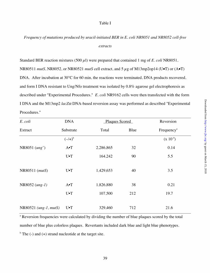
Table I
Frequency of mutations produced by uracil-initiated BER in E. coli NR8051 and NR8052 cell-free
extracts
Standard BER reaction mixtures (500 µl) were prepared that contained 1 mg of E. coli NR8051,
NR80511 mutS, NR8052, or NR80521 mutS cell extract, and 5 µg of M13mp2op14 (U•T) or (A•T)
DNA. After incubation at 30°C for 60 min, the reactions were terminated, DNA products recovered,
and form I DNA resistant to Ung/Nfo treatment was isolated by 0.8% agarose gel electrophoresis as
described under "Experimental Procedures." E. coli NR9162 cells were then transfected with the form
I DNA and the M13mp2 lacZα DNA-based reversion assay was performed as described "Experimental
Procedures."
E. coli DNA Plaques Scored Reversion
Extract Substrate Total Blue Frequencya
(–/+)b (x 10-4)
NR8051 (ung+) A•T 2,286,865 32 0.14
U•T 164,242 90 5.5
NR80511 (mutS) U•T 1,429,653 40 3.5
NR8052 (ung-1) A•T 1,826,880 38 0.21
U•T 107,500 212 19.7
NR80521 (ung-1, mutS) U•T 329,460 712 21.6
a Reversion frequencies were calculated by dividing the number of blue plaques scored by the total
number of blue plus colorless plaques. Revertants included dark blue and light blue phenotypes.
b The (-) and (+) strand nucleotide at the target site.
by guest on March 15, 2018
http://ww
w.jbc.org/
Dow
nloaded from

40
Table II
Frequency of mutations produced by uracil-initiated BER in E. coli NR8051 and E. coli NR8052 cell-
free extracts supplemented with purified Ung, Dug, or Ugi Protein
Standard BER reaction mixtures (500 µl) were prepared that contained 5 µg of M13mp2op14 (U•T)
DNA and 1 mg of E. coli cell extract, as indicated. After incubation at 30°C for 60 min, the reactions
were terminated, DNA products recovered, and Ung/Nfo-resistant form I DNA was isolated by 0.8%
agarose gel electrophoresis as described under "Experimental Procedures." The form I DNA was then
use to transfect E. coli NR9162 cells, and the M13mp2 lacZα DNA-based reversion assay was
performed as described "Experimental Procedures."
E. coli Protein Plaques Scored Reversion
Extract Allele Additiona Total Blue Frequencyb
Experiment 1 (x 10-4)
NR8051 ung+ -- 164,242 90 5.5
NR8051 ung+ Ugi 272,409 1098 40.3
NR8051 ung+ Dug 826,457 950 11.5
NR8052 ung- -- 107,500 212 19.7
NR8052 ung- Ugi 198,440 782 39.4
NR8052 ung- Ung 326,745 77 2.4
Experiment 2
BH156 ung- dug+ -- 524,716 1357 25.9
BH157 ung+ dug- -- 1,300,550 741 5.7
a Ugi (1000 units), Dug (20 pmol), or Ung (4 units) was included in the standard BER reaction as
indicated.
b Reversion frequencies were calculated by dividing the number of blue plaques scored by the total
number of blue plus colorless plaques. Revertants included dark blue and light blue phenotypes.
by guest on March 15, 2018
http://ww
w.jbc.org/
Dow
nloaded from
Jung-Suk Sung, Samuel E. Bennett and Dale W. MosbaughFidelity of uracil-initiated base excision DNA repair in Escherichia coli cell extracts
published online October 16, 2000J. Biol. Chem.
10.1074/jbc.M008147200Access the most updated version of this article at doi:
Alerts:
When a correction for this article is posted•
When this article is cited•
to choose from all of JBC's e-mail alertsClick here
by guest on March 15, 2018
http://ww
w.jbc.org/
Dow
nloaded from