ferret tnf- and ifn- immunoassays -...
TRANSCRIPT

9
Ferret TNF-α and IFN-γ Immunoassays
Alyson Ann Kelvin1, David Banner2, Ali Danesh2, Charit Seneviratne2, Atsuo Ochi2 and David Joseph Kelvin1,2,3,4
1Immune Diagnostics & Research, Toronto, Ontario, 2Division of Experimental Therapeutics, Toronto General Hospital Research Institute,
University Health Network, Toronto, Ontario, 3International Institute of Infection and Immunity,
Shantou University Medical College, Shantou, Guangdong, 4Sezione di Microbiologia Sperimentale e Clinica, Dipartimento di Scienze Biomediche,
Universita' degli Studi di Sassari, Sassari, 1,2Canada
3China 4Italy
1. Introduction
Despite the prominent use of ferrets in medical research, the immune system of ferrets remains poorly characterized (Svitek & von, V, 2007). Here we describe ferret TNF-┙ and IFN-┛ immunoassays.
Specifically, this covers the following topics:
Background: The use of ferrets in medical research
TNF-┙ and IFN-┛ cloning and sequencing
Expression and purification of recombinant ferret TNF-┙ and IFN-┛ proteins
Cytokine real-time PCR based assays
Development of IFN-┛ and TNF-┙ hybridoma clones
ELISA and ELISPOT assays for the ferret cytokine IFN-┛
2. Background
This Background describes the current use of ferrets in medical research but also includes a description of past uses. The importance of ferrets is highlighted in human disease modeling and in prophylactic and vaccine development. The various uses of ferrets in medical research demonstrate the need for immune profiling reagents and assays.
2.1 Biology of the ferret
The ferret, Mustela putorius furo, is a relatively small and inexpensive animal in terms of its potential for research use, although mice remain the traditional influenza model for virus pathogenesis.
www.intechopen.com
Trends in Immunolabelled and Related Techniques 134
Essentially, mice are low cost animals that have a broad availability of corresponding reagents for immunological investigation. Mice are easily mutated and there exists a plethora of currently available transgenic mice with immune targeted gene deletions or gene knock-ins (Belser, Szretter, Katz, & Tumpey, 2009). Two factors against the use of mice in influenza immune studies are 1) most human influenza strains must be mouse adapted prior to initiation of infection studies due to the inability of human influenza viruses to replicate in the mouse and 2) mice do not exhibit human-like clinical signs of influenza such as sneezing and temperature fluxes.
In contrast to mice, ferrets do develop respiratory illnesses that are similar to human
disease. Although the ferret is not considered a large laboratory animal, it is able to provide
many biological samples for pathological testing during an infection study. For instance,
frequent and sizable blood sampling is feasible in the ferret that is not practical in smaller
rodents such as in mice and rats.
Furthermore, clinical features of disease are easily observed. Such as clinical fever manifested as an elevation in body temperatures can be detected as early as 1 day following infection with many viruses. Our own as well as previously published studies have shown that high fevers can persist for many days following infection of viruses such as H1N1pdm influenza (Rowe et al., 2010b; Sweet et al., 1979; Zitzow et al., 2002). As well as fever, nasal discharge, sneezing and activity level can also be observed in ferrets infected with influenza viruses (Rowe et al., 2010b). Taken together, these clinical features along with the feasibility for blood and pathological sampling suggest the ferret to be an optimal animal for the study of human infectious diseases, including influenza viruses. For example, our group has recently used the ferret model successfully to characterize and compare immunopathology caused by several strains of currently circulating influenza A and B viruses (Huang et al., 2011).
2.2 Respiratory viral infections
Viral respiratory infectious diseases are a major worldwide concern which causes significant
morbidity and mortality (Kolling et al., 2001). Respiratory viral diseases such as the severe
acute respiratory syndrome coronavirus (SARS-CoV), avian influenza H5N1 and pandemic
influenza H1N1 virus are potential epidemic and/or pandemic threats (Dushoff, Plotkin,
Viboud, Earn, & Simonsen, 2006; Weiss & McMichael, 2004; Dawood, Dalton, Durrheim, &
Hope, 2009; Dawood et al., 2009). Specifically, influenza is a significant contributor to
morbidity and mortality worldwide and is the focus of our laboratory and the focus disease
of this chapter.
The World Health Organization estimates the burden of season influenza to be approximately one billion cases annually, including 3-5 million severe cases and 300,000-500,000 deaths (Girard, Cherian, Pervikov, & Kieny, 2005). Influenza illness in humans is caused by an influenza RNA virus of the Orthomyxoviridae family. The influenza virus can be categorized as one of three types: A, B, or C (Steinhauer & Skehel, 2002). Importantly, the influenza viral genome is susceptible to two primary types of genetic mutations that cause variation in the immunogenic proteins and subsequently in disease presentation and clinical features (Kasowski, Garten, & Bridges, 2011; Steinhauer & Skehel, 2002). Firstly, antigenic drift is defined by minor changes to the viral genome introduced during virus RNA
www.intechopen.com

Ferret TNF-α and IFN-γ Immunoassays 135
replication (Steinhauer & Skehel, 2002). Second, antigenic shift is when a host is infected by various influenza strains at the same time allowing entire genome segments to be reassorted during co-infection. This results in novel influenza strains markedly distinct from their progenitors (Steinhauer & Skehel, 2002; Kasowski et al., 2011). From these reassortments, novel influenza strains may arise with new clinical symptoms and disease features that have the potential to be highly pathogenic, easily transmissibility with pandemic potential.
The most significant recent reassortant to emerge was the 2009 pandemic H1N1 influenza A (H1N1pdm) strain (Perez-Padilla et al., 2009). H1N1pdm is closely related to the reassortant swine influenza A viruses previously isolated in North America, Europe, and Asia (Trifonov, Khiabanian, & Rabadan, 2009). Infection with H1N1pdm resulted in diverse clinical outcomes. The majority of reported cases were mild and self-limiting (Gilsdorf, Poggensee, & Working Group, 2009; Nicoll & Coulombier, 2009; Writing Committee of the WHO Consultation on Clinical Aspects of Pandemic, 2010) and typical symptoms include fever, sore throat, malaise, and headache (Health Protection Agency, Health, National Public Health Service for Wales, & HPA Northern Ireland Swine influenza investigation team, 2009). A small proportion of pandemic influenza cases required hospitalization and patient ventilator support (Centers for Disease Control and Prevention (CDC), 2009; Kumar et al., 2009; Perez-Padilla et al., 2009; Writing Committee of the WHO Consultation on Clinical Aspects of Pandemic, 2010). Common complications in there severe cases were severe hypoxemia, shock, pneumonia, and acute respiratory distress syndrome (ARDS)(Perez-Padilla et al., 2009; Kumar et al., 2009; Centers for Disease Control and Prevention (CDC), 2009). Nonpulmonary acute organ dysfunction has also been reported (Uyeki, Sharma, & Branda, 2009; Kumar et al., 2009). Since ferrets show signs of illness and are easily infected with human strains of H1N1pdm, we are currently investigating and have published on the immunopathogenic mechanisms and possible therapeutics for H1N1pdm illness in ferrets (Rowe et al., 2010a; Huang S.S.H. et al., 2011; Cameron et al., 2008).
2.3 Host immune responses
Host immunity can be broken down into an innate and adaptive immune response. The innate immune response is a nonspecific attack on the invading agent while the adaptive immune response is an attack tailored to the individual pathogen (Ryan & Majno, 1977). What determines the type of triggered immune response is the invading agent itself. The agent is recognized first by the innate immune arm and together the innate and adaptive immune responses lead to a unique immune signature that for each pathogen. Furthermore, the clinical outcome is the biological consequence of the immune response that has developed toward the pathogen (Belz, Bedoui, Kupresanin, Carbone, & Heath, 2007; Zheng et al., 2007).
In order to evaluate the immune response during the course of viral infection, it is important to be able to determine the activity of the immune cells. Cell identity and their activation status are distinguished by the molecules expressed at the cell surface. Furthermore, cells of the immune system can be described as innate or adaptive immune cells. Innate immune cells include neutrophils, esosinophils, basophils, macrophages and NK cells. Adaptive immune cells include T lymphocytes, B lymphocytes and Dendritic cells (Hauge, Madhun, Cox, Brokstad, & Haaheim, 2007). Many of these cells have yet to be characterized in the ferret.
www.intechopen.com
Trends in Immunolabelled and Related Techniques 136
As well as understanding the cell activation and cellular populations during an immune
response, it is also important to elucidate the intracellular activation and intercellular events
which occur following infection. Cells are often activated by cytokines, soluble extracellular
proteins that mediate signals from one cell to another. Once the cell has been in contact with
a cytokine, intracellular signalling cascades are activated. The activation of these signalling
cascades leads to cell effector function.
One of the most prominent branches of cytokine-cell signalling events is of the
inflammatory interferon (IFN) cytokines which connect the innate immune response with
the activation of the adaptive immunity. The IFN family of cytokines can be categorized as
either Type I IFN or Type II. IFN-┙ and IFN-┚ are of the Type I IFN cytokines and have a
prominent role during viral infection. IFN-┛ is of the Type II IFN family. IFN-┛ also plays a
role in viral infections but also functions during bacterial infections.
The release of IFN-┛ leads to cellular activation through signalling pathways. Ligation of the
interferon receptors 1 and 2 (IFNAR1 and IFNAR2 for IFN-┙ and IFN-┚; IFNGR1 and
IFNGR2 for IFN-┛) with an IFN cytokine induces IFN signaling pathways and promotes IFN
gene induction. Both the Type I and Type II cytokines signal through JAK-STAT pathways
to activate IFN genes and promote immune responses (Marijanovic, Ragimbeau, van der
Heyden, Uze, & Pellegrini, 2007). IFN induced JAK-STAT signalling often involves
interferon regulatory factor 9 (IRF9) (Takaoka & Yanai, 2006). The interferon stimulatory
factor 3 complex (ISGF3) binds to interferon-stimulated response element (ISRE) and
activates transcription of IFN-┙ inducible genes, including 2'–5' oligoadenylate synthase 1
(OAS1), myxovirus resistance 1 (MX1), interferon stimulated gene 15 (ISG15) and many
other IFN-response genes (IRGs) (Uddin & Platanias, 2004). The expression of IFN-┛-
induced protein IP10, or CXCL10, following IFN stimulation, is often considered a hallmark
of virus infection in host organisms. IFN-┙ stimulation ultimately promotes a cellular
antiviral state which is hallmarked by the upregulation of IRGs (Chevaliez & Pawlotsky,
2009). Although IFN signalling gene upregulation during viral infection has been the subject
of previous reports, there is little information regarding the host immune responses directly
induced by viruses versus those that are upregulated due to secondary IFN stimulation
(Chelbi-Alix & Wietzerbin, 2007; Haagmans et al., 2004; Loutfy et al., 2003; Cameron et al.,
2007). Therefore there is a need for the study of IFN signalling and IFN stimulated events
during viral infection which can be investigated using the ferret model.
TNF (Tumor Necrosis Factor) -┙ is a cytokine produced mainly by activated macrophages
and T-lymphocytes, and exerts a multitude of biological activities including cytotoxic effects
upon certain tumours and virus- infected cells, immunomodulation, and regulation of
cellular proliferation (Vilcek & Lee, 1991). TNF-┙ is a potent inhibitor of influenza
replication in vitro (Seo & Webster, 2002), and the induction of TNF-┙ expression has been
associated with ARDS-like symptoms in H5N1 infected mice (Xu et al., 2006). Depletion of
TNF-┙ in influenza or respiratory syncytial virus-infected animals significantly reduced
pulmonary inflammation and cytokine production without compromising viral clearance,
and almost completely abolished any associated weight loss and observable illness (Hussell,
Pennycook, & Openshaw, 2001). There is evidence to suggest that hyper-production of TNF-
┙ contributes to the high degree of virulence exhibited by H5N1 strains in humans. Using
primary cultures of human monocyte-derived macrophages, Cheung et al. demonstrated a
www.intechopen.com

Ferret TNF-α and IFN-γ Immunoassays 137
significant increase in TNF-┙ gene transcription and protein expression in H5N1-infected
cells compared to that of H1N1- or H3N2- infected cells (Cheung et al., 2002). Dysregulation
of cytokines, including TNF-┙, is thought to contribute to the immunopathogenesis of
influenza and SARS CoV virus infections, however, the in vivo mechanism is unknown.
2.4 Examining the ferret host immune response in respiratory diseases
When infected with respiratory viruses ferrets display many of the symptoms and pathological features seen in infected humans (Darnell et al., 2007; Martina et al., 2003; Peltola, Boyd, McAuley, Rehg, & McCullers, 2006). The ferret model has been used in influenza research since the influenza virus was first isolated (Bouvier & Lowen, 2010; Lambkin et al., 2004; Small, Jr., Waldman, Bruno, & Gifford, 1976). Importantly, ferrets and humans have similar lung physiology allowing influenza to infect both species through a comparable mechanism, sialic receptors the host receptor for influenza (Maher & DeStefano, 2004; van et al., 2007). Furthermore, as ferrets are highly susceptible to influenza virus, they can also transmit the influenza virus from infected to healthy ferrets (Smith et al., 1933).
As well as influenza, ferrets have shown promise as a model for other respiratory viruses such as Severe Acute Respiratory Syndrome Corona virus (SARS Covirus), the BSL-4 Nipah virus and morbilliviruses and other pathogens such as gastro-intestinal bacteria and prions (Bouvier, Lowen, & Palese, 2008; van den Brand et al., 2008; Bossart et al., 2009; Svitek & von, V, 2007; ter et al., 2006; Martina et al., 2003).
2.5 The use of ferrets for the investigation of influenza therapeutics
Not only are ferrets useful for infectious disease modeling but they are also a good platform for testing and developing viral therapeutics. Currently ferrets are used for influenza drug testing; for example, neuraminidase inhibitors are effective during ferret influenza infection (Mendel et al., 1998; Govorkova et al., 2007; Yun et al., 2008). As well, ferrets display immunological memory and are thus useful for testing the safety and efficacy of vaccines (Bouvier & Lowen, 2010; Maher & DeStefano, 2004; Gupta, Earl, & Deem, 2006). Ferrets have been used to investigate SARS and influenza vaccines (Bouvier & Lowen, 2010; Maher & DeStefano, 2004; Gupta et al., 2006).
Previously, we sought to elucidate the ferret immune response during viral infection and identify potential therapeutic drug targets (Danesh et al., 2011). We investigated the genetic programs and cell signaling pathways that were regulated by SARSCoV infection compared to IFN-┙2b stimulation in the ferret model (Danesh et al., 2011). The phosphorylation status of signaling molecules in IFN-┙2b-stimulated peripheral blood mononuclear cells (PBMCs) was examined with the end of identifying kinase inhibitors that may be useful in SARS pathogenesis. We found IFN-┙2b caused STAT1 phosphorylation in in vitro experiments (Danesh et al., 2011). Importantly, gene expression profiles of PBMCs as well as lung necropsies of SARS-CoV-infected ferrets identified 7 upregulated IRGs that were similarly upregulated in response to IFN-┙2b injection (Danesh et al., 2011). In summary, IFN-┙2b injection and SARS-CoV infection led to both similar as well as unique gene expression signatures (Danesh et al., 2011). Taken together, increased knowledge of these gene expression signatures and signalling pathways will improve the understanding of the ferret immune system and lead to possible therapeutic drug targets.
www.intechopen.com

Trends in Immunolabelled and Related Techniques 138
2.6 Recent advances in ferret reagent development
Although researchers are able to use direct infection of human influenza strains and monitor biological clinical signs with the ferret model, there is a paucity of reagents for influenza investigative studies in the ferret. As well, there is a lack of information on the ferret immune system that has slowed the progress of this system. Specifically, the lack of ferret specific antibodies capable of detecting surface molecules of immune cells and reagents for ferret inflammatory mediators such as cytokines has hindered the immune profiling in ferret infectious disease models.
Recently, we reported the characterization of the ferret chemokines, CXCL9, CXCL10 and CXCL11, which are important in migration of mononuclear cells to sites of infection (Danesh et al., 2008). We have previously characterized ferret cytokine and chemokine genes as well as have developed immunological assays for evaluating the ferret immune system following SARS and influenza infection (Cameron et al., 2008; Danesh et al., 2008; Ochi et al., 2008). As both IFN-┛ and TNF-┙ are significant hallmarks of adaptive immunity, these cytokines are useful markers when studying the viral immune response.
3. Ferret immunoassays
3.1 TNF-α and IFN-γ cloning and sequencing
The methods used for cloning ferret TNF-┙ and IFN-┛ genes are covered in this section.
ClustalW alignments of ferret genes with orthologues from other mammalian species such
as canine are also shown.
3.1.1 Methods
Animals
Six-month-old male ferrets (Mustela putorius furo) were obtained from Triple F Farms Inc.
(Sayre, Pa. USA). Animals were housed at Toronto General Research Institute animal facility
and the animal use protocol was approved by the animal care committee of the University
Health Network, Toronto, Ontario. Animals were quarantined and monitored for one week
before tissue, blood collection and project initiation. Animal diets are based on a low fat,
high protein regimen, recommended by Triple F Farms for small carnivores.
Total RNA purification of ferret IFN-γ and TNF-α
Ferret whole blood was diluted at a ratio of 1:1 with RPMI (Invitrogen, Mississauga, Canada) and blood was stimulated with mitogens, LPS (1ug/ml, Sigma Chemicals, St. Louise, MO, USA), PMA (50 ng/ml, Sigma), ionomycin (0.1 mM, Sigma) or poly I:C (25 μg/ml, Sigma) by incubating at 37ºC in 5% CO2 for 2, 4, 8, and 12 hrs. Following cell stimulation, RNA was isolated using the Paxgene RNA isolation method (Qiagen, Missisauga, Canada) according to manufacturer’s protocols. cDNA was synthesized from purified total RNA by reverse transcriptase II (Invitrogen) according to supplier’s instructions.
Cloning, sequencing and expression of ferret TNF-α and IFN-γ
Gene specific primers were used to amplify ferret TNF-┙ and IFN-┛ by PCR. Primers were
designed based on highly conserved regions of the nucleotide gene sequences. These
www.intechopen.com

Ferret TNF-α and IFN-γ Immunoassays 139
regions were identified through ClustalW-based multiple sequence alignments of the TNF-┙
and IFN-┛ genes from several species (ClustalW 1.83, European Bioinformatics Institute
(http://www.ebi.ac.uk/clustalw/) and are shown in Table 1. Accession numbers used for
ClustalW alignments of INF-┛ are as follows: Eurasian badger; Y11647, rabbit; P30123, cat;
P46402, dog; P42161, mouse; P01580, and human; P01579. The Gene peptide accession
numbers for IFN-┛ are: badger, CAA72346; dog, AAD314233; panda, ABE02189; cat,
BAA06309; rhinoceros, ABC18310; donkey, AAC42595; pig, ABG56234; dolphin, BAA82042;
sheep, ABD64367; buffalo, BAE75855; cow, NP_776511; armadillo, AAZ57195; woodchuck,
AAC31963; rabbit, BAA24439; human, P01579; monkey, AAM21477; mouse, P01580; rat,
NP_620235; chicken, CAA69227; zebrafish, BAD06253.
Table 1. Primers used to clone and express full length cDNA for ferret TNF-┙
To ensure correct sequencing of 3’ and 5’ cDNA ends or the genes, we used RNA ligase-
mediated rapid amplification of cDNA ends (RLM-RACE) as per manufacturer’s
instructions (FirstChoice RLM-RACE Kit, Ambion, Ausin, Texas, USA). Briefly, 1-2μg of
total RNA from mitogen-stimulated ferret blood cells (described above) was used as starting
material. The RNA was treated with calf intestinal alkaline phosphatase (CIP) and
subsequently with tobacco acid pyrophosphate (TAP). RNA adapter was ligated and the
RNA was reverse transcribed to cDNA followed by PCR amplification with nested primers
(outer and inner) to adapter and gene (for TNF-┙ see Table 1.). 3’ RACE was also performed
as per manufacturer’s protocol with gene-specific nested primers (Table 1.).
The primers were tested in silico using Primer Express (Applied Biosystems). Standard PCR
was performed using these consensus primers and template cDNA. Bands at the
appropriate size were excised, gel purified and sub-cloned into pCR2.1-TOPO vector
(Invitrogen). DNA sequences of positive clones were confirmed by sequencing with ABI
3730XL DNA analyzers (Center for Applied Genomics, Toronto, ON, Canada). Gene
identification was confirmed using Basic Local Alignment Search Tool (BLAST) analyses
against the National Centre for Biotechnology Information databases.
www.intechopen.com

Trends in Immunolabelled and Related Techniques 140
3.1.2 Results
Cloning and sequencing of the ferret TNF-α and IFN-γ genes
To determine the consensus regions for both ferret TNF-┙ and IFN-┛ ClustalW analysis was performed for human, cat and dog TNF-┙ and IFN-┛ nucleotide sequences. The coding region for all three species was predicted to encode a 702 bp transcript and a 501 bp transcript, respectively for TNF-┙ and IFN-┛.
Using the consensus sequence for TNF-┙ and IFN-┛ genes, forward and reverse primers (Table 1. for TNF-┙) were designed and used to amplify full-length ferret TNF-┙ and IFN-┛ from a ferret cDNA library derived from mitogen-stimulated ferret PBMCs. When necessary, RACE was used to identify the endogenous ferret sequences at the 5’ and 3’ ends of the transcript. The nucleotide sequence of ferret TNF-┙ and IFN-┛ (previously published (Ochi et al., 2008)) are shown in Figure 1. and Figure 2., respectively. In addition to the 702 bp coding region, we have determined the sequence of the 65 bp 5’ and 175 bp 3’ untranslated regions, respectively.
The ferret cDNA was 942 bp, and the predicted protein 314 aa. Numbers on the right indicate cDNA bp. The 5’-UTR was 65 base pairs, the complete coding region of 702 base pairs, and 175 bp of the 3’UTR. Calculated molecular weight (MW) of the predicted protein is indicated at the bottom.
Fig. 1. Full-length ferret TNF-┙ cDNA nucleotide sequence and predicted amino acid sequence.
Multiple amino acid sequence alignment revealed that the predicted ferret TNF-┙ protein shares a high level of homology with cat (91% similarity) and dog (95% similarity) sequences (Figure 3). The sequence similarity to ferret TNF-┙ was lower when compared to human (78%) and mouse (88%) TNF-┙ sequences. The TNF ligand family consists of 19 proteins characterized by a conserved C-terminal domain called the TNF-┙ homology domain (THD) (Bodmer et al., 2002). The ferret TNF-┙ THD domain was found to be significantly conserved with dog and cat TNF-┙ sequences (Figure 3).
www.intechopen.com

Ferret TNF-α and IFN-γ Immunoassays 141
Full length ferret IFN-┛ cDNA sequence including 80 base pairs in the 5’ untranslated region (UTR), 501 base pairs of coding sequence with predicted amino acid sequence, and 404 base pairs in the 3’UTR.
Fig. 2. Ferret IFN-┛ cDNA (Ochi et al., 2008).
Conserved regions of the TNF-┙ homology domains (THD) are indicated by the highlighted regions. The table below indicates the overall aa homology (including conservative substitution) with ferret TNF-┙.
Fig. 3. Multiple protein sequence alignment analysis of TNF-┙ from various species.
www.intechopen.com
Trends in Immunolabelled and Related Techniques 142
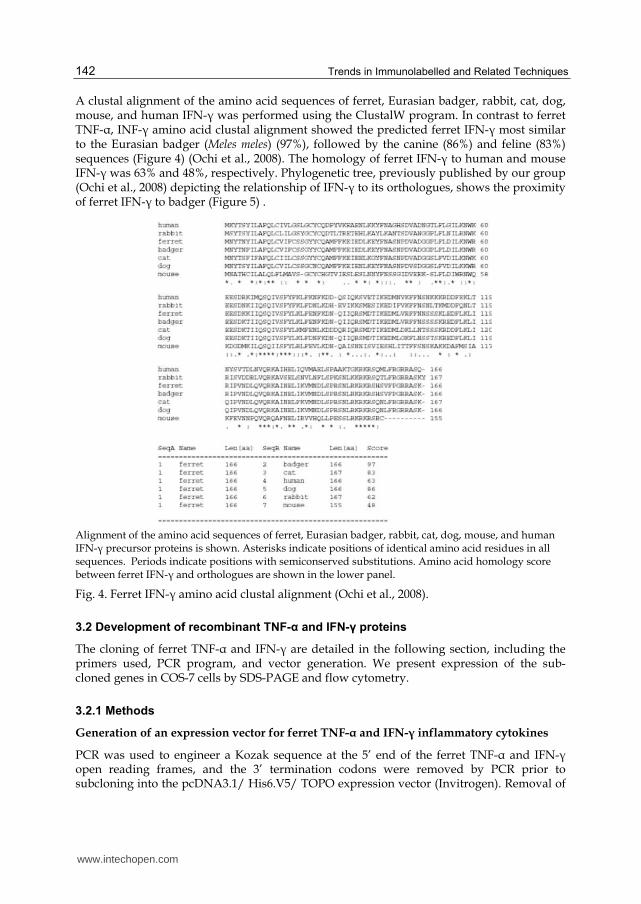
A clustal alignment of the amino acid sequences of ferret, Eurasian badger, rabbit, cat, dog, mouse, and human IFN-┛ was performed using the ClustalW program. In contrast to ferret TNF-┙, INF-┛ amino acid clustal alignment showed the predicted ferret IFN-┛ most similar to the Eurasian badger (Meles meles) (97%), followed by the canine (86%) and feline (83%) sequences (Figure 4) (Ochi et al., 2008). The homology of ferret IFN-┛ to human and mouse IFN-┛ was 63% and 48%, respectively. Phylogenetic tree, previously published by our group (Ochi et al., 2008) depicting the relationship of IFN-┛ to its orthologues, shows the proximity of ferret IFN-┛ to badger (Figure 5) .
Alignment of the amino acid sequences of ferret, Eurasian badger, rabbit, cat, dog, mouse, and human IFN-┛ precursor proteins is shown. Asterisks indicate positions of identical amino acid residues in all sequences. Periods indicate positions with semiconserved substitutions. Amino acid homology score between ferret IFN-┛ and orthologues are shown in the lower panel.
Fig. 4. Ferret IFN-┛ amino acid clustal alignment (Ochi et al., 2008).
3.2 Development of recombinant TNF-α and IFN-γ proteins
The cloning of ferret TNF-┙ and IFN-┛ are detailed in the following section, including the primers used, PCR program, and vector generation. We present expression of the sub-cloned genes in COS-7 cells by SDS-PAGE and flow cytometry.
3.2.1 Methods
Generation of an expression vector for ferret TNF-α and IFN-γ inflammatory cytokines
PCR was used to engineer a Kozak sequence at the 5’ end of the ferret TNF-┙ and IFN-┛ open reading frames, and the 3’ termination codons were removed by PCR prior to subcloning into the pcDNA3.1/ His6.V5/ TOPO expression vector (Invitrogen). Removal of
www.intechopen.com

Ferret TNF-α and IFN-γ Immunoassays 143
the termination codon enabled the cloned gene to be expressed as a fusion protein with two C-terminal epitope tags, His6 and V5.
Phylogenetic tree analysis showing the relationship between ferret and other known vertebrate IFN-┛ sequences. This tree was constructed using ClustalW and MEGA 3.1 packages and bootstrapped 10,000 times. †Bootstrapping confidence values are between 66 and 100.
Fig. 5. Ferret IFN-┛ phologenetic analysis (Ochi et al., 2008).
Transfection and purification of recombinant His-tagged TNF-α
COS-7 cells (ATCC, Manassas, Virginia, USA) were maintained in Dulbecco’s modified eagle’s medium (DMEM), supplemented with 10% fetal bovine serum (Invitrogen) at 37ºC, 5% CO2. COS-7 cells (1x10⁷) were transfected with Effectene transfection reagent according to manufacturer’s instructions (Qiagen, Mississauga, Ontario, Canada). Twenty-four to forty-eight hours following transfection, the cell culture media from the cells were run through Ni-NTA metal immobilized affinity columns to bind the His-tagged recombinant protein (Novogen, Oakville, ON, Canada). The columns were washed and the protein was eluted according to the manufacturer’s protocols. Fractions containing the recombinant proteins were pooled and dialyzed against phosphate buffered saline (PBS) at 4ºC and subsequently concentrated by spin column (Nanosep 10k OMEGA, Pall Life Science,East Hills, NY, USA). TNF-┙ and IFN-┛ protein concentrations were determined by protein assay kit (Pierce, Rockford, IL, USA).
Expression and purification of GST-IFN-γ using a bacterial expression system
Ferret IFN-┛ cDNA was subcloned into pGEX-6P1 vector (GE Healthcare) and purified as a GST fusion protein from Escherichia coli strain BL21 (DE3) by glutathione affinity chromatography.
Western Blot analysis
SDS-Poylacrylamide gel electrophoresis (10-15% SDS-PAGE) was performed with pre-cast gels (Bio-Rad, Hercules, CA, USA) according to standard protocols. Protein blots were blocked with 5% milk protein in 0.01% Tween-20 in PBS (T-PBS) for 1 hour at room
www.intechopen.com

Trends in Immunolabelled and Related Techniques 144
The expression construct encoding full-length ferret TNF-┙ was transfected into COS-7 cells and purified using with ion (Ni2+) immobilized affinity chromatography. The eluted protein fractions were subjected to SDS-PAGE and western blotting. Arrow depicts the band at predicted molecular weight of ferret TNF-┙. W3 denotes wash fraction 2, and E1 and E2 denote elution fractions 1 and 2.
Fig. 6. Purified recombinant ferret TNF-┙ protein molecular weight determined by western blot analysis.
Detection of recombinant ferret IFN-┛ by polyclonal antibody-specific immunoblotting.
Fig. 7. Detection of ferret recombinant IFN-┛.
temperature followed by an incubation of 16 hours at 4°C with mouse-anti-His6 primary antibody (Invitrogen) at 508 ng/ml concentration or monoclonal anti-V5 Ab (1:1000) (Invitrogen). The blots were washed and incubated with goat-anti-mouse-HRP secondary antibody (Santa Cruz, Santa Cruz, CA, USA). Bands were visualized using enhanced chemiluminiscent (ECL) reagents according to manufacturer’s protocol (GE Healthcare, Peterborough, Ontario, Canada).
3.2.2 Results
Expression and purification of the recombinant ferret TNF-α and IFN-γ
In order to obtain recombinant ferret TNF-┙ and IFN-┛ protein to be used in immunoassays and monoclonal antibody generation, TNF-┙ and IFN-┛ genes were first subcloned. Specifically, TNF-┙ and IFN-┛ cDNA were individually subcloned into expression plasmid to generate expression tag fusion proteins (described previously). Ferret TNF-┙ was expressed and purified from a mammalian cell line and IFN-┛ from a bacterial expression system.
The native human TNF-┙ polypeptide is cleaved at amino acid 76 to form the mature TNF-┙ signal peptide at 17215 Da (Wang et al., 1985). We predicted the ferret signal sequence cleavage site to be at the junction of 46 and 47 by SignalP 3.0 analysis. The observed molecular weight of ferret TNF-┙ according to our results was closer to that of human TNF-┙ at 25.7 kDa (including the His6 and V5 tags). In agreement, western blot analysis of the eluted fractions of ferret TNF- ┙ protein purified from COS-7 transfected cell media resulted in a single band of approximately 25.7 kDa molecular weight (Figure 6).
www.intechopen.com
Ferret TNF-α and IFN-γ Immunoassays 145
For IFN-┛, purified recombinant ferret IFN-┛ protein was expressed as a GST fusion protein and purified from chemically competent E. coli cells. Ferret recombinant IFN-┛ protein migrated as a 40 kDa band when subjected to SDS-PAGE and western blotting using a polyclonal anti-IFN-┛ antibody (Figure 7).
3.3 Cytokine real-time PCR based assay
Cytokines are important inflammatory and immune mediators. Here we present primers designed using ClustalW alignment analysis for studying ferret cytokines by real-time PCR.
3.3.1 Methods
ClustalW analysis for cytokine primer design
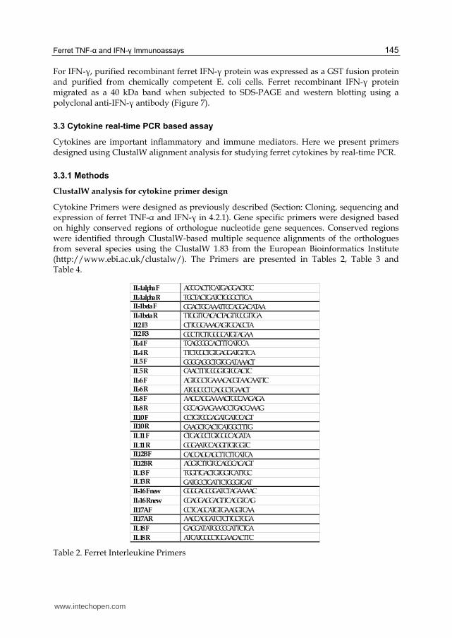
Cytokine Primers were designed as previously described (Section: Cloning, sequencing and expression of ferret TNF-┙ and IFN-┛ in 4.2.1). Gene specific primers were designed based on highly conserved regions of orthologue nucleotide gene sequences. Conserved regions were identified through ClustalW-based multiple sequence alignments of the orthologues from several species using the ClustalW 1.83 from the European Bioinformatics Institute (http://www.ebi.ac.uk/clustalw/). The Primers are presented in Tables 2, Table 3 and Table 4.
Table 2. Ferret Interleukine Primers
IL-1alpha F ACCCACTTCATGAGGACTGC
IL-1alpha R TGCTACTGATCTGGGCTTCA IL-1beta F GGACTGCAAATTCCAGGACATAA IL-1beta R TTGGTTCACACTAGTTCCGTTGA
IL2 F3 CTTCGCAAACAGTGCACCTA IL2 R3 GCCTTCTTGGGCATGTAGAA IL-4 F TCACCGGCACTTTCATCCA
IL-4 R TTCTCGCTGTGAGGATGTTCA IL 5 F GGGGAGGCTGTGGATAAACT IL 5 R CAACTTTCCGGTGTCCACTC
IL-6 F AGTGGCTGAAACACGTAACAATTC IL-6 R ATGGCCCTCAGGCTGAACT IL-8 F AAGCAGGAAAACTGCCAAGAGA
IL-8 R GCCAGAAGAAACCTGACCAAAG
IL10 F CCTGTCGGAGATGATCCAGT IL10 R CAAGCTCACTCATGGCTTTG IL 11 F CTGAGCCTGTGGCCAGATA
IL 11 R GGGAATCCAGGTTGTGGTC IL12B F CACCAGCAGCTTCTTCATCA IL12B R AGGTCTTGTCCACGCAGAGT
IL 13 F TGGTTGACTGTGGTCATTGC IL 13 R GATGCCTGATTCTGGGTGAT IL-16 F new GGGGAGCGGATCTAGAAAAC
IL-16 R new CGAGGAGGAGTTCAGGTCAG
IL17A F CCTCAGCATGTGAAGGTCAA IL17A R AACCAGGATCTCTTGCTGGA
IL 18 F GAGGATATGCCCGATTCTGA
IL 18 R ATCATGGCCTGGAACACTTC
www.intechopen.com
Trends in Immunolabelled and Related Techniques 146
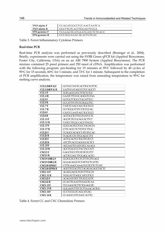
Table 3. Ferret Inflammatory Cytokine Primers
Real-time PCR
Real-time PCR analysis was performed as previously described (Bosinger et al., 2004). Briefly, experiments were carried out using the SYBR Green qPCR kit (Applied Biosystems, Foster City, California, USA) on an an ABI 7900 System (Applied Biosystems). The PCR mixture contained 25 pmol primers and 250 nmol of cDNA. Amplification was performed with the following program: pre-heating for 15 minutes at 95oC followed by 40 cycles of 95oC for 15 seconds, 60oC for 1 minute, and 72oC for 1 minute. Subsequent to the completion of PCR amplification, the temperature was raised from annealing temperature to 95oC for melting curve analysis.
Table 4. Ferret CC and CXC Chemokine Primers
TNF-alpha F CCAGATGGCCTCCAACTAATCA
TNF-alpha R GGCTTGTCACTTGGAGTTCGA IFN-gamma F TCAAAGTGATGAATGATCTCTCACC IFN-gamma R GCCGGGAAACACACTGTGAC
CCL2 (MCP-1) F GCTCCCTATTCACTTGCTGTTTC
CCL2 (MCP-1) R GATTCGATAGCCCTCCAGCTT CCL 4 F TGTGACCGTCCTTTCTCTCC CCL 4 R GAATCTTCCGCAGGGTGTAA
CCL5 F GCTGCTTTGCCTACATTTCC CCL5 R CCCATTTCTTCTGTGGGTTG CCL 7 F TATCTCAACCACCTGCTGCT
CCL 7 R GCTTGGGTTTTCTTGTCCAG CCL8 F CATCCCAATTACCTGCTGCT CCL8 R ACTGGCTGTTGGTGATCCTC
CCL 11 F AGGTCTCCGCAGCACTTCT CCL 11 R TATCCTTGGCCAGTTTGGTC CCL 17 F CGGGAGTGCTGCCTAGAGTA
CCL 17 R CTTCACCCTCTTGTCCTTGG
CCL21 F TCAGGCAGAGCTATGTGCAG CCL21 R TCAGTCCTCTTGCAGCCTTT CCL22 F ACTGCACTCCTGGTTGTCCT
CCL22 R ATCTTCACCCAGGGCACTCT CCL 23 F ACGAATTCGATGTGCAAACA CCL 23 R AGCTGGCCCCTACTACCATT
CXCL5 F GAGCTGCGTTGTGTGTGTTT CXCL5 R ACTTCCACCTTGGAGCACTG CXCL9 (MIG) F GGTGGTGTTCCTCTTTTGTTGAGT
CXCL9 (MIG) R GGAACAGCGTCTATTCCTCATTG
CXCL10 (IP10) F CTTTGAACCAAAGTGCTGTTCTTATC CXCL10 (IP10) R AGCGTGTAGTTCTAGAGAGAGGTACTC
CXCL 11 F AGAGGACGCTGTCTTTGCAT
CXCL 11 R TGGGATTTAGGCATCGTTGT CXCL12 F ACAGATGTCCTTGCCGATTC
CXCL12 R CCACTTCAATTTCGGGTCAA
CXCL 13 F TCCAAGGTGTTCTGGAGGTC CXCL 13 R GGGAATCTTTCTCTTAAACACTGG CXCL 14 F CCCTCCGGTCAGCATGAG
CXCL 14 R CCAGGCGTTGTACCACTTG
www.intechopen.com

Ferret TNF-α and IFN-γ Immunoassays 147
3.3.2 Results
Ferret cytokine and chemokine specific real-time PCR primers
Primers specific toward ferret cytokine and chemokine genes were designed by ClustalW analysis. Primers for ferret interleukins (Table 2), ferret inflammatory cytokines (Table 3) and ferret CC and CXC chemokines (Table 4) are described.
Induction of TNF-α target genes by treatment of ferret blood cells with recombinant ferret TNF-α
Once confirming that the recombinant TNF-┙ protein was of proper molecular weight, we then went on to assess the biological potential of the recombinant protein in vitro. Ferret whole peripheral blood was stimulated with recombinant ferret TNF-┙ and the RNA was extracted. Following extraction, the expression level of a panel of known TNF-┙ target cytokine/chemokine genes were measured by real-time PCR. CXCL8 (120-fold), IFN-┛ (12-fold), IL-1R (6-fold), IL-6 (3-fold) and IL-1┚ (2-fold) were increased following stimulation (Figure 8). These results indicated that the recombinant ferret TNF-┙ protein had biological activity.
Fig. 8. Real-Time PCR analysis of ferret cytokines transcript stimulated by TNF-┙
3.4 Development of IFN-γ and TNF-α hybridoma clones
The generation of monoclonal antibodies specific to ferret IFN-┛ and TNF-┙ are vital to the development of ELISAs and ELISPOTs. In this section we outline the development of these monoclonal antibodies using recombinant IFN-┛ and TNF-┙ conjugated to a carrier protein, KLH (keyhole limpet hemocyanin) using glutaraldehyde. Furthermore, the KLH-IFN-┛/TNF-┙ complexes injected and cell fusion to establish IFN-┛/TNF-┙ reactive B cell hybridomas is described along with downstream hybridoma clones slection.
3.4.1 Methods
Monoclonal anti-ferret TNF-α antibody production
Monoclonal antibodies to recombinant ferret TNF-┙ were manufactured by Open Biosystems (Birmingham, AL, USA).
Mouse B cell hybridoma preparation for anti-ferret-IFN-γ
Recombinant ferret IFN-┛ (50 µg) along with 2 mg of keyhole limpet hemocyaine (KLH)
(Calbiochem, San Diego, CA, USA) were diluted in 0.5 ml PBS. Five µl of glutaraldehyde
www.intechopen.com

Trends in Immunolabelled and Related Techniques 148
were added and the mixture was incubated for 1 hr at room temperature. The whole
mixture was washed on a spin column (Nanosep 10k OMEGA, Pall Life Science) and then
concentrated to 0.1 ml volume. PBS (0.5 ml) was then added and the mixture was
centrifuged. After two PBS washes, the mixture was filled to 0.5 ml with PBS. This mixture
was used as the priming antigen. Mice were immunized with 25 µl antigen suspension in
emulsified Complete Freund's Adjuvant and further injected at bi-weekly intervals with 5
µg of recombinant ferret IFN-┛. Three days following the third injection, spleen cells were
removed and isolated for fusion with Sp2/0-Ag14 using polyethylene glycol (Roche,
Mannheim, Germany). HAT (hypoxanthine aminopterin thymidine) resistant hybridomas
were selected. Hybridoma cells were screened for the reactivity against IFN-┛ by ELISA
using Nunc MaxiSorp 96 well plates coated with ferret IFN-┛ (100 µl, 0.1 µg/ml).
3.4.2 Results
Monoclonal ferret TNF-α antibody recognizes endogenous TNF-α isolated secreted from mitogen-stimulated ferret blood cells
A monoclonal anti-ferret TNF-┙ antibody was commercially manufactured by immunizing
mice with recombinant ferret TNF-┙. Isolated ferret peripheral blood cells were then
stimulated with the mitogens: SEB (Staphylococcal enterotoxin B), IFN-┛, ionomycin and
PMA plus ionomycin. Following stimuation, cell supernantants were run on SDS-PAGE and
analyzed by western blotting using the manufactured monoclonal ferret TNF-┙ antibody.
Endogenous ferret TNF-┙ protein was recognized in samples that had been stimulated with
SEB, IFN-┛ and PMA plus ionomycin. In contrast, TNF-┙ was not present in the supernatant
from cells treated with ionomycin alone or unstimulated cells (Figure 9). These results
suggested that the manufactured ferret monoclonal TNF-┙ antibody was able to recognize
endogenous secreted ferret TNF-┙.
PBMCs were isolated and stimulated with SEB (100 μg/ml), IFN-┛ (2000IU/ml), ionomycin alone (1 ug/ml) or PMA (50 ng/ml) with ionomycin (1 μg/ml), for 24 hrs at 37ºC. Cell supernatant was run on SDS-PAGE and subjected to western blotting with a monoclonal antibody (2μg/ml) against recombinant ferret TNF-┙.
Fig. 9. Ferret monoclonal TNF-┙ antibody recognizes secreted endogenous TNF-┙.
www.intechopen.com
Ferret TNF-α and IFN-γ Immunoassays 149
Generation of monoclonal Abs specific for ferret IFN-γ
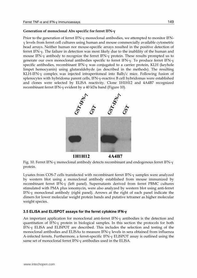
Prior to the generation of ferret IFN-┛ monoclonal antibodies, we attempted to monitor IFN-┛ levels from ferret cell cultures using human and mouse commercially available cytometric bead arrays. Neither human nor mouse-specific arrays resulted in the positive detection of ferret IFN-┛. The failure in detection was most likely due to the inablility of the human and mouse IFN-┛ antibody to recognize the ferret IFN-┛ protein. These results prompted us to generate our own monoclonal antibodies specific to ferret IFN-┛. To produce ferret IFN-┛ specific antibodies, recombinant IFN-┛ was conjugated to a carrier protein, KLH (keyhole limpet hemocyanin) using glutaraldehyde (as described in the methods). The resulting KLH-IFN-┛ complex was injected intraperitoneal into Balb/c mice. Following fusion of splenocytes with hybridoma parent cells, IFN-┛-reactive B cell hybridomas were established and clones were selected by ELISA reactivity. Clone 1H1H12 and 4A4B7 recognized recombinant ferret IFN-┛ evident by a 40 kDa band (Figure 10).
Fig. 10. Ferret IFN-┛ monoclonal antibody detects recombinant and endogenous ferret IFN-┛ protein.
Lysates from COS-7 cells transfected with recombinant ferret IFN-┛ samples were analyzed by western blot using a monoclonal antibody established from mouse immunized by recombinant ferret IFN-┛ (left panel). Supernatants derived from ferret PBMC cultures stimulated with PMA plus ionomycin, were also analyzed by western blot using anti-ferret IFN-┛ monoclonal antibody (right panel). Arrows at the right of each panel indicate the dimers for lower molecular weight protein bands and putative tetramer as higher molecular weight species.
3.5 ELISA and ELISPOT assays for the ferret cytokine IFN-γ
An important application for monoclonal anti-ferret IFN-┛ antibodies is the detection and quantitation of IFN-┛ protein in biological samples. In this section the protocols for both IFN-┛ ELISA and ELISPOT are described. This includes the selection and testing of the monoclonal antibodies and ELISAs to measure IFN-┛ levels in sera obtained from influenza A-infected ferrets. Furthermore, a ferret-specific IFN-┛ ELISPOT assay is outlined using the same set of monoclonal ferret IFN-┛ antibodies used in the ELISA.
www.intechopen.com

Trends in Immunolabelled and Related Techniques 150
3.5.1 Methods
Ferret IFN-γ-specific ELISA
ELISA plates (96-well) (MaxiSorb, Nunc) were coated with 100 µl/well with monoclonal
anti-IFN-┛ (2 µg/ml) overnight at 4oC. Wells were blocked with 150 µl 1% BSA in PBS for 1
hour at 37oC. Supernatants from mitogen-stimulated PBMC cultures or serum from
influenza A virus infected ferrets were loaded into the wells at appropriate dilutions and
incubated for 1 hour at 37oC. Wells were washed with PBS/0.5% Tween-20 and then
incubated for 1 hour at room temperature with biotin conjugated anti-IFN-┛ antibody (1
µg/ml in 0.5% Tween-20/1%BSA). Following secondary antibody incubation, the wells
were washed three times with PBS/0.5% Tween-20 before incubation with HRP-Avidin for
30 minutes. The substrate, (o-phenylenediamine, Sigma) was applied for 15 minutes at room
temperature. Colorimetric changes were quantitated using an automated ELISA reader
(µQuant, BIO-TEK Instruments, Winooski, VT, USA).
ELISA plate wells were coated with a monoclonal anti-ferret antibody. Recombinant ferret IFN-┛ was
sequentially diluted and added to the wells. Ferret IFN-┛ was detected by a second monoclonal anti-
ferret IFN-┛ biotin conjugated antibody. Logarithmic dilution was used to derive a standard curve for
downstream applications of the ELISA.
Fig. 11. Recombinant IFN-┛ protein quantification by ELISA.
Ferret IFN-γ-specific ELISPOT assay
PVDF plates (Millipore, MAIPS4510) or MaxiSorp plates (Nunc) were coated with a
monoclonal anti-ferret IFN-┛ antibody, and subsequently blocked with 1% BSA in PBS.
Isolated ferret PBMCs from peripheral blood were stimulated for 18 hours. The wells were
then washed with water to remove cells and captured IFN-┛ was detected by a biotin-
conjugated detection antibody coupled to HRP-avidin (Sigma). The ELISPOT was
developed using DAB (Vector Laboratories, Burlingame, CA, USA).
0
0.1
0.2
0.3
0.4
0.5
0.6
0.7
0.8
0.9
100 50 25 12.5 6.25 3.125 0
IFN-gamma protein concentration (ug/mL)
Ab
so
rban
ce
GST-IFNg
GST
www.intechopen.com
Ferret TNF-α and IFN-γ Immunoassays 151
3.5.2 Results
Detection of ferret IFN-γ by ELISA and ELISPOT immunoassays
An important application for monoclonal antibodies is the detection and quantitation of
protein in biological samples. Here we tested our monoclonal antibodies for use in a ferret
IFN-┛-specific ELISA by first determining the antibody pair for IFN-┛ recognition. Clone
4A4B7 was conjugated to biotin and used as the detection antibody against ferret IFN-┛.
Clone 1H1H12 was used to coat the assay wells and be used as a capture antibody.
This clone pair showed an increased optical density that correlated directly with the
concentration of purified recombinant IFN-┛ (Figure 11).
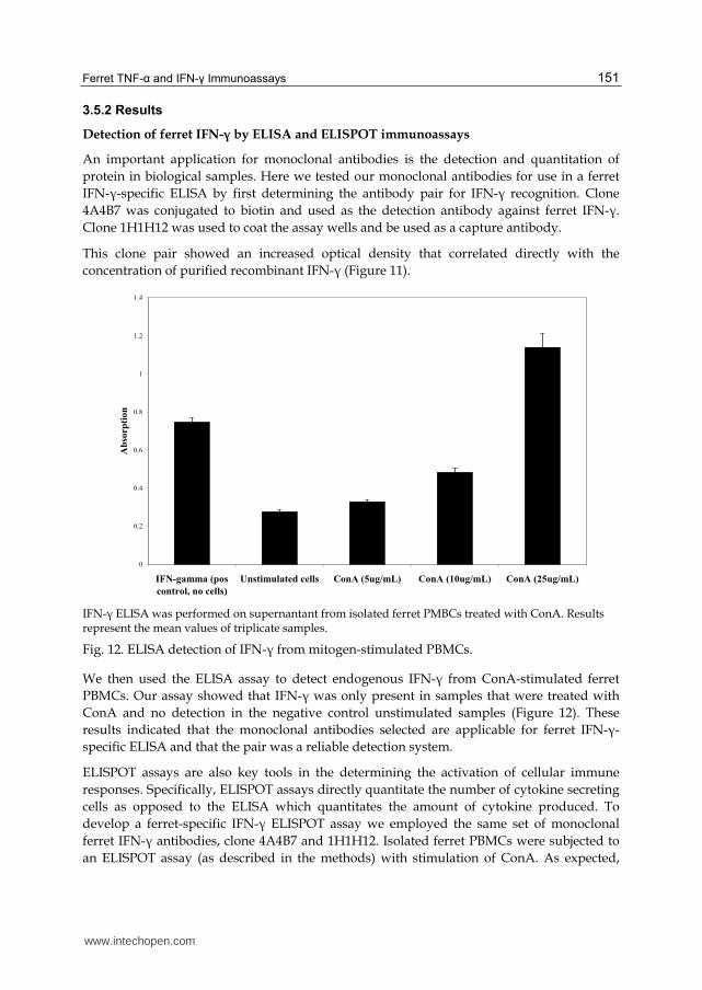
IFN-┛ ELISA was performed on supernantant from isolated ferret PMBCs treated with ConA. Results represent the mean values of triplicate samples.
Fig. 12. ELISA detection of IFN-┛ from mitogen-stimulated PBMCs.
We then used the ELISA assay to detect endogenous IFN-┛ from ConA-stimulated ferret
PBMCs. Our assay showed that IFN-┛ was only present in samples that were treated with
ConA and no detection in the negative control unstimulated samples (Figure 12). These
results indicated that the monoclonal antibodies selected are applicable for ferret IFN-┛-
specific ELISA and that the pair was a reliable detection system.
ELISPOT assays are also key tools in the determining the activation of cellular immune
responses. Specifically, ELISPOT assays directly quantitate the number of cytokine secreting
cells as opposed to the ELISA which quantitates the amount of cytokine produced. To
develop a ferret-specific IFN-┛ ELISPOT assay we employed the same set of monoclonal
ferret IFN-┛ antibodies, clone 4A4B7 and 1H1H12. Isolated ferret PBMCs were subjected to
an ELISPOT assay (as described in the methods) with stimulation of ConA. As expected,
0
0.2
0.4
0.6
0.8
1
1.2
1.4
IFN-gamma (pos
control, no cells)
Unstimulated cells ConA (5ug/mL) ConA (10ug/mL) ConA (25ug/mL)
Ab
sorp
tion
www.intechopen.com

Trends in Immunolabelled and Related Techniques 152
increasing numbers of IFN-┛ secreting cells were detected in direct proportion to the
number of stimulated cells plated. Furthermore, the number of IFN-┛ positive cells did not
increase above background when increasing numbers of unstimulated cells were plated
(Figure 13). These results suggested that 4A4B7 and 1H1H12 anti-ferret IFN-┛ monoclonal
antibodies will be useful for not only quantitating ferret IFN-┛ levels but also for
quantitating IFN-┛ producing ferret cells in an ELISPOT assay.
ELISPOT assay was performed in the same manner outlined for Figure 12 for capture and detection.
PBMCs were plated and stimulated with ConA 18 hours and IFN-┛ secreting cells were captured and
detected by biotinylated IFN-g antibody.
Fig. 13. ELISPOT assay for the IFN-┛ producing cells in mitogen-stimulated ferret PBMCs.
4. Conclusion
The development of ferret specific immune reagents will significantly improve knowledge
gained by ferret infectious disease studies. The importance of the ferret as an animal model
in the study of respiratory diseases is increasing. Currently, the use of the ferret model to
investigate the immune response as well as immunotherapeutics in respiratory infections by
viruses such as SARS CoV and influenza has been slowed by the lack of immunological
reagents and immune response assessment tools. In this chapter we highlighted immune
reagents and immunoassays that are successful at monitoring the ferret immune response,
specifically focusing on cytokines.
Currently, there is much interest to determine the immunological role of TNF-┙ during
influenza infection, as TNF-┙ is thought to play a pathogenic role in other respiratory
infections (Headley, Tolley, & Meduri, 1997). Here we have described the cloning and
sequence of the ferret TNF-┙ gene with the purpose of generating reagents and assays for
the investigation of ferret immune responses. With the generated ferret TNF-┙ expression
vector we were able to express and purify recombinant protein for downstream uses.
Importantly, the recombinant ferret TNF-┙ protein was able to stimulate ferret peripheral
blood cells and induce the expression of TNF-┙ target genes such as: CXCL-8, IFN-┛, IL-1┚,
IL-6 and IL-1R (Vassalli, 1992; Baumann & Gauldie, 1994; Dinarello, 1996; Gabay & Kushner,
1999). Furthermore, we have successfully generated monoclonal antibodies that specifically
recognized ferret TNF-┙ as well as primers that measured TNF-┙ transcripts in cells
stimulated with LPS, a known stimulant of TNF-┙ (Curnis & Corti, 2004; Mestan et al., 1986;
Wang et al., 1985). In summary, we have cloned, sequenced and expressed recombinant
ferret TNF-┙ and demonstrated its biological activity. Our ferret TNF-┙ protein has enabled
us to generate several useful reagents that will be important tools for immune modeling in
respiratory diseases.
www.intechopen.com

Ferret TNF-α and IFN-γ Immunoassays 153
IFN-┛ is an essential regulator in the viral host immune response. Here the cloning
of a full length ferret IFN-┛ and expression of the recombinant IFN-┛ protein was
described. Furthermore, we described the generation of two monoclonal antibodies
specific for ferret IFN-┛ and the subsequent development of immunoassays for the
detection of native IFN-┛. We anticipate that the IFN-┛ immunoassays established in this
study will be useful in gaining insight into ferret antiviral responses and in other immune
processes in general.
Previous studies on IFN-┛ have shown that there is a strict species-specific activity of IFN-┛.
IFN-┛ genes isolated from diverse species such as guinea pig, turkey, rhino, and catfish have
been previously described in the literature (Jeevan et al., 2006; Loa, Hsieh, Wu, & Lin, 2001;
Milev-Milovanovic et al., 2006; Morar et al., 2007; Svitek & von, V, 2007). Our monoclonal
antibodies detected recombinant IFN-┛ in Western blotting. These two clones were paired
successfully during the development of ferret IFN-┛-specific ELISA and ELISPOT bioassays.
Previously, we infected ferrets with H3N2 influenza A strain (A/Panama/2007/99) and
sampled serum from the animals 6 days post infection (Ochi et al., 2008). The ELISA showed
a marked increase in the levels of circulating IFN-┛ on day 6 post-infection compared to the
uninfected control. Specifically, the level of IFN-┛ in serum from the non-infected control
ferret was below the detection limit. These results suggested that our developed ELISA
assay for quantitation of ferret IFN-┛ will be invaluable in monitoring systemic IFN-┛
responses during a host response against virus infection.
There is an overwhelming need for experimental models that enable the evaluation of
therapeutic treatments and vaccines for use in infectious diseases such as emerging
influenza viruses. Ferrets have been used as an animal model of infection with influenza A
viruses to test the severity of the disease and also to evaluate efficacy of potential vaccines
(Suguitan, Jr. et al., 2006; Cameron et al., 2008; Danesh et al., 2011; Rowe et al., 2010b).
Specifically, TNF-┙ and IFN-┛ have been identified as major inducers of pathogenesis in
respiratory illnesses which includes SARS (Cheung et al., 2005), (Roberts & Subbarao, 2006)
and influenza virus (Maher & DeStefano, 2004; Cheung et al., 2002; de Jong et al., 2006).
From these findings comes the hypothesis that these inflammatory cytokines may be the key
to potential future therapies.
TNF-┙ antibodies have been shown to have therapeutic potential in disease either as
therapeutic targets or as biomarkers. In a mouse model of influenza-induced pneumonia, a
neutralizing antibody to TNF-┙ significantly reduced the lung pathology and prolonged
survival of infected animals thereby suggesting TNF-┙ as a therapeutic target (Peper & Van,
1995). With the antibodies generated, it is now possible to investigate the therapeutic
potential of targeting TNF-┙ to modulate the immunopathology during influenza infection.
Alternatively, IFN-┛ has been shown to be a useful biomarker. ELISPOT and ELISA assays
are excellent techniques that can be employed in studies monitoring vaccine and therapeutic
efficacy by measuring the levels of biomarkers such as IFN-┛. Taken together, these reagents
will be helpful in the assessment of vaccine efficacy against influenza A and other emerging
infectious viruses.
In conclusion, this work has expanded the potential of the ferret model for respiratory
disease investigation as well as other diseases that involved the immune response.
www.intechopen.com

Trends in Immunolabelled and Related Techniques 154
5. References
Anglen, C. S., Truckenmiller, M. E., Schell, T. D., & Bonneau, R. H. (2003). The dual role of
CD8+ T lymphocytes in the development of stress-induced herpes simplex
encephalitis. J.Neuroimmunol., 140, 13-27.
Baumann, H. & Gauldie, J. (1994). The acute phase response. Immunol.Today, 15, 74-80.
Belser, J. A., Szretter, K. J., Katz, J. M., & Tumpey, T. M. (2009). Use of animal models to
understand the pandemic potential of highly pathogenic avian influenza viruses.
Adv.Virus Res., 73, 55-97.
Belz, G. T., Bedoui, S., Kupresanin, F., Carbone, F. R., & Heath, W. R. (2007). Minimal
activation of memory CD8(+) T cell by tissue-derived dendritic cells favors the
stimulation of naive CD8(+) T cells. Nat.Immunol., 8, 1060-1066.
Boon, A. C., de Mutsert, G., Fouchier, R. A., Osterhaus, A. D., & Rimmelzwaan, G. F. (2005).
Functional profile of human influenza virus-specific cytotoxic T lymphocyte
activity is influenced by interleukin-2 concentration and epitope specificity.
Clin.Exp.Immunol., 142, 45-52.
Bossart, K. N., Zhu, Z., Middleton, D., Klippel, J., Crameri, G., Bingham, J. et al. (2009). A
neutralizing human monoclonal antibody protects against lethal disease in a new
ferret model of acute nipah virus infection. PLoS.Pathog., 5, e1000642.
Bouvier, N. M. & Lowen, A. C. (2010). Animal Models for Influenza Virus Pathogenesis and
Transmission. Viruses., 2, 1530-1563.
Bouvier, N. M., Lowen, A. C., & Palese, P. (2008). Oseltamivir-resistant influenza A viruses
are transmitted efficiently among guinea pigs by direct contact but not by aerosol.
J.Virol., 82, 10052-10058.
Brown, T. J., Crawford, S. E., Cornwall, M. L., Garcia, F., Shulman, S. T., & Rowley, A. H.
(2001). CD8 T lymphocytes and macrophages infiltrate coronary artery aneurysms
in acute Kawasaki disease. J.Infect.Dis., 184, 940-943.
Cameron, C. M., Cameron, M. J., Bermejo-Martin, J. F., Ran, L., Xu, L., Turner, P. V. et al.
(2008). Gene expression analysis of host innate immune responses during Lethal
H5N1 infection in ferrets. J.Virol., 82, 11308-11317.
Cameron, M. J., Ran, L., Xu, L., Danesh, A., Bermejo-Martin, J. F., Cameron, C. M. et al.
(2007). Interferon-mediated immunopathological events are associated with
atypical innate and adaptive immune responses in patients with severe acute
respiratory syndrome. J.Virol., 81, 8692-8706.
Centers for Disease Control and Prevention (CDC) (2009). Intensive-care patients with
severe novel influenza A (H1N1) virus infection - Michigan, June 2009. MMWR -
Morbidity & Mortality Weekly Report.58(27):749-52.
Chelbi-Alix, M. K. & Wietzerbin, J. (2007). Interferon, a growing cytokine family: 50 years of
interferon research. Biochimie, 89, 713-718.
Chen, H. C., Lai, S. Y., Sung, J. M., Lee, S. H., Lin, Y. C., Wang, W. K. et al. (2004).
Lymphocyte activation and hepatic cellular infiltration in immunocompetent mice
infected by dengue virus. J.Med.Virol., 73, 419-431.
Cheung, C. Y., Poon, L. L., Lau, A. S., Luk, W., Lau, Y. L., Shortridge, K. F. et al. (2002).
Induction of proinflammatory cytokines in human macrophages by influenza A
www.intechopen.com

Ferret TNF-α and IFN-γ Immunoassays 155
(H5N1) viruses: a mechanism for the unusual severity of human disease? Lancet,
360, 1831-1837.
Cheung, C. Y., Poon, L. L., Ng, I. H., Luk, W., Sia, S. F., Wu, M. H. et al. (2005). Cytokine
responses in severe acute respiratory syndrome coronavirus-infected
macrophages in vitro: possible relevance to pathogenesis. J.Virol., 79, 7819-
7826.
Chevaliez, S. & Pawlotsky, J. M. (2009). Interferons and their use in persistent viral
infections. Handb.Exp.Pharmacol., 203-241.
Curnis, F. & Corti, A. (2004). Production and characterization of recombinant human and
murine TNF. Methods Mol.Med., 98, 9-22.
Danesh, A., Cameron, C. M., Leon, A. J., Ran, L., Xu, L., Fang, Y. et al. (2011). Early gene
expression events in ferrets in response to SARS coronavirus infection versus direct
interferon-alpha2b stimulation. Virology, 409, 102-112.
Danesh, A., Seneviratne, C., Cameron, C. M., Banner, D., DeVries, M. E., Kelvin, A. A. et al.
(2008). Cloning, expression and characterization of ferret CXCL10. Mol.Immunol.,
45, 1288-1297.
Darnell, M. E., Plant, E. P., Watanabe, H., Byrum, R., St Claire, M., Ward, J. M. et al. (2007).
Severe acute respiratory syndrome coronavirus infection in vaccinated ferrets.
J.Infect.Dis., 196, 1329-1338.
Dawood, F. S., Dalton, C. B., Durrheim, D. N., & Hope, K. G. (2009). Rates of
hospitalisation for acute respiratory illness and the emergence of pandemic
(H1N1) 2009 virus in the Hunter New England Area Health Service. Med.J.Aust.,
191, 573-574.
Dawood, F. S., Jain, S., Finelli, L., Shaw, M. W., Lindstrom, S., Garten, R. J. et al. (2009).
Emergence of a novel swine-origin influenza A (H1N1) virus in humans.
N.Engl.J.Med., 360, 2605-2615.
de Jong, M. D., Simmons, C. P., Thanh, T. T., Hien, V. M., Smith, G. J., Chau, T. N. et al.
(2006). Fatal outcome of human influenza A (H5N1) is associated with high viral
load and hypercytokinemia. Nat.Med., 12, 1203-1207.
Dinarello, C. A. (1996). Biologic basis for interleukin-1 in disease. Blood, 87, 2095-2147.
Dushoff, J., Plotkin, J. B., Viboud, C., Earn, D. J., & Simonsen, L. (2006). Mortality due to
influenza in the United States--an annualized regression approach using multiple-
cause mortality data. Am.J.Epidemiol., 163, 181-187.
Foxwell, A. R., Kyd, J. M., Karupiah, G., & Cripps, A. W. (2001). CD8+ T cells have an
essential role in pulmonary clearance of nontypeable Haemophilus influenzae
following mucosal immunization. Infect.Immun., 69, 2636-2642.
Gabay, C. & Kushner, I. (1999). Acute-phase proteins and other systemic responses to
inflammation. N.Engl.J.Med., 340, 448-454.
Gilsdorf, A., Poggensee, G., & Working Group (2009). Influenza A(H1N1)v in Germany: the
first 10,000 cases. Euro Surveillance: Bulletin Europeen sur les Maladies
Transmissibles = European Communicable Disease Bulletin.14(34).
Girard, M. P., Cherian, T., Pervikov, Y., & Kieny, M. P. (2005). A review of vaccine research
and development: human acute respiratory infections. [Review] [107 refs].
Vaccine.23(50):5708-24.
www.intechopen.com

Trends in Immunolabelled and Related Techniques 156
Govorkova, E. A., Ilyushina, N. A., Boltz, D. A., Douglas, A., Yilmaz, N., & Webster, R. G.
(2007). Efficacy of oseltamivir therapy in ferrets inoculated with different clades of
H5N1 influenza virus. Antimicrob.Agents Chemother., 51, 1414-1424.
Gupta, V., Earl, D. J., & Deem, M. W. (2006). Quantifying influenza vaccine efficacy and
antigenic distance. Vaccine, 24, 3881-3888.
Haagmans, B. L., Kuiken, T., Martina, B. E., Fouchier, R. A., Rimmelzwaan, G. F., van, A. G.
et al. (2004). Pegylated interferon-alpha protects type 1 pneumocytes against SARS
coronavirus infection in macaques. Nat.Med., 10, 290-293.
Hauge, S., Madhun, A. S., Cox, R. J., Brokstad, K. A., & Haaheim, L. R. (2007). A
comparison of the humoral and cellular immune responses at different
immunological sites after split influenza virus vaccination of mice.
Scand.J.Immunol., 65, 14-21.
Headley, A. S., Tolley, E., & Meduri, G. U. (1997). Infections and the inflammatory response
in acute respiratory distress syndrome. Chest, 111, 1306-1321.
Health Protection Agency, Health, P. S., National Public Health Service for Wales, & HPA
Northern Ireland Swine influenza investigation team (2009). Epidemiology of new
influenza A (H1N1) virus infection, United Kingdom, April-June 2009. Euro
Surveillance: Bulletin Europeen sur les Maladies Transmissibles = European
Communicable Disease Bulletin.14(22).
Hoji, A. & Rinaldo, C. R., Jr. (2005). Human CD8+ T cells specific for influenza A virus M1
display broad expression of maturation-associated phenotypic markers and
chemokine receptors. Immunology, 115, 239-245.
Huang S.S.H., Banner, D., Fang, Y., Ng, D. C., Kanagasabai T., Kelvin, D. J. et al.
Comparative Analyses of Pandemic H1N1 and Seasonal H1N1, H3N2, and
Influenza B Infections Depict Distinct Clinical Pictures In Ferrets. PLoS One, (in
press).
HULL, R. B. & LOOSLI, C. G. (1951). Adrenocorticotrophic hormone (ACTH) in the
treatment of experimental air-borne influenza virus type A infection in the ferret.
J.Lab Clin.Med., 37, 603-614.
Hussell, T., Pennycook, A., & Openshaw, P. J. (2001). Inhibition of tumor necrosis factor
reduces the severity of virus-specific lung immunopathology. Eur.J.Immunol., 31,
2566-2573.
Jeevan, A., McFarland, C. T., Yoshimura, T., Skwor, T., Cho, H., Lasco, T. et al. (2006).
Production and characterization of guinea pig recombinant gamma interferon and
its effect on macrophage activation. Infect.Immun., 74, 213-224.
Kasowski, E. J., Garten, R. J., & Bridges, C. B. (2011). Influenza pandemic epidemiologic and
virologic diversity: reminding ourselves of the possibilities. [Review]. Clinical
Infectious Diseases.52 Suppl 1:S44-9.
Kolling, U. K., Hansen, F., Braun, J., Rink, L., Katus, H. A., & Dalhoff, K. (2001). Leucocyte
response and anti-inflammatory cytokines in community acquired pneumonia.
Thorax, 56, 121-125.
Kumar, A. M., Zarychanski, R. M., Pinto, R. P., Cook, D. J. M., Marshall, J. M., Lacroix, J. M.
et al. (2009). Critically Ill Patients With 2009 Influenza A(H1N1) Infection in
Canada. [Miscellaneous Article]. JAMA, 302, 1872-1879.
www.intechopen.com

Ferret TNF-α and IFN-γ Immunoassays 157
Lambkin, R., Oxford, J. S., Bossuyt, S., Mann, A., Metcalfe, I. C., Herzog, C. et al. (2004).
Strong local and systemic protective immunity induced in the ferret model by an
intranasal virosome-formulated influenza subunit vaccine. Vaccine, 22, 4390-
4396.
Loa, C. C., Hsieh, M. K., Wu, C. C., & Lin, T. L. (2001). Molecular identification and
characterization of turkey IFN-gamma gene. Comp Biochem.Physiol B
Biochem.Mol.Biol., 130, 579-584.
Loutfy, M. R., Blatt, L. M., Siminovitch, K. A., Ward, S., Wolff, B., Lho, H. et al. (2003).
Interferon alfacon-1 plus corticosteroids in severe acute respiratory syndrome: a
preliminary study. JAMA, 290, 3222-3228.
Maher, J. A. & DeStefano, J. (2004). The ferret: an animal model to study influenza virus. Lab
Anim (NY), 33, 50-53.
Marijanovic, Z., Ragimbeau, J., van der Heyden, J., Uze, G., & Pellegrini, S. (2007).
Comparable potency of IFNalpha2 and IFNbeta on immediate JAK/STAT
activation but differential down-regulation of IFNAR2. Biochem.J., 407, 141-
151.
Martina, B. E., Haagmans, B. L., Kuiken, T., Fouchier, R. A., Rimmelzwaan, G. F., Van
Amerongen, G. et al. (2003). Virology: SARS virus infection of cats and ferrets.
Nature, 425, 915.
Mendel, D. B., Tai, C. Y., Escarpe, P. A., Li, W., Sidwell, R. W., Huffman, J. H. et al. (1998).
Oral administration of a prodrug of the influenza virus neuraminidase inhibitor GS
4071 protects mice and ferrets against influenza infection. Antimicrob.Agents
Chemother., 42, 640-646.
Mestan, J., Digel, W., Mittnacht, S., Hillen, H., Blohm, D., Moller, A. et al. (1986).
Antiviral effects of recombinant tumour necrosis factor in vitro. Nature, 323,
816-819.
Milev-Milovanovic, I., Long, S., Wilson, M., Bengten, E., Miller, N. W., & Chinchar, V. G.
(2006). Identification and expression analysis of interferon gamma genes in channel
catfish. Immunogenetics, 58, 70-80.
Morar, D., Tijhaar, E., Negrea, A., Hendriks, J., van, H. D., Godfroid, J. et al. (2007). Cloning,
sequencing and expression of white rhinoceros (Ceratotherium simum) interferon-
gamma (IFN-gamma) and the production of rhinoceros IFN-gamma specific
antibodies. Vet.Immunol.Immunopathol., 115, 146-154.
Nicoll, A. & Coulombier, D. (2009). Europe's initial experience with pandemic (H1N1) 2009 -
mitigation and delaying policies and practices. Euro Surveillance: Bulletin
Europeen sur les Maladies Transmissibles = European Communicable Disease
Bulletin.14(29).
Ochi, A., Danesh, A., Seneviratne, C., Banner, D., Devries, M. E., Rowe, T. et al. (2008).
Cloning, expression and immunoassay detection of ferret IFN-gamma. Dev.Comp
Immunol., 32, 890-897.
Peltola, V. T., Boyd, K. L., McAuley, J. L., Rehg, J. E., & McCullers, J. A. (2006). Bacterial
sinusitis and otitis media following influenza virus infection in ferrets.
Infect.Immun., 74, 2562-2567.
www.intechopen.com

Trends in Immunolabelled and Related Techniques 158
Peper, R. L. & Van, C. H. (1995). Tumor necrosis factor as a mediator of inflammation in
influenza A viral pneumonia. Microb.Pathog., 19, 175-183.
Perez-Padilla, R., Rosa-Zamboni, D., Ponce, d. L., Hernandez, M., Quinones-Falconi, F.,
Bautista, E. et al. (2009). Pneumonia and respiratory failure from swine-origin
influenza A (H1N1) in Mexico. New England Journal of Medicine.361(7):680-9.
Pinto, R. D., Nascimento, D. S., Vale, A., & Santos, N. M. (2006). Molecular cloning and
characterization of sea bass (Dicentrarchus labrax L.) CD8alpha.
Vet.Immunol.Immunopathol., 110, 169-177.
Roberts, A. & Subbarao, K. (2006). Animal models for SARS. Adv.Exp.Med.Biol., 581, 463-
471.
Rowe, T., Banner, D., Farooqui, A., Ng, D. C., Kelvin, A. A., Rubino, S. et al. (2010a). In vivo
ribavirin activity against severe pandemic H1N1 Influenza A/Mexico/4108/2009.
J.Gen.Virol., 91, 2898-2906.
Rowe, T., Leon, A. J., Crevar, C. J., Carter, D. M., Xu, L., Ran, L. et al. (2010b). Modeling host
responses in ferrets during A/California/07/2009 influenza infection. Virology,
401, 257-265.
Ryan, G. B. & Majno, G. (1977). Acute inflammation. A review. The American journal of
pathology, 86, 183.
Seo, S. H. & Webster, R. G. (2002). Tumor necrosis factor alpha exerts powerful anti-
influenza virus effects in lung epithelial cells. J.Virol., 76, 1071-1076.
Small, P. A., Jr., Waldman, R. H., Bruno, J. C., & Gifford, G. E. (1976). Influenza infection in
ferrets: role of serum antibody in protection and recovery. Infect.Immun., 13, 417-
424.
Somamoto, T., Yoshiura, Y., Nakanishi, T., & Ototake, M. (2005). Molecular cloning and
characterization of two types of CD8alpha from ginbuna crucian carp, Carassius
auratus langsdorfii. Dev.Comp Immunol., 29, 693-702.
Steinhauer, D. A. & Skehel, J. J. (2002). Genetics of influenza viruses. [Review] [175 refs].
Annual Review of Genetics.36:305-32.
Suguitan, A. L., Jr., McAuliffe, J., Mills, K. L., Jin, H., Duke, G., Lu, B. et al. (2006). Live,
attenuated influenza A H5N1 candidate vaccines provide broad cross-protection in
mice and ferrets. PLoS Med., 3, e360.
Svitek, N. & von, M., V (2007). Early cytokine mRNA expression profiles predict
Morbillivirus disease outcome in ferrets. Virology, 362, 404-410.
Sweet, C., Bird, R. A., Cavanagh, D., Toms, G. L., Collie, M. H., & Smith, H. (1979). The local
origin of the febrile response induced in ferrets during respiratory infection with a
virulent influenza virus. Br.J.Exp.Pathol., 60, 300-308.
Takaoka, A. & Yanai, H. (2006). Interferon signalling network in innate defence. Cell
Microbiol., 8, 907-922.
ter, M. J., van den Brink, E. N., Poon, L. L., Marissen, W. E., Leung, C. S., Cox, F. et al. (2006).
Human monoclonal antibody combination against SARS coronavirus: synergy and
coverage of escape mutants. PLoS.Med., 3, e237.
Trifonov, V., Khiabanian, H., & Rabadan, R. (2009). Geographic dependence, surveillance,
and origins of the 2009 influenza A (H1N1) virus. New England Journal of
Medicine.361(2):115-9.
www.intechopen.com

Ferret TNF-α and IFN-γ Immunoassays 159
Uddin, S. & Platanias, L. C. (2004). Mechanisms of type-I interferon signal transduction.
J.Biochem.Mol.Biol., 37, 635-641.
Uyeki, T. M. M., Sharma, A. M. D., & Branda, J. A. M. (2009). Case 40-2009: A 29-Year-Old
Man with Fever and Respiratory Failure. [Miscellaneous Article]. New England
Journal of Medicine, 361, 2558-2569.
van den Brand, J. M., Haagmans, B. L., Leijten, L., van, R. D., Martina, B. E., Osterhaus, A. D.
et al. (2008). Pathology of experimental SARS coronavirus infection in cats and
ferrets. Vet.Pathol., 45, 551-562.
van, R. D., Munster, V. J., de, W. E., Rimmelzwaan, G. F., Fouchier, R. A., Osterhaus, A. D. et
al. (2007). Human and avian influenza viruses target different cells in the lower
respiratory tract of humans and other mammals. Am.J.Pathol., 171, 1215-1223.
Vassalli, P. (1992). The pathophysiology of tumor necrosis factors. Annu.Rev.Immunol., 10,
411-452.
Vikman, S., Giandomenico, V., Sommaggio, R., Oberg, K., Essand, M., & Totterman, T. H.
(2007). CD8(+) T cells against multiple tumor-associated antigens in peripheral
blood of midgut carcinoid patients. Cancer Immunol.Immunother..
Vilcek, J. & Lee, T. H. (1991). Tumor necrosis factor. New insights into the molecular
mechanisms of its multiple actions. J.Biol.Chem., 266, 7313-7316.
Wang, A. M., Creasey, A. A., Ladner, M. B., Lin, L. S., Strickler, J., Van Arsdell, J. N. et al.
(1985). Molecular cloning of the complementary DNA for human tumor necrosis
factor. Science, 228, 149-154.
Wang, Y., Lobigs, M., Lee, E., & Mullbacher, A. (2003). CD8+ T cells mediate recovery and
immunopathology in West Nile virus encephalitis. J.Virol., 77, 13323-13334.
Weiss, R. A. & McMichael, A. J. (2004). Social and environmental risk factors in the
emergence of infectious diseases. Nat.Med., 10, S70-S76.
Willemsen, R. A., Sebestyen, Z., Ronteltap, C., Berrevoets, C., Drexhage, J., & Debets, R.
(2006). CD8 alpha coreceptor to improve TCR gene transfer to treat melanoma:
down-regulation of tumor-specific production of IL-4, IL-5, and IL-10. J.Immunol.,
177, 991-998.
Writing Committee of the WHO Consultation on Clinical Aspects of Pandemic ( (2010).
Clinical Aspects of Pandemic 2009 Influenza A (H1N1) Virus Infection. [Review].
New England Journal of Medicine, 362, 1708-1719.
Xu, T., Qiao, J., Zhao, L., Wang, G., He, G., Li, K. et al. (2006). Acute respiratory distress
syndrome induced by avian influenza A (H5N1) virus in mice. Am.J.Respir.Crit
Care Med., 174, 1011-1017.
Yun, N. E., Linde, N. S., Zacks, M. A., Barr, I. G., Hurt, A. C., Smith, J. N. et al. (2008).
Injectable peramivir mitigates disease and promotes survival in ferrets and mice
infected with the highly virulent influenza virus, A/Vietnam/1203/04 (H5N1).
Virology, 374, 198-209.
Zheng, B., Zhang, Y., He, H., Marinova, E., Switzer, K., Wansley, D. et al. (2007).
Rectification of age-associated deficiency in cytotoxic T cell response to influenza a
virus by immunization with immune complexes. J.Immunol., 179, 6153-6159.
www.intechopen.com

Trends in Immunolabelled and Related Techniques 160
Zitzow, L. A., Rowe, T., Morken, T., Shieh, W. J., Zaki, S., & Katz, J. M. (2002).
Pathogenesis of avian influenza A (H5N1) viruses in ferrets. J.Virol., 76, 4420-
4429.
www.intechopen.com

Trends in Immunolabelled and Related TechniquesEdited by Dr. Eltayb Abuelzein
ISBN 978-953-51-0570-1Hard cover, 360 pagesPublisher InTechPublished online 27, April, 2012Published in print edition April, 2012
InTech EuropeUniversity Campus STeP Ri Slavka Krautzeka 83/A 51000 Rijeka, Croatia Phone: +385 (51) 770 447 Fax: +385 (51) 686 166www.intechopen.com
InTech ChinaUnit 405, Office Block, Hotel Equatorial Shanghai No.65, Yan An Road (West), Shanghai, 200040, China
Phone: +86-21-62489820 Fax: +86-21-62489821
The book is coined to provide a professional insight into the different trends of immunoassay and relatedtechniques. It encompasses 22 chapters which are grouped into two sections. The first section consists ofarticles dealing with emerging uni-and-multiplex immunolabelled methods employed in the various areas ofresearch. The second section includes review articles which introduce the researchers to someimmunolabelled techniques which are of vital significance such as the use of the conjugates of theStaphylococcus aureus protein "A" and the Streptococcus Spps. protein "G" in immunolabelled assay systems,the use of bead-based assays and an overview on the laboratory assay systems. The book providestechnological innovations that are expected to provide an efficient channel for developments inimmunolabelled and related techniques. It is also most useful for researchers and post-graduate students, inall fields, where immunolabelled techniques are applicable.
How to referenceIn order to correctly reference this scholarly work, feel free to copy and paste the following:
Alyson Ann Kelvin, David Banner, Ali Danesh, Charit Seneviratne, Atsuo Ochi and David Joseph Kelvin (2012).Ferret TNF-α and IFN-γ Immunoassays, Trends in Immunolabelled and Related Techniques, Dr. EltaybAbuelzein (Ed.), ISBN: 978-953-51-0570-1, InTech, Available from: http://www.intechopen.com/books/trends-in-immunolabelled-and-related-techniques/ferret-tnf-alpha-and-ifn-gamma-immunoassays

© 2012 The Author(s). Licensee IntechOpen. This is an open access articledistributed under the terms of the Creative Commons Attribution 3.0License, which permits unrestricted use, distribution, and reproduction inany medium, provided the original work is properly cited.