fatal infection in two icelandic stallions caused by ...keldur.is/sites/keldur.is/files/fatal...
TRANSCRIPT
S
Fg
MVÓa
b
c
d
a
ARRA
KHIHI
1
1diirTw
0d
Veterinary Parasitology 186 (2012) 523– 527
Contents lists available at SciVerse ScienceDirect
Veterinary Parasitology
jo u rn al hom epa ge : www.elsev ier .com/ locate /vetpar
hort communication
atal infection in two Icelandic stallions caused by Halicephalobusingivalis (Nematoda: Rhabditida)
atthías Eydala,∗, Slavko H. Bambira, Sigurdur Sigurdarsonb, Eggert Gunnarssona,ilhjámur Svanssona, Stefán Fridrikssonc, Ellert Thór Benediktssond,löf G. Sigurdardóttir a
Institute for Experimental Pathology, University of Iceland, Keldur, Vesturlandsvegur, 112 Reykjavík, IcelandAgricultural Authority of Iceland, Austurvegur 64, 800 Selfoss, IcelandÁrtún 9, 550 Saudárkrókur, IcelandDyralæknamidstödin ehf, Veterinary Clinic, Dynskálum 30, 850 Hella, Iceland
r t i c l e i n f o
rticle history:eceived 1 March 2011eceived in revised form 28 October 2011ccepted 7 November 2011
eywords:alicephalobus gingivalis
nfectionorses
celand
a b s t r a c t
Opportunistic infections with the free living nematode Halicephalobus gingivalis are infre-quently reported in horses but the cases are widespread geographically. The nematodesare believed to penetrate wounds and subsequently reproduce within the host tissues. Thispaper reports two cases of a fatal disease in stallions of the Icelandic breed in Iceland. Case1: a stallion, which sustained injuries to the mouth after an accident, developed severeneurological signs and had to be euthanatized. Histological examination revealed mildinflammation and malacia in the cerebellum associated with the presence of numerousH. gingivalis nematodes. Case 2: a stallion that started swerving to one side and lost balancewas euthanatized due to lack of response to therapy and rapid deterioration. Histologi-cal examination revealed numerous H. gingivalis nematodes in the cerebellum, brain stem,
cervical spinal cord and in the meninges, with minimal reactive changes. In case 1 the infec-tion presumably was acquired by nematodes from soil penetrating through wounds in themouth. The mode of the H. gingivalis infection in case 2 is uncertain. These are the firstcases of H. gingivalis infection reported from Iceland and the second report from the Nordiccountries.. Introduction
The nematode Halicephalobus gingivalis (Stefanski,954), synonym Halicephalobus deletrix and Micronemaeletrix, belongs to the order Rhabditida, which commonlynhabit soil, manure and decaying humus. Opportunisticnfections with this free living nematode are infrequently
eported but the cases are widespread geographically.here are only about 50 published cases in horses world-ide (Anon., 2005; Boswinkel et al., 2006; Blunden et al.,∗ Corresponding author. Tel.: +354 5855100; fax: +354 5673979.E-mail address: [email protected] (M. Eydal).
304-4017/$ – see front matter © 2011 Elsevier B.V. All rights reserved.oi:10.1016/j.vetpar.2011.11.024
© 2011 Elsevier B.V. All rights reserved.
1987; Bryant et al., 2006; Muller et al., 2008; Nadler et al.,2003; Takai et al., 2005; Vasconcelos et al., 2007), one inGrevy’s zebra (Isada et al., 2000) and four cases in humans(Sarah et al., 2010). H. gingivalis – like nematodes have beenreported from the brain of a cow (Montgomery and O’toole,2006) and according to Muller et al. (2008) this nematodehas been associated with encephalitis in a big horn sheep.
The pathogenesis, life cycle and route of infection ofthis nematode are not well known. The nematodes arebelieved to penetrate through contamination of oral, nasal
or skin wounds and subsequently reproduce within thehost tissues. Once in the host, these parthenogenetic nema-todes seem to proliferate rapidly. Pathological effects inhorses commonly include granulomatous inflammation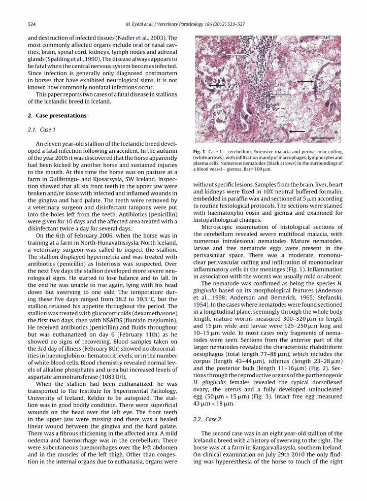
Parasitology 186 (2012) 523– 527
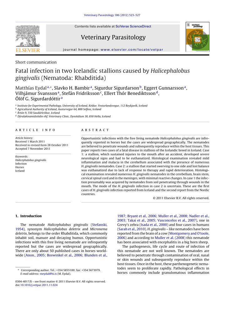
Fig. 1. Case 1 – cerebellum. Extensive malacia and perivascular cuffing
524 M. Eydal et al. / Veterinary
and destruction of infected tissues (Nadler et al., 2003). Themost commonly affected organs include oral or nasal cav-ities, brain, spinal cord, kidneys, lymph nodes and adrenalglands (Spalding et al., 1990). The disease always appears tobe fatal when the central nervous system becomes infected.Since infection is generally only diagnosed postmortemin horses that have exhibited neurological signs, it is notknown how commonly nonfatal infections occur.
This paper reports two cases of a fatal disease in stallionsof the Icelandic breed in Iceland.
2. Case presentations
2.1. Case 1
An eleven year-old stallion of the Icelandic breed devel-oped a fatal infection following an accident. In the autumnof the year 2005 it was discovered that the horse apparentlyhad been kicked by another horse and sustained injuriesto the mouth. At this time the horse was on pasture at afarm in Gullbringu- and Kjosarsysla, SW Iceland. Inspec-tion showed that all six front teeth in the upper jaw werebroken and/or loose with infected and inflamed wounds inthe gingiva and hard palate. The teeth were removed bya veterinary surgeon and disinfectant tampons were putinto the holes left from the teeth. Antibiotics (penicillin)were given for 10 days and the affected area treated with adisinfectant twice a day for several days.
On the 6th of February 2006, when the horse was intraining at a farm in North-Hunavatnssysla, North Iceland,a veterinary surgeon was called to inspect the stallion.The stallion displayed hypermetria and was treated withantibiotics (penicillin) as listeriosis was suspected. Overthe next five days the stallion developed more severe neu-rological signs. He started to lose balance and to fall. Inthe end he was unable to rise again, lying with his headdown but swerving to one side. The temperature dur-ing these five days ranged from 38.2 to 39.5 ◦C, but thestallion retained his appetite throughout the period. Thestallion was treated with glucocorticoids (dexamethasone)the first two days, then with NSAIDS (flunixin meglumin).He received antibiotics (penicillin) and fluids throughoutbut was euthanatized on day 6 (February 11th) as heshowed no signs of recovering. Blood samples taken onthe 3rd day of illness (February 8th) showed no abnormal-ities in haemoglobin or hematocrit levels, or in the numberof white blood cells. Blood chemistry revealed normal lev-els of alkaline phosphates and urea but increased levels ofaspartate aminotransferase (1083 U/l).
When the stallion had been euthanatized, he wastransported to The Institute for Experimental Pathology,University of Iceland, Keldur to be autopsied. The stal-lion was in good bodily condition. There were superficialwounds on the head over the left eye. The front teethin the upper jaw were missing and there was a healedlinear wound between the gingiva and the hard palate.There was a fibrous thickening in the affected area. A mild
oedema and haemorrhage was in the cerebellum. Therewere subcutaneous haemorrhages over the left abdomenand in the muscles of the left thigh. Other than conges-tion in the internal organs due to euthanasia, organs were(white arrows), with infiltration mainly of macrophages, lymphocytes andplasma cells. Numerous nematodes (black arrows) in the surroundings ofa blood vessel – giemsa. Bar = 100 �m.
without specific lesions. Samples from the brain, liver, heartand kidneys were fixed in 10% neutral buffered formalin,embedded in paraffin wax and sectioned at 5 �m accordingto routine histological protocols. The sections were stainedwith haematoxylin eosin and giemsa and examined forhistopathological changes.
Microscopic examination of histological sections ofthe cerebellum revealed severe multifocal malacia, withnumerous intralesional nematodes. Mature nematodes,larvae and free nematode eggs were present in theperivascular space. There was a moderate, mononu-clear perivascular cuffing and infiltration of mononuclearinflammatory cells in the meninges (Fig. 1). Inflammationin association with the worms was usually mild or absent.
The nematode was confirmed as being the species H.gingivalis based on its morphological features (Andersonet al., 1998; Anderson and Bemerick, 1965; Stefanski,1954). In the cases where nematodes were found sectionedin a longitudinal plane, seemingly through the whole bodylength, mature worms measured 300–320 �m in lengthand 15 �m wide and larvae were 125–250 �m long and10–15 �m wide. In most cases only fragments of nema-todes were seen. Sections from the anterior part of thelarger nematodes revealed the characteristic rhabditiformoesophagus (total length 77–88 �m), which includes thecorpus (length 43–44 �m), isthmus (length 23–28 �m)and the posterior bulb (length 11–16 �m) (Fig. 2). Sec-tions through the reproductive organs of the parthenogenicH. gingivalis females revealed the typical dorsoflexedovary, the uterus and a fully developed uninucleatedegg (50 �m × 15 �m) (Fig. 3). Intact free egg measured43 �m × 18 �m.
2.2. Case 2
The second case was in an eight year-old stallion of the
Icelandic breed with a history of swerving to the right. Thehorse was at a farm in Rangarvallasysla, southern Iceland.On clinical examination on July 29th 2010 the only find-ing was hyperesthesia of the horse to touch of the right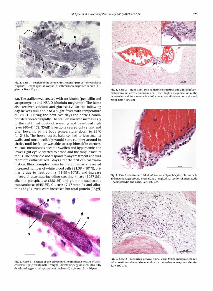
M. Eydal et al. / Veterinary Parasitology 186 (2012) 523– 527 525
Fgg
esadottfbfwcMlttiimiatm
Fcd
Fig. 4. Case 2 – brain stem. Two nematode structures and a mild inflam-mation around a vessel in brain stem. Inset: higher magnification of thenematodes and the mononuclear inflammatory cells – haemotoxylin andeosin. Bars = 100 �m.
Fig. 5. Case 2 – brain stem. Mild infiltration of lymphocytes, plasma cells
ig. 2. Case 1 – section of the cerebellum. Anterior part of Halicephalobusingivalis. Oesophagus (a), corpus (b), isthmus (c) and posterior bulb (d) –iemsa. Bar = 10 �m.
ar. The stallion was treated with antibiotics (penicillin andtreptomycin) and NSAID (flunixin meglumin). The horselso received calcium and glucose i.v. On the followingay he was dull and had a slight fever with temperaturef 38.6 ◦C. During the next two days the horse’s condi-ion deteriorated rapidly. The stallion swerved increasinglyo the right, had bouts of sweating and developed highever (40–41 ◦C). NSAID injections caused only slight andrief lowering of the body temperature, down to 39 ◦Cor 2–3 h. The horse lost its balance, had to lean againstalls, and uncontrollably would start running around in
ircles until he fell or was able to stop himself in corners.ucous membranes became swollen and hyperaemic, the
ower right eyelid started to droop and the tongue lost itsonus. The horse did not respond to any treatment and washerefore euthanatized 5 days after the first clinical exam-nation. Blood samples taken before euthanasia revealedncreased number of white blood cells (21.38 × 10⁹/l), pri-
arily due to neutrophilia (18.99 × 10⁹/l), and increasen several enzymes, including creatine kinase (1057 U/l),
lkaline phosphatase (288 U/l) and glutamic-oxaloaceticransaminase (645 U/l). Glucose (7.47 mmol/l) and albu-in (52 g/l) levels were increased but total protein (36 g/l)
ig. 3. Case 1 – section of the cerebellum. Reproductive organs of Hali-ephalobus gingivalis female. Ovary (a), developing eggs in uterus (b), fullyeveloped egg (c) and a prominent nucleus (d) – giemsa. Bar = 10 �m.
and macrophages around a vessel and a longitudinal section of a nematode– haemotoxylin and eosin. Bar = 100 �m.
Fig. 6. Case 2 – meninges, cervical spinal cord. Mixed mononuclear cellinflammation and several nematode structures – haemotoxylin and eosin.Bar = 100 �m.
526 M. Eydal et al. / Veterinary Parasitology 186 (2012) 523– 527
ed brain.
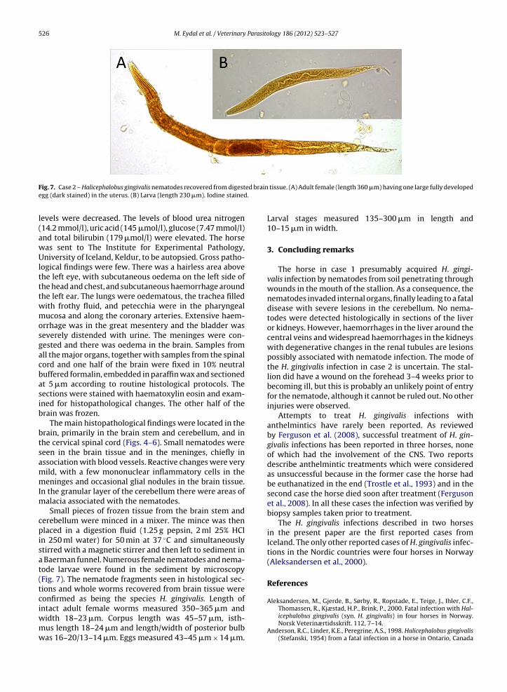
Fig. 7. Case 2 – Halicephalobus gingivalis nematodes recovered from digestegg (dark stained) in the uterus. (B) Larva (length 230 �m). Iodine stained
levels were decreased. The levels of blood urea nitrogen(14.2 mmol/l), uric acid (145 �mol/l), glucose (7.47 mmol/l)and total bilirubin (179 �mol/l) were elevated. The horsewas sent to The Institute for Experimental Pathology,University of Iceland, Keldur, to be autopsied. Gross patho-logical findings were few. There was a hairless area abovethe left eye, with subcutaneous oedema on the left side ofthe head and chest, and subcutaneous haemorrhage aroundthe left ear. The lungs were oedematous, the trachea filledwith frothy fluid, and petecchia were in the pharyngealmucosa and along the coronary arteries. Extensive haem-orrhage was in the great mesentery and the bladder wasseverely distended with urine. The meninges were con-gested and there was oedema in the brain. Samples fromall the major organs, together with samples from the spinalcord and one half of the brain were fixed in 10% neutralbuffered formalin, embedded in paraffin wax and sectionedat 5 �m according to routine histological protocols. Thesections were stained with haematoxylin eosin and exam-ined for histopathological changes. The other half of thebrain was frozen.
The main histopathological findings were located in thebrain, primarily in the brain stem and cerebellum, and inthe cervical spinal cord (Figs. 4–6). Small nematodes wereseen in the brain tissue and in the meninges, chiefly inassociation with blood vessels. Reactive changes were verymild, with a few mononuclear inflammatory cells in themeninges and occasional glial nodules in the brain tissue.In the granular layer of the cerebellum there were areas ofmalacia associated with the nematodes.
Small pieces of frozen tissue from the brain stem andcerebellum were minced in a mixer. The mince was thenplaced in a digestion fluid (1.25 g pepsin, 2 ml 25% HClin 250 ml water) for 50 min at 37 ◦C and simultaneouslystirred with a magnetic stirrer and then left to sediment ina Baerman funnel. Numerous female nematodes and nema-tode larvae were found in the sediment by microscopy(Fig. 7). The nematode fragments seen in histological sec-tions and whole worms recovered from brain tissue wereconfirmed as being the species H. gingivalis. Length of
intact adult female worms measured 350–365 �m andwidth 18–23 �m. Corpus length was 45–57 �m, isth-mus length 18–24 �m and length/width of posterior bulbwas 16–20/13–14 �m. Eggs measured 43–45 �m × 14 �m.tissue. (A) Adult female (length 360 �m) having one large fully developed
Larval stages measured 135–300 �m in length and10–15 �m in width.
3. Concluding remarks
The horse in case 1 presumably acquired H. gingi-valis infection by nematodes from soil penetrating throughwounds in the mouth of the stallion. As a consequence, thenematodes invaded internal organs, finally leading to a fataldisease with severe lesions in the cerebellum. No nema-todes were detected histologically in sections of the liveror kidneys. However, haemorrhages in the liver around thecentral veins and widespread haemorrhages in the kidneyswith degenerative changes in the renal tubules are lesionspossibly associated with nematode infection. The mode ofthe H. gingivalis infection in case 2 is uncertain. The stal-lion did have a wound on the forehead 3–4 weeks prior tobecoming ill, but this is probably an unlikely point of entryfor the nematode, although it cannot be ruled out. No otherinjuries were observed.
Attempts to treat H. gingivalis infections withanthelmintics have rarely been reported. As reviewedby Ferguson et al. (2008), successful treatment of H. gin-givalis infections has been reported in three horses, noneof which had the involvement of the CNS. Two reportsdescribe anthelmintic treatments which were consideredas unsuccessful because in the former case the horse hadbe euthanatized in the end (Trostle et al., 1993) and in thesecond case the horse died soon after treatment (Fergusonet al., 2008). In all these cases the infection was verified bybiopsy samples taken prior to treatment.
The H. gingivalis infections described in two horsesin the present paper are the first reported cases fromIceland. The only other reported cases of H. gingivalis infec-tions in the Nordic countries were four horses in Norway(Aleksandersen et al., 2000).
References
Aleksandersen, M., Gjerde, B., Sørby, R., Ropstade, E., Teige, J., Ihler, C.F.,
Thomassen, R., Kjæstad, H.P., Brink, P., 2000. Fatal infection with Hal-icephalobus gingivalis (syn. H. gingivalis) in four horses in Norway.Norsk Veterinærtidsskrift. 112, 7–14.Anderson, R.C., Linder, K.E., Peregrine, A.S., 1998. Halicephalobus gingivalis(Stefanski, 1954) from a fatal infection in a horse in Ontario, Canada
Parasito
A
A
B
B
B
F
I
M
M. Eydal et al. / Veterinary
with comments on the validity of H. deletrix and a review of the genus.Parasite 5, 255–261.
nderson, R.V., Bemerick, W.J., 1965. Micronema deletrix n. sp., asaprophagous nematode inhabiting a nasal tumor of a horse. Proc.Helminthol. Soc. Wash. 32, 74–75.
non., 2005. The armed forces institute of pathology. Departmentof veterinary pathology. In: Wednesday Slide Conference. Con-ference 1, 7 September 2005 (Conference moderator Dr. DaleDunn), Available from: http://vp4.afip.org/wsc/wsc05/05wsc01.pdf(accessed 23.02.11).
lunden, A.S., Khalil, L.F., Webbon, P.M., 1987. Halicephalobus gingivalisinfection in a horse. Equine Vet. J. 19, 255–260.
oswinkel, M., Neyens, I.J.S., Sloet van Oldruitenborgh, M.M., 2006. Hali-cephalobus gingivalis infection in a 5-year-old Tinker gelding. Tijdschr.Diergeneesk. 131, 74–80.
ryant, U.K., Lyons, E.T., Bain, F.T., Hong, C.B., 2006. Halicephalobusgingivalis-associated meningoencephalitis in a thoroughbred foal. J.Vet. Diagn. Invest. 18, 612–615.
erguson, R., van Dreumel, T., Keystone, J.S., Manning, A., Malatestinic, A.,Caswell, J.L., Peregrine, A.S., 2008. Unsuccessful treatment of a horsewith mandibular granulomatous osteomyelitis due to Halicephalobusgingivalis. Can. Vet. J. 49, 1099–1103.
sada, R., Schiller, C.A., Stover, J., Smith, P.J., Greiner, E.C., 2000. Hali-
cephalobus gingivalis (Nematoda) infection in a Grevy’s zebra (Equusgrevyi). J. Zoo Wildlife Med. 31, 77–81.ontgomery, D., O’toole, D., April 2006. Neurological Disease in a Cowin Big Horn Basin due to a Free-living Nematode. Wyoming StateVeterinary Laboratory Newsletter, pp. 3–4.
logy 186 (2012) 523– 527 527
Muller, S., Grzybowski, M., Sager, H., Bornand, V., Brehm, W., 2008. A nodu-lar granulomatous posthitis cased by Halicephalobus sp. in a horse. Vet.Dermatol. 19, 44–48.
Nadler, S.A., Carreno, R.A., Adams, B.J., Kinde, H., Baldwin, J.G., Mundo-Ocampo, M., 2003. Molecular phylogenetics and diagnosis of soil andclinical isolates of Halicephalobus gingivalis (Nematoda: Cephalobina:Panagrolaimoidea), an opportunistic pathogen of horses. Int. J. Para-sitol. 33, 1115–1125.
Sarah, L., Ondrejka, D.O., Gary, W., Procop, M.D., Keith, K., Lai, M.D., Richard,A., Prayson, M.D., 2010. Fatal parasitic meningoencephalomyeli-tis caused by Halicephalobus deletrix. Arch. Pathol. Lab. Med. 134,625–629.
Spalding, M.G., Greiner, E.C., Green, S.L., 1990. Halicephalobus (Micronema)deletrix infection in two half-sibling foals. J. Am. Vet. Med. Assoc. 196,1127–1129.
Stefanski, W., 1954. Rhabditis gingivalis sp. n. Parasite trouvé dans ungranulome de la gencive chez un cheval. Acta Parasitol. Pol. 1,329–336.
Takai, H., Shibaharta, T., Murakami, T., Hayashi, M., Kadota, K., 2005. Repeatoccurrence of equine Halicephalobus infection at an equestrian club. J.Jpn. Vet. Med. Assoc. 58, 105–108.
Trostle, S.S., Wilson, D.G., Steinberg, H., Dzata, G., Dubielzig, R.R.,1993. Antemortem diagnosis and attempted treatment of (Hali-
cephalobus) Micronema deletrix infection in a horse. Can. Vet. J. 34,117–118.Vasconcelos, R.deO., Lemos, K.R., de Morales, J.R.E., Borges, V.P., 2007. Hal-icephalobus gingivalis (H. deletrix) in the brain of a horse. Ciência Rural,Santa Maria 37, 1185–1187.