fat embolism
TRANSCRIPT

FAT EMBOLISMA topic presentation at Amala Institute of Medical Sciences
by
Dr. Libin Thomas Manathara

Background
• For those who manage major trauma victims, the topic of fatembolism weighs heavily on the mind
• The incidence of this problem can approach 90% in patients who havesustained major injuries
• If it progresses to the rare clinical entity known as fat embolismsyndrome (FES), a systemic inflammatory cascade affecting multipleorgan systems, morbidity and mortality are high
• Accordingly, swift diagnosis and treatment of fat embolism areparamount for ensuring the survival of this patient population

Background
• Ernst Von Bergmann, in 1873, was the first person credited withmaking a clinical diagnosis of fat embolism
• He did this on the basis of knowledge gathered from experimentswith cats 10 years previously, in which he injected them withintravenous oils
• Von Bergmann later described a patient who fell off a roof andsustained a comminuted fracture of the distal femur; 60 hours afterthe injury, the patient developed dyspnea, cyanosis, and coma
Background
• The diagnosis of FES is mainly a clinical one
• It is dependent on clinical identification of dyspnea, petechiae, andcognitive dysfunction in the first few days following trauma, longbone fracture, or intramedullary surgery
• Various laboratory studies and imaging modalities exist to aid in itsdiscovery
• Supportive measures are the mainstay of treatment; thus, efforts aretargeted at prevention, early diagnosis, and symptom management
Pathophysiology
• The exact mechanism of fat embolism and its evolution to the entityknown as FES has not been fully elucidated, but a number ofexperimental models have been proposed
• Asymptomatic fat embolism to the pulmonary circulation almostalways occurs with major trauma, including elective surgicalprocedures such as intramedullary nailing of long bones, which hasbeen demonstrated with echocardiography
• The development of FES is rare, occurring in 0.5-11% of cases
Pathophysiology
• Although poorly understood, the development of FES is attributed toa series of biochemical cascades resulting from the mechanical insultsustained in major trauma
• Release of fat emboli leads to occlusion of the microvasculature,triggering an inflammatory response that is clinically manifested bydermatologic, pulmonary, and neurologic dysfunction
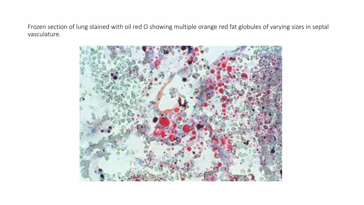
Frozen section of lung stained with oil red O showing multiple orange red fat globules of varying sizes in septal vasculature.
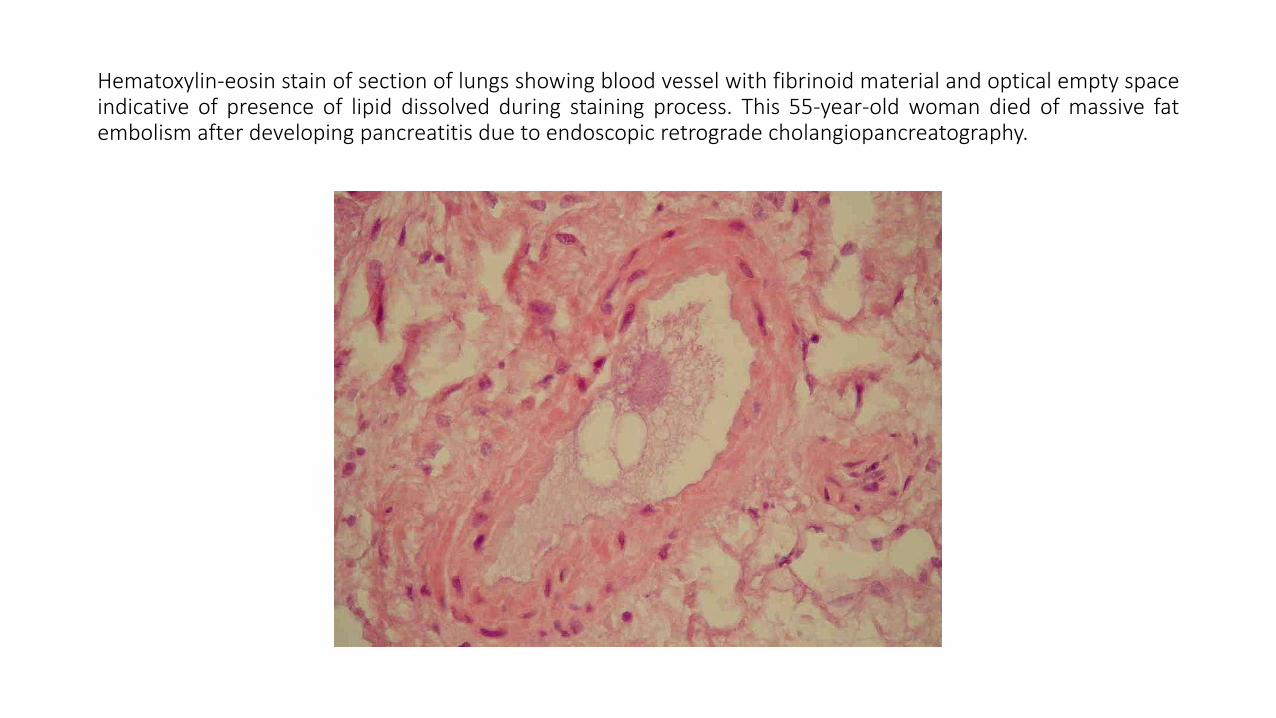
Hematoxylin-eosin stain of section of lungs showing blood vessel with fibrinoid material and optical empty spaceindicative of presence of lipid dissolved during staining process. This 55-year-old woman died of massive fatembolism after developing pancreatitis due to endoscopic retrograde cholangiopancreatography.

Pathophysiology
• Pulmonary consequences of FES are clinically similar to those of acuterespiratory distress syndrome (ARDS) and almost always occur
• They are usually the initial manifestation of FES, typically appearingwithin 24 hours after the traumatic insult
• They result from injury to the pulmonary capillary endotheliumcaused by free fatty acids that were hydrolyzed by lipoprotein lipase,releasing local toxic mediators
• These mediators cause increased vascular permeability, resulting inalveolar hemorrhage and edema and causing respiratory failure andARDS

Pathophysiology
• Approximately 20-30% of the population have a patent foramenovale; this may explain how fat emboli that pass through thepulmonary circulation end up with systemic manifestations of FES,particularly involving the brain and kidneys
• As a result of the occluded cerebral vasculature, patients exhibit grossencephalopathy, localized cerebral edema, and white-matter changes
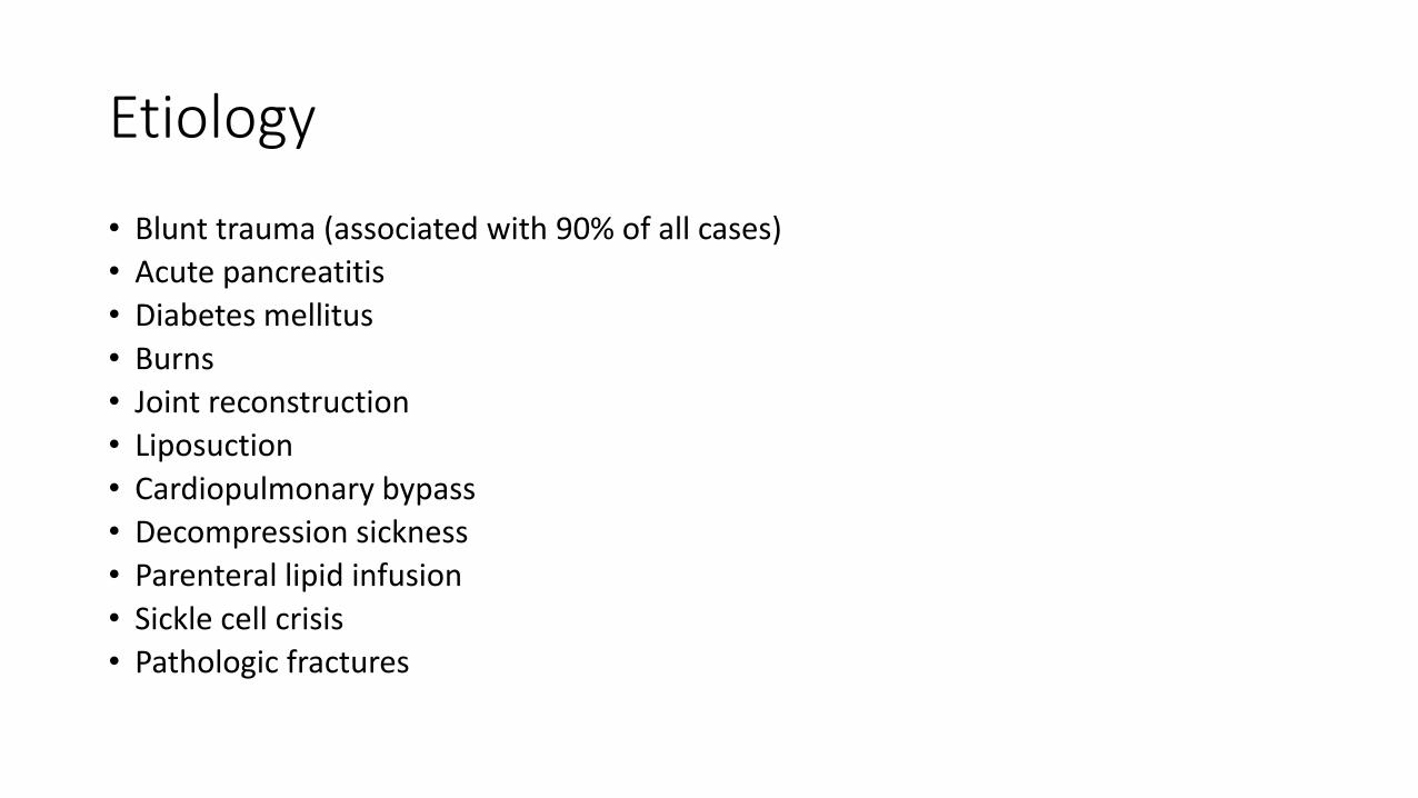
Etiology
• Blunt trauma (associated with 90% of all cases)
• Acute pancreatitis
• Diabetes mellitus
• Burns
• Joint reconstruction
• Liposuction
• Cardiopulmonary bypass
• Decompression sickness
• Parenteral lipid infusion
• Sickle cell crisis
• Pathologic fractures
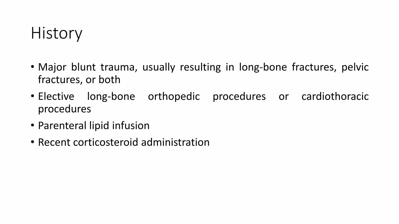
History
• Major blunt trauma, usually resulting in long-bone fractures, pelvicfractures, or both
• Elective long-bone orthopedic procedures or cardiothoracicprocedures
• Parenteral lipid infusion
• Recent corticosteroid administration
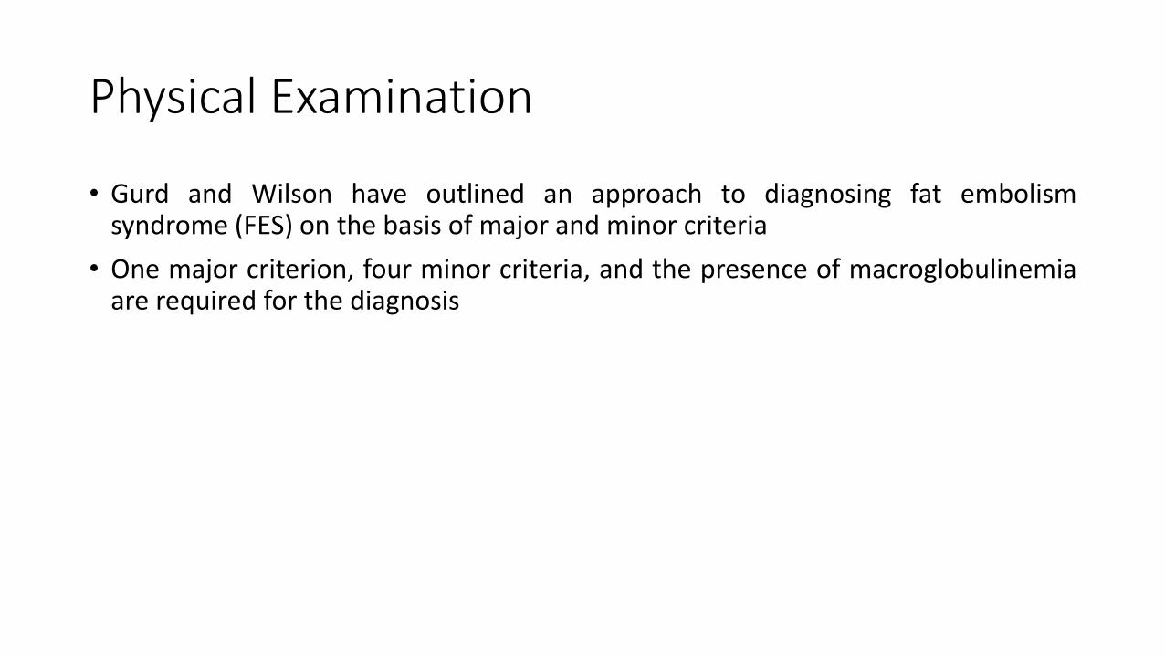
Physical Examination
• Gurd and Wilson have outlined an approach to diagnosing fat embolismsyndrome (FES) on the basis of major and minor criteria
• One major criterion, four minor criteria, and the presence of macroglobulinemiaare required for the diagnosis
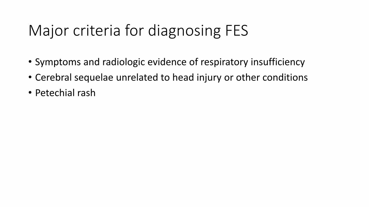
Major criteria for diagnosing FES
• Symptoms and radiologic evidence of respiratory insufficiency
• Cerebral sequelae unrelated to head injury or other conditions
• Petechial rash

Minor criteria
• Tachycardia (heart rate >110 beats/min)
• Pyrexia (temperature >38.5° C)
• Retinal changes of fat or petechiae
• Renal dysfunction
• Jaundice
• Acute drop in hemoglobin level
• Sudden thrombocytopenia
• Elevated erythrocyte sedimentation rate
• Fat microglobulinemia
Physical Examination
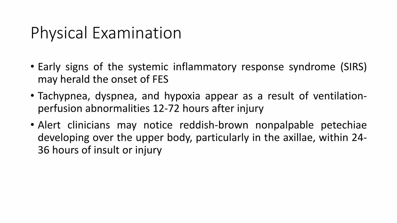
• Early signs of the systemic inflammatory response syndrome (SIRS)may herald the onset of FES
• Tachypnea, dyspnea, and hypoxia appear as a result of ventilation-perfusion abnormalities 12-72 hours after injury
• Alert clinicians may notice reddish-brown nonpalpable petechiaedeveloping over the upper body, particularly in the axillae, within 24-36 hours of insult or injury

Physical Examination
• These petechiae occur in 20-50% of patients and resolve quickly, butthey are virtually diagnostic in the right clinical setting
• Subconjunctival and oral hemorrhages and petechiae can also appear
• Central nervous system dysfunction initially manifests as agitation ordelirium but may progress to stupor, seizures, or coma and isfrequently unresponsive to correction of hypoxia
• Retinal hemorrhages with intra-arterial fat globules are visible uponfunduscopic examination

Diagnostic Considerations
• Every effort should be made to look for treatable or life-threateningdisorders before the diagnosis of fat embolism syndrome (FES) ismade
• Computed tomography (CT) of the head is necessary to rule outintracranial pathology
• A careful search for infectious agents and possibly the institution ofempiric antibiotics are necessary until an infectious source is ruledout
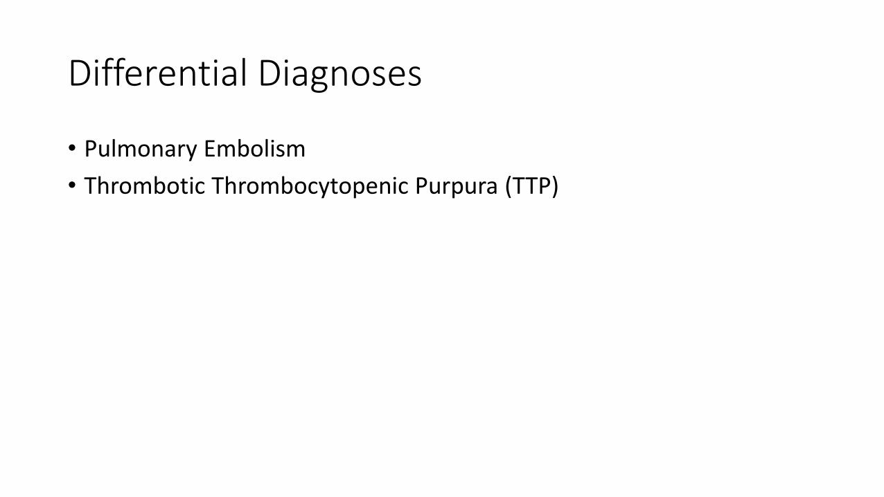
Differential Diagnoses
• Pulmonary Embolism
• Thrombotic Thrombocytopenic Purpura (TTP)
Workup- Laboratory Studies
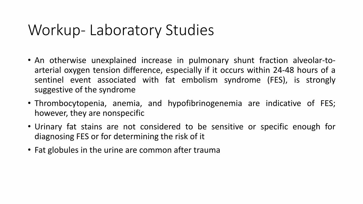
• An otherwise unexplained increase in pulmonary shunt fraction alveolar-to-arterial oxygen tension difference, especially if it occurs within 24-48 hours of asentinel event associated with fat embolism syndrome (FES), is stronglysuggestive of the syndrome
• Thrombocytopenia, anemia, and hypofibrinogenemia are indicative of FES;however, they are nonspecific
• Urinary fat stains are not considered to be sensitive or specific enough fordiagnosing FES or for determining the risk of it
• Fat globules in the urine are common after trauma
Workup- Laboratory Studies
• Preliminary investigations of the cytology of pulmonary capillaryblood obtained from a wedged pulmonary artery catheter revealedfat globules in patients with FES and showed that this method may bebeneficial in early detection of patients at risk
• In the future, genotyping for polymorphisms associated withincreased susceptibility to inflammatory stimuli may help identifythose at risk for FES
• Specific antibody therapy targeting inflammatory molecules has notbeen useful
Radiography and Computed Tomography
• Serial chest radiographs reveal increasing diffuse bilateral pulmonary infiltrateswithin 24-48 hours of the onset of clinical findings
• Findings from noncontrast computed tomography (CT) of the head performedbecause of alterations in mental status may be normal or may reveal diffusewhite-matter petechial hemorrhages consistent with microvascular injury
• Because the embolic particles are lodged in the capillary beds, helical CT findingsmay be normal
• Parenchymal changes consistent with lung contusion, acute lung injury, or acuterespiratory distress syndrome (ARDS) may be evident
• Nodular or ground-glass opacities in the setting of trauma suggest fat embolism
Ultrasonography
• In a small case study, five patients with trauma were monitored withintracranial Doppler ultrasonography, two during intraoperativenailing of long-bone fractures
• Cerebral microembolic signals were detected as long as 4 days afterinjury
• Transesophageal echocardiography (TEE) may be of use in evaluatingthe intraoperative release of marrow contents into the bloodstreamduring intramedullary reaming and nailing
Ultrasonography
• The density of the echogenic material passing through the right sideof the heart correlates with the degree of reduction in arterial oxygensaturation
• Repeated showers of emboli on TEE have been noted to increase rightheart and pulmonary artery pressures
• Embolization of marrow contents through a patent foramen ovalealso has been noted
• However, evidence of embolization obtained by means of TEE is notcorrelated with the actual development of FES

Bronchoalveolar Lavage With Staining for Fat
• Bronchoalveolar lavage (BAL) specimens have been evaluated intrauma patients and sickle-cell patients with acute chest syndrome,and the results have been mixed
• Lipid inclusions commonly appear in patients with traumatic andnontraumatic respiratory failure; the standard cutoff in the BALstudies—5% fat-containing macrophages—results in a low specificityfor the test
• To improve specificity, some authors suggest raising the cutoff to 30%
• At present, using BAL to aid in the diagnosis of FES or to predict itslikelihood is controversial
Other Studies
• Scant data exist regarding magnetic resonance imaging (MRI) findingsin patients with FES; however, in one small patient group, multiplenonconfluent, hyperintense lesions were seen on proton-density–and T2-weighted images
• Nuclear ventilation-perfusion imaging of the lungs may be performedwhen pulmonary embolism is suspected
• The findings from this scan may be normal or may demonstratesubsegmental perfusion defects
Supportive Medical Care
• Specific medical therapy for fat embolism and fat embolism syndrome (FES) doesnot exist at this time, and supportive measures have not been tested in adequaterandomized, controlled trials
• Treatments such as heparin, dextran, and steroids have not been shown to helpreduce morbidity and mortality, but methylprednisolone given prophylacticallymay have beneficial effects
Supportive Medical Care
• Current care of patients with fat embolism is aimed at supporting physiologicderangements and includes the following:
• Maintenance of adequate oxygenation and ventilation with open lung strategies such as theuse of airway pressure release ventilation (APRV)
• Maintenance of hemodynamic stability
• Administration of blood products as clinically indicated
• Hydration
• Prophylaxis of deep venous thrombosis and stress-related gastrointestinal bleeding
• Nutrition
Supportive Medical Care
• Judicious use of crystalloids, colloids, and diuretics is necessary; volume depletionmay precipitate shock and organ dysfunction, but volume overload may worsenthe hypoxia
• Continuous pulse oximetry monitoring in at-risk patients (eg, patients with long-bone fractures and multiple trauma), may facilitate early detection ofdesaturation, allowing prophylactic administration of oxygen and possiblysteroids, thereby decreasing the chances of hypoxic injury and systemiccomplications of FES
• At-risk patients should be placed in a monitored setting, and appropriate servicesshould be consulted
• If a patient has sustained major traumatic injuries, transfer to the nearest traumacenter with 24-hour in-house surgical intensive care is essential
Surgical Management
• Early stabilization of long bone fractures is recommended to minimize bonemarrow embolization into the venous system
• Rigid fixation within 24 hours has been shown to yield a fivefold reduction in theincidence of FES
• Appropriate surgical technique, particularly in reaming or nailing the marrow,may help reduce the volume of fat embolization
• Utilization of a vacuum or venting during reaming has been shown to decreasethe incidence of fat embolization
• Prophylactic placement of inferior vena cava filters may help reduce the volumeof fat that reaches the heart in at-risk patients
Prevention
• Several studies performed in the late 1970s attempted to show that use ofmethylprednisolone as a “membrane stabilizer” would reduce the incidence ofFES, but follow-up work has yet to reproduce these findings
• A meta-analysis of randomized trials studying corticosteroid use as a preventiveadjunct in patients with long-bone fractures uncovered 104 studies, of which onlyseven met the authors' eligibility criteria for analysis
• Although the pooled analysis of 389 patients found that corticosteroids reducedthe risk of FES by 78%, the authors warned that these studies were of poorquality and held to standards of the 1970s
Prevention
• The use of heparin has been shown to reduce the degree ofpulmonary comprise and intravascular coagulation despite the risk ofhemorrhage and intravascular lipolysis; however, this practice has notbeen shown to yield a statistically significant benefit
• Ethanol (which decreases lipolysis) and dextrose (which decreasesfree fatty acid mobilization) have been used as prevention modalities,but at present, there is little to no evidence to support the use ofthese agents in FES

Medication Summary
• The goals of pharmacotherapy for fat embolism syndrome (FES) are to reducemorbidity and prevent complications
• Corticosteroids may be used in certain cases
• The best dosing protocol for corticosteroids in the prophylaxis of FES has notbeen established, and currently, there is no treatment regimen
Corticosteroids
• Corticosteroids have anti-inflammatory properties and causeprofound and varied metabolic effects
• They modify the body’s immune response to diverse stimuli
Methylprednisolone (Depo-Medrol, Medrol,Solu-Medrol, A-Methapred)• Methylprednisolone is most often used for the prophylaxis of FES in
at-risk patients
• Currently, there are no good data to support the use of this agentover the use of any other steroids
Isotonic Crystalloids
• Isotonic sodium chloride solution (normal saline [NS]) and lactatedRinger (LR) solution are isotonic crystalloids, the standard intravenous(IV) fluids used for initial volume resuscitation
• They expand the intravascular and interstitial fluid spaces
• Typically, about 30% of administered isotonic fluid stays intravascular;therefore, large quantities may be required to maintain adequatecirculating volume
Isotonic Crystalloids
• Both fluids are isotonic, and they have equivalent volume-restorativeproperties
• Whereas some differences exist between the metabolic changesobserved with the administration of large quantities of one fluid andthose observed with high-volume administration of the other, forpractical purposes and in most situations, these differences areclinically irrelevant
• No demonstrable difference in hemodynamic effect, morbidity, ormortality exists between resuscitation with NS and resuscitation withLR solution
Normal saline (NS, 0.9% NaCl)
• NS restores interstitial and intravascular volume
• It is used in initial volume resuscitation

Lactated Ringer
• RL solution restores interstitial and intravascular volume
• It is used in initial volume resuscitation
Colloids
• Colloids are used to provide oncotic expansion of plasma volume
• They expand plasma volume to a greater degree than isotonic crystalloids andreduce the tendency of pulmonary and cerebral edema
• About 50% of the administered colloid stays intravascular
Albumin (Buminate, Albuminar-5, Albuminar-25, Plasbumin-5, Plasbumin-25)• Albumin has been recommended for volume resuscitation
• It is useful for plasma volume expansion and maintenance of cardiacoutput
• It also binds with the fatty acids and may thus decrease the extent oflung injury
• Five-percent solutions are indicated to expand plasma volume,whereas 25% solutions are indicated to raise oncotic pressure

THANK YOU