extraintestinal manifestations of inflammatory bowel diseasebib_01a199a06c14.p001/ref.pdf ·...
TRANSCRIPT
CLINICAL REVIEW ARTICLE
Extraintestinal Manifestations of Inflammatory Bowel DiseaseStephan R. Vavricka, MD,*,† Alain Schoepfer, MD,‡ Michael Scharl, MD,* Peter L. Lakatos, MD,§
Alexander Navarini, MD,k and Gerhard Rogler, MD*
Abstract: Extraintestinal manifestations (EIM) in inflammatory bowel disease (IBD) are frequent and may occur before or after IBD diagnosis. EIM mayimpact the quality of life for patients with IBD significantly requiring specific treatment depending on the affected organ(s). They most frequently affect joints,skin, or eyes, but can also less frequently involve other organs such as liver, lungs, or pancreas. Certain EIM, such as peripheral arthritis, oral aphthous ulcers,episcleritis, or erythema nodosum, are frequently associated with active intestinal inflammation and usually improve by treatment of the intestinal activity. OtherEIM, such as uveitis or ankylosing spondylitis, usually occur independent of intestinal inflammatory activity. For other not so rare EIM, such as pyodermagangrenosum and primary sclerosing cholangitis, the association with the activity of the underlying IBD is unclear. Successful therapy of EIM is essential forimproving quality of life of patients with IBD. Besides other options, tumor necrosis factor antibody therapy is an important therapy for EIM in patients with IBD.
(Inflamm Bowel Dis 2015;21:1982–1992)
Key Words: extraintestinal manifestations, inflammatory bowel disease, arthritis, uveitis
I nflammatory bowel disease (IBD), which includes Crohn’s disease(CD) and ulcerative colitis (UC), should be regarded as a systemic
disorder not limited to the gastrointestinal tract because many patientswill develop extraintestinal symptoms. Extraintestinal symptoms mayinvolve virtually any organ system with a potentially detrimentalimpact on the patient’s functional status and quality of life.
Extraintestinal symptoms can be divided in 2 groups: extra-intestinal manifestations (EIM) and extraintestinal complications.EIM most frequently affect joints (peripheral and axial arthropathies),the skin (erythema nodosum, pyoderma gangrenosum, Sweet’s syn-drome, aphthous stomatitis), the hepatobiliary tract (primary scleros-ing cholangitis [PSC]), and the eye (episcleritis, uveitis) (Fig. 1). Lessfrequently, EIM also affect the lungs, the heart, the pancreas, or the
vascular system. Extraintestinal complications are mainly caused bythe disease itself and include conditions such as malabsorption withconsequent micronutrient deficiencies, osteoporosis, peripheral neu-ropathies, kidney stones, gallstones, and IBD drug-related side effects.
This article focuses on the clinical features of EIM. CertainEIM such as pauciarticular arthritis, oral aphthous ulcers, erythemanodosum, or episcleritis usually occur with increased intestinaldisease activity.1,2 Other EIM such as ankylosing spondylitis anduveitis usually follow an independent course from IBD disease activ-ity.1,2 And finally, some EIM such as PSC and pyoderma gangreno-sum may or may not be related to IBD disease activity (Table 1).2–4
EIM in IBD are reported with frequencies ranging from 6% to47%.5–13 Multiple EIM may occur concomitantly, and the presenceof 1 EIM confers a higher likelihood to develop other EIM.14
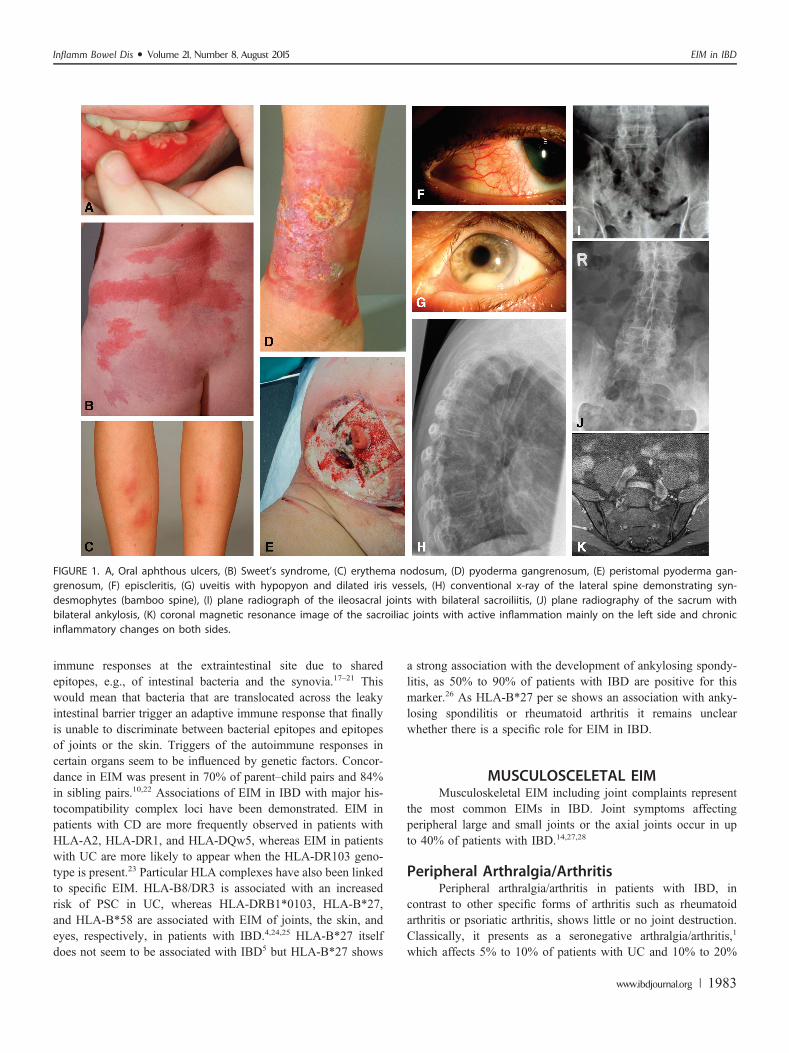
Recently, we reported based on data from the Swiss IBD Cohortstudy that up to 1 quarter of EIM-affected patients with IBD tend tosuffer from a combination of several EIMs (up to 5).14 Furthermore,our group recently published data regarding the chronological orderof EIM appearance relative to IBD diagnosis.15 A summary of thechronologic appearance of EIMs relative to IBD diagnosis is pre-sented in Figure 2. In 25.8% of cases, a first EIM occurred beforeIBD was diagnosed (median time 5 mo before IBD diagnosis; range,0–25 mo). In 74.2% of cases, the first EIM manifested after IBDdiagnosis was made (median, 92 mo; range, 29–183 mo) (Fig. 2).We found that up to 4 different EIM occurred before IBD wasdiagnosed, and that at 30 years after IBD diagnosis, 50% of patientshad suffered from at least 1 EIM. Perianal CD, colonic involvement,and cigarette smoking increased the likelihood to suffer from EIMs.16
PATHOGENESIS OF EIMThe pathogenesis of EIM in IBD is not well understood. It
is believed that the diseased gastrointestinal mucosa may trigger
Received for publication November 6, 2014; Accepted February 9, 2015.
From the *Department of Medicine, Division of Gastroenterology and Hepatol-ogy, University Hospital Zurich, Zurich, Switzerland; †Department of Medicine,Division of Gastroenterology and Hepatology, Triemlispital Zurich, Zurich,Switzerland; ‡Department of Medicine, Division of Gastroenterology and Hepatol-ogy, Centre Hospitalier Universitaire Vaudois (CHUV), Lausanne, Switzerland; §1stDepartment of Medicine, Semmelweis University, Budapest, Hungary; andkDepartment of Dermatology, University Hospital Zurich, Zurich, Switzerland.
Supported by research grants from the Swiss National Science Foundation(33CSC0_134274 [Swiss IBD Cohort Study] to G. Rogler, 320000-114009/3 and32473B_135694/1 to S. R. Vavricka, and 314730-146204 and CRSII3_154488/1 toM. Scharl) and the Zurich Center for Integrative Human Physiology of theUniversity of Zurich (to G. Rogler, M. Scharl, and S. R. Vavricka).
The authors have no conflicts of interest to disclose.
Reprints: Stephan R. Vavricka, MD, Department of Internal Medicine, Divisionof Gastroenterology, Triemli Hospital, Birmensdorferstrasse 497, CH-8063 Zurich,Switzerland (e-mail: [email protected]).
Copyright © 2015 Crohn’s & Colitis Foundation of America, Inc. This is anopen access article distributed under the Creative Commons Attribution License4.0 (CCBY), which permits unrestricted use, distribution, and reproduction in anymedium, provided the original work is properly cited.
DOI 10.1097/MIB.0000000000000392
Published online 2 July 2015.
1982 | www.ibdjournal.org Inflamm Bowel Dis � Volume 21, Number 8, August 2015
immune responses at the extraintestinal site due to sharedepitopes, e.g., of intestinal bacteria and the synovia.17–21 Thiswould mean that bacteria that are translocated across the leakyintestinal barrier trigger an adaptive immune response that finallyis unable to discriminate between bacterial epitopes and epitopesof joints or the skin. Triggers of the autoimmune responses incertain organs seem to be influenced by genetic factors. Concor-dance in EIM was present in 70% of parent–child pairs and 84%in sibling pairs.10,22 Associations of EIM in IBD with major his-tocompatibility complex loci have been demonstrated. EIM inpatients with CD are more frequently observed in patients withHLA-A2, HLA-DR1, and HLA-DQw5, whereas EIM in patientswith UC are more likely to appear when the HLA-DR103 geno-type is present.23 Particular HLA complexes have also been linkedto specific EIM. HLA-B8/DR3 is associated with an increasedrisk of PSC in UC, whereas HLA-DRB1*0103, HLA-B*27,and HLA-B*58 are associated with EIM of joints, the skin, andeyes, respectively, in patients with IBD.4,24,25 HLA-B*27 itselfdoes not seem to be associated with IBD5 but HLA-B*27 shows
a strong association with the development of ankylosing spondy-litis, as 50% to 90% of patients with IBD are positive for thismarker.26 As HLA-B*27 per se shows an association with anky-losing spondilitis or rheumatoid arthritis it remains unclearwhether there is a specific role for EIM in IBD.
MUSCULOSCELETAL EIMMusculoskeletal EIM including joint complaints represent
the most common EIMs in IBD. Joint symptoms affectingperipheral large and small joints or the axial joints occur in upto 40% of patients with IBD.14,27,28
Peripheral Arthralgia/ArthritisPeripheral arthralgia/arthritis in patients with IBD, in
contrast to other specific forms of arthritis such as rheumatoidarthritis or psoriatic arthritis, shows little or no joint destruction.Classically, it presents as a seronegative arthralgia/arthritis,1
which affects 5% to 10% of patients with UC and 10% to 20%
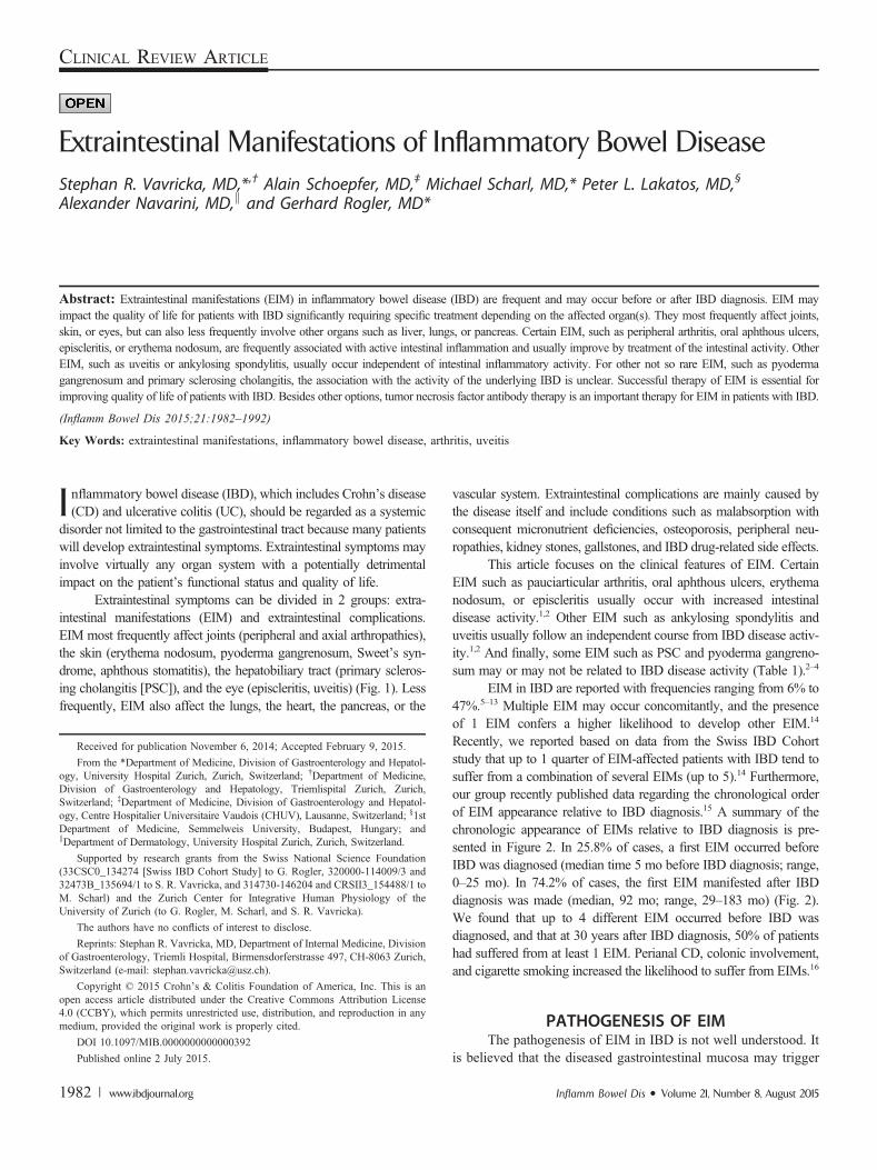
FIGURE 1. A, Oral aphthous ulcers, (B) Sweet’s syndrome, (C) erythema nodosum, (D) pyoderma gangrenosum, (E) peristomal pyoderma gan-grenosum, (F) episcleritis, (G) uveitis with hypopyon and dilated iris vessels, (H) conventional x-ray of the lateral spine demonstrating syn-desmophytes (bamboo spine), (I) plane radiograph of the ileosacral joints with bilateral sacroiliitis, (J) plane radiography of the sacrum withbilateral ankylosis, (K) coronal magnetic resonance image of the sacroiliac joints with active inflammation mainly on the left side and chronicinflammatory changes on both sides.
Inflamm Bowel Dis � Volume 21, Number 8, August 2015 EIM in IBD
www.ibdjournal.org | 1983
of patients with CD.29 A higher risk for peripheral arthralgia/arthritis is seen in patients with IBD with colonic involvementand in patients suffering from perianal disease, erythema nodo-sum, stomatitis, uveitis, and pyoderma gangrenosum.1,20
Peripheral arthralgia/arthritis has been classified into 2entities (Table 2): type I (pauciarticular) arthralgie/arthritis usuallyaffects less than 5 large joints, such as ankles, knees, hips, wrists,elbows, and shoulders and is often acute, asymmetrical, andmigratory. The knee is commonly involved. Approximately,20% to 40% of all patients have more than 1 episode of arthral-gia/arthritis. Pauciarticular arthralgia/arthritis is usually related toIBD activity and self-limiting with a maximum duration of up to10 weeks.29 Consequently, medical or surgical treatment of theunderlying intestinal inflammation (i.e., colitis) is usually associ-ated with improvement of type I arthritis. Type II (polyarticular)
arthralgia/arthritis frequently is a symmetrical arthritis involving 5or more small joints. It is not related with intestinal disease activ-ity and may precede IBD diagnosis. Type II arthropathy canpersist for years (median of 3 yr).29 The metacarpophalangealjoint is most commonly involved. Type II arthritis is associatedwith an increased risk of uveitis but not erythema nodosum.29
The diagnosis/classification of type 1 and type 2 arthro-pathies is purely clinical, as imaging is most often normal with noevidence of significant inflammation or joint destruction.31 Bothtypes are seronegative (i.e., rheumatoid factor-negative), but mayrepresent immunogenetically distinct entities. Type 1 peripheralarthropathy is associated with HLA-B27, HLA-B35, and HLA-DR103, whereas type 2 is associated with HLA-B44.25
As type II peripheral arthropathy usually occurs indepen-dently from intestinal activity and anti-inflammatory treatmentmay not be successful, physiotherapy and treatment of associatedpain is the main treatment option in those cases.
Table 3 summarizes treatment options of EIM in IBD.Other treatment modalities include rest, and intra-articular ste-roid injections. Use of sulphasalazine has been reported toimprove peripheral arthropathies.45 Therapy with nonsteroidalanti-inflammatory drugs (NSAIDs) in the management ofIBD-associated peripheral arthropathies requires caution
FIGURE 2. Chronology of EIM in patients with IBD. In one quarter ofpatients with IBD, up to 4 EIM appeared before the time of IBDdiagnosis. The median time before IBD diagnosis is 5 mo (range, 0–25mo). In 75% of cases, the first EIM manifested after IBD diagnosis(median, 92 mo; range, 29–183 mo). Thirty years after diagnosis up to50% of patients with IBD have suffered from at least 1 EIM.15
TABLE 1. Relationship Between EIM Activity andIntestinal Activity
EIM
Parallel
Course
of IBD
Separate
Course of
IBD
May or May
Not Parallel
Disease Activity
Axial arthropathy ✓
Peripheral arthropathy ✓ ✓
(Type I) (Type II)Erythema nodosum ✓
Pyoderma gangrenosum ✓
Sweet’s syndrome ✓
Oral aphthous ulcers ✓
Episcleritis ✓
Uveitis ✓
PSC ✓
Adapted from Trikudanathan et al.2
TABLE 2. Classification of Peripheral ArthropathyAssociated with IBD
Type 1 (Pauciarticular) Type 2 (Polyarticular)
Prevalence in UC, 35% Prevalence in UC, 24%
Prevalence in CD, 29% Prevalence in CD, 20%
Less than 5 joints Five or more joints
Mainly large joints Mainly small joints
Knee . ankle . wrist .elbow . MCP .hip . shoulder
MCP . knees . PIP . wrist .ankle . elbow . shoulder
Asymmetric involvement It can be symmetric or asymmetric,may be erosive
Parallels intestinal disease activity Clinical course independent of IBDactivity
Self-limited episodes that last,10 wk
Persistent inflammation for monthsor even years
High frequency of other EIM(erythema nodosum and uveitis)
Associated only with uveitis
Associated with HLA-B27, B35,and DR103
Associated with HLA-B44
PIP, proximal interphalangeal joint; MCP, metacarpophalangeal joint.Combined use of the Assessment of SpondyloArthritis International Society (ASAS)criteria for axial spondyloarthritis (SpA) and the ASAS criteria for peripheral SpA in theentire SpA population. In patients with predominantly axial involvement (back pain) withor without peripheral manifestations, the ASAS criteria for axial SpA6 are applied. Inpatients with peripheral manifestations only, the ASAS criteria for peripheral SpA areapplied. In the entire ASAS population of 975 patients’ sensitivity and specificity of thecombined use of the 2 sets of criteria were 79.5% and 83.3%, respectively.Adapted from Su et al,10 Orchard et al,29 and Rodriguez-Reyna et al.30
Vavricka et al Inflamm Bowel Dis � Volume 21, Number 8, August 2015
1984 | www.ibdjournal.org
because of the reported association of exacerbation of IBD withNSAID use.46–50
In a study by Takeuchi et al, up to 25% of patients inremission experienced a disease flare when provided certainNSAIDs. It seems that COX-2 inhibitors may show a better safetyprofile and might be used with caution in patients with IBDsuffering from peripheral arthropathies.49,51–54
Whether a discrimination of type I and type II arthropathy isclinically useful and meaningful has never been studied in detail. Inmost large IBD centers, this discrimination is not used. In fact,affection of small joints may disappear as well with the treatment ofthe underlying disease, whereas inflammation of the large joints mayalso occur as side effect of anti-TNF therapy. A careful documen-tation of joint affections, which is standard in rheumatology, certainlywould be helpful and could improve the outcome of patients withIBD. This should be requested as a standard in IBD centers.
Axial ArthropathiesAxial arthropathies are less frequent than peripheral
arthralgia/arthritis in patients with IBD, occurring in 3% to 5%
of patients although frequencies of up to 25% have beenreported.9–13,55 Males are more frequently affected than females.In contrast to peripheral arthralgia/arthritis (at least in contrast totype I arthropathy), axial arthropathies are usually independent ofthe intestinal IBD activity. Axial arthropathies can be categorizedinto ankylosing spondylitis and sacroiliitis. Ankylosing spondyli-tis in patients with IBD occurs in 5% to 10% of patients and ismainly HLA-B27–positive.10,56,57 Patients with ankylosing spon-dylitis often experience severe onset of back pain at young age,usually associated with morning stiffness or pain exacerbation byperiods of rest. Physical examination reveals limited spinal flexion(Schober’s test) and reduced chest expansion. Radiographs inearly stages may be normal or show only minimal sclerosis.The disease course is usually progressive, resulting in permanentskeletal damage. Patients with IBD with advanced ankylosingspondylitis may show squaring of vertebral bodies, marginal syn-desmophytes, bone proliferation, and ankylosis, features classi-cally described as bamboo spine.
Sacroiliitis is observed radiographically in up to 25% ofpatients.6,48 Most patients with sacroiliitis are HLA-B27–negative
TABLE 3. Treatment Options of EIM in IBD
EIM Organ Specific EIM First-line Therapy Second-line Therapy References
Joints Peripheral arthritis Intraarticular/oral steroids,sulfasalazine, immunomodulators,COX-2 inhibitors; treatmentof IBD flare (type 1)
IFX, adalimumab Generini et al32
Type 1 (large joints) Herfarth et al33
Type 2 (small joints) Atzeni et al34
Axial arthropathies Physiotherapy, COX-2 inhibitors,MTX, sulfasalazine
IFX, adalimumab Sarzi-Puttini et al35
Ankylosing spondylitis Kaufmann et al36
Sacroileitis Generini et al32
Skin Pyoderma gangrenosum Oral steroids, cyclosporine,immunosupressives
IFX, adalimumab Brooklyn et al37
Kaufmann et al36
Regueiro et al38
Erythema nodosum Treatment of IBD flare IFX, adalimumab In Bechet’s disease
Tanida et al39
Sweet’s syndrome Topical/systemic steroids IFX Vanbiervliet et al40
Aphthous ulcers Treatment of IBD flare, topical steroids, oralsteroids, topical lidocaine
IFX Kaufman et al35
Liver PSC Endoscopic retrograde cholangiographyfor dilatation of dominant strictures,UDCA up to 15 m/kg, controversialfor high dose
Transplantation Singh et al41
Eyes Uveitis Topical/systemic steroids, cyclosporine IFX Fries et al42
Hernandez Garfella43
In Bechet’s disease
Episcleritis Treatment of IBD flare, topical steroids Lakatos44
Adapted from Lakatos et al.44
Inflamm Bowel Dis � Volume 21, Number 8, August 2015 EIM in IBD
www.ibdjournal.org | 1985
and do not progress to ankylosing spondylitis. Patients with theradiographic finding of bilateral sacroiliitis are more likely toprogress to ankylosing spondylitis.58
Treatment options of peripheral arthralgia/arthritis and axialarthropathies in IBD are summarized in Table 3. Therapeutic agentsfor axial arthropathies that have been reported include sulfasalazine,mesalamine, methotrexate (MTX), azathioprine, thalidomide, andanti-tumor necrosis factor therapy.32,59–61 TNF-antibodies such asinfliximab and adalimumab have shown an improvement of axialarthropathies in several studies in patients with IBD and should beused especially in refractory cases.31,32,44,61–64
Axial arthropathies can impact work ability and cause anadditional burden for patients with IBD. The diagnosis of axialarthropathies is followed by an access to medications thatfrequently are not approved for the treatment of patients withIBD. The treatment of axial arthropathies frequently is initiated byrheumatologists. However, it needs to be highlighted that this hasto happen in close collaboration with the gastroenterologist.Enbrel, which has been found not to be effective in IBD, is nota good treatment option for ankylosing spondylitis in patientswith IBD.
EIM OF THE SKINThe diagnosis of cutaneous EIM in IBD is based on the
clinical picture and on their characteristic features and theexclusion of other specific skin disorders. Cutaneous disordersassociated with IBD occur in up to 15% of patients.11,14
Erythema NodosumErythema nodosum occurs in up to 15% of patients with
CD and 10% of patients with UC.11 Other publications reporta considerably lower frequency.4,14,65 Preponderance in femalepatients has been suggested.65,66 Furthermore, erythema nodosumis frequently associated with eye and joint involvement, isolatedcolonic involvement, and pyoderma gangrenosum.65
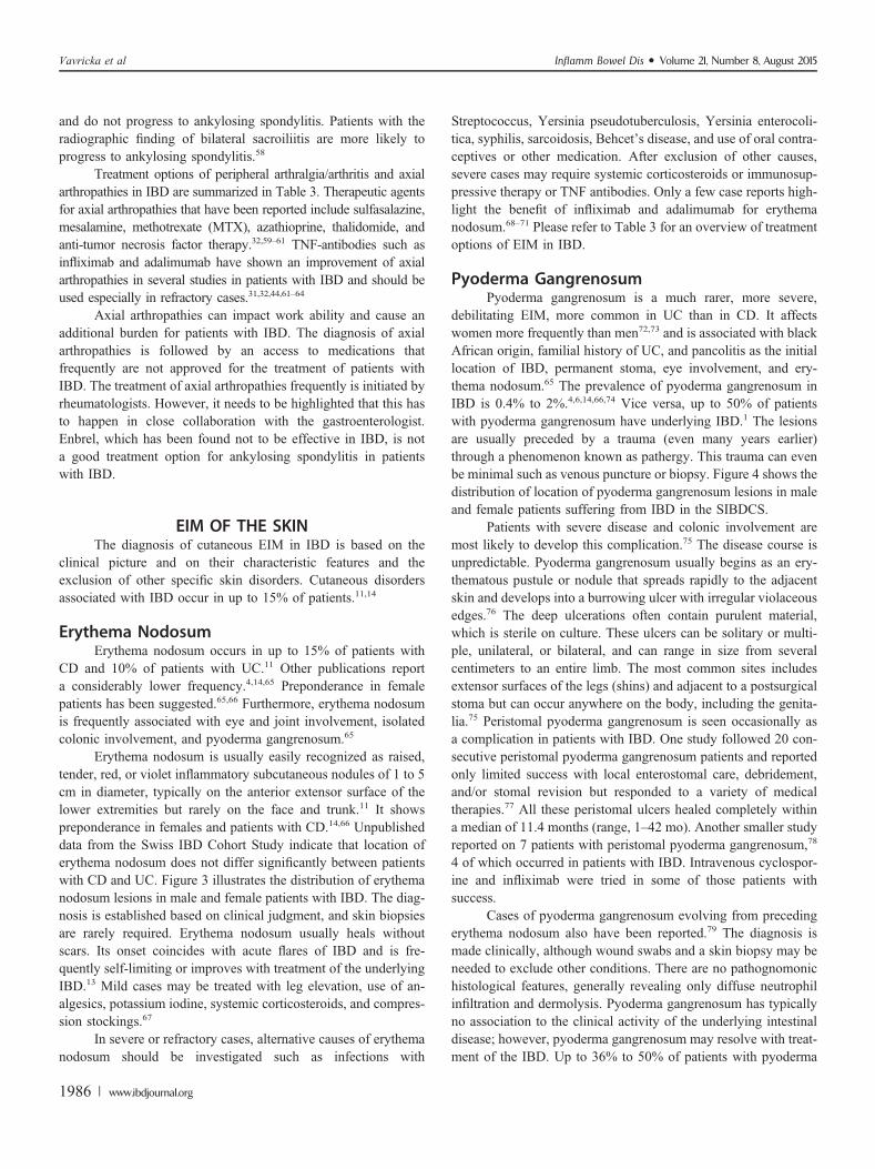
Erythema nodosum is usually easily recognized as raised,tender, red, or violet inflammatory subcutaneous nodules of 1 to 5cm in diameter, typically on the anterior extensor surface of thelower extremities but rarely on the face and trunk.11 It showspreponderance in females and patients with CD.14,66 Unpublisheddata from the Swiss IBD Cohort Study indicate that location oferythema nodosum does not differ significantly between patientswith CD and UC. Figure 3 illustrates the distribution of erythemanodosum lesions in male and female patients with IBD. The diag-nosis is established based on clinical judgment, and skin biopsiesare rarely required. Erythema nodosum usually heals withoutscars. Its onset coincides with acute flares of IBD and is fre-quently self-limiting or improves with treatment of the underlyingIBD.13 Mild cases may be treated with leg elevation, use of an-algesics, potassium iodine, systemic corticosteroids, and compres-sion stockings.67
In severe or refractory cases, alternative causes of erythemanodosum should be investigated such as infections with
Streptococcus, Yersinia pseudotuberculosis, Yersinia enterocoli-tica, syphilis, sarcoidosis, Behcet’s disease, and use of oral contra-ceptives or other medication. After exclusion of other causes,severe cases may require systemic corticosteroids or immunosup-pressive therapy or TNF antibodies. Only a few case reports high-light the benefit of infliximab and adalimumab for erythemanodosum.68–71 Please refer to Table 3 for an overview of treatmentoptions of EIM in IBD.
Pyoderma GangrenosumPyoderma gangrenosum is a much rarer, more severe,
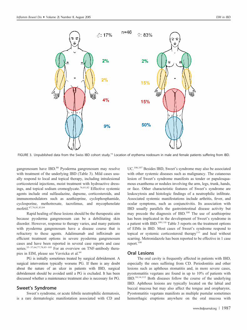
debilitating EIM, more common in UC than in CD. It affectswomen more frequently than men72,73 and is associated with blackAfrican origin, familial history of UC, and pancolitis as the initiallocation of IBD, permanent stoma, eye involvement, and ery-thema nodosum.65 The prevalence of pyoderma gangrenosum inIBD is 0.4% to 2%.4,6,14,66,74 Vice versa, up to 50% of patientswith pyoderma gangrenosum have underlying IBD.1 The lesionsare usually preceded by a trauma (even many years earlier)through a phenomenon known as pathergy. This trauma can evenbe minimal such as venous puncture or biopsy. Figure 4 shows thedistribution of location of pyoderma gangrenosum lesions in maleand female patients suffering from IBD in the SIBDCS.
Patients with severe disease and colonic involvement aremost likely to develop this complication.75 The disease course isunpredictable. Pyoderma gangrenosum usually begins as an ery-thematous pustule or nodule that spreads rapidly to the adjacentskin and develops into a burrowing ulcer with irregular violaceousedges.76 The deep ulcerations often contain purulent material,which is sterile on culture. These ulcers can be solitary or multi-ple, unilateral, or bilateral, and can range in size from severalcentimeters to an entire limb. The most common sites includesextensor surfaces of the legs (shins) and adjacent to a postsurgicalstoma but can occur anywhere on the body, including the genita-lia.75 Peristomal pyoderma gangrenosum is seen occasionally asa complication in patients with IBD. One study followed 20 con-secutive peristomal pyoderma gangrenosum patients and reportedonly limited success with local enterostomal care, debridement,and/or stomal revision but responded to a variety of medicaltherapies.77 All these peristomal ulcers healed completely withina median of 11.4 months (range, 1–42 mo). Another smaller studyreported on 7 patients with peristomal pyoderma gangrenosum,78
4 of which occurred in patients with IBD. Intravenous cyclospor-ine and infliximab were tried in some of those patients withsuccess.
Cases of pyoderma gangrenosum evolving from precedingerythema nodosum also have been reported.79 The diagnosis ismade clinically, although wound swabs and a skin biopsy may beneeded to exclude other conditions. There are no pathognomonichistological features, generally revealing only diffuse neutrophilinfiltration and dermolysis. Pyoderma gangrenosum has typicallyno association to the clinical activity of the underlying intestinaldisease; however, pyoderma gangrenosum may resolve with treat-ment of the IBD. Up to 36% to 50% of patients with pyoderma
Vavricka et al Inflamm Bowel Dis � Volume 21, Number 8, August 2015
1986 | www.ibdjournal.org
gangrenosum have IBD.80 Pyoderma gangrenosum may resolvewith treatment of the underlying IBD (Table 3). Mild cases usu-ally respond to local and topical therapy, including intralesionalcorticosteroid injections, moist treatment with hydroactive dress-ings, and topical sodium cromoglycate.76,81,82 Effective systemicagents include oral sulfasalazine, dapsone, corticosteroids, andimmunomodulators such as azathioprine, cyclophosphamide,cyclosporine, methotrexate, tacrolimus, and mycophenolatemofetil.67,76,81,83,84
Rapid healing of these lesions should be the therapeutic aimbecause pyoderma gangrenosum can be a debilitating skindisorder. However, response to therapy varies, and many patientswith pyoderma gangrenosum have a disease course that isrefractory to these agents. Adalimumab and infliximab areefficient treatment options in severe pyoderma gangrenosumcases and have been reported in several case reports and caseseries.35–37,64,77,78,85–105 For an overview on TNF-antibody thera-pies in EIM, please see Vavricka et al.64
PG is initially sometimes treated by surgical debridement. Asurgical intervention typically worsens PG. If there is any doubtabout the nature of an ulcer in patients with IBD, surgicaldebridement should be avoided until a PG is excluded. It has beendiscussed whether a maintenance treatment also is necessary for PG.
Sweet’s SyndromeSweet’s syndrome, or acute febrile neutrophilic dermatosis,
is a rare dermatologic manifestation associated with CD and
UC.106,107 Besides IBD, Sweet’s syndrome may also be associatedwith other systemic diseases such as malignancy. The cutaneouslesion of Sweet’s syndrome manifests as tender or papulosqua-mous exanthema or nodules involving the arm, legs, trunk, hands,or face. Other characteristic features of Sweet’s syndrome areleukocytosis and histologic findings of a neutrophilic infiltrate.Associated systemic manifestations include arthritis, fever, andocular symptoms, such as conjunctivitis. Its association withIBD usually parallels the gastrointestinal disease activity butmay precede the diagnosis of IBD.108 The use of azathioprinehas been implicated in the development of Sweet’s syndrome ina patient with IBD.109,110 Table 3 reports on the treatment optionsof EIMs in IBD. Most cases of Sweet’s syndrome respond totopical or systemic corticosteroid therapy111 and heal withoutscarring. Metronidazole has been reported to be effective in 1 casereport.108
Oral LesionsThe oral cavity is frequently affected in patients with IBD,
especially the ones suffering from CD. Periodontitis and otherlesions such as aphthous stomatitis and, in more severe cases,pyostomatitis vegetans are found in up to 10% of patients withIBD.10,14,112 Both diseases follow the course of the underlyingIBD. Aphthous lesions are typically located on the labial andbuccal mucosa but may also affect the tongue and oropharynx.Pyostomatitis vegetans manifests as multiple pustular sometimeshemorrhagic eruptions anywhere on the oral mucosa with
FIGURE 3. Unpublished data from the Swiss IBD cohort study.15 Location of erythema nodosum in male and female patients suffering from IBD.
Inflamm Bowel Dis � Volume 21, Number 8, August 2015 EIM in IBD
www.ibdjournal.org | 1987
a cobblestone pattern. Therapy includes antiseptic mouthwashesand topical steroids (Table 3).67,105
OCULAR MANIFESTATIONSBeside joints and skin, the eye is the third major tissue type
predisposed to immune-mediated EIMs. Nearly, 2% to 5% ofpatients with IBD experience ocular manifestations,10,13,113 particu-larly associated with concomitant musculoskeletal manifestations.4
Ocular manifestations are reported more frequently in patients withCD (3.5%–6.3%) than patients with UC (1.6%–4.6%) and includeepiscleritis and uveitis.9,11,13,14,55 Patients older than 40 years havemore likely iritis/uveitis than those younger than 40 years.6
Episcleritis is defined as painless hyperemia of the conjunc-tiva and sclera without changes of virus and often parallels theactivity of the underlying IBD. Besides episcleritis, anterior uveitisis the most common ocular manifestations of IBD. The differenttypes of uveitis are divided as follows: (1) anterior uveitis has itsprimary site of inflammation in the anterior chamber, (2) interme-diate uveitis with its primary site of inflammation being thevitreous, (3) posterior uveitis with its primary site of inflammationbeing the retina and the choroid, and (4) panuveitis with its primarysite of inflammation including anterior chamber, vitreous, retina,and choroid. Uveitis occurs independently of disease activity and isdefined as inflammation of the middle chamber of the eye. Uveitisoccurs acutely or subacutely and is usually very painful. Anterior
uveitis is also referred to as iritis, which typically presents as pain,photophobia, and red eye and can be associated with blurry visionor floaters. Diagnosis is confirmed by slit-lamp examination. Anincreasing number of case reports and pilot studies exist on thetherapy of uveitis and episcleritis; however, only few reports focuson patients with IBD.42,114–124
Episcleritis and ScleritisEpiscleritis is more common in CD than in UC.1 It is char-
acterized by acute hyperemia, irritation, burning, and tenderness.Episcleritis usually does not need specific treatment other thanthose for the underlying disease. Scleritis affects the deeper layersof the eye and can cause visual impairment if not diagnosed early.Patients often complain of severe pain associated with tendernessto palpation.125 Recurrent scleritis can lead to scleromalacia, ret-inal detachment, or optic nerve swelling. If therefore mandatesaggressive treatment. Disease-specific treatment and topical ste-roid therapy usually provide prompt relief of symptoms (Table 3).
In case of impairment of vision, the presence of scleritismust be suspected, and prompt referral to an ophthalmologist ismandatory to avoid vision loss.
UveitisUveitis is less common than episcleritis and occurs in 0.5%
to 3% of patients with IBD.4 When associated with UC, it is
FIGURE 4. Unpublished data from the Swiss IBD cohort study.15 Location of pyoderma gangrenosum in male and female patients suffering from IBD.
Vavricka et al Inflamm Bowel Dis � Volume 21, Number 8, August 2015
1988 | www.ibdjournal.org

frequently bilateral, insidious in onset, and long-lasting.4 Itpresents as ocular pain, blurred vision, photophobia, and head-aches. In contrast to episcleritis, the temporal correlation of uveitiswith IBD is less predictable, and its occurrence may precede thediagnosis of IBD. On slit-lamp examination, uveitis presents asa perilimbic edema and inflammatory flare in the anterior cham-ber.125 Prompt diagnosis and treatment with topical and systemiccorticosteroids is necessary to prevent progression to blindness.Steroid refractory cases are treated with cyclosporine A (Table 3).Successful use of infliximab for IBD-associated uveitis was dem-onstrated in a patient with CD with uveitis and sacroileitis.42
HEPATOBILIARY EIMUp to 50% of patients with IBD are affected by hepato-
biliary manifestations during the course of their disease.5 PSC,small-duct PSC, fatty liver disease, granulomatous hepatitis, auto-immune liver and pancreas disease, cholestasis, gallstone forma-tion, and liver injury are hepatobiliary manifestations of IBD.126
PSC Is the Most Frequent BiliaryManifestation of IBD
It is more common in patients with UC than in CD.Approximately, 2.4% to 7.5% of patients with UC are diagnosedwith PSC.127,128 Conversely, 75% of patients with PSC sufferfrom IBD, typically UC.129,130 PSC manifests with inflammationand fibrosis of the biliary system that presents clinically witha chronic cholestatic disease. A cholestatic biochemical profileis seen, and characteristic features are frequently found on chol-angiography. These include multifocal bile duct strictures andsegmental dilatation. PSC can precede the diagnosis of IBD; how-ever, some patients are even diagnosed with PSC several yearsafter proctocolectomy due to UC.3
Patients with PSC should undergo colonoscopy to evaluateconcomitant IBD. Extensive involvement of the colon with rectalsparing, backwash ileitis in UC, and predominance in malepatients are typical features of PSC.3,130 Patients with PSC candevelop bouts of acute cholangitis and ultimately progress tocirrhosis, portal hypertension, and acute decompensation.131 Inter-estingly, the diagnosis of PSC seems to influence the course ofIBD, as patients with both PSC and UC are suggested to havea milder course of their colitis with less histological inflammationof the colon than patients without PSC.132 Nevertheless, the pres-ence of PSC is an independent risk factor for the development ofcolorectal dysplasia and/or cancer in patients with IBD, leading tothe recommendation for annual surveillance colonoscopies inaffected patients from the time of first diagnosis of IBD.133–135
The natural course of PSC is independent of IBD, and the bileduct damage is irreversible and nonresponsive to medication. Ur-sodeoxycholic acid is used widely in patients with PSC; however,only limited effect has been shown. Ursodeoxycholic acid is re-ported to improve liver enzymes; however, the disease course ofPSC is not changed.136 Some patients with dominant strictures onendoscopic retrograde cholangiography might improve with
dilatation. The majority of patients with PSC ultimately requireliver transplantation.
CONCLUSIONSIBD is a systemic disease, and EIM are the proof that IBD
is not only limited to the gut. Those EIM may affect multipleorgans beyond the intestine. Sometimes, these EIM can even bemore debilitating than the intestinal disease. Careful screening forEIMs in these patients and early appropriate diagnosis areimperative to prevent morbidity. In EIM responding to theunderlying IBD, sufficient IBD therapy and careful monitoringof the EIM is often enough to improve symptoms of the EIM. InEIM not after the activity of the underlying IBD, a multidisciplin-ary approach is often needed. Clinicians who care for patientswith IBD must recognize those various systemic manifestations,as failure to diagnose and treat them early may result in majormorbidity.
REFERENCES1. Rothfuss KS, Stange EF, Herrlinger KR. Extraintestinal manifestations
and complications in inflammatory bowel diseases. World J Gastroen-terol. 2006;12:4819–4831.
2. Trikudanathan G, Venkatesh PG, Navaneethan U. Diagnosis and thera-peutic management of extra-intestinal manifestations of inflammatorybowel disease. Drugs. 2012;72:2333–2349.
3. Navaneethan U, Shen B. Hepatopancreatobiliary manifestations andcomplications associated with inflammatory bowel disease. InflammBowel Dis. 2010;16:1598–1619.
4. Orchard TR, Chua CN, Ahmad T, et al. Uveitis and erythema nodosumin inflammatory bowel disease: clinical features and the role of HLAgenes. Gastroenterology. 2002;123:714–718.
5. Danese S, Semeraro S, Papa A, et al. Extraintestinal manifestations ininflammatory bowel disease. World J Gastroenterol. 2005;11:7227–7236.
6. Bernstein CN, Blanchard JF, Rawsthorne P, et al. The prevalence ofextraintestinal diseases in inflammatory bowel disease: a population-based study. Am J Gastroenterol. 2001;96:1116–1122.
7. Ricart E, Panaccione R, Loftus EV Jr, et al. Autoimmune disorders andextraintestinal manifestations in first-degree familial and sporadic inflam-matory bowel disease: a case-control study. Inflamm Bowel Dis. 2004;10:207–214.
8. Mendoza JL, Lana R, Taxonera C, et al. [Extraintestinal manifestationsin inflammatory bowel disease: differences between Crohn’s disease andulcerative colitis [Article in Spanish]. Med Clin (Barc). 2005;125:297–300.
9. Rankin GB, Watts HD, Melnyk CS, et al. National Cooperative Crohn’sDisease Study: extraintestinal manifestations and perianal complications.Gastroenterology. 1979;77:914–920.
10. Su CG, Judge TA, Lichtenstein GR. Extraintestinal manifestations ofinflammatory bowel disease. Gastroenterol Clin North Am. 2002;31:307–327.
11. Greenstein AJ, Janowitz HD, Sachar DB. The extra-intestinal complica-tions of Crohn’s disease and ulcerative colitis: a study of 700 patients.Medicine. 1976;55:401–412.
12. Farmer RG, Hawk WA, Turnbull RB Jr. Clinical patterns in Crohn’sdisease: a statistical study of 615 cases. Gastroenterology. 1975;68:627–635.
13. Veloso FT, Carvalho J, Magro F. Immune-related systemic manifesta-tions of inflammatory bowel disease. A prospective study of 792 pa-tients. J Clin Gastroenterol. 1996;23:29–34.
14. Vavricka SR, Brun L, Ballabeni P, et al. Frequency and risk factors forextraintestinal manifestations in the Swiss inflammatory bowel diseasecohort. Am J Gastroenterol. 2011;106:110–119.
15. Vavricka SR, Gantenbein C, Spoerri M, et al. Chronological order ofappearance of extraintestinal manifestations relative to the time of IBD
Inflamm Bowel Dis � Volume 21, Number 8, August 2015 EIM in IBD
www.ibdjournal.org | 1989
diagnosis in the Swiss Inflammatory Bowel Disease Cohort. InflammBowel Dis. 2015;21:1794–1800.
16. Ardizzone S, Puttini PS, Cassinotti A, et al. Extraintestinal mani-festations of inflammatory bowel disease. Dig Liver Dis. 2008;40(suppl 2):S253–S259.
17. Biancone L, Mandal A, Yang H, et al. Production of immunoglobulin Gand G1 antibodies to cytoskeletal protein by lamina propria cells inulcerative colitis. Gastroenterology. 1995;109:3–12.
18. Das KM, Sakamaki S, Vecchi M, et al. The production and character-ization of monoclonal antibodies to a human colonic antigen associatedwith ulcerative colitis: cellular localization of the antigen by using themonoclonal antibody. J Immunol. 1987;139:77–84.
19. Geng X, Biancone L, Dai HH, et al. Tropomyosin isoforms in intestinalmucosa: production of autoantibodies to tropomyosin isoforms in ulcer-ative colitis. Gastroenterology. 1998;114:912–922.
20. Bhagat S, Das KM. A shared and unique peptide in the human colon,eye, and joint detected by a monoclonal antibody. Gastroenterology.1994;107:103–108.
21. Das KM, Vecchi M, Sakamaki S. A shared and unique epitope(s) onhuman colon, skin, and biliary epithelium detected by a monoclonalantibody. Gastroenterology. 1990;98:464–469.
22. Satsangi J, Grootscholten C, Holt H, et al. Clinical patterns of familialinflammatory bowel disease. Gut. 1996;38:738–741.
23. Roussomoustakaki M, Satsangi J, Welsh K, et al. Genetic markers maypredict disease behavior in patients with ulcerative colitis. Gastroenter-ology. 1997;112:1845–1853.
24. Ott C, Scholmerich J. Extraintestinal manifestations and complications inIBD. Nat Rev Gastroenterol Hepatol. 2013;10:585–595.
25. Orchard TR, Thiyagaraja S, Welsh KI, et al. Clinical phenotype is relatedto HLA genotype in the peripheral arthropathies of inflammatory boweldisease. Gastroenterology. 2000;118:274–278.
26. Mallas EG, Mackintosh P, Asquith P, et al. Histocompatibility antigensin inflammatory bowel disease. Their clinical significance and their asso-ciation with arthropathy with special reference to HLA-B27 (W27). Gut.1976;17:906–910.
27. Smale S, Natt RS, Orchard TR, et al. Inflammatory bowel disease andspondylarthropathy. Arthritis Rheum. 2001;44:2728–2736.
28. Gravallese EM, Kantrowitz FG. Arthritic manifestations of inflammatorybowel disease. Am J Gastroenterol. 1988;83:703–709.
29. Orchard TR, Wordsworth BP, Jewell DP. Peripheral arthropathies ininflammatory bowel disease: their articular distribution and natural his-tory. Gut. 1998;42:387–391.
30. Rodriguez-Reyna TS, Martinez-Reyes C, Yamamoto-Furusho JK. Rheu-matic manifestations of inflammatory bowel disease. World J Gastro-enterol. 2009;15:5517–5524.
31. Brakenhoff LK, van der Heijde DM, Hommes DW. IBD and arthropa-thies: a practical approach to its diagnosis and management. Gut. 2011;60:1426–1435.
32. Generini S, Giacomelli R, Fedi R, et al. Infliximab in spondyloarthrop-athy associated with Crohn’s disease: an open study on the efficacy ofinducing and maintaining remission of musculoskeletal and gut manifes-tations. Ann Rheum Dis. 2004;63:1664–1669.
33. Herfarth H, Obermeier F, Andus T, et al. Improvement of arthritis andarthralgia after treatment with infliximab (Remicade) in a German pro-spective, open-label, multicenter trial in refractory Crohn’s disease. Am JGastroenterol. 2002;97:2688–2690.
34. Atzeni F, Ardizzone S, Bertani L, et al. Combined therapeutic approach:inflammatory bowel diseases and peripheral or axial arthritis. World JGastroenterol. 2009;15:2469–2471.
35. Sarzi-Puttini P, Antivalle M, Marchesoni A, et al. Efficacy and safetyof anti-TNF agents in the Lombardy rheumatoid arthritis network(LORHEN). Reumatismo. 2008;60:290–295.
36. Kaufman I, Caspi D, Yeshurun D, et al. The effect of infliximab onextraintestinal manifestations of Crohn’s disease. Rheumatol Int. 2005;25:406–410.
37. Brooklyn TN, Dunnill MG, Shetty A, et al. Infliximab for the treatmentof pyoderma gangrenosum: a randomised, double blind, placebo con-trolled trial. Gut. 2006;55:505–509.
38. Regueiro M, Valentine J, Plevy S, et al. Infliximab for treatment ofpyoderma gangrenosum associated with inflammatory bowel disease.Am J Gastroenterol. 2003;98:1821–1826.
39. Tanida S, Inoue N, Kobayashi K, et al. Adalimumab for the treatment ofJapanese patients with intestinal Behcet’s disease. Clin GastroenterolHepatol. 2015;13:940–948.
40. Vanbiervliet G, Anty R, Schneider S, et al. Sweet’s syndrome and ery-thema nodosum associated with Crohn’s disease treated by infliximab[Article in French]. Gastroenterol Clin Biol. 2002;26:295–297.
41. Singh S, Khanna S, Pardi DS, et al. Effect of ursodeoxycholic acid useon the risk of colorectal neoplasia in patients with primary sclerosingcholangitis and inflammatory bowel disease: a systematic review andmeta-analysis. Inflamm Bowel Dis. 2013;19:1631–1638.
42. Fries W, Giofre MR, Catanoso M, et al. Treatment of acute uveitisassociated with Crohn’s disease and sacroileitis with infliximab. Am JGastroenterol. 2002;97:499–500.
43. Calvo-Rio V, Blanco R, Beltran E, et al. Anti-TNF-alpha therapy inpatients with refractory uveitis due to Behcet’s disease: a 1-yearfollow-up study of 124 patients. Rheumatology (Oxford). 2014;53:2223–2231.
44. Lakatos PL, Lakatos L, Kiss LS, et al. Treatment of extraintestinalmanifestations in inflammatory bowel disease. Digestion. 2012;86(suppl 1):28–35.
45. Clegg DO, Reda DJ, Abdellatif M. Comparison of sulfasalazine andplacebo for the treatment of axial and peripheral articular manifestationsof the seronegative spondylarthropathies: a Department of Veterans Af-fairs cooperative study. Arthritis Rheum. 1999;42:2325–2329.
46. Bjarnason I, Hayllar J, MacPherson AJ, et al. Side effects of nonsteroidalanti-inflammatory drugs on the small and large intestine in humans.Gastroenterology. 1993;104:1832–1847.
47. Kaufmann HJ, Taubin HL. Nonsteroidal anti-inflammatory drugs acti-vate quiescent inflammatory bowel disease. Ann Intern Med. 1987;107:513–516.
48. Miner PB Jr. Factors influencing the relapse of patients with inflamma-tory bowel disease. Am J Gastroenterol. 1997;92:1S–4S.
49. Kefalakes H, Stylianides TJ, Amanakis G, et al. Exacerbation of inflam-matory bowel diseases associated with the use of nonsteroidal anti-inflammatory drugs: myth or reality? Eur J Clin Pharmacol. 2009;65:963–970.
50. Takeuchi K, Smale S, Premchand P, et al. Prevalence and mechanism ofnonsteroidal anti-inflammatory drug-induced clinical relapse in patientswith inflammatory bowel disease. Clin Gastroenterol Hepatol. 2006;4:196–202.
51. Sandborn WJ, Stenson WF, Brynskov J, et al. Safety of celecoxibin patients with ulcerative colitis in remission: a randomized,placebo-controlled, pilot study. Clin Gastroenterol Hepatol. 2006;4:203–211.
52. Reinisch W, Miehsler W, Dejaco C, et al. An open-label trial of theselective cyclo-oxygenase-2 inhibitor, rofecoxib, in inflammatory boweldisease-associated peripheral arthritis and arthralgia. Aliment PharmacolTher. 2003;17:1371–1380.
53. Mahadevan U, Loftus EV Jr, Tremaine WJ, et al. Safety of selectivecyclooxygenase-2 inhibitors in inflammatory bowel disease. Am J Gas-troenterol. 2002;97:910–914.
54. El Miedany Y, Youssef S, Ahmed I, et al. The gastrointestinal safetyand effect on disease activity of etoricoxib, a selective cox-2 inhib-itor in inflammatory bowel diseases. Am J Gastroenterol. 2006;101:311–317.
55. Monsen U, Sorstad J, Hellers G, et al. Extracolonic diagnoses in ulcer-ative colitis: an epidemiological study. Am J Gastroenterol. 1990;85:711–716.
56. Fornaciari G, Salvarani C, Beltrami M, et al. Muscoloskeletal manifes-tations in inflammatory bowel disease. Can J Gastroenterol. 2001;15:399–403.
57. Brewerton DA, James DC. The histocompatibility antigen (HL-A 27)and disease. Semin Arthritis Rheum. 1975;4:191–207.
58. Braun J, Sieper J. The sacroiliac joint in the spondyloarthropathies. CurrOpin Rheumatol. 1996;8:275–287.
59. Breban M, Gombert B, Amor B, et al. Efficacy of thalidomide in thetreatment of refractory ankylosing spondylitis. Arthritis Rheum. 1999;42:580–581.
60. Keyser FD, Mielants H, Veys EM. Current use of biologicals for thetreatment of spondyloarthropathies. Expert Opin Pharmacother. 2001;2:85–93.
Vavricka et al Inflamm Bowel Dis � Volume 21, Number 8, August 2015
1990 | www.ibdjournal.org

61. Van den Bosch F, Kruithof E, De Vos M, et al. Crohn’s disease asso-ciated with spondyloarthropathy: effect of TNF-alpha blockade withinfliximab on articular symptoms. Lancet. 2000;356:1821–1822.
62. Ellman MH, Hanauer S, Sitrin M, et al. Crohn’s disease arthritis treatedwith infliximab: an open trial in four patients. J Clin Rheumatol. 2001;7:67–71.
63. Zochling J, van der Heijde D, Dougados M, et al. Current evidence forthe management of ankylosing spondylitis: a systematic literature reviewfor the ASAS/EULAR management recommendations in ankylosingspondylitis. Ann Rheum Dis. 2006;65:423–432.
64. Vavricka SR, Scharl M, Gubler M, et al. Which extraintestinal manifestationsof IBD respond to biologics? Curr Drug Targets. 2014;15:1064–1073.
65. Farhi D, Cosnes J, Zizi N, et al. Significance of erythema nodosum andpyoderma gangrenosum in inflammatory bowel diseases: a cohort studyof 2402 patients. Medicine. 2008;87:281–293.
66. Freeman HJ. Erythema nodosum and pyoderma gangrenosum in 50patients with Crohn’s disease. Can J Gastroenterol. 2005;19:603–606.
67. Timani S, Mutasim DF. Skin manifestations of inflammatory boweldisease. Clin Dermatol. 2008;26:265–273.
68. Kugathasan S, Miranda A, Nocton J, et al. Dermatologic manifestationsof Crohn disease in children: response to infliximab. J Pediatr Gastro-enterol Nutr. 2003;37:150–154.
69. Ortego-Centeno N, Callejas-Rubio JL, Sanchez-Cano D, et al. Refractorychronic erythema nodosum successfully treated with adalimumab. J EurAcad Dermatol Venereol. 2007;21:408–410.
70. Quin A, Kane S, Ulitsky O. A case of fistulizing Crohn’s disease anderythema nodosum managed with adalimumab. Nat Clin Pract Gastro-enterol Hepatol. 2008;5:278–281.
71. Clayton TH, Walker BP, Stables GI. Treatment of chronic erythemanodosum with infliximab. Clin Exp Dermatol. 2006;31:823–824.
72. Jorizzo JL, Solomon AR, Zanolli MD, et al. Neutrophilic vascular re-actions. J Am Acad Dermatol. 1988;19:983–1005.
73. Bennett ML, Jackson JM, Jorizzo JL, et al. Pyoderma gangrenosum. Acomparison of typical and atypical forms with an emphasis on time toremission. Case review of 86 patients from 2 institutions. Medicine.2000;79:37–46.
74. Nguyen GC, Torres EA, Regueiro M, et al. Inflammatory bowel diseasecharacteristics among African Americans, Hispanics, and non-HispanicWhites: characterization of a large North American cohort. Am J Gastro-enterol. 2006;101:1012–1023.
75. Orchard T. Extraintestinal complications of inflammatory bowel disease.Curr Gastroenterol Rep. 2003;5:512–517.
76. Callen JP. Pyoderma gangrenosum. Lancet. 1998;351:581–585.77. Sheldon DG, Sawchuk LL, Kozarek RA, et al. Twenty cases of peristo-
mal pyoderma gangrenosum: diagnostic implications and management.Arch Surg. 2000;135:564–568; discussion 568–569.
78. Hughes AP, Jackson JM, Callen JP. Clinical features and treatment ofperistomal pyoderma gangrenosum. JAMA. 2000;284:1546–1548.
79. Gellert A, Green ES, Beck ER, et al. Erythema nodosum progressing topyoderma gangrenosum as a complication of Crohn’s disease. PostgradMed J. 1983;59:791–793.
80. Chow RK, Ho VC. Treatment of pyoderma gangrenosum. J Am AcadDermatol. 1996;34:1047–1060.
81. Powell RJ, Holbrook MR, Stevens A. Pyoderma gangrenosum and itstreatment. Lancet. 1997;350:1720–1721.
82. Trost LB, McDonnell JK. Important cutaneous manifestations of inflam-matory bowel disease. Postgrad Med J. 2005;81:580–585.
83. Friedman S, Marion JF, Scherl E, et al. Intravenous cyclosporine inrefractory pyoderma gangrenosum complicating inflammatory boweldisease. Inflamm Bowel Dis. 2001;7:1–7.
84. Wollina U, Haroske G. Pyoderma gangraenosum. Curr Opin Rheumatol.2011;23:50–56.
85. Arnott ID, McDonald D, Williams A, et al. Clinical use of Infliximab inCrohn’s disease: the Edinburgh experience. Aliment Pharmacol Ther.2001;15:1639–1646.
86. Batres LA, Mamula P, Baldassano RN. Resolution of severe peristomalpyoderma gangrenosum with infliximab in a child with Crohn disease.J Pediatr Gastroenterol Nutr. 2002;34:558–560.
87. Ferkolj I, Hocevar A, Golouh R, et al. Infliximab for treatment of resis-tant pyoderma gangrenosum associated with Crohn’s disease. Acta Der-matovenerol Alp Pannonica Adriat. 2006;15:173–177.
88. Grange F, Djilali-Bouzina F, Weiss AM, et al. Corticosteroid-resistantpyoderma gangrenosum associated with Crohn’s disease: rapid cure withinfliximab. Dermatology. 2002;205:278–280.
89. Juillerat P, Christen-Zach S, Troillet FX, et al. Infliximab for thetreatment of disseminated pyoderma gangrenosum associated withulcerative colitis. Case report and literature review. Dermatology.2007;215:245–251.
90. Kiran RP, O’Brien-Ermlich B, Achkar JP, et al. Management ofperistomal pyoderma gangrenosum. Dis Colon Rectum. 2005;48:1397–1403.
91. Kouklakis G, Moschos J, Leontiadis GI, et al. Infliximab for treatment ofpyoderma gangrenosum associated with clinically inactive Crohn’s dis-ease. A case report. Rom J Gastroenterol. 2005;14:401–403.
92. Ljung T, Staun M, Grove O, et al. Pyoderma gangrenosum associatedwith crohn disease: effect of TNF-alpha blockade with infliximab. ScandJ Gastroenterol. 2002;37:1108–1110.
93. Fonder MA, Cummins DL, Ehst BD, et al. Adalimumab therapy forrecalcitrant pyoderma gangrenosum. J Burns Wounds. 2006;5:e8.
94. Martin D, Handler T, McDermott J. Leucocytoclastic vasculitis in severeulcerative colitis. Mil Med. 2011;176:581–583.
95. Lopez San Roman A, Bermejo F, Aldanondo I, et al. Pyoderma gangre-nosum associated with ulcerative colitis: response to infliximab. Rev EspEnferm Dig. 2004;96:420–422; 422-424.
96. Sapienza MS, Cohen S, Dimarino AJ. Treatment of pyoderma gangre-nosum with infliximab in Crohn’s disease. Dig Dis Sci. 2004;49:1454–1457.
97. Singh M, Andrew SM, Lear JT. Infliximab as a treatment for recalcitrantpyoderma gangrenosum. Clin Exp Dermatol. 2004;29:196–197.
98. Tan MH, Gordon M, Lebwohl O, et al. Improvement of Pyodermagangrenosum and psoriasis associated with Crohn disease with anti-tumor necrosis factor alpha monoclonal antibody. Arch Dermatol.2001;137:930–933.
99. Triantafillidis JK, Cheracakis P, Sklavaina M, et al. Favorable responseto infliximab treatment in a patient with active Crohn disease and pyo-derma gangrenosum. Scand J Gastroenterol. 2002;37:863–865.
100. Zaccagna A, Bertone A, Puiatti P, et al. Anti-tumor necrosis factor alphamonoclonal antibody (infliximab) for the treatment of Pyoderma gangre-nosum associated with Crohn’s disease. Eur J Dermatol. 2003;13:258–260.
101. Alkhouri N, Hupertz V, Mahajan L. Adalimumab treatment for peristo-mal pyoderma gangrenosum associated with Crohn’s disease. InflammBowel Dis. 2009;15:803–806.
102. Pomerantz RG, Husni ME, Mody E, et al. Adalimumab for treatment ofpyoderma gangrenosum. Br J Dermatol. 2007;157:1274–1275.
103. Zold E, Nagy A, Devenyi K, et al. Successful use of adalimumab fortreating fistulizing Crohn’s disease with pyoderma gangrenosum: twobirds with one stone. World J Gastroenterol. 2009;15:2293–2295.
104. Mimouni D, Anhalt GJ, Kouba DJ, et al. Infliximab for peristomalpyoderma gangrenosum. Br J Dermatol. 2003;148:813–816.
105. Thrash B, Patel M, Shah KR, et al. Cutaneous manifestations of gastro-intestinal disease: part II. J Am Acad Dermatol. 2013;68:211 e211-233;quiz 244–216.
106. Becuwe C, Delaporte E, Colombel JF, et al. Sweet’s syndrome associ-ated with Crohn’s disease. Acta Derm Venereol. 1989;69:444–445.
107. Benton EC, Rutherford D, Hunter JA. Sweet’s syndrome and pyodermagangrenosum associated with ulcerative colitis. Acta Derm Venereol.1985;65:77–80.
108. Banet DE, McClave SA, Callen JP. Oral metronidazole, an effectivetreatment for Sweet’s syndrome in a patient with associated inflamma-tory bowel disease. J Rheumatol. 1994;21:1766–1768.
109. Treton X, Joly F, Alves A, et al. Azathioprine-induced Sweet’s syn-drome in Crohn’s disease. Inflamm Bowel Dis. 2008;14:1757–1758.
110. Ali M, Duerksen DR. Ulcerative colitis and Sweet’s syndrome: a casereport and review of the literature. Can J Gastroenterol. 2008;22:296–298.
111. Kemmett D, Hunter JA. Sweet’s syndrome: a clinicopathologic review oftwenty-nine cases. J Am Acad Dermatol. 1990;23:503–507.
112. Vavricka SR, Manser CN, Hediger S, et al. Periodontitis and gingivitis ininflammatory bowel disease: a case-control study. Inflamm Bowel Dis.2013;19:2768–2777.
113. Petrelli EA, McKinley M, Troncale FJ. Ocular manifestations of inflam-matory bowel disease. Ann Ophthalmol. 1982;14:356–360.
Inflamm Bowel Dis � Volume 21, Number 8, August 2015 EIM in IBD
www.ibdjournal.org | 1991
114. Foeldvari I, Nielsen S, Kummerle-Deschner J, et al. Tumor necrosisfactor-alpha blocker in treatment of juvenile idiopathic arthritis-associated uveitis refractory to second-line agents: results of a multina-tional survey. J Rheumatol. 2007;34:1146–1150.
115. Hale S, Lightman S. Anti-TNF therapies in the management of acute andchronic uveitis. Cytokine. 2006;33:231–237.
116. Joseph A, Raj D, Dua HS, et al. Infliximab in the treatment of refractoryposterior uveitis. Ophthalmology. 2003;110:1449–1453.
117. Lindstedt EW, Baarsma GS, Kuijpers RW, et al. Anti-TNF-alpha therapyfor sight threatening uveitis. Br J Ophthalmol. 2005;89:533–536.
118. Rajaraman RT, Kimura Y, Li S, et al. Retrospective case review ofpediatric patients with uveitis treated with infliximab. Ophthalmology.2006;113:308–314.
119. Sfikakis PP, Theodossiadis PG, Katsiari CG, et al. Effect of infliximabon sight-threatening panuveitis in Behcet’s disease. Lancet. 2001;358:295–296.
120. Murphy CC, Ayliffe WH, Booth A, et al. Tumor necrosis factor alphablockade with infliximab for refractory uveitis and scleritis. Ophthalmol-ogy. 2004;111:352–356.
121. Saurenmann RK, Levin AV, Rose JB, et al. Tumour necrosis factor alphainhibitors in the treatment of childhood uveitis. Rheumatology. 2006;45:982–989.
122. Kahn P, Weiss M, Imundo LF, et al. Favorable response to high-doseinfliximab for refractory childhood uveitis. Ophthalmology. 2006;113:860–864.e862.
123. Biester S, Deuter C, Michels H, et al. Adalimumab in the therapy ofuveitis in childhood. Br J Ophthalmol. 2007;91:319–324.
124. Braun J, Baraliakos X, Listing J, et al. Decreased incidence of anterioruveitis in patients with ankylosing spondylitis treated with the anti-tumornecrosis factor agents infliximab and etanercept. Arthritis Rheum. 2005;52:2447–2451.
125. Mintz R, Feller ER, Bahr RL, et al. Ocular manifestations of inflamma-tory bowel disease. Inflamm Bowel Dis. 2004;10:135–139.
126. Yarur AJ, Czul F, Levy C. Hepatobiliary manifestations of inflammatorybowel disease. Inflamm Bowel Dis. 2014;20:1655–1667.
127. Olsson R, Danielsson A, Jarnerot G, et al. Prevalence of primary scle-rosing cholangitis in patients with ulcerative colitis. Gastroenterology.1991;100:1319–1323.
128. Fausa O, Schrumpf E, Elgjo K. Relationship of inflammatory boweldisease and primary sclerosing cholangitis. Semin Liver Dis. 1991;11:31–39.
129. Wiesner RH, LaRusso NF. Clinicopathologic features of the syndromeof primary sclerosing cholangitis. Gastroenterology. 1980;79:200–206.
130. Broome U, Bergquist A. Primary sclerosing cholangitis, inflammatorybowel disease, and colon cancer. Semin Liver Dis. 2006;26:31–41.
131. Tischendorf JJ, Hecker H, Kruger M, et al. Characterization, outcome,and prognosis in 273 patients with primary sclerosing cholangitis: a sin-gle center study. Am J Gastroenterol. 2007;102:107–114.
132. Joo M, Abreu-e-Lima P, Farraye F, et al. Pathologic features of ulcera-tive colitis in patients with primary sclerosing cholangitis: a case-controlstudy. Am J Surg Pathol. 2009;33:854–862.
133. Soetikno RM, Lin OS, Heidenreich PA, et al. Increased risk of colorectalneoplasia in patients with primary sclerosing cholangitis and ulcerativecolitis: a meta-analysis. Gastrointest Endosc. 2002;56:48–54.
134. Loftus EV Jr, Harewood GC, Loftus CG, et al. PSC-IBD: a unique formof inflammatory bowel disease associated with primary sclerosing chol-angitis. Gut. 2005;54:91–96.
135. Talwalkar JA, Lindor KD. Primary sclerosing cholangitis. InflammBowel Dis. 2005;11:62–72.
136. Lindor KD. Ursodiol for primary sclerosing cholangitis. Mayo PrimarySclerosing Cholangitis-Ursodeoxycholic Acid Study Group. N Engl JMed. 1997;336:691–695.
Vavricka et al Inflamm Bowel Dis � Volume 21, Number 8, August 2015
1992 | www.ibdjournal.org