external morphology of flea larvae (siphonaptera) and its significance in taxonomy
TRANSCRIPT
External Morphology of Flea Larvae (Siphonaptera) and Its Significance in TaxonomyAuthor(s): R. L. C. PilgrimSource: The Florida Entomologist, Vol. 74, No. 3 (Sep., 1991), pp. 386-395Published by: Florida Entomological SocietyStable URL: http://www.jstor.org/stable/3494831 .
Accessed: 14/06/2014 01:48
Your use of the JSTOR archive indicates your acceptance of the Terms & Conditions of Use, available at .http://www.jstor.org/page/info/about/policies/terms.jsp
.JSTOR is a not-for-profit service that helps scholars, researchers, and students discover, use, and build upon a wide range ofcontent in a trusted digital archive. We use information technology and tools to increase productivity and facilitate new formsof scholarship. For more information about JSTOR, please contact [email protected].
.
Florida Entomological Society is collaborating with JSTOR to digitize, preserve and extend access to TheFlorida Entomologist.
http://www.jstor.org
This content downloaded from 62.122.72.154 on Sat, 14 Jun 2014 01:48:34 AMAll use subject to JSTOR Terms and Conditions
386 Florida Entomologist 74(3) September, 1991
EXTERNAL MORPHOLOGY OF FLEA LARVAE (SIPHONAPTERA) AND ITS SIGNIFICANCE IN TAXONOMY,
R.L.C. PILGRIM
Department of Zoology, University of Canterbury Christchurch, New Zeland
ABSTRACT
More than 180 species and subspecies of larvae from all except 4 small families of fleas, and including at least 30% of known genera/subgenera, were examined with phase- contrast and/or scanning electron microscopy. A much greater variety of detailed form was revealed than appears in the literature. Especially significant are: numbers and patterns of setae and their topographical relations to sensilla on the thorax and abdomen; form of the setae, of which at least 9 types can be distinguished on the head, thorax and abdomen; form, arrangement and surface texture of sensory papillae on the antennal mound. Some of these features appear to be characteristic of, and restricted to, genera, subfamilies or higher taxonomic categories. Others, such as the shape of the mandible and their numbers of teeth, show considerable variation which does not clearly follow recognized classifications based on adult fleas; these may be more related to factors in the biology of specific larvae.
RESUMEN
Con excepcion de 4 familias, se examinaron mas de 180 especies y subespecies en estado larvario de pulgas. Se incluyo el 30% de los generos/subgeneros conocidos y se examinaron con el microscopio de escanografia electronica con fase de contraste. Se encontro una gran variedad de formas en comparacion con lo que aparece en la literatura. Se encontraron diferencias significativas en: numeros y patrones de setas y sus relaciones topograficas con la sensilla del torax y abdomen; forma de las setas, de las cuales, al menos 9 tipos pueden ser distinguidos en la cabeza, torax y abdomen; la forma y distribu- cion, y la textura de la papilla sensorial en la antena. Algunos de estos rasgos parecen ser caracteristicos o restringidos a un genero, subfamilia o categorias taxonomicas altas. Otras, tales como la forma de la mandibula y los numeros de molares, mostraron una variacion considerable, la cual no sigue exactamente las clasificaciones reconocidas, las cuales se basan en descripciones de las pulgas adultas; estas pueden estar mas re- lacionadas con factores de la biologia de las larvas.
The desirability of being able to identify flea larvae in the absence of the corresponding adult(s) is not one of mere academic interest. It is of intense practical value to be able to categorize a host's nest as to whether or not it contains certain flea species which are potential vectors of bubonic plague or other diseases. This will be particularly useful in the case of abandoned nests, or of nests examined in the temporary absence of the hosts. It also makes allowance for seasonality factors in surveying the fauna of a nest, i.e. in respect of "summer" and "winter" fleas, whereby the larvae may be present in the near or complete absence of the adult flea(s). As well, a taxonomy of their larvae can provide a base for extending studies on the biology of fleas; it could also add data for consideration along with adult morphology in elucidating the taxonomy and phylogeny of the order.
The life-cycle of a flea comprises: egg - larva (with usually 3 instars) - pupa - adult. About 5% of adult fleas are parasitic on birds, the remainder on mammals; they are
This content downloaded from 62.122.72.154 on Sat, 14 Jun 2014 01:48:34 AMAll use subject to JSTOR Terms and Conditions
Pilgrim: External Morphology of Flea Larvae 387
found closely associated with the hosts or in their nests. Over 2200 species and subspecies are known; they are classified on the basis of adult morphology in 15 families, grouped into 5 superfamilies (Smit 1982). The families are of very disparate size, ranging from Malacopsyllidae with 2 monotypic genera to Ctenophthalmidae with more than 630 species/subspecies and Ceratophyllidae with over 750.
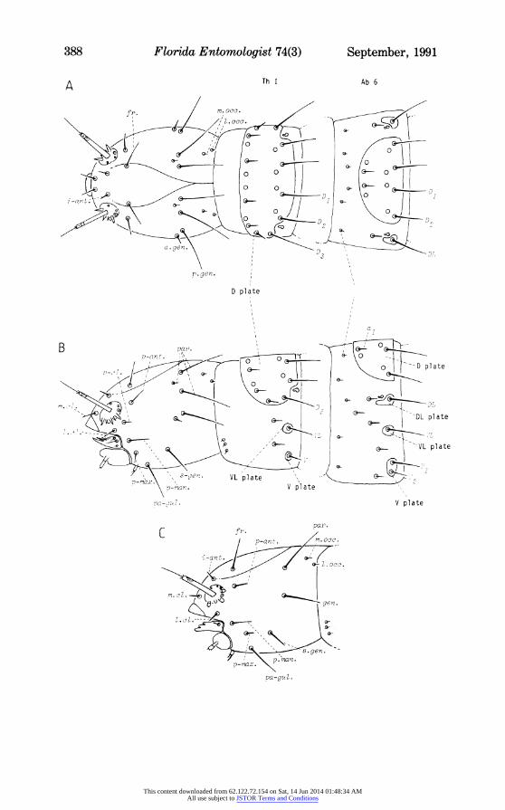
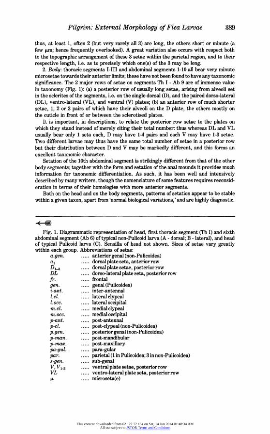
The larvae are, with very rare exceptions, active scavengers living non-parasitically in nest debris of the hosts. They are 1.5-10 mm long, legless, eyeless, maggot-like with a firmly sclerotised head capsule. The head bears prominent antennae, well sclerotised labrum, mandibles and maxillae, and a weakly developed labium. The body is usually considered to comprise 3 thoracic and 10 abdominal segments, the last of which bears a pair of anal struts projecting backwards and downwards from the swollen anal mounds. Numerous long and short setae are present on the head capsule and on the body segments; many setae on the body arise from alveoli in well-defined plate-like sclerotisations (Fig. 1).
This study was initiated to seek a key to the taxa of flea larvae over as wide a range as possible, and to provide a basis for suggesting phylogenetic relationships within the order as exhibited by the larvae. Before these aims could be realized, it was found necessary to provide for a more consistent terminology for the larval morphology, since there is much confusion between published accounts. Some features in the morphology have been described in considerable detail in the literature for a number of species, from many genera. These published accounts were based on light microscope examination of slide-mounted specimens, and some patterns of taxonomic significance have emerged. Re-examination of such material in the present study, together with supporting evidence from scanning electron microscopy, now shows that some of these features have been incorrectly interpreted. As well, attention is here drawn to a number of structures which had been inadequately studied, dismissed as not significant, or simply overlooked; they can now be shown to have considerable taxonomic value.
This account is based on examination of more than 3000 specimens from over 180 spp./sspp. of larvae in 84 generalsubgenera, taken from all superfamilies (sensu Smit 1982). The larvae of several small families remain unknown (Stephanocircidae, Ancitrop- syllidae, Xiphiopsyllidae, Malacopsyllidae) as do the larvae of a few subfamilies/tribes among the larger families. Generalizations made now must be subject to modification as more widely representative collections are examined; almost every feature so far described has been found to have some exception or enhancement when a fresh taxon is studied. Uropsylla tasmanica Rothschild, 1905 (Pygiopsyllidae) is specifically excluded from the generalizations which follow: its larva is so grossly modified in association with its parasitic mode of life that almost every feature in its morphology would require a separate statement.
PATTERNS OF SETATION
1. Head: most published accounts describe the majority of setae behind the antennae as distributed in 2 rows, anterior and posterior. These 'rows' are not very impressive, several setae being variously displaced from anything resembling a linear arrangement; and schools of writers are not in complete agreement as to the designation of the lowermost setae. I propose to abandone the 'row' concept and to redefine the setal nomenclature in terms of regions of the head capsule on which the setae lie; see Fig. 1. Major distinctions are immediately evident between Pulicoidea and all remaining super- families: the non-Pulicoidea possess, on each side of the head, 2 additional parietal setae, a post-clypeal seta and an anterior genal seta beyond those found in Pulicoidea. (These are basic patterns within which some variations occur, such as an extra pair of para-gular setae in Stenoponia, and reduced setation in Tunga.)
Many head capsule setae show important differences in length between different groups, but the parietal setae are especially useful taxonomically in the non-Pulicoidea:
This content downloaded from 62.122.72.154 on Sat, 14 Jun 2014 01:48:34 AMAll use subject to JSTOR Terms and Conditions
388 Florida Entomologist 74(3) September, 1991
A Th I Ab 6
M.en f D ' 3
0~~~~~~~ 0 0
-\ I
a gen. a 9
D plate
x ad
0~~~~~~~~~~~V 0 Dplate
m.,
-
, 1 -
s- veY . VL pl ateplt
\ s-r7an. V pl ate \ V.
\ \<r,
p - n. VV plate 7-an V plate
[.pan.
.rman. --ant./ pa- an\
a- laca.
This content downloaded from 62.122.72.154 on Sat, 14 Jun 2014 01:48:34 AMAll use subject to JSTOR Terms and Conditions
Pilgrim: External Morphology of Flea Larvae 389
thus, at least 1, often 2 (but very rarely all 3) are long, the others short or minute (a few ,um; hence frequently overlooked). A great variation also occurs with respect both to the topographic arrangement of these 3 setae within the parietal region, and to their respective length, i.e. as to precisely which one(s) of the 3 may be long.
2. Body: thoracic segments I-III and abdominal segments 1-10 all bear very minute microsetae towards their anterior limits; these have not been found to have any taxonomic significance. The 2 major rows of setae on segments Th I - Ab 9 are of immense value in taxonomy (Fig. 1): (a) a posterior row of usually long setae, arising from alveoli set in the sclerites of the segments, i.e. on the single dorsal (D), and the paired dorso-lateral (DL), ventro-lateral (VL), and ventral (V) plates; (b) an anterior row of much shorter setae, 1, 2 or 3 pairs of which have their alveoli on the D plate, the others mostly on the cuticle in front of or between the sclerotised plates.
It is important, in descriptions, to relate the posterior row setae to the plates on which they stand instead of merely citing their total number: thus whereas DL and VL usually bear only 1 seta each, D may have 1-4 pairs and each V may have 1-3 setae. Two different larvae may thus have the same total number of setae in a posterior row but their distribution between D and V may be markedly different, and this forms an excellent taxonomic character.
Setation of the 10th abdominal segment is strikingly different from that of the other body segments; together with the form and setation of the anal mounds it provides much information for taxonomic differentiation. As such, it has been well and intensively described by many writers, though the nomenclature of some features requires reconsid- eration in terms of their homologies with more anterior segments.
Both on the head and on the body segments, patterns of setation appear to be stable within a given taxon, apart from 'normal biological variations,' and are highly diagnostic.
Fig. 1. Diagrammatic representation of head, first thoracic segment (Th I) and sixth abdominal segment (Ab 6) of typical non-Pulicoid larva (A - dorsal; B - lateral), and head of typical Pulicoid larva (C). Sensilla of head not shown. Sizes of setae vary greatly within each group. Abbreviations of setae:
a.gen. ..... anterior genal (non-Pulicoidea) a1 ..... dorsal plate seta, anterior row D1__3 ..... dorsal plate setae, posterior row DL ..... dorso-lateral plate seta, posterior row fr. frontal gen. ..... genal (Pulicoidea) i-ant. ..... inter-antennal Lcl. ..... lateral clypeal l.occ. ..... lateral occipital m.c1. ..... medial clypeal m.occ. ..... medial occipital p-ant. ..... post-antennal p-cl. ..... post-clypeal (non-Pulicoidea) p.gen. ..... posterior genal (non-Pulicoidea) p-man. ..... post-mandibular p-mnax. ..... post-maxillary pa-gul. ..... para-gular par. ..... parietal (1 in Pulicoidea; 3 in non-Pulicoidea) s-gen. ..... sub-genal V, Vl2 ..... ventral plate setae, posterior row VL ..... ventro-lateral plate seta, posterior row ,u ..... microseta(e)
This content downloaded from 62.122.72.154 on Sat, 14 Jun 2014 01:48:34 AMAll use subject to JSTOR Terms and Conditions
390 Florida Entomologist 74(3) September, 1991
o 0
/ 0 - - D
Xenopsyl inae
loss of
D 1
0
anterior
\0O D Pul]icinae
\easi7 Iam PULICOIDEA-PULICIDAE
mr.si o zr -
aaLP oPfaml es\
a lateral to sensi 11 umr Spilopsyll inae
(birds)
0 Q 7 loss of \ 9
\t A~~~~~~~~~rchaeopsyll1i nae
CERA TOPHfYL L OIDEA\ \ ~ ~ - -
0
Spirhaopsyl 1 inae ( mammals 1 )
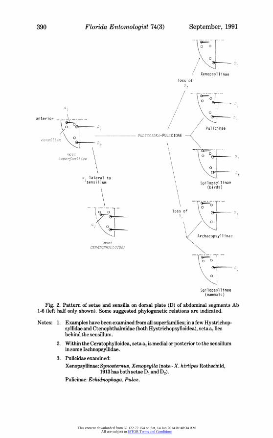
Fig. 2. Pattern of setae and sensilla on dorsal plate (D) of abdominal segments Ab 1-6 (left half only shown). Some suggested phylogenetic relations are indicated.
Notes: 1. Examples have been examined from all superfamilies; in a few Hystrichop- syllidae and Ctenophthalmidae (both Hystrichopsylloidea), seta a, lies behind the sensillum.
2. Within the Ceratophylloidea, seta a, is medial or posterior to the sensillum in some Ischnopsyllidae.
3. Pulicidae examined: Xenopsyllinae: Synosternus, Xenopsylla (note - X. hirtipes Rothschild,
1913 has both setae D1 and D2). Pulicinae: Echidnophwga, Pulex.
This content downloaded from 62.122.72.154 on Sat, 14 Jun 2014 01:48:34 AMAll use subject to JSTOR Terms and Conditions

Pilgrim: External Morphology of Flea Larvae 391
Contrary to some published opinions, I find that the patterns retain their characteristics throughout the larval instars, the main changes being a progressive relative increase in size of the setae at each moult. Even the pattern of parietal and occipital setae does not become disturbed in the presence of the egg tooth in the first instar.
DIFFERENT FORMS OF SETAE
Most accounts refer to differences in length of various setae, but there are also great variations in their shape and structure. The majority of setae are acicular, tapering regularly to a fine tip. In several families, e.g. Hystrichopsyllidae, Pygiopsyllidae, at least some body setae are obtuse; they are much stouter and their tip is blunt; they may be short (Hystrichopsylla s.1.) or very long (Notiopsylla) and they are present not only on the large larvae of these genera but also on the quite small Pagipsylla. Whereas most head capsule setae are acicular, in Chiastopsylla they are attenuate, narrowing rather abruptly to a finer almost filamentous nature at about mid-length. Ischnopsyllidae larvae so far examined (3 genera, 7 species) have all their long body setae capitate, or swollen at the tip. Similar, but perhaps only analogous, spatulate setae occur in many Pygiopsyllidae, the terminal swelling tending to be more gradually developed from the shaft. In both these types, the bulbous swellings are thin-walled and may perhaps be devoted to the exchange of water or solutes with the substrate, but their function has not been investigated.
In the above types of setae, the shaft may be quite smooth even at high magnification in the SEM; in the following types the shaft is highly structured, the modifications being visible at low power in light microscopy. Branched setae, with a few spine-like processes irregularly arranged occur in Pygiopsylla; spiculate setae, with a series of finer processes along one side of the shaft, are widespread in Pygiopsyllidae and they occur in Chiastop- sylla and in Ischnopsyllidae as well. In pilose setae the processes densely clothe the shaft, the tip of which may be truncated; they are very common in Pygiopsyllidae.
Individual setae may show a combination of types, and larvae may bear more than 1 type of seta: in Ischnopsyllidae, capitate setae towards the posterior end of the abdomen have spiculate shafts; in Pygiopsyllidae the head capsule setae are acicular, but the body segments variously bear acicular, obtuse, spatulate, branched, spiculate, and pilose setae.
In Rhopalopsyllus the body setae appear acicular in light microscopy but at high magnifications (SEM, x 2000) a helically wound series of fine grooves is visible producing a striated appearance. This has not been seen in comparable SEM examination of any other acicular setae and may be an idiosyncratic feature of Rhopalopsyllus (but note that no other member of the Rhopalopsyllinae has been studied).
SENSILLA
Clearly-defined, translucent, circular areas (ca. 4 ,um diameter), presumably sensory, are visible on many parts of the body and the head, including the mouth appendages. On the head capsule their patterns have not yet been completely elucidated though
Spilopsyllinae (birds): Actenopsylla, Ornithopsylla. Archaeopsyllinae: Archaeopsylla, Ctenocephalides. Spilopsyllinae (mammals): Cediopsylla, Euhoplopsyllus, Spilopsyllus.
This content downloaded from 62.122.72.154 on Sat, 14 Jun 2014 01:48:34 AMAll use subject to JSTOR Terms and Conditions
392 Florida Entomologist 74(3) September, 1991
there appear to be consistently more sensilla in the non-Pulicoid genera. On the body segments they are confined to the dorsal plates (Fig. 1), apart from 1 on the ventral face of each anal strut. In both the thoracic and the abdominal segments the sensilla are closely associated, topographically, with setae of the anterior and posterior rows.
The combination of arrangements of these setae and the sensilla is taxonomically useful and may also point to phylogenetic considerations. For example, in most super- families the basic pattern over the first 6 abdominal segments is, on each half dorsal plate (Fig. 2): a sensillum lying laterally of the single anterior seta (a1) and a sensillum lying antero-medially of each of the 2 posterior setae (D1 and D2). It is characteristic of all Ceratophylloidea so far examined (except some Ischopsyllidae) that the anterior seta lies laterad of its associated sensillum. Within the Pulicoidea, the basic pattern is present in Pulicinae and in those Spilopsyllinae parasitizing birds. In Xenopsyllinae it appears that seta D1 is absent from the posterior row (except in X. hirtipes Rothschild, 1913, which may be primitive in at least this respect); conversely, it is seta D2 which is missing in Archaeopsyllinae and the lagomorph-infesting genera of Spilopsyllinae.
This would suggest the Spilopsyllinae be separated into 2 groups, which neatly parallel the host preferences: (1) those whose adults are parasites of birds (Actenopsylla, Ornithopsylla) and (2) those whose adults are parasites of lagomorph mammals (Cediop- sylla, Euhooplopsyllus, Spilopsyllua [Hoplopsyllus has not been examined]). This sep- aration is supported by other differences: in group (1) the antennal mound papillae are rounded (type A of Bacot & Ridewood 1914), and the integument bears small scales with minute posteriorly directed spiny tips--this combination of characters also obtains throughout the Xenopsyllinae, Pulicinae, and Archaeopsyllinae. In Spilopsyllinae group (2), however, the antennal mound papillae are pointed (type B) and the integumentary scales have long spines, as great as the length of the scale proper in Spilopsyllus.
ANTENNAL MOUND
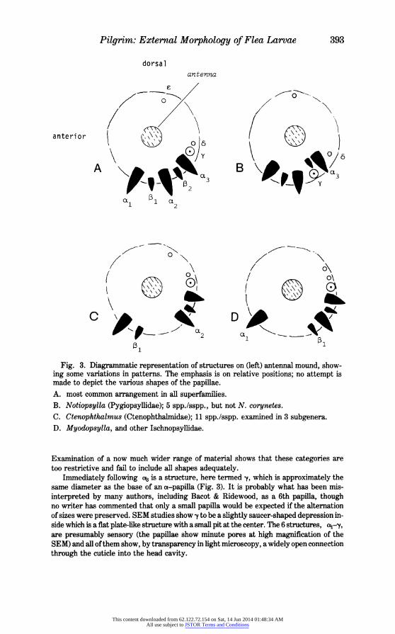
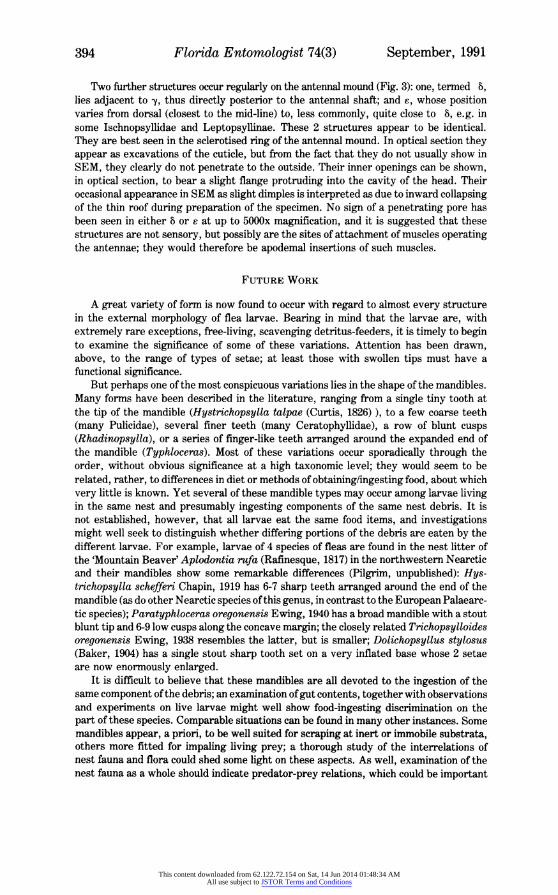
One of the features which has commonly been incorrectly described is the series of papillae on the mound at the base of the antennal shaft (Fig. 1). These were first referred to in detail by Bacot & Ridewood (1914) who stated that there were 3 large papillae, alternating with "3 (rarely 2") smaller ones; with few exceptions, subsequent authors have followed this account, describing and even illustrating 6 papillae. Examination with SEM shows that there are only 5 papillae on each side of the head and that variations from this number are extremely rare abnormalities. The papillae lie postero-laterally on the mound in an arc of about 1/3-1/2 the circumference. The 2 sizes of papillae are of roughly the same shape, the larger (a-papillae) being a few to many times the bulk of the smaller (e-papillae). On each side of the head they are here numbered, commencing from the most anterior:
(a1-t1-t-~a3 (Fig. 3).
In the great majority of species, the 5 papillae are regularly spaced in their row, but certain variations from this appear to have taxonomic significance (Fig. 3). In Ischnop- syllidae (3 genera examined) a wide gap occurs between al and P1; a similar gap occurs between ,13 and e in Ctenophthalmus (11 spp./sspp., 3 subgenera examined) but has not otherwise been seen in the very large family Ctenophthalmidae or elsewhere. Among the Pygiopsyllidae, species of Notiopsylla have very large papillae and in all but one (N. corynetes Smit, 1979) the row becomes bent away from the usual arc, with 2 and to lie much closer to the antennal shaft.
Differentiation of the shape of the papillae was made by Bacot & Ridewood who distinguished A-type (rounded tip) and B-type (pointed tip); Karadina (1964) further suggested a sub-division of B-type, separating straight-sided from convex-sided cones.
This content downloaded from 62.122.72.154 on Sat, 14 Jun 2014 01:48:34 AMAll use subject to JSTOR Terms and Conditions
Pilgrim: External Morphology of Flea Larvae 393
dorsal antenna
anterior C )6 B
A OL \t5 5ia~~~~O3 t t t 3
1 ~~~~12 1 1 2
0'1 / O2 OL \ *
Fig. 3. Diagrammatic representation of structures on (left) antennal mound, show- ing some variations in patterns. The emphasis is on relative positions; no attempt is made to depict the various shapes of the papillae. A. most common arrangement in all superfamilies. B. Notiopsylla (Pygiopsyllidae); 5 spp./sspp., but not N. corynetes.
C. Ctenophthalmus (Ctenophthalmidae); 11 spp./sspp. examined in 3 subgenera. D. Myodopsylla, and other Ischnopsyllidae.
Examination of a now much wider range of material shows that these categories are too restrictive and fail to include all shapes adequately.
Immediately following aB is a structure, here termed y, which is approximately the same diameter as the base of an a-papilla (Fig. 3). It is probably what has been mis- interpreted by many authors, including Bacot & Ridewood, as a 6th papilla, though no writer has commented that only a small papilla would be expected if the alternation of sizes were preserved. SEM studies show y to be a slightly saucer-shaped depression in- side which is a flat plate-like structure with a small pit at the center. The 6 structures, a4-y, are presumably sensory (the papillae show minute pores at high magnification of the SEM) and all of them show, by transparency in light microscopy, a widely open connection through the cuticle into the head cavity.
This content downloaded from 62.122.72.154 on Sat, 14 Jun 2014 01:48:34 AMAll use subject to JSTOR Terms and Conditions
394 Florida Entomologist 74(3) September, 1991
Two further structures occur regularly on the antennal mound (Fig. 3): one, termed 8, lies adjacent to y, thus directly posterior to the antennal shaft; and ?, whose position varies from dorsal (closest to the mid-line) to, less commonly, quite close to i, e.g. in some Ischnopsyllidae and Leptopsyllinae. These 2 structures appear to be identical. They are best seen in the sclerotised ring of the antennal mound. In optical section they appear as excavations of the cuticle, but from the fact that they do not usually show in SEM, they clearly do not penetrate to the outside. Their inner openings can be shown, in optical section, to bear a slight flange protruding into the cavity of the head. Their occasional appearance in SEM as slight dimples is interpreted as due to inward collapsing of the thin roof during preparation of the specimen. No sign of a penetrating pore has been seen in either 8 or e at up to 5000x magnification, and it is suggested that these structures are not sensory, but possibly are the sites of attachment of muscles operating the antennae; they would therefore be apodemal insertions of such muscles.
FUTURE WORK
A great variety of form is now found to occur with regard to almost every structure in the external morphology of flea larvae. Bearing in mind that the larvae are, with extremely rare exceptions, free-living, scavenging detritus-feeders, it is timely to begin to examine the significance of some of these variations. Attention has been drawn, above, to the range of types of setae; at least those with swollen tips must have a functional significance.
But perhaps one of the most conspicuous variations lies in the shape of the mandibles. Many forms have been described in the literature, ranging from a single tiny tooth at the tip of the mandible (Hystrichopsylla talpae (Curtis, 1826) ), to a few coarse teeth (many Pulicidae), several finer teeth (many Ceratophyllidae), a row of blunt cusps (Rhadinopsylla), or a series of finger-like teeth arranged around the expanded end of the mandible (Typhloceras). Most of these variations occur sporadically through the order, without obvious significance at a high taxonomic level; they would seem to be related, rather, to differences in diet or methods of obtaining/ingesting food, about which very little is known. Yet several of these mandible types may occur among larvae living in the same nest and presumably ingesting components of the same nest debris. It is not established, however, that all larvae eat the same food items, and investigations might well seek to distinguish whether differing portions of the debris are eaten by the different larvae. For example, larvae of 4 species of fleas are found in the nest litter of the 'Mountain Beaver' Aplodontia rufa (Rafinesque, 1817) in the northwestern Nearctic and their mandibles show some remarkable differences (Pilgrim, unpublished): Hys- trichopsylla schefferi Chapin, 1919 has 6-7 sharp teeth arranged around the end of the mandible (as do other Nearctic species of this genus, in contrast to the European Palaearc- tic species); Paratyphloceras oregonensis Ewing, 1940 has a broad mandible with a stout blunt tip and 6-9 low cusps along the concave margin; the closely related Trichopsylloides oregonensis Ewing, 1938 resembles the latter, but is smaller; Dolichopsyllus stylosus (Baker, 1904) has a single stout sharp tooth set on a very inflated base whose 2 setae are now enormously enlarged.
It is difficult to believe that these mandibles are all devoted to the ingestion of the same component of the debris; an examination of gut contents, together with observations and experiments on live larvae might well show food-ingesting discrimination on the part of these species. Comparable situations can be found in many other instances. Some mandibles appear, a priori, to be well suited for scraping at inert or immobile substrata, others more fitted for impaling living prey; a thorough study of the interrelations of nest fauna and flora could shed some light on these aspects. As well, examination of the nest fauna as a whole should indicate predator-prey relations, which could be important
This content downloaded from 62.122.72.154 on Sat, 14 Jun 2014 01:48:34 AMAll use subject to JSTOR Terms and Conditions
Pilgrim: External Morphology of Flea Larvae 395
in the natural control of flea populations via larval mortality. Little work has been done on this aspect, though considerable effort has gone into examining physical factors obtaining in larval environments. It is acknowledged, of course, that a number of species of larvae have been successfully reared in laboratories on 'artificial' diets including added yeasts, blood or blood derivatives, such diets or pabula being 'fauna-free'. In not all attempts, however, has it been possible to rear all instars and the failure might be due in some measure to the absence of a dietary requisite.
With regard to the debris factor, it might be interesting to compare conditions-phys- ical and biotic-in nests of mammals with those of birds. Admittedly there are wide differences between nest conditions within these 2 groups of hosts, but the question arises as to whether the litter in a bird's nest might not show qualitative distinctions from that of a mammal's, bearing in mind that a bird's nest is usually occupied by the host for a short time (the breeding period only): the sudden accumulation of organic detritus, including a high nitrogen content, must surely affect the environment in which the larvae find themselves. In contrast, the mammal's nest is often one of long-term occupancy with host-breeding superimposed on it at intervals; the change(s) in physical conditions may be much less marked at those times and the larvae subject to less drastic changes. The physical conditions in the 2 cases are probably accompanied by differences in the in-fauna/flora between bird and mammal nest.
In this context, it would be interesting to compare the detailed morphology of larvae of closely related taxa, in which some are inhabitants of birds' nests whereas their close relatives live in mammals' nests. For example, most species of Xenopsylla are mammal parasites butX. gratiosaJ. & R., 192, X. moucheti Smit, 1958 andX. trispinisWaterston, 1911 are found on birds. Similarly, 2 genera of Ceratophyllidae include both mammal and bird parasites (Smit 1983): within Ceratophyllus s.l., the subgenera Ceratophyllus s.s., Celeophilus, and Emmareus are associated with birds, as is the subgenus Orneacus of Callopsylla s.l. The 1 species of Echidnophaga (gallinacea (Westwood, 1875) ) living commonly on birds also parasitises many mammals, but its larva might be compared with those of other strictly mammal-infesting species of the genus. Many other examples could be examined, mainly between genera, in families such as Rhopalopsyllidae.
ACKNOWLEDGMENT
This study was partly supported by grants from the New Zealand Lottery Board, to whom I extend my grateful thanks.
REFERENCES CITED
BACOT, A. W., AND RIDEWOOD, W. G. 1914. Observations on the larvae of fleas. Parasitology 7(2): 157-175.
KARANDINA, R. S. 1964. [The larvae of fleas of the red-tailed Libyan Jird and other rodents]. Trudy Armianskaia protivochumnaia stantsiia 3: 473-504 (In Russian).
SMIT, F.G.A.M. 1982. Siphonaptera, in 'Synopsis and Classification of Living Or- ganisms', S. P. Parker (ed.) 2: 557-563.
SMIT, F.G.A.M. 1983. in 'The Ceratophyllidae: Key to the genera and host relationships' R. Traub, M. Rothschild andJ. F. Haddow. London: Rothschild and Traub: 1-36.
This content downloaded from 62.122.72.154 on Sat, 14 Jun 2014 01:48:34 AMAll use subject to JSTOR Terms and Conditions