external morphological differences ... - folia.paru.cas.cz · 211 external morphological...
TRANSCRIPT
211
External morphological differences between Crepidostomum farionisand Crepidostomum metoecus (Trematoda: Allocreadiidae),parasites of salmonids, as revealed by SEM
František Moravec
Institute of Parasitology, Academy of Sciences of the Czech Republic, Branišovská 31, 370 05 České Budějovice, CzechRepublic
Key words: Trematoda, Crepidostomum farionis, Crepidostomum metoecus, surface morphology, Salmo trutta,Czech Republic
Abstract. Scanning electron microscopy examinations of trematode specimens belonging to Crepidostomum farionis (O.F.Müller, 1784) and C. metoecus Braun, 1900, collected from brown trout, Salmo trutta fario L., in the Czech Republic, made itpossible to study their surface morphology including details not described previously. The tegument of both species bearsnumerous characteristic papillae around the oral sucker (in C. metoecus also around the ventral sucker) and the ventral and dorsalsurfaces of the forebody, exhibiting a high degree of variability in numbers and arrangement, with tegumental bosses forminglateral fields on the forebody and minute sensory receptors with submerged cilia scattered on the surface of the dorsal part of theoral sucker. In addition to marked differences in the size, shape and position of the oral muscular lobes, both species distinctlydiffer in the number of genital pores: two separate pores in C. farionis and a single pore in C. metoecus.
In European salmonids, trematodes of the genusCrepidostomum Braun, 1900 are represented mainly bytwo species, C. farionis (O.F. Müller, 1784) and C.metoecus Braun, 1900, that are also reported frompalaearctic Asia and North America and the former alsofrom North Africa (Ślusarski 1958a, Yamaguti 1971,Caira 1989). In Europe, both species are among themost common and widely distributed freshwater para-sites of brown trout, Salmo trutta L., and other speciesof salmonids, frequently occurring in them in commoninfections. Occasionally they are found in cottids(Cottus spp.), balitorids [Barbatula barbatula (L.)] andsome other fishes sharing the same environment withsalmonids. Both species are morphologically rathersimilar and, as mentioned by Ślusarski (1958a, b) andErgens (1963), in the past C. metoecus was frequentlymistaken for C. farionis.
Ślusarski (1958a) and Ergens (1963) carried out adetailed comparison of the morphology of C. farionisand C. metoecus based on the light microscopicalexaminations of extensive materials from salmonidsfrom the Vistula River basin and the South Baltic inPoland and from all three main river basins (the RiversElbe, Oder and Danube basins) in former Czecho-slovakia. Ergens (1963) considered the following fourcharacters to be reliable differentiating features: positionof the cirrus sac opening relative to the caecal bifurca-tion; relative size of ventrolateral, dorsal and dorso-lateral muscular oral lobes; size of the pharynx; and sizeratio of the pharynx and the oral sucker.
A recent study (including scanning electron micro-scopy) by Choudhury and Nelson (2000) of Crepido-stomum opeongoensis Caira, 1985, a parasite of NorthAmerican hiodontids, has shown that surface morphol-ogy, especially the presence and distribution of varioustegumental structures, may provide additional specificcharacters in trematodes of this genus. Because similardata on C. farionis and C. metoecus from Europeansalmonids have not yet been published, both specieshave recently been examined by the scanning electronmicroscope (SEM) and the results obtained are pre-sented herein.
MATERIALS AND METHODS
In this study, 10 gravid specimens of C. farionis and 10gravid specimens of C. metoecus, both from the intestinal tractof brown trout, Salmo trutta fario L., were used; specimens ofthe former were collected from brown trout of the ŘasniceRiver near Hniliště, southern Bohemia, on 27 June 2000,whereas those of the latter from the Homolský Brook in VelkéBřezno, northern Bohemia, on 16 August 2000; both streamsare in the Czech Republic and belong to the Elbe Riverdrainage system. After washing in physiological saline, thetrematodes were fixed in hot 4% formaldehyde, postfixed in1% osmium tetroxide, dehydrated though graded ethanol andacetone and then subjected to critical point drying. Thespecimens were sputter-coated with gold and examined with aJEOL JSM-6300 scanning electron microscope at an accelerat-ing voltage of 15 kV.
FOLIA PARASITOLOGICA 49: 211-217, 2002
Address for correspondence: F. Moravec, Institute of Parasitology, Academy of Sciences of the Czech Republic, Branišovská 31, 370 05 ČeskéBudějovice, Czech Republic. Phone: ++420 38 777 5432; Fax: ++420 38 5300 388; E-mail: [email protected]
212
RESULTS
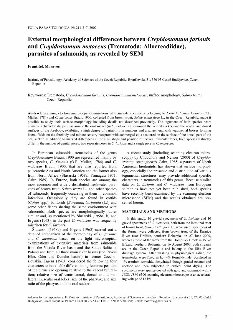
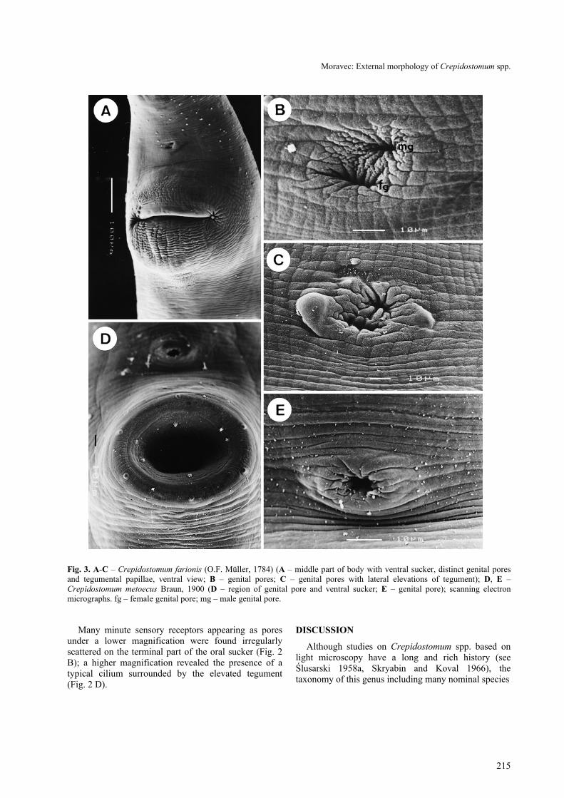
Crepidostomum farionis (O. F. Müller, 1784) Figs. 1, 3 A-C
The body is elongate-oval, with the maximum widthin the region of the ventral sucker. The tegument isunarmed, transversely striated. The anterior end bearsthe subterminal oral sucker provided with six muscularlobes (3 pairs); whereas the ventrolateral pair is formedby rather large, laterally oriented and in en face viewalmost triangular lobes distinctly projecting outwardfrom the oral sucker (Fig. 1 B, C), both dorsal anddorsolateral pairs are formed by small, transversely ovalpapilla-like lobes not protruding or slightly protrudingout from the body surface (Fig. 1 D, E, F); the lattermay be very reduced as is visible in Fig. 1 D; lobes arerather far from each other. The bases of ventrolaterallobes are continuous with the inner margin of the dorsalpart of the sucker, which appears to form a narrow butdistinct edge (Fig. 1 E). The oral opening is oval.
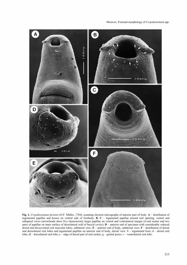
The ventral sucker is approximately twice as large asthe oral sucker, very broad, situated approximately atthe border of the first and the second fourths of the bodylength. In all specimens studied the body region of theventral sucker was markedly elevated, with a broad, slit-like opening situated transversely to the body axis (Fig.1 A). No tegumental papillae were observed around thissucker. There is not one common genital pore, but themale and the female pores are distinctly separated,located close to each other (Fig. 3 B, C); the femalepore is median, whereas the male one is situatedobliquely anterior to the former (shifted to the left side);tegument around both genital pores may form twolateral elevations (Fig. 3 C) in some specimens. Thegenital pores are situated ventrally, slightly posterior tomid-line between the two suckers.
Three types of tegumental formations, papillae,bosses and minute sensory receptors with submergedcilia, were observed. The papillae are distributed aroundthe oral opening and on the forebody (Fig. 1 A); nopapillae were found in the acetabular region or on thehindbody. The papillae around the oral opening and onthe forebody are numerous and are of two types, largerand smaller (Fig. 1 B, D, E). Although many of themare symmetrically arranged, there are many that are notand, apparently, a considerable variability exists in theirnumbers and arrangement. Nevertheless, there arealways five characteristic larger papillae on the ventraland ventrolateral margin of the oral sucker and twopairs of papillae on the inner surface of the dorsolateralwall of the buccal cavity (Fig. 1 B); as can be seen inFig. 1 D and 1 E, the arrangement and numbers ofpapillae on the dorsal and dorsolateral part of the oralsucker may differ considerably. The numerous tegu-mental papillae on the ventral side extend posteriorly tothe level of the ventral sucker; on the dorsal side of the
body they extend posteriorly approximately to the levelof genital pores, but they are less numerous.
Tegumental bosses are weakly developed and arelimited to two ventrolateral fields between ventrolateraloral lobes and sides of the ventral sucker (Fig. 1 A); nobosses were observed on the dorsal side of the body.Numerous minute sensory receptors appearing as poresunder a lower magnification were found irregularlyscattered on the terminal part of the oral sucker.
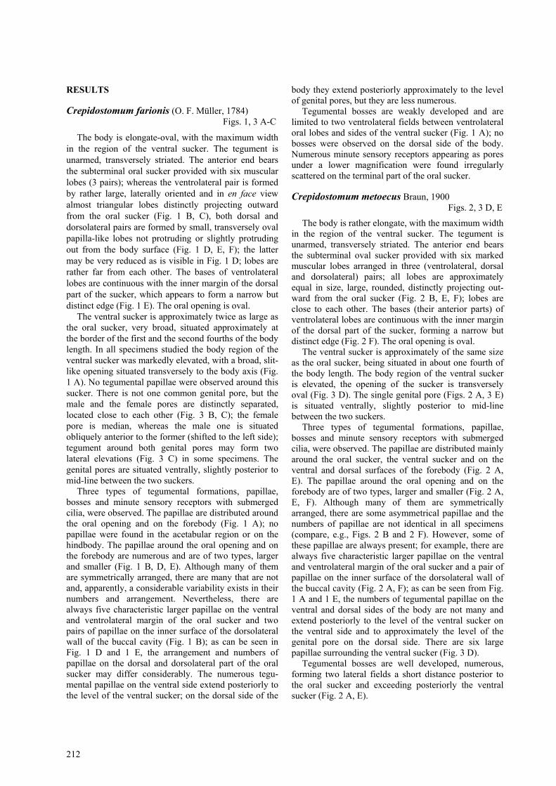
Crepidostomum metoecus Braun, 1900 Figs. 2, 3 D, E
The body is rather elongate, with the maximum widthin the region of the ventral sucker. The tegument isunarmed, transversely striated. The anterior end bearsthe subterminal oval sucker provided with six markedmuscular lobes arranged in three (ventrolateral, dorsaland dorsolateral) pairs; all lobes are approximatelyequal in size, large, rounded, distinctly projecting out-ward from the oral sucker (Fig. 2 B, E, F); lobes areclose to each other. The bases (their anterior parts) ofventrolateral lobes are continuous with the inner marginof the dorsal part of the sucker, forming a narrow butdistinct edge (Fig. 2 F). The oral opening is oval.
The ventral sucker is approximately of the same sizeas the oral sucker, being situated in about one fourth ofthe body length. The body region of the ventral suckeris elevated, the opening of the sucker is transverselyoval (Fig. 3 D). The single genital pore (Figs. 2 A, 3 E)is situated ventrally, slightly posterior to mid-linebetween the two suckers.
Three types of tegumental formations, papillae,bosses and minute sensory receptors with submergedcilia, were observed. The papillae are distributed mainlyaround the oral sucker, the ventral sucker and on theventral and dorsal surfaces of the forebody (Fig. 2 A,E). The papillae around the oral opening and on theforebody are of two types, larger and smaller (Fig. 2 A,E, F). Although many of them are symmetricallyarranged, there are some asymmetrical papillae and thenumbers of papillae are not identical in all specimens(compare, e.g., Figs. 2 B and 2 F). However, some ofthese papillae are always present; for example, there arealways five characteristic larger papillae on the ventraland ventrolateral margin of the oral sucker and a pair ofpapillae on the inner surface of the dorsolateral wall ofthe buccal cavity (Fig. 2 A, F); as can be seen from Fig.1 A and 1 E, the numbers of tegumental papillae on theventral and dorsal sides of the body are not many andextend posteriorly to the level of the ventral sucker onthe ventral side and to approximately the level of thegenital pore on the dorsal side. There are six largepapillae surrounding the ventral sucker (Fig. 3 D).
Tegumental bosses are well developed, numerous,forming two lateral fields a short distance posterior tothe oral sucker and exceeding posteriorly the ventralsucker (Fig. 2 A, E).
Moravec: External morphology of Crepidostomum spp.
213
Fig. 1. Crepidostomum farionis (O.F. Müller, 1784), scanning electron micrographs of anterior part of body. A – distribution oftegumental papillae and bosses on ventral side of forebody; B, C – tegumental papillae around oral opening, ventral andsubapical views (arrowheads show five characteristic larger papillae on ventral and ventrolateral margin of oral sucker and twopairs of papillae on inner surface of dorsolateral wall of buccal cavity); D – anterior end of specimen with considerably reduceddorsal and dorsoventral oral muscular lobes, sublateral view; E – anterior end of body, subdorsal view; F – distribution of dorsaland dorsolateral oral lobes and tegumental papillae on anterior end of body, dorsal view. b – tegumental boss; d – dorsal orallobe; dl – dorsolateral oral lobe; e – edge of dorsal part of oral sucker; g – genital pores; v – ventrolateral oral lobe.
214
Fig. 2. Crepidostomum metoecus Braun, 1900, scanning electron micrographs of anterior part of body. A – distribution oftegumental papillae around oral opening and on ventral side of forebody (arrowheads show five characteristic larger papillae onventral and ventrolateral margin of oral sucker); B, C – anterior body end with marked oral muscular lobes, two types oftegumental papillae and sensory receptors, apical and sublateral views; D – sensory receptor with submerged cilium; E –distribution of tegumental papillae and bosses on dorsal side of anterior end of body, dorsal view; F – apical view of anotherspecimen with different number and arrangement of tegumental papillae on dorsal part of oral sucker (arrowheads show acharacteristic pair of papillae on inner surface of dorsolateral wall of buccal cavity). b – tegumental boss; d – dorsal oral lobe;dl – dorsolateral oral lobe; e – edge of dorsal part of oral sucker; g – genital pore; p – tegumental papilla; r – sensory receptorwith cilium; v – ventrolateral oral lobe.
Moravec: External morphology of Crepidostomum spp.
215
Fig. 3. A-C – Crepidostomum farionis (O.F. Müller, 1784) (A – middle part of body with ventral sucker, distinct genital poresand tegumental papillae, ventral view; B – genital pores; C – genital pores with lateral elevations of tegument); D, E –Crepidostomum metoecus Braun, 1900 (D – region of genital pore and ventral sucker; E – genital pore); scanning electronmicrographs. fg – female genital pore; mg – male genital pore.
Many minute sensory receptors appearing as poresunder a lower magnification were found irregularlyscattered on the terminal part of the oral sucker (Fig. 2B); a higher magnification revealed the presence of atypical cilium surrounded by the elevated tegument(Fig. 2 D).
DISCUSSION
Although studies on Crepidostomum spp. based onlight microscopy have a long and rich history (seeŚlusarski 1958a, Skryabin and Koval 1966), thetaxonomy of this genus including many nominal species
216
parasitic mainly in freshwater fishes is unsatisfactoryand the species identification is complicated byinsufficient knowledge of the morphology of theseforms. Until Ślusarski (1958a) provided detailed rede-scriptions of Crepidostomum farionis and C. metoecus,probably the only two species of this genus parasitisingsalmonids in Europe, these two were frequently con-fused and, consequently, many earlier published data onthe geographical distribution, hosts and biology of C.farionis are unreliable, because, in fact, they may eithercompletely or partly concern the more frequent C.metoecus.
It has been mentioned that Ślusarski (1958a) andErgens (1963) carried out a detailed morphometricalcomparison of these two species based on light micro-scopical examinations, oriented mainly to the internalstructure of the worms. The present study shows thatthere are additional differences in the superficial struc-tures of these species visible by SEM not previouslyreported
The SEM study confirmed the previous observationsby Ślusarski (1958a) and some subsequent authors thatthe morphology (size, shape, location) of oral muscularlobes is conspicuously different in these two species andis one of the best interspecific differentiating features.
A very important interspecific difference found nowby SEM is that there are two separate genital pores in C.farionis, whereas only a single genital pore (opening ofa common genital atrium) is found in C. metoecus. Thepresence of a single genital pore is stated in the genericdiagnoses of Crepidostomum given by Hopkins (1933),Skryabin and Koval (1966) and Yamaguti (1971) and inthe description of C. farionis provided, e.g., by Nicoll(1909). According to Ślusarski (1958a), C. farionis pos-sesses a pocket-like genital atrium, although he illus-trated two (male and female) separate genital poresopening directly to the body surface.
To date, there are almost no data on the tegumentalstructures in Crepidostomum spp. In her revisionarywork dealing with North American papillose Allo-creadiidae, Caira (1989) provided scanning electronmicrographs of the anterior end of eight North Ameri-can Crepidostomum spp. including C. farionis and C.metoecus (these two based on specimens from BritishColumbia, Canada), mentioning the presence of tegu-mental papillae in them. Choudhury and Nelson (2000)found by SEM that the North American species Crepi-dostomum opeongoensis possessed three types oftegumental structures: tegumental papillae (around theoral and ventral suckers and on the ventral and dorsalsurfaces of the body), tegumental bosses (on the dorsalsurface of the forebody), and minute pores of sub-tegumental ducts (on ventral oral lobes and along edgesof dorsolateral and dorsal lobes).
In this study, tegumental papillae and bosses werefound in both C. farionis and C. metoecus. The papillaeare more numerous in C. farionis as compared to thosein C. metoecus. In both species they extend posteriorlyto the level of the ventral sucker on the ventral side andto the level of the genital pores on the dorsal side. Incontrast to what was found in C. opeongoensis, thetegumental papillae in both congeneric European spe-cies are much more numerous, highly variable in theirnumbers and arrangement, although some papillae arepaired or have a characteristic position around the oralsucker; this intraspecific variability is also apparentwhen scanning electron micrographs of conspecificNorth American specimens (Caira 1989) are comparedto those of the present European material; characteristicsix papillae around the ventral sucker found in C.metoecus were not observed in C. farionis. Five largepapillae around the ventral side of the oral sucker werepresent both in the two European species studied and inC. opeongoensis (Choudhury and Nelson 2000).
The tegumental bosses were poorly developed in C.farionis and better developed in C. metoecus, in bothspecies forming two lateral fields on the forebody, ex-tending slightly to ventral and dorsal sides. In contrast,bosses in C. opeongoensis are on the dorsal surface(Choudhury and Nelson 2000).
The series of closely-set openings of subtegumentalducts were not observed in C. farionis and C. metoecus,but their presence on the anterior edge of the dorsal partof the oral sucker cannot be excluded in these species.On the other hand, minute sensory receptors with sub-merged cilia, which appear as pores, were found ir-regularly scattered on the dorsal part of the oral suckerin both species. As far as the author knows, thesereceptors were not previously found in adult Crepido-stomum spp., although they have recently been reportedin the cercaria of a Crepidostomum sp. by Bogéa andCaira (2001).
Acknowledgements. The author’s thanks are due to themanagement of the Board of the National Park and ProtectedLandscape Šumava in Vimperk and to the Association ofCzech Fishermen in Ústí nad Labem for permission to carryout ichthyoparasitological investigations in the respectivelocalities. The author is also grateful to the staff of theLaboratory of Electron Microscopy of the Institute of Para-sitology, ASCR, in České Budějovice for their technicalassistance and to Mrs. I. Husáková from the Laboratory ofHelminth Biology of the same Institute for her help with thepreparation of illustrations. Prof. J.N. Caira from the Uni-versity of Connecticut (USA) kindly provided a copy of herrevisional publication on North American papillose allo-creadiids. This study was supported by the grant No. 524/00/0267 from the Grant Agency of the Czech Republic.
Moravec: External morphology of Crepidostomum spp.
217
REFERENCES
BOGÉA T., CAIRA J.N. 2001: Ultrastructure and chaetotaxyof sensory receptors in the cercariae of a species ofCrepidostomum Braun, 1900 and Bunodera Railliet, 1896(Digenea: Allocreadiidae). J. Parasitol. 87: 273-286.
CAIRA J.N. 1989: A revision of the North American papilloseAllocreadiidae (Digenea) with independent cladisticanalyses of larval and adult forms. Bull. Univ. NebraskaState Mus. 11: 1-58 + 195 Figs.
CHOUDHURY A., NELSON P.A. 2000: Redescription ofCrepidostomum opeongoensis Caira, 1985 (Trematoda:Allocreadiidae) from fish hosts Hiodon alosoides andHiodon tergisus (Osteichthyes: Hiodontidae). J. Parasitol.86: 1305-1312.
ERGENS R. 1963: Revision of the helminth fauna of fishesfrom the territory of Czechoslovakia I. Genus Crepido-stomum Braun 1900 (Trematoda: Allocreadiidae). Čs.Parasitol. 10: 81-88. (In Czech, German summary.)
HOPKINS S.H. 1933: The morphology, life histories andrelationships of the papillose Allocreadiidae (Trematoda).(Preliminary report.) Zool. Anz. 103: 65-74.
NICOLL W. 1909: Studies on the structure and classificationof the digenetic trematodes. Q. J. Microsc. Sci. 53: 391-487.
SKRYABIN K.I., KOVAL V.P. 1966: Family BunoderidaeNicoll, 1914. In: K.I. Skryabin (Ed.): Trematodes ofAnimals and Man. Essentials of trematodology 22. Nauka,Moscow, pp. 311-456. (In Russian.)
ŚLUSARSKI W. 1958a: The adult Digenea from Salmonidaeof the basin of the Vistula and of the South Baltic. ActaParasitol. Pol. 6: 247-528.
ŚLUSARSKI W. 1958b: Distribution of two species of thegenus Crepidostomum Braun, 1900 (Digenea: Allo-creadiidae) from Salmonidae in the basin of Vistula. Wiad.Parazytol. 4: 647-650.
YAMAGUTI S. 1971: Synopsis of Digenetic Trematodes ofVertebrates, I, II. Keigaku Publishing Co., Tokyo, 1074pp. + 349 Plts.
Received 21 September 2001 Accepted 26 November 2001
FOLIA PARASITOLOGICA 49: 217, 2002
GIBSONNEMA NOM. N., A NEW NAME FOR THE NEMATODE GENUSPARASEURATOIDES MORAVEC, SALGADO-MALDONADO ET AGUILAR-AGUILAR, 2002
František Moravec1,3, Guillermo Salgado-Maldonado2 and Rogelio Aguilar-Aguilar2
1Institute of Parasitology, Academy of Sciences of the Czech Republic, Branišovská 31, 370 05 České Budějovice, CzechRepublic;
2Institute of Biology, National Autonomous University of Mexico, A.P. 70-153, 04510 Mexico, D.F., Mexico;3Corresponding author
We have found that the generic name ParaseuratoidesMoravec, Salgado-Maldonado et Aguilar-Aguilar, 2002 is ajunior homonym to Paraseuratoides Wang, 1984. Therefore, anew name, Gibsonnema nom. n., is proposed to replace it (thenew genus is named in honour of David I. Gibson, a well-known English helminthologist).
These two genera are monotypic, belonging to theseuratoid family Quimperiidae Gendre, 1928, and both theirtype species are parasitic in swamp-eels (Synbranchidae,Synbranchiformes). However, they are well separatedmorphologically. In contrast to Gibsonnema, the genusParaseuratoides is characterised mainly by a toothless buccal
cavity, a bulbously inflated anterior end of the oesophagus andthe presence of caudal alae in the male.
ReferencesMoravec F., Salgado-Maldonado G., Aguilar-Aguilar R. 2002:Two new nematodes, Paraseuratoi-des ophisterni gen. et sp.n. (Quimperiidae) and Philo-metra ophisterni sp. n.(Philometridae), from the swamp-eel Ophisternonaenigmaticum in Mexico. Folia Parasitol. 49: 109-117.Wang P.-q. 1984: Some nematodes of fishes from FujianProvince, China. Acta Zootaxon. Sin. 9: 228-237. (In Chinese,Engl. summary.)
Received 29 July 2002 Accepted 30 July 2002