expression of smooth muscle and extracellular matrix proteins in relation to airway function in...
TRANSCRIPT
Expression of smooth muscle and extracellular matrixproteins in relation to airway function in asthma
Annelies M. Slats, MD,a Kirsten Janssen, BHe,a Annemarie van Schadewijk, MSc,a Dirk T. van der Plas, BSc,a
Robert Schot, BSc,a Joost G. van den Aardweg, MD, PhD,b Johan C. de Jongste, MD, PhD,c Pieter S. Hiemstra, PhD,a
Thais Mauad, MD,d Klaus F. Rabe, MD, PhD,a and Peter J. Sterk, MD, PhDa,e Leiden, Alkmaar, Rotterdam, and Amsterdam,
The Netherlands, and Sao Paulo, Brazil
Background: Smooth muscle content is increased within theairway wall in patients with asthma and is likely to play a role inairway hyperresponsiveness. However, smooth muscle cellsexpress several contractile and structural proteins, and each ofthese proteins may influence airway function distinctly.Objective: We examined the expression of contractile andstructural proteins of smooth muscle cells, as well asextracellular matrix proteins, in bronchial biopsies of patientswith asthma, and related these to lung function, airwayhyperresponsiveness, and responses to deep inspiration.Methods: Thirteen patients with asthma (mild persistent,atopic, nonsmoking) participated in this cross-sectional study.FEV1% predicted, PC20 methacholine, and resistance of therespiratory system by the forced oscillation technique duringtidal breathing and deep breath were measured. Within 1 week,a bronchoscopy was performed to obtain 6 bronchial biopsiesthat were immunohistochemically stained for a-SM-actin,desmin, myosin light chain kinase (MLCK), myosin, calponin,vimentin, elastin, type III collagen, and fibronectin. The level ofexpression was determined by automated densitometry.Results: PC20 methacholine was inversely related to theexpression of a-smooth muscle actin (r 5 20.62), desmin(r 5 20.56), and elastin (r 5 20.78). In addition, FEV1%predicted was positively related and deep inspiration-inducedbronchodilation inversely related to desmin (r 5 20.60), MLCK(r 5 20.60), and calponin (r 5 20.54) expression.Conclusion: Airway hyperresponsiveness, FEV1% predicted,and airway responses to deep inspiration are associated with
From athe Department of Pulmonology, Leiden University Medical Center; bthe Depart-
ment of Pulmonology, Medical Center Alkmaar; cthe Department of Pediatrics, Eras-
mus University Medical Center, Sophia Children’s Hospital, Rotterdam; dthe
Department of Pathology, Sao Paulo University Medical School; and ethe Department
of Respiratory Medicine, Academic Medical Center, University of Amsterdam.
Supported by the Netherlands Asthma Foundation (3.2.02.34).
Disclosure of potential conflict of interest: J. C. de Jongste has received research support
from Aerocrine. K. F. Rabe has consulting arrangements with AstraZeneca,
Boehringer Ingelheim, Novartis, Pfizer, Altana, GlaxoSmithKline, and Roche; has
received research support from AstraZeneca, Merck, Altana, and Boehringer
Ingelheim; and is on the speakers’ bureau for AstraZeneca, Boehringer Ingelhim,
Novartis, Pfizer, Altana, GlaxoSmithKline, and Roche. P. S. Hiemstra has received
research support from AltanaPharma, Novartis, Bayer, AstraZeneca, Pfizer, Merck,
Exhale Therapeutics, Boehringer Ingelheim, Roche, and GlaxoSmithKline. The rest of
the authors have declared that they have no conflict of interest.
Received for publication August 9, 2007; revised January 3, 2008; accepted for publica-
tion February 14, 2008.
Available online April 14, 2008.
Reprint requests: Annelies M. Slats, MD, Leiden University Medical Center, Department
of Pulmonology (C2-P-62), PO Box 9600, 2300 RC Leiden, The Netherlands. E-mail:
0091-6749/$34.00
� 2008 American Academy of Allergy, Asthma & Immunology
doi:10.1016/j.jaci.2008.02.017
1196
selective expression of airway smooth muscle proteins andcomponents of the extracellular matrix. (J Allergy ClinImmunol 2008;121:1196-202.)
Key words: Actin, desmin, elastin, airway smooth muscle, extracel-lular matrix, lung function, hyperresponsiveness, deep inspiration-induced bronchodilation, bronchial biopsies
Asthma is characterized by chronic airway inflammation,which is presumed to contribute to variable airways obstructionand bronchial hyperresponsiveness.1 However, recent studieshave led to a reappraisal of the role of airway smooth muscle inasthma pathophysiology.2 Because smooth muscle contractionleads to airway narrowing, abnormalities in airway smooth mus-cle size, mass, or function could easily lead to exaggerated airwaynarrowing. In addition, mast cells within the airway smoothmuscle bundles have been associated with airway hyperrespon-siveness,3 and more recently, we observed a similar associationwith impaired deep inspiration-induced bronchodilation inpatients with asthma.4
Increased smooth muscle mass has been demonstrated inbronchial biopsies5-7 as well as in resected lung tissue8,9 from pa-tients with asthma compared with healthy subjects. Mathematicalmodels have shown that increased smooth muscle mass canexplain exaggerated airway narrowing to contractile stimuli in pa-tients with asthma,10 especially at high lung volumes.11 Interest-ingly, although increased smooth muscle area in bronchialbiopsies has been associated with impaired lung function,5,7 norelationship was found with airway hyperresponsiveness. Never-theless, in vitro studies have shown that smooth muscle cells ob-tained from bronchial biopsies of patients with asthma exhibit anincrease in isotonic shortening12 and shortening velocity com-pared with controls without asthma.13
Smooth muscle cells express several contractile and structuralproteins.14,15 Cultured airway smooth muscle cells with a con-tractile phenotype are relatively rich in smooth muscle myosinheavy chain (sm-MHC), a-smooth muscle actin (a-SM-actin),calponin, desmin, and myosin light chain kinase (MLCK),whereas when proliferating they express less sm-MHC, calponin,a-SM-actin, and desmin, and significantly more vimentin. Be-nayoun et al5 examined the expression of some of these contrac-tile proteins in bronchial biopsies in relation to asthma severity.MLCK expression correlated inversely with lung function, butthis was not the case for the proteins a-SM-actin or myosin.This suggests that the level of expression of these proteins mayhave different functional consequences. We selected several con-tractile and structural proteins that may influence airway respon-siveness and function, namely a-SM-actin, myosin, desmin,vimentin, calponin, and MLCK. Furthermore, it has been shown
J ALLERGY CLIN IMMUNOL
VOLUME 121, NUMBER 5
SLATS ET AL 1197
Abbreviations used
a-SM-actin: a-Smooth muscle actin
MLCK: Myosin light chain kinase
Rrs: Resistance of the respiratory system
RrsExp: Resistance of the respiratory system during tidal
expirations
RrsInsp: Resistance of the respiratory system during tidal
inspirations
sm-MHC: Smooth muscle myosin heavy chain
that stretch of smooth muscle cells can increase the expression ofcontractile proteins.16,17 Because smooth muscle cells are mostlikely stretched during deep inspiration, we analyzed the relation-ship between protein expression and airway responses to deepinspiration.
In addition, the smooth muscle bundles are embedded in andalso contain extracellular matrix. The amount and the composi-tion of the matrix may have functional consequences by alteringthe physical properties of the airway wall18-20 and can also influ-ence the proliferation of smooth muscle cells.21-23 Therefore, weanalyzed the expression of different extracellular matrix proteins(type III collagen, fibronectin, and elastin) within and surround-ing the smooth muscle bundles.
We hypothesized that a higher level of expression of the selectedcontractile and structural proteins of smooth muscle cells, as wellas components of the extracellular matrix, in bronchial biopsies areassociated with increased airway hyperresponsiveness and im-paired deep inspiration–induced bronchodilation in asthma. Theaim of this study was to relate FEV1% predicted, airway hyperres-ponsiveness, and deep inspiration–induced changes in resistance ofthe respiratory system as measured by forced oscillation techniqueto the level of expression of a-SM-actin, myosin, desmin, vimen-tin, calponin, and MLCK, as well as type III collagen, fibronectinand elastin, in bronchial biopsies of patients with asthma.
METHODS
SubjectsThis study was performed in the framework of a previously published
project.4 Thirteen patients with mild persistent asthma (Global Initiative for
Asthma steps 1 and 224) were recruited for this study. All patients had a history
of episodic chest tightness or wheezing. Their baseline FEV1 was more than
70% of predicted.25 The PC20 methacholine was less than 8 mg/mL.26 All pa-
tients were atopic, as determined by a positive skin prick test result (�3mm
wheal) to 1 or more of 10 common aeroallergen extracts (ALK-Abello, Nieu-
wegein, The Netherlands). The patients were clinically stable, nonsmokers or
exsmokers with less than 2 pack-years, and did not have a recent (�2 weeks)
upper respiratory tract infection or other relevant diseases. None of the pa-
tients had used inhaled or oral corticosteroids within 3 months before or during
the study. The protocol was approved by the institutional review board for hu-
man studies, and before entering the study, the patients gave their written in-
formed consent.
Study designIn this cross-sectional study, measurements were performed on 4 separate
days within 3 to 4 weeks. On the first visit, medical history was taken, atopy
was determined, and FEV1% predicted and resistance of the respiratory sys-
tem (Rrs) were measured before and after 400 mg salbutamol. On 2 additional
visits, the patients returned for a methacholine challenge to determine airway
hyperresponsiveness and deep inspiration–induced bronchodilation after a
single dose of methacholine that induced a 20% fall in FEV1.4 Within
1 week of the last visit, a bronchoscopy was performed to obtain 6 bronchial
biopsies.
Airway hyperresponsivenessMethacholine bromide in normal saline was used for the bronchial
challenges that were performed by standardized methodology.26 At 5-minute
intervals, aerosolized serial doubling concentrations of methacholine (0.15-
40 mmol/L) were inhaled by tidal breathing (DeVilbiss, Somerset, Pa) for 2
minutes with the nose clipped. The challenge was stopped when FEV1 drop-
ped by more than 20% from baseline, and the response was expressed as the
provocative concentration causing a 20% fall in FEV1 PC20.
Airway responses to deep inspirationDeep inspiration–induced bronchodilation was measured using a single-
dose methacholine challenge to induce a fall in FEV1 of 20% in the absence of
deep inspirations before methacholine inhalation. Baseline measurements of
FEV1 and Rrs were followed by a period of 20 minutes without deep inspira-
tions. A single dose of methacholine (approximately the cumulative dose of
the PC20 of the previous challenge) was inhaled, and 2 minutes later, Rrs
was measured during tidal breathing, a deep inspiration to total lung capacity,
a passive expiration, and again tidal breathing. This was directly followed by
spirometry to measure the fall in FEV1. The forced oscillation technique with
an applied oscillation frequency of 8 Hz and an amplitude of 61 cmH2O was
used to measure Rrs continuously during tidal breathing and a deep inspiration
(Woolcock Institute, Sydney, Australia).4 Deep inspiration–induced broncho-
dilation was expressed as the reduction in Rrs during tidal breathing induced
by the deep inspiration.
Bronchoscopy, immunohistochemistry, and image
analysisBronchoscopy was performed by experienced pulmonologists according to a
standardized and validated protocol.27 All patients received 400 mg salbutamol
30 minutes before bronchoscopy. Six biopsies were taken at the (sub)segmental
level using disposable forceps (radial edge; Boston Scientific, Boston, Mass).
The biopsies were fixed for 24 hours in 4% neutral buffered formaldehyde,
processed, and embedded in paraffin. Sections 4 mm thick were cut, and hema-
toxylin-eosin staining was used to evaluate morphologic quality (intact retic-
ular basal membrane and submucosa without crushing artifacts, blood clots, or
only epithelial scrapings). Two sections per subject were selected on the qual-
ity of the submucosa, and not on the quantity of smooth muscle area. This was
done to avoid a selection bias with regard to the main outcome parameter. In
addition, the observers were blinded with regard to the subject number and
their disease. The latter was chosen to avoid a selection bias with regard to
the main outcome parameter. Antigen retrieval was performed on paraffin-em-
bedded sections with citrate (desmin, myosin, and MLCK) or trypsin (type III
collagen). a-SM-actin, calponin, vimentin, and fibronectin did not need anti-
gen retrieval. The sections were incubated with mouse mAbs directed against
a-SM-actin (1:50,000, clone 1A; Santa Cruz Biotechnology, Santa Cruz, Ca-
lif), myosin (1:40, clone 1A4; Sigma-Aldrich, St Louis, Mo), desmin (1:200,
clone D33; Dako UK Ltd, Cambridgeshire, United Kingdom [UK]), vimentin
(1:1000, clone V9; Dako), calponin (1:10,000, clone hCP; Sigma), MLCK
(1:4000, clone k36; Sigma), type III collagen (1:2000, clone III-53; Merck
Calbiochem, Darmstadt, Germany), and fibronectin (1:100, clone 568; Novo-
castra, Newcastle upon Tyne, UK). As a secondary antibody, Envision-HRP
(Dako), was used. Positive cells stained red after development with NovaRed
(Vector Laboratories, Burlingame, Vt). Sections were counterstained with
Mayer hematoxylin. As a negative control, the primary antibody was omitted
from this procedure. For elastin expression, we used Weigert staining with
Oxone (Klinipath BV, Duiven, The Netherlands).28
Morphometry was performed by means of digital image analysis.29 The ex-
pression of the smooth muscle proteins (a-SM-actin, myosin, desmin, vimen-
tin, MLCK) was determined in the total biopsy area (including the epithelial
layer and glands). Type III collagen, fibronectin, and elastin were measured
J ALLERGY CLIN IMMUNOL
MAY 2008
1198 SLATS ET AL
within the smooth muscle bundles and the area surrounding the smooth muscle
bundles separately. We used the desmin-stained adjacent biopsy sections to
detect manually the positive stained area that appeared in bundles . Protein ex-
pression was quantified by fully automated densitometry (KS400; Zeiss, Ober-
kochen, Germany).27,29 This was performed by using a linear combination of
red-filtered and blue-filtered grayscale images to derive a grayscale image
(range, 0-255) in which the brown-red staining of interest is highlighted above
a uniform background (white 5 gray value 255). This resulted in a narrow and
peaked gray value distribution of background pixels with a longer tail on the
left, which represented the positive stained pixels. The distribution was nor-
malized toward the background peak, and subsequently inversed to obtain a
zero value for the white background peak (white 5 gray value 0).
Data and statistical analysesAirway hyperresponsiveness was expressed as PC20. Reversibility was de-
fined as the change in FEV1% predicted or Rrs by 400 mg salbutamol. Rrs was
calculated from all the data points, within the 95% CI, during 3 tidal inspira-
tions (RrsInsp) and during 3 tidal expirations (RrsExp) before and after deep in-
spiration. Deep inspiration–induced bronchodilation was expressed as the
difference between Rrs after deep inspiration and Rrs before deep inspiration4;
thus, a negative value indicates bronchodilation. This was performed for
RrsInsp and RrsExp separately because the airways may behave differently dur-
ing inspiration and expiration.30
Positive staining intensity was expressed as mean density (gray value).
The outcome parameters were (log)transformed if necessary to obtain a
normal distribution. Within-group differences were analyzed by 2-tailed
paired t tests or Wilcoxon ranks test. Spearman rank correlation coefficient
was used to explore associations between the expression of the proteins
and the functional parameters. P values <.05 were considered statistically
significant.
RESULTS
Smooth muscle protein expressionThe density of the smooth muscle protein staining was
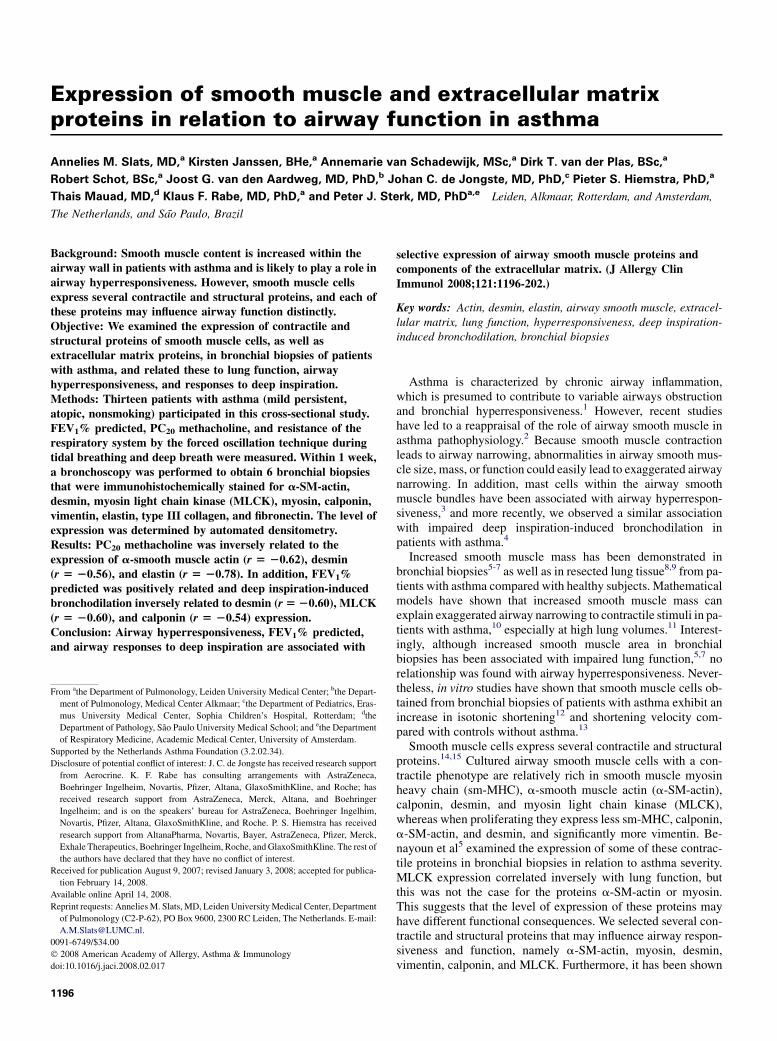
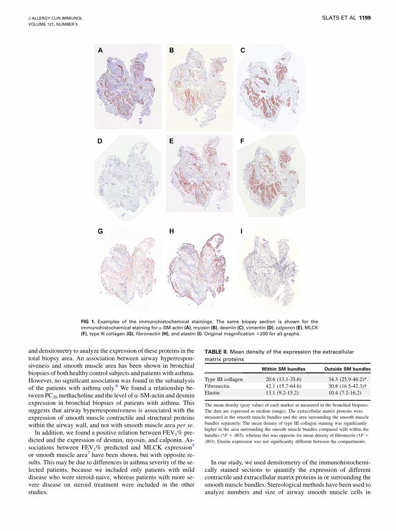
determined in the whole biopsy section. The mean density(gray value) for each marker is given in Table I. All available sec-tions (2 per patient) were used in the analysis. Fig 1 presents ex-amples of the immunohistochemical staining in the same biopsysection of 1 subject. All markers, except for vimentin, stronglystained the airway smooth muscle cells. Vimentin was negativeor weakly expressed in the smooth muscle cells. Outside thesmooth muscle bundles, there was staining of the contractilemarkers, mainly around vessels, around submucosal glands (my-oepithelial cells), and in scattered mesenchymal cells (fibroblastsand myofibroblasts).
Extracellular matrix expressionThe density of the extracellular matrix protein expression of
type III collagen, fibronectin, and elastin was determined in the
TABLE I. Mean density of the expression the smooth muscle
proteins
Smooth muscle protein Mean density
a-SM-actin 20.8 (11.9-38.9)
Myosin 14.3 (5.9-18.1)
Desmin 14.4 (6.1-26.7)
Vimentin 13.2 (9.7-19.0)
Calponin 20.2 (9.8-40.4)
MLCK 15.9 (6.5-26.0)
The mean density (gray value) of each marker as measured in the bronchial biopsies.
The smooth muscle proteins were measured in the total biopsy area. The data are
expressed as median (range).
smooth muscle bundles and the area surrounding the smoothmuscle bundles separately. The proteins were expressed both inthe lamina propria as well as within the airway smooth musclebundles, with a fibrillar pattern for type III collagen and elastinand more diffuse pattern for fibronectin. Fibronectin and typeIII collagen stained the subepithlial basal membrane as well. Air-way smooth muscle cells stained negatively for these markers.There were significant differences in density of type III collagenand fibronectin between the smooth muscle bundles and the areasurrounding the muscle (Table II).
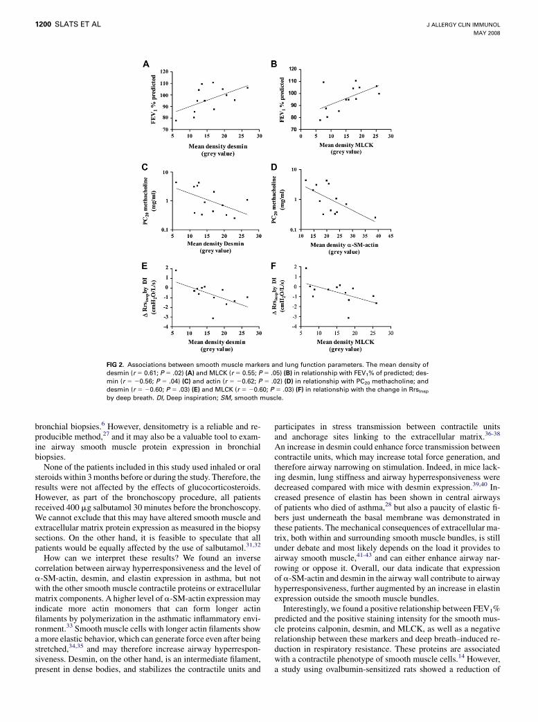
Airways obstructionFEV1% predicted correlated positively with the mean density
of calponin (r 5 0.58), desmin (r 5 0.61), and MLCK (r 5
0.55; P < .05; Fig 2, A and B). There was a borderline significantcorrelation between FEV1% predicted and the expression ofa-SM-actin (r 5 0.58), myosin (r 5 0.53), and fibronectin (withinthe smooth muscle bundles; r 5 0.56; P 5 .06). None of thesecorrelations was seen for FEV1% predicted after salbutamol. Inaddition, mean density of both a-SM-actin and calponin corre-lated inversely with FEV1 reversibility (r 5 20.54 and r 5
20.61, respectively; P < .05). Also, the change in RrsInsp andRrsexp by salbutamol correlated positively with mean density ofa-SM-actin and calponin (r > 0.70; P < .01).
Dynamics of airway narrowingPC20 methacholine was inversely related to the expression of
the contractile smooth muscle protein a-SM-actin (r 5 20.62;P < .05; Fig 2, D), the structural smooth muscle protein desmin(r 5 20.56; P <.05; Fig 2, C), and the extracellular matrix proteinelastin outside the smooth muscle bundles (r 5 20.78; P < .01).PC20 methacholine was not significantly related to expression ofelastin within the smooth muscle bundles.
Airway responses to deep inspirationThe reduction in Rrsinsp by deep inspiration was inversely re-
lated to the expression of desmin (r 5 20.60; Fig 2, E), MLCK(r 5 20.60; Fig 2, F), and calponin (r 5 20.54) in the bronchialbiopsies.
DISCUSSIONOur results demonstrate an inverse association between PC20
methacholine and the level of expression of a-SM-actin, desmin,and elastin in bronchial biopsies in patients with asthma. Also, weshowed that FEV1% predicted was positively related, and deep in-spiration–induced reduction in respiratory resistance inversely re-lated to calponin, desmin, and MLCK expression. Thus, airwayhyperresponsiveness, lung function, and airway responses todeep inspiration are associated with the level of expression ofsome, but not all, of the smooth muscle contractile and structuralproteins, as well as the composition of the extracellular matrixwithin the airway wall. This suggests that the dynamics of airwayfunction are influenced by the expression of several distinctsmooth muscle and extracellular matrix proteins.
To our knowledge, this is the first study showing an associationbetween airway hyperresponsiveness and smooth muscle cellprotein expression in patients with asthma. We used markers ofdifferent functional components of smooth muscle cells
J ALLERGY CLIN IMMUNOL
VOLUME 121, NUMBER 5
SLATS ET AL 1199
FIG 1. Examples of the immunohistochemical stainings. The same biopsy section is shown for the
immunohistochemical staining for a-SM-actin (A), myosin (B), desmin (C), vimentin (D), calponin (E), MLCK
(F), type III collagen (G), fibronectin (H), and elastin (I). Original magnification 3200 for all graphs.
and densitometry to analyze the expression of these proteins in thetotal biopsy area. An association between airway hyperrespon-siveness and smooth muscle area has been shown in bronchialbiopsies of both healthy control subjects and patients with asthma.However, no significant association was found in the subanalysisof the patients with asthma only.6 We found a relationship be-tween PC20 methacholine and the level of a-SM-actin and desminexpression in bronchial biopsies of patients with asthma. Thissuggests that airway hyperresponsiveness is associated with theexpression of smooth muscle contractile and structural proteinswithin the airway wall, and not with smooth muscle area per se.
In addition, we found a positive relation between FEV1% pre-dicted and the expression of desmin, myosin, and calponin. As-sociations between FEV1% predicted and MLCK expression5
or smooth muscle area7 have been shown, but with opposite re-sults. This may be due to differences in asthma severity of the se-lected patients, because we included only patients with milddisease who were steroid-naive, whereas patients with more se-vere disease on steroid treatment were included in the otherstudies.
In our study, we used densitometry of the immunohistochemi-cally stained sections to quantify the expression of differentcontractile and extracellular matrix proteins in or surrounding thesmooth muscle bundles. Stereological methods have been used toanalyze numbers and size of airway smooth muscle cells in
TABLE II. Mean density of the expression the extracellular
matrix proteins
Within SM bundles Outside SM bundles
Type III collagen 20.6 (13.1-35.6) 34.3 (25.9-46.2)*
Fibronectin 42.1 (15.7-64.6) 30.8 (16.5-42.3)�Elastin 13.1 (9.2-15.2) 10.4 (7.2-16.2)
The mean density (gray value) of each marker as measured in the bronchial biopsies.
The data are expressed as median (range). The extracellular matrix proteins were
measured in the smooth muscle bundles and the area surrounding the smooth muscle
bundles separately. The mean density of type III collagen staining was significantly
higher in the area surrounding the smooth muscle bundles compared with within the
bundles (*P 5 .003), whereas this was opposite for mean density of fibronectin (�P 5
.003). Elastin expression was not significantly different between the compartments.
J ALLERGY CLIN IMMUNOL
MAY 2008
1200 SLATS ET AL
FIG 2. Associations between smooth muscle markers and lung function parameters. The mean density of
desmin (r 5 0.61; P 5 .02) (A) and MLCK (r 5 0.55; P 5 .05) (B) in relationship with FEV1% of predicted; des-
min (r 5 20.56; P 5 .04) (C) and actin (r 5 20.62; P 5 .02) (D) in relationship with PC20 methacholine; and
desmin (r 5 20.60; P 5 .03) (E) and MLCK (r 5 20.60; P 5 .03) (F) in relationship with the change in RrsInsp
by deep breath. DI, Deep inspiration; SM, smooth muscle.
bronchial biopsies.6 However, densitometry is a reliable and re-producible method,27 and it may also be a valuable tool to exam-ine airway smooth muscle protein expression in bronchialbiopsies.
None of the patients included in this study used inhaled or oralsteroids within 3 months before or during the study. Therefore, theresults were not affected by the effects of glucocorticosteroids.However, as part of the bronchoscopy procedure, all patientsreceived 400 mg salbutamol 30 minutes before the bronchoscopy.We cannot exclude that this may have altered smooth muscle andextracellular matrix protein expression as measured in the biopsysections. On the other hand, it is feasible to speculate that allpatients would be equally affected by the use of salbutamol.31,32
How can we interpret these results? We found an inversecorrelation between airway hyperresponsiveness and the level ofa-SM-actin, desmin, and elastin expression in asthma, but notwith the other smooth muscle contractile proteins or extracellularmatrix components. A higher level of a-SM-actin expression mayindicate more actin monomers that can form longer actinfilaments by polymerization in the asthmatic inflammatory envi-ronment.33 Smooth muscle cells with longer actin filaments showa more elastic behavior, which can generate force even after beingstretched,34,35 and may therefore increase airway hyperrespon-siveness. Desmin, on the other hand, is an intermediate filament,present in dense bodies, and stabilizes the contractile units and
participates in stress transmission between contractile unitsand anchorage sites linking to the extracellular matrix.36-38
An increase in desmin could enhance force transmission betweencontractile units, which may increase total force generation, andtherefore airway narrowing on stimulation. Indeed, in mice lack-ing desmin, lung stiffness and airway hyperresponsiveness weredecreased compared with mice with desmin expression.39,40 In-creased presence of elastin has been shown in central airwaysof patients who died of asthma,28 but also a paucity of elastic fi-bers just underneath the basal membrane was demonstrated inthese patients. The mechanical consequences of extracellular ma-trix, both within and surrounding smooth muscle bundles, is stillunder debate and most likely depends on the load it provides toairway smooth muscle,41-43 and can either enhance airway nar-rowing or oppose it. Overall, our data indicate that expressionof a-SM-actin and desmin in the airway wall contribute to airwayhyperresponsiveness, further augmented by an increase in elastinexpression outside the smooth muscle bundles.
Interestingly, we found a positive relationship between FEV1%predicted and the positive staining intensity for the smooth mus-cle proteins calponin, desmin, and MLCK, as well as a negativerelationship between these markers and deep breath–induced re-duction in respiratory resistance. These proteins are associatedwith a contractile phenotype of smooth muscle cells.14 However,a study using ovalbumin-sensitized rats showed a reduction of
J ALLERGY CLIN IMMUNOL
VOLUME 121, NUMBER 5
SLATS ET AL 1201
50% to 60% in the smooth muscle proteins a-SM-actin, smoothmuscle 1 smooth muscle-myosin heavy chain, and smooth mus-cle-MLCK 24 hours after allergen (ovalbumin) challenge.44,45
Expression of smooth muscle-(myosin heavy chain)1, calponin,and sm-MLCK was also reduced after 35 days of allergen chal-lenge.44 This suggests that allergen exposure may lead to a changein smooth muscle phenotype from contractile to proliferative, inparallel with impaired lung function. When extrapolating theseresults, our findings may indicate that the patients, who were allatopic, with lower lung function had more smooth muscle cellsof the proliferative phenotype, and thus less expression of con-tractile proteins. On the other hand, it has been shown that cul-tured smooth muscle cells with increased tone produceenhanced levels of contractile proteins, such as myosin, MLCKand desmin, when cultured under cyclic stretch conditions.16,17,46
The positive correlations between FEV1% predicted and deepbreath–induced bronchodilation could therefore also reflect theeffect of stretch on contractile protein production in these patientswith asthma, rather than the influence of increased expression ofthese contractile markers on lung function. However, both sug-gestions are purely speculative and require further investigation.
What is the clinical implication of our study? Our data showthat an increased expression of different components of thecontractile unit of the smooth muscle cell, as well as of elastinin the surrounding extracellular matrix, may lead to an increasein the response of the airways to methacholine. Symptoms inmild persistent asthma are most likely the result of airwaysmooth muscle stimulation by direct or indirect stimuli. Eventhough it is controversial whether selective destruction ofsmooth muscle by thermoplasty can improve hyperresponsive-ness,47,48 it has been shown to reduce symptoms, asthma con-trol, prebronchodilator FEV1, and exacerbations, and improvequality of life for as long as 12 months.48,49 Also, long-actinganticholinergic treatment in a guinea pig model of ongoingasthma prohibited the allergen induced increase in airwaysmooth muscle cell proliferation and contractility.50 In addition,glucocorticosteroids have been shown to influence airwaysmooth muscle function as well, in particular actin-filament dy-namics.51,52 Understanding the structure and function of airwaysmooth muscle cells in asthma could therefore lead to new tar-geted therapeutic strategies.
We conclude that airway hyperresponsiveness is associatedwith the level of expression of a-SM-actin, desmin, and elastinwithin the bronchial wall, but not with myosin, calponin, vimen-tin, type III collagen, or fibronectin. This suggests that expressionof each of the contractile and structural smooth muscle proteins,as well as components of the extracellular matrix, influencesdynamic airway function distinctly.
Clinical implications: This study underlines the role of airwaysmooth muscle in relation to the dynamics of airway functionin asthma.
REFERENCES
1. Busse WW, Lemanske RF Jr. Asthma. N Engl J Med 2001;344:350-62.
2. An SS, Bai TR, Bates JH, Black JL, Brown RH, Brusasco V, et al. Airway smooth
muscle dynamics: a common pathway of airway obstruction in asthma. Eur Respir
J 2007;29:834-60.
3. Brightling CE, Bradding P, Symon FA, Holgate ST, Wardlaw AJ, Pavord ID. Mast-
cell infiltration of airway smooth muscle in asthma. N Engl J Med 2002;346:
1699-705.
4. Slats AM, Janssen K, van Schadewijk A, van der Plas DT, Schot R, van den Aard-
weg JG, et al. Bronchial inflammation and airway responses to deep inspiration in
asthma and COPD. Am J Respir Crit Care Med 2007;176:121-8.
5. Benayoun L, Druilhe A, Dombret MC, Aubier M, Pretolani M. Airway structural
alterations selectively associated with severe asthma. Am J Respir Crit Care Med
2003;167:1360-8.
6. Woodruff PG, Dolganov GM, Ferrando RE, Donnelly S, Hays SR, Solberg OD,
et al. Hyperplasia of smooth muscle in mild to moderate asthma without
changes in cell size or gene expression. Am J Respir Crit Care Med 2004;
169:1001-6.
7. Pepe C, Foley S, Shannon J, Lemiere C, Olivenstein R, Ernst P, et al. Differences in
airway remodeling between subjects with severe and moderate asthma. J Allergy
Clin Immunol 2005;116:544-9.
8. Ebina M, Yaegashi H, Chiba R, Takahashi T, Motomiya M, Tanemura M. Hyper-
reactive site in the airway tree of asthmatic patients revealed by thickening of bron-
chial muscles: a morphometric study. Am Rev Respir Dis 1990;141:1327-32.
9. Carroll N, Elliot J, Morton A, James A. The structure of large and small airways in
nonfatal and fatal asthma. Am Rev Respir Dis 1993;147:405-10.
10. Lambert RK, Wiggs BR, Kuwano K, Hogg JC, Pare PD. Functional significance of
increased airway smooth muscle in asthma and COPD. J Appl Physiol 1993;74:
2771-81.
11. Macklem PT. A theoretical analysis of the effect of airway smooth muscle load on
airway narrowing. Am J Respir Crit Care Med 1996;153:83-9.
12. Thomson RJ, Bramley AM, Schellenberg RR. Airway muscle stereology: implications
for increased shortening in asthma. Am J Respir Crit Care Med 1996;154:749-57.
13. Stephens NL, Li W, Jiang H, Unruh H, Ma X. The biophysics of asthmatic airway
smooth muscle. Respir Physiol Neurobiol 2003;137:125-40.
14. Halayko AJ, Salari H, Ma X, Stephens NL. Markers of airway smooth muscle cell
phenotype. Am J Physiol 1996;270:L1040-51.
15. Hirst SJ. Regulation of airway smooth muscle cell immunomodulatory function:
role in asthma. Respir Physiol Neurobiol 2003;137:309-26.
16. Smith PG, Moreno R, Ikebe M. Strain increases airway smooth muscle contractile
and cytoskeletal proteins in vitro. Am J Physiol 1997;272:L20-7.
17. Smith PG, Deng L, Fredberg JJ, Maksym GN. Mechanical strain increases cell
stiffness through cytoskeletal filament reorganization. Am J Physiol Lung Cell
Mol Physiol 2003;285:L456-63.
18. Bramley AM, Thomson RJ, Roberts CR, Schellenberg RR. Hypothesis: excessive
bronchoconstriction in asthma is due to decreased airway elastance. Eur Respir J
1994;7:337-41.
19. Meiss RA. Influence of intercellular tissue connections on airway muscle mechan-
ics. J Appl Physiol 1999;86:5-15.
20. Meiss RA, Pidaparti RM. Mechanical state of airway smooth muscle at very short
lengths. J Appl Physiol 2004;96:655-67.
21. Hirst SJ, Twort CH, Lee TH. Differential effects of extracellular matrix proteins on
human airway smooth muscle cell proliferation and phenotype. Am J Respir Cell
Mol Biol 2000;23:335-44.
22. Black JL, Burgess JK, Johnson PR. Airway smooth muscle-its relationship to the
extracellular matrix. Respir Physiol Neurobiol 2003;137:339-46.
23. Johnson PR, Burgess JK, Underwood PA, Au W, Poniris MH, Tamm M, et al. Ex-
tracellular matrix proteins modulate asthmatic airway smooth muscle cell prolifer-
ation via an autocrine mechanism. J Allergy Clin Immunol 2004;113:690-6.
24. NHLBI/WHO workshop report. Bethesda (MD): National Institutes of Health;
1991. Pub no. 95-3659. Global Initiative for Asthma Management and Prevention.
(Update November 2006). Available at: http://www.ginasthma.org. Accessed July
2007.
25. Quanjer PH, Tammeling GJ, Cotes JE, Pedersen OF, Peslin R, Yernault JC. Lung
volumes and forced ventilatory flows. Report Working Party Standardization of
Lung Function Tests, European Community for Steel and Coal. Official Statement
of the European Respiratory Society. Eur Respir J Suppl 1993;16:5-40.
26. Sterk PJ, Fabbri LM, Quanjer PH, Cockcroft DW, O’Byrne PM, Anderson SD,
et al. Airway responsiveness: standardized challenge testing with pharmacological,
physical and sensitizing stimuli in adults. Report Working Party Standardization of
Lung Function Tests, European Community for Steel and Coal. Official Statement
of the European Respiratory Society. Eur Respir J Suppl 1993;16:53-83.
27. de Kluijver J, Schrumpf JA, Evertse CE, Sont JK, Roughley PJ, Rabe KF, et al.
Bronchial matrix and inflammation respond to inhaled steroids despite ongoing
allergen exposure in asthma. Clin Exp Allergy 2005;35:1361-9.
28. Mauad T, Xavier AC, Saldiva PH, Dolhnikoff M. Elastosis and fragmentation of fibers
of the elastic system in fatal asthma. Am J Respir Crit Care Med 1999;160:968-75.
29. Sont JK, De Boer WI, van Schadewijk WA, Grunberg K, van Krieken JH, Hiemstra
PS, et al. Fully automated assessment of inflammatory cell counts and cytokine ex-
pression in bronchial tissue. Am J Respir Crit Care Med 2003;167:1496-503.
30. Thorpe CW, Salome CM, Berend N, King GG. Modeling airway resistance dynam-
ics after tidal and deep inspirations. J Appl Physiol 2004;97:1643-53.
J ALLERGY CLIN IMMUNOL
MAY 2008
1202 SLATS ET AL
31. Jarjour NN, Peters SP, Djukanovic R, Calhoun WJ. Investigative use of bronchos-
copy in asthma. Am J Respir Crit Care Med 1998;157:692-7.
32. Jeffery P, Holgate S, Wenzel S. Methods for the assessment of endobronchial biop-
sies in clinical research: application to studies of pathogenesis and the effects of
treatment. Am J Respir Crit Care Med 2003;168:S1-17.
33. Solway J, Bellam S, Dowell M, Camoretti-Mercado B, Dulin N, Fernandes D, et al.
Actin dynamics: a potential integrator of smooth muscle (dys-)function and con-
tractile apparatus gene expression in asthma. Parker B. Francis lecture. Chest
2003;123(suppl 3):392S-8S.
34. Lakser OJ, Lindeman RP, Fredberg JJ. Inhibition of the p38 MAP kinase pathway
destabilizes smooth muscle length during physiological loading. Am J Physiol
Lung Cell Mol Physiol 2002;282:L1117-21.
35. Wang L, Chitano P, Murphy TM. Length oscillation induces force potentiation in
infant guinea pig airway smooth muscle. Am J Physiol Lung Cell Mol Physiol
2005;289:L909-15.
36. Gunst SJ, Tang DD. The contractile apparatus and mechanical properties of airway
smooth muscle. Eur Respir J 2000;15:600-16.
37. Kuo KH, Herrera AM, Seow CY. Ultrastructure of airway smooth muscle. Respir
Physiol Neurobiol 2003;137:197-208.
38. Paulin D, Li Z. Desmin: a major intermediate filament protein essential for the
structural integrity and function of muscle. Exp Cell Res 2004;301:1-7.
39. Shardonofsky FR, Capetanaki Y, Boriek AM. Desmin modulates lung elastic re-
coil and airway responsiveness. Am J Physiol Lung Cell Mol Physiol 2006;290:
L890-6.
40. Sjuve R, Arner A, Li Z, Mies B, Paulin D, Schmittner M, et al. Mechanical alter-
ations in smooth muscle from mice lacking desmin. J Muscle Res Cell Motil 1998;
19:415-29.
41. Pare PD. Airway hyperresponsiveness in asthma: geometry is not everything! Am J
Respir Crit Care Med 2003;168:913-4.
42. Wang L, McParland BE, Pare PD. The functional consequences of structural
changes in the airways: implications for airway hyperresponsiveness in asthma.
Chest 2003;123(suppl 3):356S-62S.
43. Bai TR, Knight DA. Structural changes in the airways in asthma: observations and
consequences. Clin Sci (Lond) 2005;108:463-77.
44. Moir LM, Leung SY, Eynott PR, McVicker CG, Ward JP, Chung KF, et al. Repeated
allergen inhalation induces phenotypic modulation of smooth muscle in bronchioles
of sensitized rats. Am J Physiol Lung Cell Mol Physiol 2003;284:L148-59.
45. McVicker CG, Leung SY, Kanabar V, Moir LM, Mahn K, Chung KF, et al. Re-
peated allergen inhalation induces cytoskeletal remodeling in smooth muscle
from rat bronchioles. Am J Respir Cell Mol Biol 2007;36:721-7.
46. Maksym GN, Deng L, Fairbank NJ, Lall CA, Connolly SC. Beneficial and harmful
effects of oscillatory mechanical strain on airway smooth muscle. Can J Physiol
Pharmacol 2005;83:913-22.
47. Cox G, Miller JD, McWilliams A, Fitzgerald JM, Lam S. Bronchial thermoplasty
for asthma. Am J Respir Crit Care Med 2006;173:965-9.
48. Pavord ID, Cox G, Thomson NC, Rubin AS, Corris PA, Niven RM, et althe RISA
Trial Study Group. Safety and efficacy of bronchial thermoplasty in symptomatic,
severe asthma. Am J Respir Crit Care Med 2007;176:1185-91.
49. Cox G, Thomson NC, Rubin AS, Niven RM, Corris PA, Siersted HC, et al. Asthma
control during the year after bronchial thermoplasty. N Engl J Med 2007;356:1327-37.
50. Gosens R, Bos IS, Zaagsma J, Meurs H. Protective effects of tiotropium bromide in
the progression of airway smooth muscle remodeling. Am J Respir Crit Care Med
2005;171:1096-102.
51. Hirst SJ, Lee TH. Airway smooth muscle as a target of glucocorticoid action in the
treatment of asthma. Am J Respir Crit Care Med 1998;158:S201-6.
52. Goldsmith AM, Hershenson MB, Wolbert MP, Bentley JK. Regulation of airway
smooth muscle alpha-actin expression by glucocorticoids. Am J Physiol Lung
Cell Mol Physiol 2007;292:L99-106.