expression and clinical significance of long-non-coding ...€¦ · expression level and biological...
TRANSCRIPT
5081
Abstract. – OBJECTIVE: To investigate the expression level and biological function of long non-coding RNA gastric carcinoma high ex-pressed transcript 1 (lncRNA-GHET1) in pancre-atic ductal adenocarcinoma (pancreatic cancer for short), to analyze the correlation between the expression of GHET1 and clinicopathologi-cal features and to explore the role and clinical significance of GHET1 in the development and progression of pancreatic cancer.
PATIENTS AND METHODS: The relative ex-pression of GHET1 in 5 human pancreatic cancer cell lines was detected by quantitative Real-time polymerase chain reaction (qRT-PCR). The spe-cific interference sequence of GHET1 was de-signed and transiently transfected into pancreat-ic cancer cells. qRT-PCR assay was used to de-tect the interference efficiency. Cell counting kit-8 (CCK-8) assay was used to detect the effect of the interference with GHET1 on the proliferation of pancreatic cancer cells. Flow cytometry was used to detect the effect of the interference with GHET1 on the cycle distribution and apoptosis. qRT-PCR was used to detect the relative expres-sion of GHET1 in pancreatic cancer tissues com-pared with that in cancer-adjacent tissues. The correlation between the expression of GHET1 and the pathological features of pancreatic can-cer patients was analyzed.
RESULTS: The expression of GHET1 in human pancreatic cancer cells was relatively high. The results of CCK-8 showed that the proliferation of tumor cells was inhibited after the interfer-ence with GHET1 expression. The results of flow cytometry showed that the expression of GHET1 was blocked at G1/G0 phase, and the apoptosis rate was increased. The results of qRT-PCR showed that the expression of GHET1 was upregulated in pancreatic cancer tissues of 49 out of 64 patients compared with that in cancer-adjacent tissues, and the highly ex-pressed GHET1 was positively correlated with the tumor, node and metastasis (TNM) staging of pancreatic cancer.
CONCLUSIONS: Highly expressed GHET1 promotes the proliferation of pancreatic cancer, inhibits apoptosis and is related to TNM staging. The expression of GHET1 can be used as a po-tential molecular marker for the prognosis and therapeutic target of pancreatic cancer.
Key Words:Pancreatic cancer, lncRNA GHET1, Occurrence and
development, Clinical significance.
Introduction
Pancreatic cancer is one of the most malignant tumors with rapid progress, low surgical resection rate and poor prognosis. The overall 5-year sur-vival rate is less than 5%1, and it ranks 4th among all the malignant tumors in fatality rate2. Most pa-tients with pancreatic cancer are often diagnosed with the disease accompanied with lymphatic or blood transfer, thus losing the chance of radical surgery. To understand the occurrence mechanism of malignant biological behavior of pancreatic can-cer and to give appropriate intervention measures are of great clinical value to improve the clinical diagnosis and treatment of pancreatic cancer and improve the prognosis of patients with pancreatic cancer. Long non-coding RNA (lncRNA) is a kind of RNA molecule with non-coding protein func-tion and with a length greater than 200 nucleotides. It was initially considered to be a byproduct of genomic transcription3. Gradually, studies4 have shown that lncRNA regulates gene expression at the epigenetic level, transcription level and post-transcriptional level. More and more evidence has shown that lncRNA plays an important role in human disease, especially in tumors5-7, and it also plays an important role as a potential oncogene or
European Review for Medical and Pharmacological Sciences 2017; 21: 5081-5088
H.-Y. ZHOU, H. ZHU, X.-Y. WU, X.-D. CHEN, Z.-G. QIAO, X. LING, X.-M. YAO, J.-H. TANG
Department of Gastroenterology, Affiliated Wujiang Hospital of Nantong University, Suzhou, China
Corresponding Author: Xuemin Yao, MM; e-mail: [email protected]; Jinhai Tang, MM; [email protected]
Expression and clinical significance of long-non-coding RNA GHET1 in pancreatic cancer
H.-Y. Zhou, H. Zhu, X.-Y. Wu, X.-D. Chen, Z.-G. Qiao, X. Ling, X.-M. Yao, J.-H. Tang
5082
tumor suppressor gene in the occurrence and de-velopment of tumors. For example, prostate-specif-ic antigen 3 (PCA3) is a prostate-specific lncRNA. Lemos et al8 pointed out that PCA3 is upregu-lated in prostate cancer cells and has potential application value that can be used as a tumor marker for early diagnosis of prostate cancer. LncRNA HOTAIR is a kind of antisense RNA from HOX locus, which is upregulated in non-small cell lung cancer, gastric cancer, esophageal cancer and other tumors, and promotes tumor growth. The therapeutic target for lncRNA HO-TAIR provides important ideas for clinically re-versing tumor malignant phenotype9-11. LncRNA gastric carcinoma high expressed transcript 1 (lncRNA-GHET1) is a kind of lncRNA with the length of 1913 nt, located at chromosome 7q36.1 position in the human genome12. Studies have shown that GHET1 is highly expressed in gastric cancer and is associated with tumor size, invasion and poor prognosis. It plays an important role in the proliferation of gastric cancer cells based on the stability and expression of c-Myc mRNA13. In colon and bladder cancer, the expression level of GHET1 is also upregulated, which can promote tumor cell proliferation and invasion14,15. Howev-er, the expression of GHET1 in pancreatic cancer and its biological effects has not been reported. Therefore, in this study, we explored the expres-sion of GHET1 in pancreatic cancer tissues and cells by using qRT-PCR, CCK-8, flow cytometer and other molecular biology experiments at the pancreatic cancer tissue and cell level. The cor-relation between GHET1 and clinicopathological features of patients with pancreatic cancer was analyzed. Besides, we conducted a preliminary study of the clinical significance of GHET1 as a tumor marker for pancreatic cancer.
Patients and Methods
Tissue Specimen and Cell Culture All clinical specimens in this study were from
64 patients with pancreatic cancer who were ad-mitted to Affiliated Wujiang Hospital of Nantong University from January 2013 to December 2015. All the patients underwent surgical resection and were diagnosed with pancreatic cancer by rapid pathology. All the patients or their clients signed the informed consent for the applied specimens. All studies involving human specimens were approved by the Ethics Committee of Affiliated Wujiang Hospital of Nantong University.
Human pancreatic cancer cell lines SWl990, CFPAC-1, Panc-1, BxPC-3 and L3.6P1 were pur-chased from the Institute of Biochemistry and Cell Biology, Shanghai Institutes for Biological Sciences, Chinese Academy of Sciences (Shang-hai, China). Human normal pancreatic ductal epithelial cell line HPDE6c7 was purchased from American Type Culture Collection (ATCC) (Manassas, VA, USA). Cells were cultured in Dulbecco’s Modified Eagle’s medium (DMEM) or 1640 (Gibco, Grand Island, NY, USA) medium containing 10% fetal bovine serum (FBS), 100 U/mL penicillin and 100 mg/mL streptomycin (Invitrogen, Carlsbad, CA, USA), and the cell suspension was placed in a constant temperature incubator with 5% CO2
at 37°C.
Detection of the Expression Level of GHET1 by qRT-PCR
The total RNA in pancreatic cancer and the corresponding cancer-adjacent tissues was ex-tracted by using TRIzol kits (Invitrogen, Carls-bad, CA, USA). The concentration of RNA was measured by an ultraviolet spectrophotometer (Shanghai Metash Instruments Co., Ltd., Shang-hai, China). Complementary DNA (cDNA) was synthesized according to the described proce-dures of the PrimeScript™ RT Master Mix kits (Eurofins MWG Operon, Ebersberg, Germany). qRT-PCR system (20 μL): 10 μL SYBR qPCR Mix, 0.8 μL (10 μmol/L) upstream and 0.8 μL (10 μmol/L) downstream primers, 2 μL cDNA products and 0.4 μL 50 × ROX reference dyes. Reaction conditions: after 1 min pre-change at 95°C, the reaction was lasted for 30 s at 95°C and 40 s at 60°C, and the whole process was repeated for 40 cycles. Three parallel wells were designed, and all samples were tested for three times. The relative expression of the target gene was ex-pressed by -ΔCt and 2-ΔΔCt, respectively, using the relative quantification method.
Synthesis of GHET1 Small Interfering RNA (siRNA) and qRT-PCR Primers
The effective interference sequences of GHET1: si-GHET1 1# GAGAAAUAGUCUGUGU UG-CCCUGAA, si-GHET1 2# CAGCCGGAUACA-GAGUGAAUAGUUA and si-GHET1 3# UGUG-CCUUUGUACUCAGCA AUUCUU were used for transfection. The upstream and downstream primer sequences of qRT-PCR: GHET1 F-TAC-CACACCCTTTCTTGCCC and R-GGGAG-CCAAAAGGGTCA; GAPDH F-GGGAGC-CAAAAGGGTCAT and R-GAGTCCTTCCAC-
Expression and clinical significance of long-non-coding RNA GHET1 in pancreatic cancer
5083
GATACCAA. The above sequences were de-signed and synthesized by Invitrogen Co., Ltd (Carlsbad, CA, USA).
Detection of the Cell Proliferation Capacity by CCK-8 Assay
The cell suspension was collected in the log-arithmic growth phase, and the cell concentra-tion was adjusted to 3.5×10⁴/mL. The cells were uniformly inoculated into a 96-well culture plate with 100 μL per well. The cells were divided into the control group and the experimental group, and 3 repeatedly used wells were set in each group. Phosphate-buffered saline (PBS) was add-ed around the wells of the culture plate to prevent the influence of medium evaporation on the test. After the incubation at 0 h, 24 h, 48 h, 72 h, 96 h, the culture plate was taken out. The cell culture medium was removed and the plate was washed twice with PBS. 110 mL CCK-8 (Keygen Biotech, Nanjing, China) and serum-free medium (1:10) mixture were added into each well by a multi-tip liquid emitter. The tip of the emitter was moved slowly along the well wall so as not to produce bubbles affecting the observation of optical den-sity (OD) value. Finally, the cell growth curve was drawn.
Detection of Periodic Apoptosis Change by Flow Cytometry
The concentration of pancreatic cancer cells in the logarithmic growth phase was adjusted to 3 × 10⁵/mL, and the cells were inoculated into 6-well plates at 2 mL/well. After the 48-hour transfec-tion, the cells were divided into the experimental group and the control group. After further 48-hour culture, cells in the two groups were collected. The Allophycocyanin Annexin V/propidium io-dide (APC-Annexin V/PI) staining was conduct-ed. Cells were placed in a dark place for 10 min. Apoptosis rate was measured by flow cytometry. Cells that were collected by the same method and under the same condition were re-suspended with pre-cooled 75% ethanol and fixed at -20°C overnight. The intracellular DNA content was de-tected by PI flow cytometric assay. The cell cycle was divided into G1/G0 phase, S phase and G2/M phase, and the percentage of each time phase was calculated by special software.
Statistical AnalysisStatistical Product and Service Solutions
(SPSS) 17.0 (SPSS Inc., Chicago, IL, USA) and GraphPad Prism 5.0 (La Jolla, CA, USA) were
used for analysis. Count data were detected by using χ2 test or Fisher’s exact test to evaluate whether there was a correlation between the ex-pression of GHET1 and clinical pathology vari-ables in patients with pancreatic cancer. p < 0.05 was considered as statistically significant.
Results
Upregulated GHET1 Expression Level in Pancreatic Cancer Cells
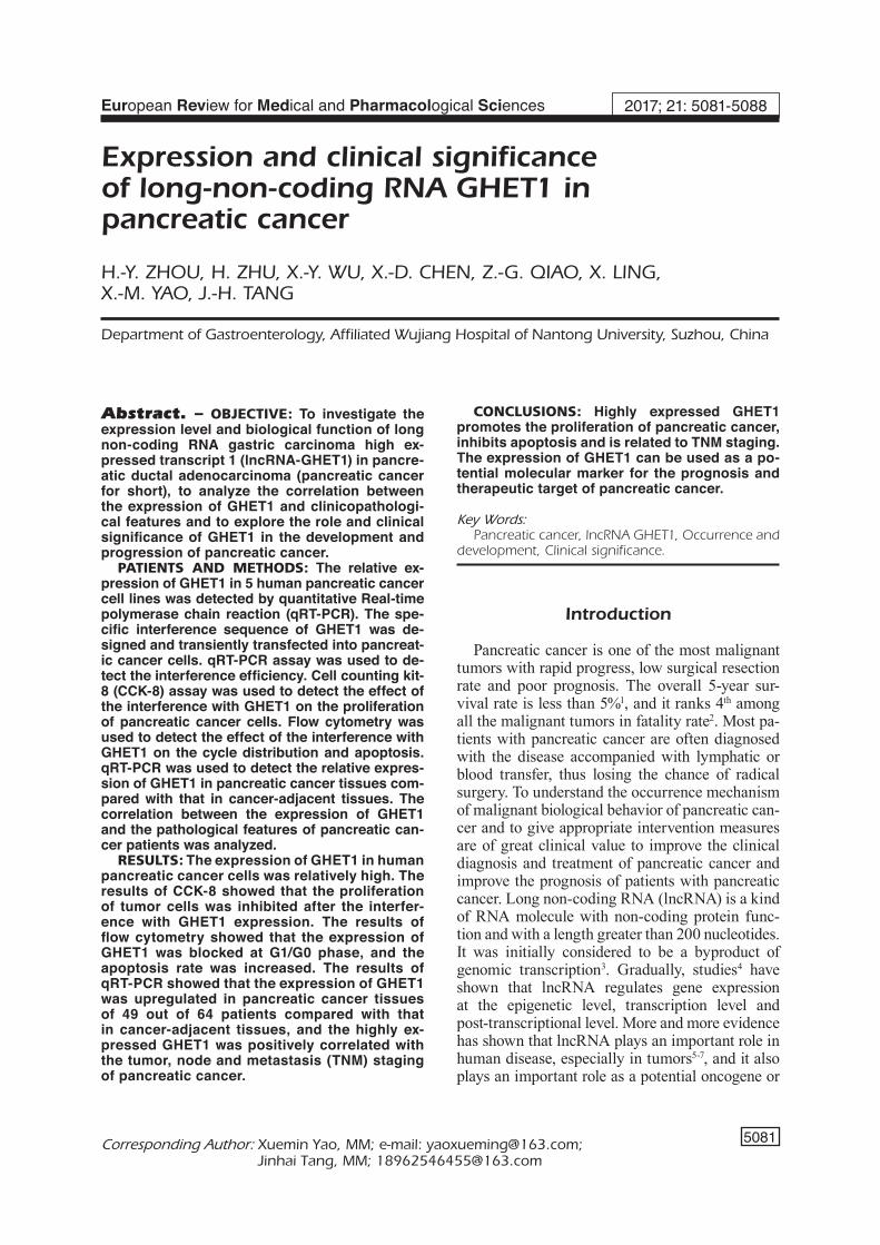
In order to study the biological function of GHET1 in pancreatic cancer, we first conduct-ed the detection by qRT-PCR assay. The results showed that the expression level of GHET1 in 5 kinds of pancreatic cancer cell lines was upreg-ulated compared with that in normal pancreatic duct epithelial cells (Figure 1A). We selected two cell lines Panc-1 and L3.6Pl with the high-est upregulation degree as the model cells, and transiently transfected si-GHET1 into the model cells. 48 hours later, the interference efficiency was detected by qRT-PCR, and the results showed that the 3# sequence had the highest transfection efficiency (Figure 1B).
Influence of GHET1 on the Biological Functions of Pancreatic Cancer Cells
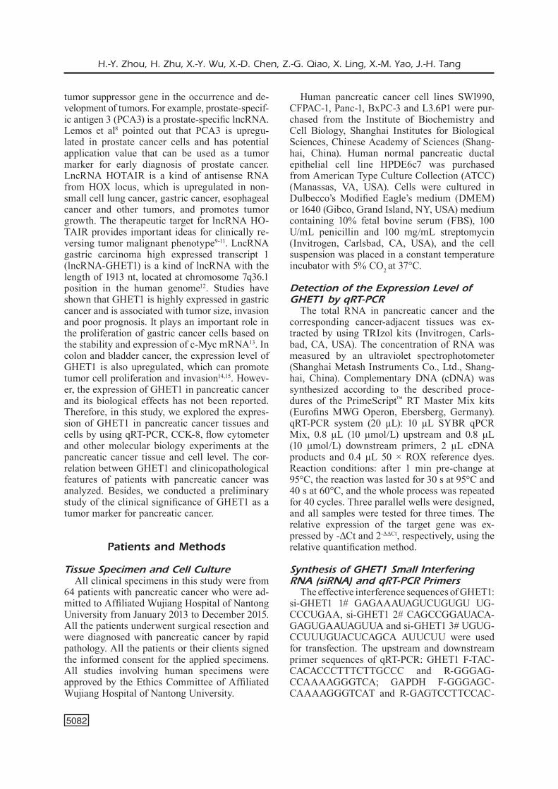
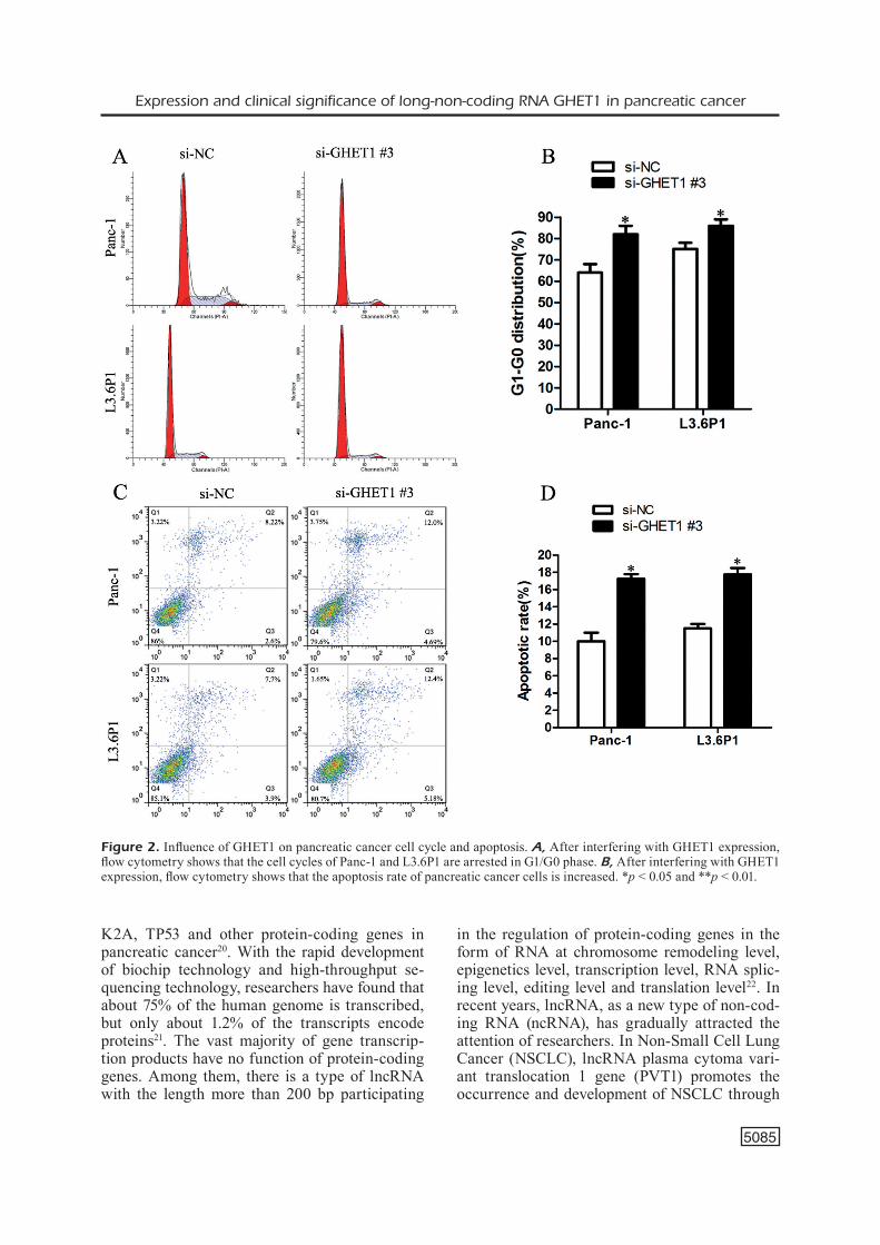
3# sequence was transfected into Panc-1 and L3.6P1. The results of CCK-8 showed that there was no significant difference in the number of viable cells between the two groups (p > 0.05) after 24-hour transfection. 24 hours later, with the extension of time, the cell proliferation ability of the experimental group was significantly inhibited compared with that of the control group (Figure 1C, D). Then, we examined the influence of GHET1 on cell cycle distribution and apoptosis of pancreatic cancer cells by flow cytometry. 3# interference se-quence was transfected into Panc-1 and L3.6Pl cells. Cells were collected 48 hours after the transfection. Results of flow cytometry showed that the cell cycle progression of pancreatic cancer was arrested in G1/G0 phase (Figure 2A, B), and the apoptosis rate was also increased (Figure 2C, D).
The Correlation Between the Expression Level of GHET1 in Human Pancreatic Cancer Tissues and Clinicopathological Features of Patients
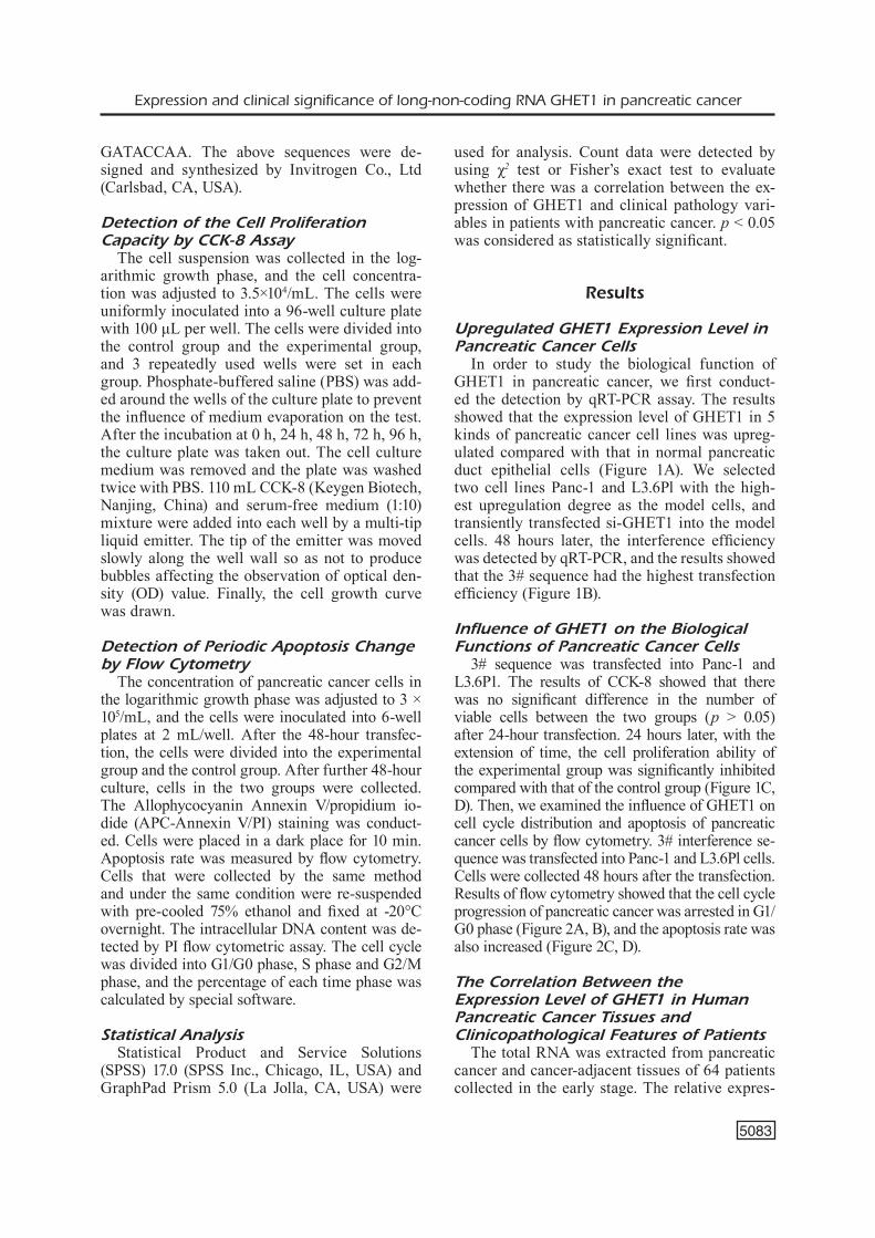
The total RNA was extracted from pancreatic cancer and cancer-adjacent tissues of 64 patients collected in the early stage. The relative expres-
H.-Y. Zhou, H. Zhu, X.-Y. Wu, X.-D. Chen, Z.-G. Qiao, X. Ling, X.-M. Yao, J.-H. Tang
5084
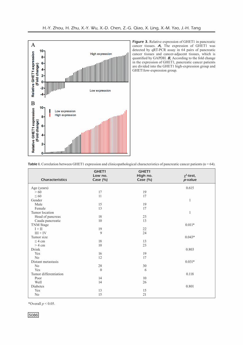
sion level of GHET1 in 49 out of 64 patients was upregulated (Figure 3A). Then, we took the me-dian of the fold change as a cut-off to divide 58 patients into two groups: GHET1 high-expression group (n = 36, fold change > 4.5) and GHET1 low-expression group (n = 28, fold change < 4.5) (Figure 3B). The results of χ2-test showed that the expression level of GHET1 was closely correlated with TNM staging (p = 0.011), but not with age, sex, whether alcohol drinking or not and other variables (Table I).
Discussion
In recent 30 years, the incidence rate of pancre-atic cancer has been at a high level. Around the
world, every 100,00016 people, there are 1-10 that are diagnosed with pancreatic cancer. About 85% of them were diagnosed with pancreatic ductal adenocarcinoma17; the incidence rate in devel-oped countries is higher than that in developing countries. In recent years, although the surgery, neoadjuvant chemotherapy, radiotherapy and oth-er treatment methods have been developed, the prognosis of pancreatic cancer still remains poor. Therefore, it is urgent to study the mechanism of pancreatic cancer and to find molecular markers for the early diagnosis of pancreatic cancer.
The incidence and development of pancreatic cancer are complex processes, in which many factors are involved in regulation18,19. Over a long period in the past, researchers mainly studied the action mechanism of KRAS, SMAD4, CDN-
Figure 1. The expression level of GHET1 in pancreatic cancer cells. A, qRT-PCR assay is used to detect the relative expression level of GHET1 in pancreatic cancer cells compared with that in normal pancreatic ductal epithelial cells. B, The interference efficiency of Panc-1 and L3.6Pl cells transfected with siRNA. C, After Panc-1 and L3.6Pl cells are transfected with si-GHET1, CCK8 assay is used to detect the change in cell proliferation capacity. All experiments are repeated for three times independently. *p < 0.05 and **p < 0.01.
Expression and clinical significance of long-non-coding RNA GHET1 in pancreatic cancer
5085
K2A, TP53 and other protein-coding genes in pancreatic cancer20. With the rapid development of biochip technology and high-throughput se-quencing technology, researchers have found that about 75% of the human genome is transcribed, but only about 1.2% of the transcripts encode proteins21. The vast majority of gene transcrip-tion products have no function of protein-coding genes. Among them, there is a type of lncRNA with the length more than 200 bp participating
in the regulation of protein-coding genes in the form of RNA at chromosome remodeling level, epigenetics level, transcription level, RNA splic-ing level, editing level and translation level22. In recent years, lncRNA, as a new type of non-cod-ing RNA (ncRNA), has gradually attracted the attention of researchers. In Non-Small Cell Lung Cancer (NSCLC), lncRNA plasma cytoma vari-ant translocation 1 gene (PVT1) promotes the occurrence and development of NSCLC through
Figure 2. Influence of GHET1 on pancreatic cancer cell cycle and apoptosis. A, After interfering with GHET1 expression, flow cytometry shows that the cell cycles of Panc-1 and L3.6P1 are arrested in G1/G0 phase. B, After interfering with GHET1 expression, flow cytometry shows that the apoptosis rate of pancreatic cancer cells is increased. *p < 0.05 and **p < 0.01.
H.-Y. Zhou, H. Zhu, X.-Y. Wu, X.-D. Chen, Z.-G. Qiao, X. Ling, X.-M. Yao, J.-H. Tang
5086
Figure 3. Relative expression of GHET1 in pancreatic cancer tissues. A, The expression of GHET1 was detected by qRT-PCR assay in 64 pairs of pancreatic cancer tissues and cancer-adjacent tissues, which is quantified by GAPDH. B, According to the fold change in the expression of GHET1, pancreatic cancer patients are divided into the GHET1 high-expression group and GHET1low-expression group.
*Overall p < 0.05.
Table I. Correlation between GHET1 expression and clinicopathological characteristics of pancreatic cancer patients (n = 64).
GHET1 GHET1 Low no. High no. χ2-test, Characteristics Case (%) Case (%) p-value
Age (years) 0.615 > 60 17 19 ≤ 60 11 17 Gender 1 Male 15 19 Female 13 17 Tumor location 1 Head of pancreas 18 23 Cauda pancreatic 10 13 TNM Stage 0.011* I + II 19 22 III + IV 9 24 Tumor size 0.043* ≤ 4 cm 18 13 > 4 cm 10 23 Drink 0.803 Yes 16 19 No 12 17 Distant metastasis 0.031* No 28 30 Yes 0 6 Tumor differentiation 0.118 Poor 14 10 Well 14 26 Diabetes 0.801 Yes 13 15 No 15 21
Expression and clinical significance of long-non-coding RNA GHET1 in pancreatic cancer
5087
the apparent regulation of the expression of large tumor suppressor homolog 2 (LATS2)23. In gas-tric cancer, lncRNA urothelial carcinoma-asso-ciated 1 (UCA1) can promote cell proliferation by activating protein kinase B (AKT) signaling pathway24.
In recent years, the role of GHET1 in tumors has rarely been reported. Initially, Yang et al13 have found that GHET1 is upregulated in gastric cancer and promoted the occurrence and devel-opment of tumors for the first time. They also demonstrated that highly expressed GHET1 in gastric cancer promotes the multidrug resistance of tumor cells25. Some scholars14,15 have gradually found that GHET1 also plays a role similar to that of oncogene in colorectal cancer and bladder cancer, but the relationship between GHET1 and pancreatic cancer has not been reported.
Conclusions
This study explored the expression and bio-logical functions of GHET1 in pancreatic cancer for the first time. We showed that GHET1 was relatively highly expressed in pancreatic can-cer tissues and cells, and the highly expressed GHET1 was positively correlated with TNM staging. The inhibition of GHET1 expression inhibited the proliferation capacity of tumor cells, arrested cell cycle in G1/G0 phase and promoted apoptosis. The results of this study are of great clinical significance for clinical screening of the best prognostic markers, selecting an appropriate treatment program, avoiding side effects caused by unnecessary treatments.
Conflict of InterestThe Authors declare that they have no conflict of interests.
References
1) Tempero m. Active systemic treatment of pancreat-ic cancer. J Natl Compr Canc Netw 2017; 15: 723-725.
2) Siegel rl, miller KD, Jemal a. Cancer statistics, 2016. CA Cancer J Clin 2016; 66: 7-30.
3) NagaNo T, FraSer p. No-nonsense functions for long noncoding RNAs. Cell 2011; 145: 178-181.
4) Heery r, FiNN Sp, CuFFe S, gray Sg. Long non-cod-ing RNAs: key regulators of epithelial-mesenchy-mal transition, tumour drug resistance and cancer stem cells. Cancers (Basel) 2017; 9: pii: E38.
5) Xu Z, yaN y, QiaN l, goNg Z. Long non-coding RNAs act as regulators of cell autophagy in diseases (Review). Oncol Rep 2017; 37: 1359-1366.
6) CHeN y, ZHou J. LncRNAs: macromolecules with big roles in neurobiology and neurological diseas-es. Metab Brain Dis 2017; 32: 281-291.
7) aTiaNaND mK, CaFFrey Dr, FiTZgeralD Ka. Immuno-biology of long noncoding RNAs. Annu Rev Im-munol 2017; 35: 177-198.
8) lemoS ae, Ferreira lB, BaToreu Nm, De FreiTaS pp, BoNamiNo mH, gimBa er. PCA3 long noncoding RNA modulates the expression of key cancer-re-lated genes in LNCaP prostate cancer cells. Tu-mour Biol 2016; 37: 11339-11348.
9) liu XH, liu Zl, SuN m, liu J, WaNg ZX, De W. The long non-coding RNA HOTAIR indicates a poor prognosis and promotes metastasis in non-small cell lung cancer. BMC Cancer 2013; 13: 464.
10) liu yW, SuN m, Xia r, ZHaNg eB, liu XH, ZHaNg ZH, Xu Tp, De W, liu Br, WaNg ZX. LincHOTAIR epige-netically silences miR34a by binding to PRC2 to promote the epithelial-to-mesenchymal transition in human gastric cancer. Cell Death Dis 2015; 6: e1802.
11) WaNg W, He X, ZHeNg Z, ma X, Hu X, Wu D, WaNg m. Serum HOTAIR as a novel diagnostic biomark-er for esophageal squamous cell carcinoma. Mol Cancer 2017; 16: 75.
12) HuaNg H, liao W, ZHu X, liu H, Cai l. Knockdown of long noncoding RNA GHET1 inhibits cell acti-vation of gastric cancer. Biomed Pharmacother 2017; 92: 562-568.
13) yaNg F, Xue X, ZHeNg l, Bi J, ZHou y, ZHi K, gu y, FaNg g. Long non-coding RNA GHET1 promotes gastric carcinoma cell proliferation by increasing c-Myc mRNA stability. FEBS J 2014; 281: 802-813.
14) li lJ, ZHu Jl, Bao WS, CHeN DK, HuaNg WW, WeNg Zl. Long noncoding RNA GHET1 promotes the development of bladder cancer. Int J Clin Exp Pathol 2014; 7: 7196-7205.
15) ZHou J, li X, Wu m, liN C, guo y, TiaN B. Knock-down of long noncoding RNA GHET1 inhibits cell proliferation and invasion of colorectal cancer. Oncol Res 2016; 23: 303-309.
16) QiN CF, ZHao Fl. Long non-coding RNA TUG1 can promote proliferation and migration of pancreatic cancer via EMT pathway. Eur Rev Med Pharma-col Sci 2017; 21: 2377-2384.
17) WaNg C, liu p, Wu H, Cui p, li y, liu y, liu Z, gou S. MicroRNA-323-3p inhibits cell invasion and me-tastasis in pancreatic ductal adenocarcinoma via direct suppression of SMAD2 and SMAD3. Onco-target 2016; 7: 14912-14924.
18) ryaN Dp, HoNg TS, BarDeeSy N. Pancreatic adeno-carcinoma. N Engl J Med 2014; 371: 2140-2141.
19) VargHeSe am, loWery ma, yu KH, o’reilly em. Cur-rent management and future directions in met-astatic pancreatic adenocarcinoma. Cancer Am Cancer Soc 2016; 122: 3765-3775.
H.-Y. Zhou, H. Zhu, X.-Y. Wu, X.-D. Chen, Z.-G. Qiao, X. Ling, X.-M. Yao, J.-H. Tang
5088
20) CiCeNaS J, KVeDeraViCiuTe K, meSKiNyTe i, meSKiNy-Te-KauSilieNe e, SKeBerDyTe a, CiCeNaS J. KRAS, TP53, CDKN2A, SMAD4, BRCA1, and BRCA2 mutations in pancreatic cancer. Cancers (Basel) 2017; 9: pii: E42.
21) DJeBali S, DaViS Ca, merKel a, DoBiN a, laSSmaNN T, morTaZaVi a, TaNZer a, lagarDe J, liN W, SCHleSiNger F, Xue C, mariNoV gK, KHaTuN J, WilliamS Ba, Zale-SKi C, roZoWSKy J, roDer m, KoKoCiNSKi F, aBDelHamiD rF, alioTo T, aNToSHeCHKiN i, Baer mT, Bar NS, Ba-TuT p, Bell K, Bell i, CHaKraBorTTy S, CHeN X, CHraST J, CuraDo J, DerrieN T, DreNKoW J, DumaiS e, DumaiS J, DuTTagupTa r, FalCoNNeT e, FaSTuCa m, FeJeS-ToTH K, Ferreira p, FoiSSaC S, FullWooD mJ, gao H, goN-ZaleZ D, gorDoN a, guNaWarDeNa H, HoWalD C, JHa S, JoHNSoN r, KapraNoV p, KiNg B, KiNgSWooD C, luo oJ, parK e, perSauD K, preall JB, riBeCa p, riSK B, roByr D, SammeTH m, SCHaFFer l, See lH, SHaHaB a, SKaNCKe J, SuZuKi am, TaKaHaSHi H, TilgNer H, TrouT D, WalTerS N, WaNg H, WroBel J, yu y, ruaN X, Ha-yaSHiZaKi y, HarroW J, gerSTeiN m, HuBBarD T, rey-moND a, aNToNaraKiS Se, HaNNoN g, giDDiNgS mC,
ruaN y, WolD B, CarNiNCi p, guigo r, giNgeraS Tr. Landscape of transcription in human cells. Nature 2012; 489: 101-108.
22) SHeNg Sr, Wu JS, TaNg yl, liaNg XH. Long noncod-ing RNAs: emerging regulators of tumor angio-genesis. Future Oncol 2017; 13: 1551-1562.
23) WaN l, SuN m, liu gJ, Wei CC, ZHaNg eB, KoNg r, Xu Tp, HuaNg mD, WaNg ZX. Long noncoding RNA PVT1 promotes non-small cell lung cancer cell pro-liferation through epigenetically regulating LATS2 expression. Mol Cancer Ther 2016; 15: 1082-1094.
24) WaNg ZQ, Cai Q, Hu l, He Cy, li JF, QuaN ZW, liu By, li C, ZHu Zg. Long noncoding RNA UCA1 in-duced by SP1 promotes cell proliferation via re-cruiting EZH2 and activating AKT pathway in gas-tric cancer. Cell Death Dis 2017; 8: e2839.
25) ZHaNg X, Bo p, liu l, ZHaNg X, li J. Overexpression of long non-coding RNA GHET1 promotes the de-velopment of multidrug resistance in gastric can-cer cells. Biomed Pharmacother 2017; 92: 580-585.