exaggeration and cooption of innate immunity for social ... · exaggeration and cooption of innate...
TRANSCRIPT

Exaggeration and cooption of innate immunity forsocial defenseMayako Kutsukakea,1, Minoru Moriyamaa,b, Shuji Shigenobuc, Xian-Ying Menga, Naruo Nikohd, Chiyo Nodae,Satoru Kobayashif, and Takema Fukatsua,g,h,1
aBioproduction Research Institute, National Institute of Advanced Science and Technology, 305-8566 Tsukuba, Japan; bComputational Bio Big Data OpenInnovation Laboratory, National Institute of Advanced Science and Technology, 305-8566 Tsukuba, Japan; cCore Research Facilities, National Institute forBasic Biology, 444-8585 Okazaki, Japan; dDepartment of Liberal Arts, The Open University of Japan, 261-8586 Chiba, Japan; eOkazaki Institute forIntegrative Bioscience, National Institute for Basic Biology, 444-8787 Okazaki, Japan; fLife Science Center for Survival Dynamics, Tsukuba Advanced ResearchAlliance, University of Tsukuba, 305-8577 Tsukuba, Japan; gDepartment of Biological Sciences, Graduate School of Science, University of Tokyo, 113-0033Tokyo, Japan; and hGraduate School of Life and Environmental Sciences, University of Tsukuba, 305-8572 Tsukuba, Japan
Edited by Nancy A. Moran, University of Texas at Austin, Austin, TX, and approved March 14, 2019 (received for review January 17, 2019)
Social insects often exhibit striking altruistic behaviors, of whichthe most spectacular ones may be self-destructive defensive be-haviors called autothysis, “self-explosion,” or “suicidal bombing.”In the social aphid Nipponaphis monzeni, when enemies damagetheir plant-made nest called the gall, soldier nymphs erupt to dis-charge a large amount of body fluid, mix the secretion with theirlegs, and skillfully plaster it over the plant injury. Dozens of sol-diers come out, erupt, mix, and plaster, and the gall breach ispromptly sealed with the coagulated body fluid. What molecularand cellular mechanisms underlie the self-sacrificing nest repairwith body fluid for the insect society? Here we demonstrate thatthe body cavity of soldier nymphs is full of highly differentiatedlarge hemocytes that contain huge amounts of lipid droplets andphenoloxidase (PO), whereas their hemolymph accumulates hugeamounts of tyrosine and a unique repeat-containing protein (RCP).Upon breakage of the gall, soldiers gather around the breach andmassively discharge the body fluid. The large hemocytes ruptureand release lipid droplets, which promptly form a lipidic clot, and,concurrently, activated PO converts tyrosine to reactive quinones,which cross-link RCP and other macromolecules to physically re-inforce the clot to seal the gall breach. Here, soldiers’ humoral andcellular immune mechanisms for wound sealing are extremely up-regulated and utilized for colony defense, which provides a strikingcase of direct evolutionary connection between individual immunityand social immunity and highlights the importance of exaggerationand cooption of preexisting traits to create evolutionary novelties.
social aphid | self-sacrificing gall repair | phenoloxidase |hemocyte | tyrosine
Animals are always under the risk of infections with bacteria,fungi, and other parasites, and immune systems are therefore
essential for their survival in the natural environment. Whereas theIg-based adaptive immunity is specific to vertebrates, innate im-mune mechanisms are generally found in diverse animals encom-passing vertebrates and invertebrates (1). Upon breakage of surfacebarrier and invasion of microbes or other nonself entities, bodyfluid coagulation, hardening, and melanization immediately occurto stop bleeding and to localize the invasion, in which enzymes withprotein cross-linking activities such as phenoloxidases (POs) andtransglutaminases play important roles (2, 3). Subsequently, spe-cialized hemocytes are recruited to participate in wound sealing aswell as phagocytosis and encapsulation of the intruders (4, 5).Finally, antimicrobial peptides and other potent effector mole-cules are transiently and drastically induced and released into thebody fluid to kill the intruders (1, 6).Conventionally, immunology has focused on molecular and
cellular mechanisms against noxious biological agents to ensuresurvival of individuals. Recently, the notion of social immunityhas emerged, which highlights traits and mechanisms of socialinsects and other group-living animals to combat against path-ogens, parasites, and other natural enemies to ensure survival of
their colony or society as a whole (7, 8). Although the concep-tualization of some fundamental commonalities between indi-vidual immunity and social immunity across the organismalhierarchical levels may be insightful in understanding the generalaspect of biological systems, the mechanistic bases of social im-munity, which are mainly behavioral, physiological, and organi-zational ones, are, needless to say, distinct from the molecularand cellular immune mechanisms (7, 8).Here we report a case of social immunity in which innate
humoral and cellular immune mechanisms at the individual levelare extremely exaggerated and directly utilized for colony de-fense at the group level in an ecological context. In addition towell-known social insects such as ants, bees, wasps, and termites,some aphids are known to be social, producing morphologicallyspecialized or nonspecialized individuals called soldiers, whichperform altruistic tasks including colony defense against preda-tors and cleaning and repairing of their plant-made nests known
Significance
Conventionally, immunology has focused on molecular andcellular mechanisms against pathogens and parasites to ensuresurvival of individuals. Recently, the notion of social immunityhas emerged, which highlights the mechanisms in social ani-mals to combat against pathogens, parasites, and other ene-mies to ensure survival of their society as a whole. Conceptually,social immunity is analogous to but distinct from individualimmunity. However, we discovered that, in the social aphidNipponaphis monzeni, molecular and cellular immune compo-nents of soldier individuals are extremely up-regulated andmassively excreted via “body eruption” upon gall breakage, andthe “hyperclotting” body fluid repairs the damaged gall forcolony defense, which uncovers unexpected molecular, cellular,and evolutionary commonalities across individual immunity andsocial immunity.
Author contributions: M.K. and T.F. designed research; M.K., M.M., S.S., X.-Y.M., and C.N.performed research; S.S. and S.K. contributed new reagents/analytic tools; M.K., M.M.,S.S., and N.N. analyzed data; and M.K. and T.F. wrote the paper.
The authors declare no conflict of interest.
This article is a PNAS Direct Submission.
This open access article is distributed under Creative Commons Attribution-NonCommercial-NoDerivatives License 4.0 (CC BY-NC-ND).
Data deposition: The sequence data reported in this paper have been deposited in theDNA Data Bank Japan (https://www.ddbj.nig.ac.jp/index-e.html) under the following ac-cession nos.: raw data obtained from RNA-sequencing experiments (DRA007668–DRA007698); gene sequences (LC436897–LC436906, IAEA01000001–IAEA01000019); andsymbiont genome sequences (AP019379–AP019381).1To whom correspondence may be addressed. Email: [email protected] or [email protected].
This article contains supporting information online at www.pnas.org/lookup/suppl/doi:10.1073/pnas.1900917116/-/DCSupplemental.
Published online April 15, 2019.
8950–8959 | PNAS | April 30, 2019 | vol. 116 | no. 18 www.pnas.org/cgi/doi/10.1073/pnas.1900917116

as galls (9, 10). The social aphid Nipponaphis monzeni formslarge galls on the tree Distylium racemosum (Fig. 1A) in whichhundreds to thousands of insects proliferate by sucking plant sapfrom the inner wall (Fig. 1 B and C) (11). In N. monzeni, allnymphs exhibit an extended first instar stage, perform social tasksas soldiers, and then grow and reproduce (11, 12). In spring, younggalls of N. monzeni are often attacked by lepidopteran enemies(13), whose larvae tunnel and damage gall tissues and also con-sume inhabiting aphids. Upon invasion of the predator, mono-morphic first-instar soldiers attack the enemy by stinging withtheir stylets (Fig. 1D). Then, many soldiers gather around the holeon the gall wall and erupt to discharge a large amount of whitishbody fluid from their cornicles on the abdominal tip (Fig. 1 E andF and Movie S1). The shriveled soldiers actively mix the secretionwith their legs and skillfully plaster it over the plant injury, and thesecretion promptly solidifies (Fig. 1G and Movie S1). Dozens ofsoldiers come out, erupt, mix, and plaster, and the hole is com-pletely plugged by the coagulated body fluid (Fig. 1 H and I andMovie S1) (12, 14). Here, the soldier nymphs of N. monzeniperform not only a soldier-type aggressive task of attacking ene-mies but also a worker-type housekeeping task of repairing theirplant-made nest, which entails a series of unique self-destructivebehaviors. The molecular and cellular mechanisms that underliethe self-sacrificing nest repair with the use of body fluid for theinsect society is of great interest, which prompted us to investigatethe biochemistry, physiology, cell and molecular biology, and de-velopmental and evolutionary aspects of the gall-repairing bodyfluid produced by soldier nymphs of N. monzeni.
Results and DiscussionDarkening upon Clotting, Large Globular Cells, and a Few MajorProteins in Body Fluid of Soldier Nymphs. The soldiers’ secretedbody fluid turned black as its solidification proceeded (Fig. 1I–L). Mechanical stimulation of soldier nymphs in a salinesolution elicited the body fluid secretion (Fig. 1M and Movie
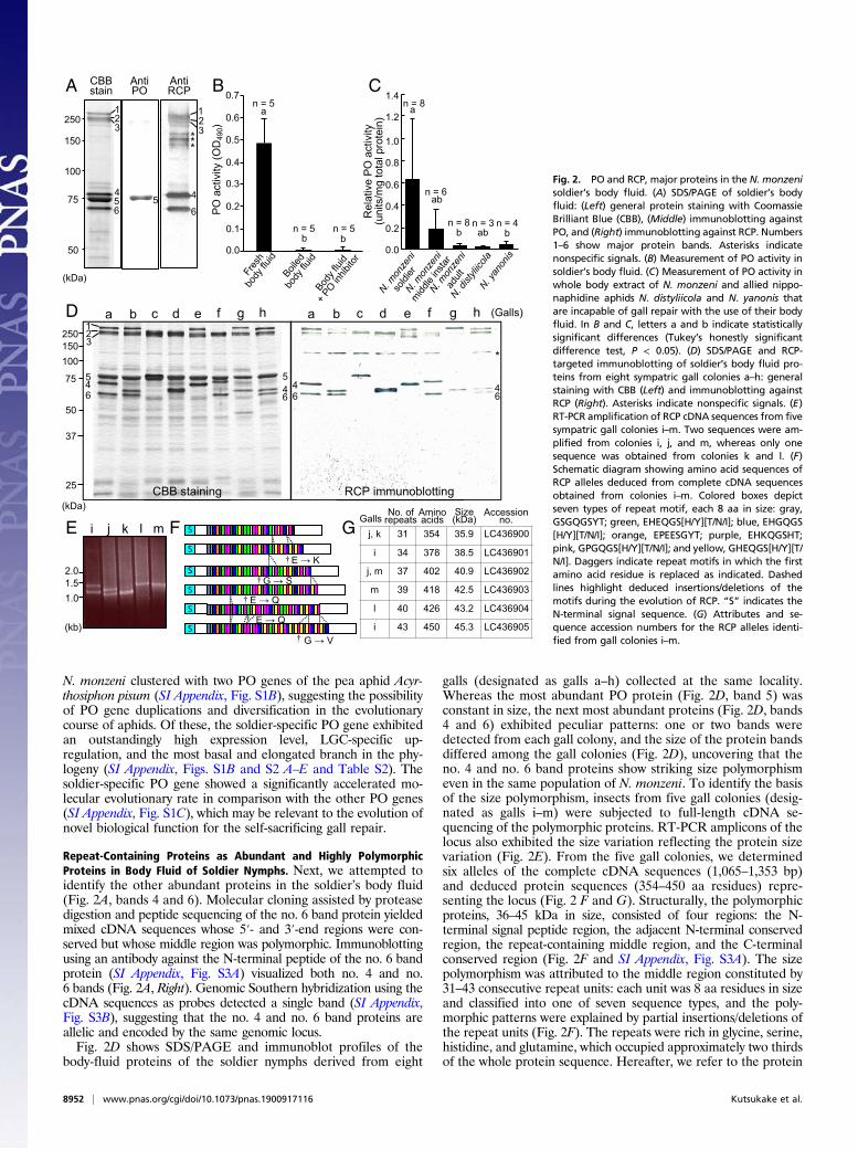
S2), which demonstrated that the body fluid contains numer-ous peculiar large cells, called the large globular cells (LGCs)(14), that fill up the soldier’s body cavity (Fig. 1 N and O).Histological examination of the hole-filling plugs identifiednuclei and granules derived from LGCs (Fig. 1P). SDS/PAGEof the soldiers’ body fluid detected only a few major proteinbands (Fig. 2A, Left), suggesting that these proteins may playsome roles in the self-sacrificing gall repair by soldier nymphs.
Phenoloxidase as a Predominant Protein in Body Fluid of SoldierNymphs. The darkening of the coagulated plugs (Fig. 1 I–L)suggested progressive melanin formation in the secreted bodyfluid, where the melanin synthesis pathway driven by PO may beactivated (3, 15). Immunoblotting identified one of the pre-dominant proteins in the soldier’s body fluid as PO (Fig. 2A,Center, band 5). An enzymatic activity assay detected drasticallyhigh PO activities in the soldier nymphs of N. monzeni (Fig. 2 Band C). Molecular cloning of the PO cDNA assisted by proteasedigestion and peptide sequencing of the no. 5 band proteinyielded a full-length cDNA sequence 2,355 bp in size containingan ORF encoding a protein of 685 aa residues (SI Appendix, Fig.S1A). Putative copper-binding histidine residues, which are gen-erally conserved in arthropod POs (16), were present, whereas aconserved proteolytic cleavage site for pro-PO activation, namelyarginine-phenylalanine at approximately 50 residues from the Nterminus (15), was not identified (SI Appendix, Fig. S1A). Peptidemass fingerprinting of the no. 5 band protein identified anN-terminal propeptide fragment of typical insect pro-PO, in-dicating that the PO protein is mainly in a proenzyme form invivo. RNA sequencing (RNA-seq) analysis of N. monzeni (SIAppendix, Tables S1 and S2) detected that, in addition to thesoldier-specific PO that exhibited extremely high expressionlevels preferentially in the soldier nymphs, four additional POgenes were expressed at low levels in N. monzeni (SI Appendix,Fig. S2 B–E). On the phylogenetic tree, the five PO genes of
A B C D
E F G H
I J K L
M N O P
Fig. 1. Self-sacrificing gall repair by soldier nymphsof N. monzeni. (A) A gall formed on the tree D.racemosum. (B) An inside view of a gall. (C) A mag-nified image of gall contents: white arrow, first-instarsoldier nymph; black arrow, adult; small white arrow,powdery aggregate consisting of excreted wax;small black arrow, aphid cadaver. (D) Soldier nymphsattacking a moth larva by stinging with their stylet.(E) A gall-repairing soldier nymph discharging bodyfluid. (F) Scanning EM image of a soldier nymph(ventral view); arrows indicate droplets of body fluiddischarged from cornicles. (G) Soldier nymphs plas-tering their own body fluid onto plant injury (MovieS1). (H) A gall with a naturally repaired hole (arrow).(I–L) An experimentally bored hole filled by bodyfluid of soldier nymphs at 0 h (I), 1 h (J), 2 h (K), and6 h after plugging (L). (M) LGCs discharged from asoldier nymph (Movie S2). (N) An enlarged image ofLGCs. (O) An abdominal cross-section of a soldiernymph whose body cavity is full of LGCs: bc, bacter-iome; gu, midgut; ov, ovary. (P) A thin section of asolidified soldier’s body fluid 3 d after gall repair;white arrows indicate unruptured LGCs and blackarrows indicate nuclei of ruptured LGCs.
Kutsukake et al. PNAS | April 30, 2019 | vol. 116 | no. 18 | 8951
EVOLU
TION
N. monzeni clustered with two PO genes of the pea aphid Acyr-thosiphon pisum (SI Appendix, Fig. S1B), suggesting the possibilityof PO gene duplications and diversification in the evolutionarycourse of aphids. Of these, the soldier-specific PO gene exhibitedan outstandingly high expression level, LGC-specific up-regulation, and the most basal and elongated branch in the phy-logeny (SI Appendix, Figs. S1B and S2 A–E and Table S2). Thesoldier-specific PO gene showed a significantly accelerated mo-lecular evolutionary rate in comparison with the other PO genes(SI Appendix, Fig. S1C), which may be relevant to the evolution ofnovel biological function for the self-sacrificing gall repair.
Repeat-Containing Proteins as Abundant and Highly PolymorphicProteins in Body Fluid of Soldier Nymphs. Next, we attempted toidentify the other abundant proteins in the soldier’s body fluid(Fig. 2A, bands 4 and 6). Molecular cloning assisted by proteasedigestion and peptide sequencing of the no. 6 band protein yieldedmixed cDNA sequences whose 5′- and 3′-end regions were con-served but whose middle region was polymorphic. Immunoblottingusing an antibody against the N-terminal peptide of the no. 6 bandprotein (SI Appendix, Fig. S3A) visualized both no. 4 and no.6 bands (Fig. 2A, Right). Genomic Southern hybridization using thecDNA sequences as probes detected a single band (SI Appendix,Fig. S3B), suggesting that the no. 4 and no. 6 band proteins areallelic and encoded by the same genomic locus.Fig. 2D shows SDS/PAGE and immunoblot profiles of the
body-fluid proteins of the soldier nymphs derived from eight
galls (designated as galls a–h) collected at the same locality.Whereas the most abundant PO protein (Fig. 2D, band 5) wasconstant in size, the next most abundant proteins (Fig. 2D, bands4 and 6) exhibited peculiar patterns: one or two bands weredetected from each gall colony, and the size of the protein bandsdiffered among the gall colonies (Fig. 2D), uncovering that theno. 4 and no. 6 band proteins show striking size polymorphismeven in the same population of N. monzeni. To identify the basisof the size polymorphism, insects from five gall colonies (desig-nated as galls i–m) were subjected to full-length cDNA se-quencing of the polymorphic proteins. RT-PCR amplicons of thelocus also exhibited the size variation reflecting the protein sizevariation (Fig. 2E). From the five gall colonies, we determinedsix alleles of the complete cDNA sequences (1,065–1,353 bp)and deduced protein sequences (354–450 aa residues) repre-senting the locus (Fig. 2 F and G). Structurally, the polymorphicproteins, 36–45 kDa in size, consisted of four regions: the N-terminal signal peptide region, the adjacent N-terminal conservedregion, the repeat-containing middle region, and the C-terminalconserved region (Fig. 2F and SI Appendix, Fig. S3A). The sizepolymorphism was attributed to the middle region constituted by31–43 consecutive repeat units: each unit was 8 aa residues in sizeand classified into one of seven sequence types, and the poly-morphic patterns were explained by partial insertions/deletions ofthe repeat units (Fig. 2F). The repeats were rich in glycine, serine,histidine, and glutamine, which occupied approximately two thirdsof the whole protein sequence. Hereafter, we refer to the protein
A
D
E F G
B C
Fig. 2. PO and RCP, major proteins in the N. monzenisoldier’s body fluid. (A) SDS/PAGE of soldier’s bodyfluid: (Left) general protein staining with CoomassieBrilliant Blue (CBB), (Middle) immunoblotting againstPO, and (Right) immunoblotting against RCP. Numbers1–6 show major protein bands. Asterisks indicatenonspecific signals. (B) Measurement of PO activity insoldier’s body fluid. (C) Measurement of PO activity inwhole body extract of N. monzeni and allied nippo-naphidine aphids N. distyliicola and N. yanonis thatare incapable of gall repair with the use of their bodyfluid. In B and C, letters a and b indicate statisticallysignificant differences (Tukey’s honestly significantdifference test, P < 0.05). (D) SDS/PAGE and RCP-targeted immunoblotting of soldier’s body fluid pro-teins from eight sympatric gall colonies a–h: generalstaining with CBB (Left) and immunoblotting againstRCP (Right). Asterisks indicate nonspecific signals. (E)RT-PCR amplification of RCP cDNA sequences from fivesympatric gall colonies i–m. Two sequences were am-plified from colonies i, j, and m, whereas only onesequence was obtained from colonies k and l. (F)Schematic diagram showing amino acid sequences ofRCP alleles deduced from complete cDNA sequencesobtained from colonies i–m. Colored boxes depictseven types of repeat motif, each 8 aa in size: gray,GSGQGSYT; green, EHEQGS[H/Y][T/N/I]; blue, EHGQGS[H/Y][T/N/I]; orange, EPEESGYT; purple, EHKQGSHT;pink, GPGQGS[H/Y][T/N/I]; and yellow, GHEQGS[H/Y][T/N/I]. Daggers indicate repeat motifs in which the firstamino acid residue is replaced as indicated. Dashedlines highlight deduced insertions/deletions of themotifs during the evolution of RCP. “S” indicates theN-terminal signal sequence. (G) Attributes and se-quence accession numbers for the RCP alleles identi-fied from gall colonies i–m.
8952 | www.pnas.org/cgi/doi/10.1073/pnas.1900917116 Kutsukake et al.

as a repeat-containing protein (RCP). The RCP gene showed anoutstandingly high expression level and LGC-specific up-regulation insoldier nymphs of N. monzeni (SI Appendix, Fig. S2F and Table S2).Oddly, SDS/PAGE estimated the molecular mass of RCP as
60–80 kDa (Fig. 2D), which was discrepant with the actual mo-lecular mass determined by molecular cloning (36–45 kDa forgalls i–m; Fig. 2G). MALDI-TOF MS analysis of the soldier’sbody fluid proteins supported the latter molecular mass of RCP(27–43 kDa for galls a–h; SI Appendix, Fig. S3C), corroboratingthe unusual electrophoretic mobility of RCP. We speculate thatthe atypical structure of the large repeat region might be re-sponsible for this peculiar feature of RCP.
Localization of PO and RCP in LGCs and Body Cavity of SoldierNymphs. In situ hybridization and immunohistochemistry visual-ized signals of PO and RCP in LGCs that occupied the bodycavity of soldier nymphs and soldier-to-be embryos in the ma-ternal body (Fig. 3 A–C and SI Appendix, Fig. S4 A–C). In earlysoldier embryos, LGCs had a large nucleus and ribosome-richcytoplasm (Fig. 3D). As the embryonic and nymphal developmentproceeded, LGCs accumulated lipid droplets in the cytoplasm(Fig. 3 E and F). In mature soldier nymphs, LGCs were full oflipid droplets with an atrophied nucleus and some cytoplasmremaining between the lipid droplets (Fig. 3G). By immuno-EM,PO was preferentially localized to the cytoplasm of LGCs (Fig. 3Hand SI Appendix, Fig. S4D), whereas RCP was detected mainly inthe hemocoel outside LGCs (Fig. 3I and SI Appendix, Fig. S4E).The extracellular localization of RCP is consistent with the pres-ence of the secretion signal sequence at the N terminus of theRCP gene (SI Appendix, Fig. S3A).
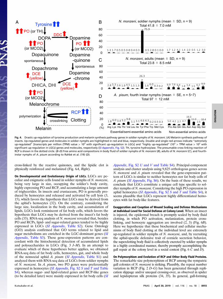
Up-Regulation of PO-Related Melanization Pathway Genes in LGCs ofSoldier Nymphs. In insects, PO-mediated melanin formation isinvolved in a variety of biological functions such as innate im-munity, hemolymph clotting, and wound sealing (1–3, 15, 17).Fig. 4A shows the melanin synthesis pathway in insects: PO ortyrosine hydroxylase converts tyrosine to dihydroxyphenylalanine(DOPA); DOPA decarboxylase (DDC) catalyzes the reactionfrom DOPA to dopamine; PO and multicopper oxidase 2 (MCO2)catalyze the reaction from DOPA or dopamine to dopaquinoneor dopamine-quinone, respectively; dopaquinone and dopamine-quinone spontaneously become dopachrome and dopaminechrome,respectively; dopachrome and dopamine-quinone are converted to5,6-dihydroxyindole (DHI)-2-carboxylic acid (DHICA) and/orDHI by dopachrome conversion enzyme (DCE; encoded byyellow family genes); and DHICA or DHI is converted to mel-anochrome by PO and MCO2 and polymerized to form melanin.Our RNA-seq data revealed that, in addition to PO, DDC andDCE were conspicuously up-regulated in LGCs (SI Appendix,Fig. S2 I and J and Table S2), indicating that enzymes consti-tuting the PO cascade are generally and drastically up-regulatedin LGCs of soldier nymphs (Fig. 4A). It is also notable that aserine protease gene and a serpin gene, which are known to beinvolved in the proteolytic cascade of PO activation (18), werehighly expressed in LGCs (SI Appendix, Fig. S2 K and L andTable S2), although their actual involvement in the PO activationcascade should be verified in future studies.
Extraordinarily Abundant Tyrosine in Body Fluid of Soldier Nymphs.As depicted in Fig. 4A, tyrosine is the main and starting substratefor the PO-mediated melanization pathway. Notably, tyrosinewas extraordinarily predominant among free amino acids in thebody fluid of soldier nymphs of N. monzeni, measuring 31.7 ±5.5 mM and accounting for 75.9% of total free amino acids (Fig.4B). We found that (i) tyrosine was the most abundant freeamino acid in the body fluid of N. monzeni irrespective of de-velopmental stage, (ii) the level of tyrosine was more than twiceas high in soldier nymphs than in N. monzeni adults, and (iii)
such high tyrosine titers are not observed in nonsocial aphids likeA. pisum (19) (Fig. 4 B–D). In soldier nymphs of N. monzeni, ty-rosine was detected not only from hemolymph (0.62 ± 0.21 nmolper insect, n = 9) but also from LGCs (0.14 ± 0.08 nmol per insect,n = 9). DOPA was negligible in soldiers’ freshly secreted bodyfluid (0.02 ± 0.03 mM, n = 7) but became detectable 15 min aftersecretion (2.3 ± 1.8 mM, n = 7), indicating conversion of tyrosineto DOPA in the solidifying body fluid.
In Vitro Clotting Assay Using PO, RCP, and Tyrosine. In the PO-mediated melanin synthesis pathway, tyrosine is converted tohighly reactive quinone molecules (Fig. 4A), which kill parasitesand pathogens through free-radical reactions (20) and cross-linkproteins for hemolymph clotting and cuticular tanning (21, 22).The extreme up-regulation of PO, RCP, and tyrosine in soldiernymphs of N. monzeni prompted us to hypothesize that thesemolecular components may play pivotal roles in the body fluidcoagulation and melanization upon self-sacrificing gall repair (Fig.1 and Movie S1). To test this hypothesis, we attempted to re-construct the chemical reactions by using defined molecular com-ponents. Recombinant PO protein was produced by using Sf9insect cells and baculovirus expression system and successfully pu-rified and activated by an addition of 2-propanol (23) (SI Appendix,Fig. S4 F–H). Recombinant RCP protein was produced and puri-fied by using Escherichia coli and pET system (SI Appendix, Fig.S4I). In the soldier’s body fluid, estimated concentrations of PO,RCP, and tyrosine were 15.3 ± 5.6 μg/μL (n = 3), 5.3 ± 0.6 μg/μL(n = 3), and 31.7 ± 5.5 mM (n = 9), respectively. As these proteinconcentrations are too high to be handled practically, we per-formed the reconstruction experiments at a 1/10 concentrationscale, whereby the basic reaction mixture (6 μL) contained ap-proximately 1.5 μg/μL recombinant PO, 0.5 μg/μL recombinantRCP, and 3.3 mM tyrosine. In the presence of 20% 2-propanol, POwas immediately activated, and the solution turned brownish within1 min and then blackish within 10 min. Concurrently, RCP wascross-linked and polymerized, causing disappearance of the RCPband and accumulation of a high–molecular-mass protein band atthe upper end of the SDS/PAGE separating gels (Fig. 5A). After1 h, a lot of black clots formed in the solution (Fig. 5D), and, after16 h, all RCP protein was consumed and black precipitates formed(Fig. 5A). In the absence of tyrosine, by contrast, neither RCPconsumption nor clot formation was observed (Fig. 5 B and E),corroborating the idea that PO-mediated conversion of tyrosine toreactive quinones should be responsible for protein cross-linkingand clotting (Fig. 4A). When RCP was replaced by BSA, cross-linking and clotting certainly occurred, but the levels of proteinconsumption and cross-linking of BSA were much less efficientthan those of RCP (Fig. 5 C and F), indicating that RCP is a moresusceptible substrate for PO/tyrosine-mediated cross-linking incomparison with BSA. Notably, the black clots formed not onlywith RCP (Fig. 5D), but also with BSA (Fig. 5F), and even withoutboth RCP and BSA (Fig. 5G). Considering that PO is the mostabundant (1.5 μg/μL) protein component in the reaction mixtureand PO bands also quickly disappeared as the reaction proceeded(Fig. 5 A–C), PO itself must be cross-linked and contribute to theclot formation. Note that activated PO is known as a very “sticky”protein that is easily lost in experimental handling (15). Whenquantities of PO and tyrosine were reduced, the clot formationwas suppressed conspicuously (Fig. 5H–K), whereas the quantitiesof black clots were apparently not affected by increase or decreaseof RCP (Fig. 5 L and M). These results strongly suggest that theup-regulated production of PO, RCP, and tyrosine underlies themelanization and hardening of the secreted body fluid upon self-sacrificing gall repair by soldier nymphs.
Abundant Lipids in Clotted Body Fluid and LGCs. Here it shouldbe noted that soldiers’ excreted body fluid is full of LGCs (Fig. 1M–O), and LGCs are full of lipid droplets (Fig. 3G). Chloroform
Kutsukake et al. PNAS | April 30, 2019 | vol. 116 | no. 18 | 8953
EVOLU
TION

extraction of hole-filling plugs collected from repaired gallsrevealed that lipids accounted for 82.5 ± 3.1% (n = 12) of theirdry weight, indicating that lipids quantitatively constitute themajor component of soldiers’ coagulated body fluid. Corniclesare a pair of tube- or pore-like defensive organs specificallyfound on the abdominal tip of aphids (24, 25). When stimulatedor threatened, aphids secrete sticky liquid from the cornicles,which contain waxy substances and alarm pheromones, to deterpredatory attacks and to elicit escaping behavior of colony mates(26–28). Previous studies reported that triglycerides are amongthe major components of the aphid’s cornicle secretion (29–31).Our lipid analysis revealed that (i) the lipids in the secretionwere mostly triglycerides whose estimated total concentrationwas as high as 528 ± 85 mM (n = 7), (ii) the major componentswere triglycerides consisting of sorboyl and dimyristic acids(C6:2, C14:0, C14:0; 35.0%) and sorboyl, myristic, and palmitoylacids (C6:2, C14:0, C16:0; 28.7%), (iii) the lipid composition did
not change before and after body fluid coagulation, and (iv) thefatty acids separated by alkaline hydrolysis of soldiers’ excretedbody fluid were almost the same as those constituting the tri-glycerides (SI Appendix, Fig. S5).
Two-Step Model for the Mechanism of Gall Repair: Immediate Formationof Lipidic Soft Clot Followed by PO-Mediated Clot Hardening. On thebasis of these results, we propose a model for the molecular andcellular mechanisms underlying the self-sacrificing gall repair bysoldier nymphs of N. monzeni. In the soldier’s body cavity, inactivePO and lipid droplets are stored in LGCs, whereas tyrosine andRCP are accumulated in hemolymph (Fig. 6A, Left). Upon dis-charge of the body fluid, LGCs rupture and release lipid droplets,which promptly form a lipidic soft clot, and PO is concurrently ac-tivated, presumably by hemolymphal enzymes, and initiates the POcascade, by which tyrosine is converted to reactive quinones (Fig.6A, Middle). Subsequently, proteins and other macromolecules are
A
D E F G
H I J
K M
L
B C
Fig. 3. In vivo localization of PO, RCP, lipids, and polysaccharides in N. monzeni. (A and B) In situ hybridization of PO gene expression (A) and RCP geneexpression (B) on abdominal tissue sections of adult insects. PO and RCP genes are preferentially expressed in LGCs in the body cavity of soldier-to-be embryos,whereas neither PO nor RCP is detected in fat body cells in the maternal body cavity. In N. monzeni, when soldier nymphs start growing, LGCs in the bodycavity are lost and replaced by fat body cells and ovaries, by which resource allocation from defense to reproduction proceeds. (C) Immunohistochemistry ofPO protein on an abdominal tissue section of a soldier nymph. Brownish PO signals are detected in the cytoplasm of LGCs, whereas nuclei are counterstainedwith hematoxylin in purple. Negative control images corresponding to A–C are shown in SI Appendix, Fig. S4 A–C. (D–G) Transmission EM images of LGCs in anearly soldier-to-be embryo (D), a late soldier-to-be embryo (E), a young soldier nymph (F), and a mature soldier nymph (G). (H and I) Immuno-EM images of PO(H) and RCP (I) in LGCs of soldier nymphs. Negative control images corresponding to H and I are shown in SI Appendix, Fig. S4 D and E. (J) Lipid staining of afrozen tissue section of a soldier-to-be embryo with Oil Red, counterstained with hematoxylin in purple. (K) Control staining of an adjacent section withoutOil Red. (L) Polysaccharide staining of a sectioned soldier-to-be embryo with periodic acid-Schiff (PAS) reagent in red, counterstained with hematoxylin inpurple. (M) Control staining of an adjacent section without PAS. bc, bacteriocyte; br, brain; fb, fat body; gu, gut; hc, hemocoel; ld, lipid droplet; lgc, largeglobular cell; n, nucleus; tg, thoracic ganglion.
8954 | www.pnas.org/cgi/doi/10.1073/pnas.1900917116 Kutsukake et al.
cross-linked by the reactive quinones, and the lipidic clot isphysically reinforced and melanized (Fig. 6A, Right).
On Developmental and Evolutionary Origin of LGCs. LGCs are pe-culiar and enigmatic cells found in soldier nymphs of N. monzeni,being very large in size, occupying the soldier’s body cavity,highly expressing PO and RCP, and accumulating a large amountof triglycerides. In insects and crustaceans, PO is generally pro-duced by hemocytes and released and activated by cell lysis (3,15), which favors the hypothesis that LGCs may be derived fromthe aphid’s hemocytes (32). On the contrary, considering thelarge size, localization in the body cavity, and accumulation oflipids, LGCs look reminiscent of fat body cells, which favors thehypothesis that LGCs may be derived from the insect’s fat bodycells (33). RNA-seq analysis of N. monzeni revealed that, besidesPO and RCPs, lipid- and sugar-related genes tended to be highlyexpressed in LGCs (SI Appendix, Table S2). Gene Ontology(GO) analysis confirmed that GO terms related to lipid andsugar metabolisms are enriched in the LGC-dominant genes (SIAppendix, Table S3). These gene-expression patterns are con-cordant with the histochemical detection of accumulated lipidsand polysaccharides in LGCs (Fig. 3 J–M). In an attempt toevaluate which of these hypotheses better applies, we obtainedRNA-seq data of fat body cells, hemocytes, gut, and whole bodyof the nonsocial aphid A. pisum (SI Appendix, Table S1) andanalyzed them with RNA-seq data of LGCs from soldier nymphsof N. monzeni. In A. pisum, two PO genes were preferentiallyexpressed in hemocytes (SI Appendix, Fig. S2 S and T and TableS4), whereas sugar- and lipid-related genes and RCP-like genes(to be detailed later) were mainly expressed in fat body cells (SI
Appendix, Fig. S2 U and V and Table S4). Principal-componentanalysis and cluster analysis using 9,825 orthologous genes acrossN. monzeni and A. pisum revealed that the gene-expression pat-tern of LGCs is similar to neither hemocytes nor fat body cells ofA. pisum (SI Appendix, Fig. S6). On the basis of these results, weconclude that LGCs constitute a unique cell type specific to sol-dier nymphs ofN. monzeni. Considering the high PO expression inaphid hemocytes (SI Appendix, Fig. S2 S and T and Table S4), itseems plausible that LGCs represent highly differentiated hemo-cytes with fat body-like features.
Exaggeration and Cooption of Wound Sealing and Defense Mechanismsat Individual Level for Social Defense at Colony Level.When an insectis injured, the epidermal breach is promptly sealed by body fluidclotting, in which PO activation, melanization, protein cross-linking, and hemocyte aggregation are involved (2, 3) (Fig. 6B).Here we hypothesize that these biochemical and cellular mecha-nisms of body fluid clotting at the individual level are extremelyup-regulated in soldier nymphs of N. monzeni, and, by recruitingthe aphid-specific defensive trait of cornicle secretion behavior,the superclotting body fluid is collectively excreted by soldier nymphsin a highly coordinated manner, thereby promptly accomplishing thegall repair at the colony level in a social context (Fig. 6 C and D).
On Polymorphism and Evolution of RCP and Other Body Fluid Proteins.The remarkable size polymorphism of RCP among the sympatricgall colonies of N. monzeni is striking. Plausibly, the repeat numbervariation in RCP (Fig. 2 D–G) has been generated through repli-cation slippage and/or unequal crossing-over, as observed in spiderand lepidopteran silk proteins (34, 35). In general, gall-forming
A B
C
D
Fig. 4. Drastic up-regulation of tyrosine production and melanin synthesis pathway genes in soldier nymphs of N. monzeni. (A) Melanin synthesis pathway ofinsects. Up-regulated genes and molecules in soldier nymphs are highlighted in red and blue, respectively. Double and single red arrows indicate “extremelyup-regulated” [transcripts per million (TPM) value > 103 with significant up-regulation in LGCs] and “highly up-regulated” (103 > TPM value > 102 withsignificant up-regulation in LGCs) genes and molecules, respectively (SI Appendix, Fig. S2). TH, tyrosine hydroxylase. The presumable cross-linking reaction ofRCP is shown in the dotted circle. (B–D) Free amino acid compositions in body fluid of soldier nymphs of N. monzeni (B), adults of N. monzeni (C), and fourth-instar nymphs of A. pisum according to Rahbé et al. (19) (D).
Kutsukake et al. PNAS | April 30, 2019 | vol. 116 | no. 18 | 8955
EVOLU
TION

aphids exhibit cyclical parthenogenesis, in which sexual females andmales appear and mate in autumn to produce overwintering eggs.The fertilized eggs hatch in spring as gall-forming parthenogeneticfemales called fundatrices or stem mothers (36). It should be notedthat, after sexual reproduction, the fundatrices are expected not tobe clonal even when they are derived from the same population oreven from the same mother. As N. monzeni forms a completelyclosed gall (11, 37), clonal mixing across the gall colonies resultingfrom intergall migration, as observed in social aphids forming opengalls (38, 39), is unlikely to occur in N. monzeni. Hence, differentgall colonies ofN. monzeni likely represent different genotypes. Theconspicuous repeat number variation may be attributable to func-tional constraints imposed on the histidine-rich amino acid com-position rather than on the repeat number (Fig. 2F). Note that, ininsect cuticular sclerotization, histidine and other amino acid resi-dues of cuticular proteins react with quinone molecules, by whichcross-linking and hardening of cuticle proceed (40).BLAST searches against the DNA databases retrieved no
genes with significant sequence similarity to RCP. Meanwhile,cDNA cloning of the RCP gene also identified a smaller gene(654–702 bp in size and encoding an ORF of 217–233 aa residues),designated as RCP small (RCP-S), whose N- and C-terminal regionsexhibited sequence similarities to those of RCP, with the middle
region consisting of only two to four repeat units (SI Appendix,Fig. S3 A and E). RCP-S gene also exhibited soldier-specific highexpression and LGC-specific up-regulation (SI Appendix, Figs.S2G and S3D and Table S2). It seems likely that RCP-S alsoparticipates in the body fluid coagulation upon gall repair, al-though its exact function is to be established in future studies. Inthe transcriptomic data of the pea aphid A. pisum, we identi-fied five genes (ACYPI005732, ACYPI069330, ACYPI086030,ACYPI081606, ACYPI088785) presumably related to RCP inthat they possess a signal sequence at the N terminus (75–80%identical to that of RCP) followed by a highly repeated sequenceregion, although the region showed no remarkable sequencesimilarity to RCP. Of these, two genes (ACYPI086030 andACYPI005732) exhibited up-regulation in fat body cells (SIAppendix, Fig. S2 U and V), whereas three genes (ACYPI081606,ACYPI088785, and ACYPI069330) were expressed in hemo-cytes at low levels (SI Appendix, Fig. S2 W–Y). On the basis ofthese results, we speculate that RCP has experienced dynamicevolutionary trajectories in aphids, which must have entailedgene duplications, repeat acquisitions, and amplifications. Theoriginal biological function of RCP-like genes in aphids is still anenigma and is to be addressed in future studies.Molecular cloning assisted by protease digestion and peptide
sequencing of the no. 3 band protein identified a large 7,170-bpgene, which encodes a fatty acid synthase of 2,389 aa residueswith expected molecular mass of 259 kDa as observed in SDS/PAGE and immunoblotting (SI Appendix, Fig. S3F), althoughthe gene exhibited no conspicuous expression levels in LGCs andsoldier nymphs (SI Appendix, Fig. S2H). As protease digestionand peptide sequencing of no. 1 and no. 2 band proteins failed,their molecular identity has not yet been determined. On thebasis that the no. 1 and no. 2 band proteins reacted to anti-RCPantibody (Fig. 2 A and D), although speculative, these bands mayrepresent some protein complexes containing RCP as a com-ponent. Functional aspects of these large-sized body fluid pro-teins of soldier nymphs deserve future studies.
On the Origin of Tyrosine: Attributable to Bacterial Symbiont? Theorigin of the huge amount of tyrosine in the soldier’s body fluid(Fig. 4B), which plays a pivotal role in the gall repair by soldiernymphs (Fig. 5), is of great interest. Initially, we suspected thatthe bacteriocyte symbiont Buchnera may play a primary role inthe overproduction of tyrosine because the symbiont is wellknown to provide the host with essential amino acids (41, 42),and some insect symbionts are known to be specialized for theprovision of tyrosine (43). However, our sequencing of the 0.59Mb Buchnera Nmo genome (Fig. 7A) uncovered no genomicfeatures specialized for tyrosine production. The tyrosine syn-thesis genes were neither amplified nor encoded on plasmids, butlooked like the same as those found in other Buchnera genomes,retaining most of the shikimate pathway genes for convertingerythrose-4-phosphate and phosphoenolpyruvate into phenylal-anine via chorismate and phenylpyruvate, but lacking the finalstep gene for conversion of phenylalanine to tyrosine (44, 45)(Fig. 7B). RNA-seq analysis of bacteriocytes of N. monzeni de-tected no conspicuous up-regulation of these symbiont genes (SIAppendix, Table S5), plausibly reflecting the paucity of tran-scriptional regulators in the streamlined Buchnera genome (46,47). On the contrary, phenylalanine-4-monooxygenase, the hostenzyme catalyzing the conversion of phenylalanine to tyrosine,was drastically up-regulated in bacteriocytes but not in LGCs (SIAppendix, Figs. S2M and S7A), highlighting the host’s regulationof tyrosine overproduction in the symbiotic cells, as reported inweevil Nardonella endosymbiosis (43). It is also notable thattransaldolase and phosphoenol-pyruvate carboxykinase (PEPCK),the host enzymes to generate erythrose-4-phosphate and phos-phoenolpyruvate, respectively, were up-regulated in bacteriocytesand LGCs (SI Appendix, Figs. S2 N and O and S7 E and F),
A
D E F G
H I J K
L M
B C
Fig. 5. In vitro clotting assay using recombinant PO and RCP with tyrosine.(A–C) Protein cross-linking assay on SDS/PAGE gels. (A) Reaction of activatedPO, RCP, and tyrosine. (B) Reaction of activated PO and RCP without tyrosine.(C) Reaction of activated PO, BSA, and tyrosine. Black arrows show the upperend of separating gels where cross-linked high–molecular-weight proteinsaccumulate. Red, blue, and green arrowheads indicate PO, RCP, and BSA,respectively. (D–M) Clotting assay in plastic tubes. Images are reaction mix-tures after 1 h incubation: (D) all components consisting of activated PO,RCP, and tyrosine; (E) without tyrosine; (F) BSA in place of RCP; (G) withoutRCP; (H) 50% PO; (I) 20% PO; (J) 50% tyrosine; (K) 20% tyrosine; (L) 150%RCP; and (M) 50% RCP.
8956 | www.pnas.org/cgi/doi/10.1073/pnas.1900917116 Kutsukake et al.

suggesting the speculative possibility that the starting substratesfor the tyrosine synthesis pathway are overproduced by the hostcells. Taken together, the massive tyrosine accumulation in soldiernymphs of N. monzenimay be mainly attributable to up-regulationof the host’s metabolic genes, whereas the tyrosine synthesispathway is constituted by the host genes and the symbiont genes(Fig. 7B). In addition, how tyrosine and its precursors are trans-ported across the host and symbiont cellular compartments, inwhich some specific transporter molecules should be involved (48,49), is to be established in future studies.
Conclusion and Perspective. In conclusion, the body fluid of soldiernymphs of N. monzeni consists of two distinct components: thecellular component, LGCs, as highly differentiated, PO-producing,and lipid-accumulating hemocytes; and the humoral component,hemolymph, that accumulates huge amounts of tyrosine and RCP.The hemocytes and the PO/tyrosine-mediated cross-linking cas-cade are normal components underpinning innate immunity andwound sealing in insects. In soldier nymphs of N. monzeni, thesemechanisms are extremely up-regulated, and, in combination withthe aphid-specific defensive behavior of cornicle secretion and the
highly coordinated social behavior of soldier nymphs, the super-clotting body fluid is massively excreted outside the insect bodyand used to seal the gall breach. In this way, molecular and cel-lular mechanisms of innate immunity at the individual level aredirectly coopted for colony defense at the group level in an eco-logical context, thereby realizing an amazingly unique style ofsocial immunity.To gain further insight into the evolutionary process leading to
the molecular and cellular specializations in soldier nymphs of N.monzeni, it is necessary to better understand the innate immunemechanisms, in particular those concerning body fluid clottingand PO-expressing hemocytes, in allied nonsocial aphids. Cur-rently, however, even in the model aphid A. pisum, very little isknown about humoral and cellular aspects of innate immunity(32) except for the genomic and transcriptomic revelation of theaphid’s exceptionally reduced immune system, in which manyconserved immune components, including IMD pathway genes,peptidoglycan recognition protein genes, and antimicrobial peptidegenes, are lacking (50). In Drosophila melanogaster, cross-linkingenzymes, PO and transglutaminase, and structural proteins,hemolectin and fondue, were reported to be involved in hemolymph
A
B C
D
Fig. 6. Hypothetical model for the evolution of self-sacrificing gall repair in N. monzeni. (A) Molecular and cellular mechanisms underlying the body fluidclotting for self-sacrificing gall repair by soldier nymphs of N. monzeni. PO, tyrosine, and triglycerides are stored in LGCs, whereas RCP and tyrosine areaccumulated in body fluid. The soldier’s body fluid excretion results in immediate formation of a lipidic soft clot and activation of the PO-mediated melaninsynthesis cascade. Activated PO converts tyrosine into reactive quinones, which cross-link RCP and other proteins to physically stabilize and harden the clot. (B)General molecular and cellular mechanisms of body fluid clotting and wound sealing in insects, in which PO activation, body fluid clotting, and hemocyteaggregation play pivotal roles. (C) Hypothetical evolutionary scenario for gall-repairing soldier nymphs of N. monzeni, in which preexisting clotting mech-anisms are extremely exaggerated—PO drastically up-regulated, tyrosine overproduced, and specialized LGCs proliferated—to cause massive clotting outsidethe insect body. (D) Self-sacrificing gall repair by soldier nymphs of N. monzeni, in which body fluid clotting mechanisms at the individual level are coopted forsocial defense at the colony level.
Kutsukake et al. PNAS | April 30, 2019 | vol. 116 | no. 18 | 8957
EVOLU
TION

clotting (51–54). In the transcriptomic data of N. monzeni, in con-trast to the extremely up-regulated PO (SI Appendix, Fig. S2A),transglutaminases and hemolectin were expressed at marginal levels(SI Appendix, Fig. S2 P–R) and fondue was not detected, whichreflects the diversity of clotting mechanisms among the differentinsect groups.
Social insects often exhibit remarkable altruistic behaviors (55).Among them, the most spectacular ones may be self-destructivedefensive behaviors called autothysis, “self-explosion,” or “suicidalbombing” in some ants, termites, and aphids (56). Initially, thesestriking phenomena were regarded as the subject of curiosity inthe field of natural history, and then reinterpreted in the context
A
B
Fig. 7. Tyrosine synthesis genes of N. monzeni and bacteriocyte symbiont Buchnera. (A) The Buchnera Nmo genome consisting of a chromosome and twoplasmids. In total, 439 protein-coding genes (blue on + strand, pink on − strand), 31 tRNA genes (green), and 2 rRNA genes (orange) are identified. (B) A hy-pothetical model of tyrosine synthesis metabolic pathways operating in soldier nymphs of N. monzeni. Genes and metabolic pathways of Buchnera are shown inblue. Up-regulated host genes of interest are highlighted in red. In the Buchnera cell, the shikimate pathway genes are constitutively expressed, thereby convertingthe starting substrates, erythrose-4-phosphate (E4P) and phosphoenolpyruvate (PEP), into phenylpyruvate (PHP) and phenylalanine. In the bacteriocyte, thephenylalanine-4-monooxygenase gene is highly and specifically up-regulated (SI Appendix, Figs. S2M and S7A), by which phenylalanine is converted to tyrosine.Aspartate aminotransferase genes are not up-regulated in the bacteriocyte (SI Appendix, Fig. S7 B–D), suggesting that the tyrosine synthesis pathway via PHP maybe less important in the host cytoplasm. The transaldolase (Tal) gene, which constitutes the pentose phosphate pathway and produces E4P, is up-regulated in thebacteriocyte and the LGC, whereas the PEPCK gene, which constitutes the gluconeogenesis pathway and produces PEP, is up-regulated in the LGC (SI Appendix,Figs. S2 N and O and S7 E and F), which suggests the possibility of host supply of starting substrates for the tyrosine synthesis pathway in the symbiotic system. Notethat the enolase (Eno) gene, which constitutes the glycolysis pathway and produces PEP, is not up-regulated in the bacteriocyte and the LGC (SI Appendix, Fig. S7G).
8958 | www.pnas.org/cgi/doi/10.1073/pnas.1900917116 Kutsukake et al.

of evolutionary ecology and sociobiology. In this study, we un-covered the immunity-derived molecular and cellular bases of theself-sacrificing colony defense in a social aphid, thereby high-lighting the importance of exaggeration and cooption of preex-isting traits in the creation of evolutionary novelties and pointingto a way in which molecular and cellular biology can contribute toour understanding of ecology and evolution from a viewpoint ofimmunity and defense.
Materials and MethodsGalls of N. monzeni were collected from D. racemosum trees at Shin-Kiba,Tokyo, Japan. Insects collected from the galls were subjected to experimentsimmediately or preserved in an ultracold freezer until use. Galls of related
aphid species, Nipponaphis distyliicola and Neothoracaphis yanonis, werealso collected from D. racemosum trees at the same locality. The pea aphidA. pisum strain ApL, which was used for RNA-seq analysis, was collected andestablished in Sapporo, Hokkaido, Japan. Further details on the study ma-terials and methods are provided in SI Appendix, SI Materials and Methods.
ACKNOWLEDGMENTS. We thank Keigo Uematsu and Harunobu Shibao foraphid collection; Tsunaki Asano, Hiroaki Sato, and Tomohiro Tamura forprotein analysis; Ryuichi Koga and Kazuhiro E. Fujimori for histology;Kazutoshi Yoshitake and Ryo Futahashi for bioinformatics; and Ryo Futahashifor comments on the manuscript. This work was supported by Japan Societyfor the Promotion of Science Grants-in-Aid for Scientific Research JP18K06373,JP23770278, JP18770222 (to M.K.), and JP17H06388 (to T.F.); and a HayashiMemorial Foundation for Female Natural Scientists Research Grant (to M.K.).
1. Lemaitre B, Hoffmann J (2007) The host defense of Drosophila melanogaster. AnnuRev Immunol 25:697–743.
2. Cerenius L, Söderhäll K (2011) Coagulation in invertebrates. J Innate Immun 3:3–8.3. Eleftherianos I, Revenis C (2011) Role and importance of phenoloxidase in insect
hemostasis. J Innate Immun 3:28–33.4. Jiravanichpaisal P, Lee BL, Söderhäll K (2006) Cell-mediated immunity in arthropods:
Hematopoiesis, coagulation, melanization and opsonization. Immunobiology 211:213–236.
5. Strand MR (2008) The insect cellular immune response. Insect Sci 15:1–14.6. De Gregorio E, Spellman PT, Tzou P, Rubin GM, Lemaitre B (2002) The Toll and Imd
pathways are the major regulators of the immune response in Drosophila. EMBO J 21:2568–2579.
7. Cremer S, Armitage SAO, Schmid-Hempel P (2007) Social immunity. Curr Biol 17:R693–R702.
8. Stroeymeyt N, Casillas-Pérez B, Cremer S (2014) Organisational immunity in socialinsects. Curr Opin Insect Sci 5:1–15.
9. Stern DL, Foster WA (1996) The evolution of soldiers in aphids. Biol Rev Camb PhilosSoc 71:27–79.
10. Aoki S, Kurosu U (2010) A review of the biology of Cerataphidini (Hemiptera, Aphi-didae, Hormaphidinae), focusing mainly on their life cycles, gall formation, and sol-diers. Psyche (Stuttg) 2010:1–34.
11. Kurosu U, Aoki S (2009) Extremely long-closed galls of a social aphid. Psyche (Stuttg)2009:1–9.
12. Kutsukake M, Shibao H, Uematsu K, Fukatsu T (2009) Scab formation and woundhealing of plant tissue by soldier aphid. Proc Biol Sci 276:1555–1563.
13. Ito Y, Hattori I (1983) Relationship between Nola innocua Butler (Lepidoptera: Nolidae),a kleptoparasite, and aphids which cause galls on Distylium racemosum trees. ApplEntomol Zool (Jpn) 18:361–370.
14. Kurosu U, Aoki S, Fukatsu T (2003) Self-sacrificing gall repair by aphid nymphs. ProcBiol Sci 270(Suppl 1):S12–S14.
15. Kanost MR, Gorman MJ (2008) Phenoloxidases in insect immunity. Insect Immunology,ed Beckage N (Academic, Waltham, MA), pp 69–96.
16. Kawabata T, Yasuhara Y, Ochiai M, Matsuura S, Ashida M (1995) Molecular cloning ofinsect pro-phenol oxidase: A copper-containing protein homologous to arthropodhemocyanin. Proc Natl Acad Sci USA 92:7774–7778.
17. Xu L, Ma L, Wang W, Li L, Lu Z (2018) Phenoloxidases are required for the pea aphid’sdefence against bacterial and fungal infection. Insect Mol Biol 28:176–186.
18. Cerenius L, Kawabata S, Lee BL, Nonaka M, Söderhäll K (2010) Proteolytic cascadesand their involvement in invertebrate immunity. Trends Biochem Sci 35:575–583.
19. Rahbé Y, et al. (2002) Metabolic and symbiotic interactions in amino acid pools of thepea aphid, Acyrthosiphon pisum, parasitized by the braconid Aphidius ervi. J InsectPhysiol 48:507–516.
20. Nappi AJ, Christensen BM (2005) Melanogenesis and associated cytotoxic reactions:Applications to insect innate immunity. Insect Biochem Mol Biol 35:443–459.
21. Kramer JK, et al. (2001) Oxidative conjugation of catechols with proteins in insectskeletal systems. Tetrahedron 57:385–392.
22. Suderman RJ, Dittmer NT, Kanost MR, Kramer KJ (2006) Model reactions for insectcuticle sclerotization: Cross-linking of recombinant cuticular proteins upon theirlaccase-catalyzed oxidative conjugation with catechols. Insect Biochem Mol Biol 36:353–365.
23. Asada N, Fukumitsu T, Fujimoto K, Masuda K (1993) Activation of prophenoloxidasewith 2-propanol and other organic compounds in Drosophila melanogaster. InsectBiochem Mol Biol 23:515–520.
24. Wynn GG, Boudreaux HB (1972) Structure and function of aphid cornicles. AnnEntomol Soc Am 65:157–166.
25. Miyazaki M (1987) “Morphology of Aphids.” Aphids: Their Biology, Natural Enemiesand Control, eds Minks AK, Harrewijn P (Elsevier, Amsterdam), Vol 2A, pp 1–25.
26. Edwards JJ (1966) Defence by smear: Supercooling in the cornicle wax of aphids.Nature 211:73–74.
27. Uematsu K, Kutsukake M, Fukatsu T, Shimada M, Shibao H (2010) Altruistic colonydefense by menopausal female insects. Curr Biol 20:1182–1186.
28. Abbot P, Tooker J, Lawson SP (2018) Chemical ecology and sociality in aphids: Op-portunities and directions. J Chem Ecol 44:770–784.
29. Callow RK, Greenway AR, Griffiths DC (1973) Chemistry of the secretion from thecornicles of various species of aphids. J Insect Physiol 19:737–748.
30. Greenway AR, Griffiths DC (1973) A comparison of triglycerides from aphids and theircornicle secretions. J Insect Physiol 19:1649–1655.
31. Alfaress S, Hijaz F, Killiny N (2016) Chemical composition of cornicle secretion of thebrown citrus aphid Toxoptera citricida. Physiol Entomol 41:38–47.
32. Schmitz A, et al. (2012) The cellular immune response of the pea aphid to foreignintrusion and symbiotic challenge. PLoS One 7:e42114.
33. Arrese EL, Soulages JL (2010) Insect fat body: Energy, metabolism, and regulation.Annu Rev Entomol 55:207–225.
34. Mita K, Ichimura S, James TC (1994) Highly repetitive structure and its organization ofthe silk fibroin gene. J Mol Evol 38:583–592.
35. Hayashi CY, Lewis RV (2000) Molecular architecture and evolution of a modular spidersilk protein gene. Science 287:1477–1479.
36. Moran NA (1992) The evolution of aphid life cycles. Annu Rev Entomol 37:321–348.37. Kutsukake M, et al. (2012) An insect-induced novel plant phenotype for sustaining
social life in a closed system. Nat Commun 3:1187.38. Abbot P, Withgott JH, Moran NA (2001) Genetic conflict and conditional altruism in
social aphid colonies. Proc Natl Acad Sci USA 98:12068–12071.39. Johnson PCD, Whitfield JA, Foster WA, Amos W (2002) Clonal mixing in the soldier-
producing aphid Pemphigus spyrothecae (Hemiptera: Aphididae). Mol Ecol 11:1525–1531.
40. Andersen SO (2012) Cuticular sclerotization and tanning. Insect Molecular Biologyand Biochemistry, ed Gilbert LI (Academic, Cambridge, MA), pp 167–192.
41. Wilson AC, et al. (2010) Genomic insight into the amino acid relations of the peaaphid, Acyrthosiphon pisum, with its symbiotic bacterium Buchnera aphidicola. InsectMol Biol 19:249–258.
42. Hansen AK, Moran NA (2011) Aphid genome expression reveals host-symbiont co-operation in the production of amino acids. Proc Natl Acad Sci USA 108:2849–2854.
43. Anbutsu H, et al. (2017) Small genome symbiont underlies cuticle hardness in beetles.Proc Natl Acad Sci USA 114:E8382–E8391.
44. Shigenobu S, Wilson ACC (2011) Genomic revelations of a mutualism: The pea aphidand its obligate bacterial symbiont. Cell Mol Life Sci 68:1297–1309.
45. Rabatel A, et al. (2013) Tyrosine pathway regulation is host-mediated in the peaaphid symbiosis during late embryonic and early larval development. BMC Genomics14:235.
46. Wilcox JL, Dunbar HE, Wolfinger RD, Moran NA (2003) Consequences of reductiveevolution for gene expression in an obligate endosymbiont. Mol Microbiol 48:1491–1500.
47. Moran NA, Dunbar HE, Wilcox JL (2005) Regulation of transcription in a reducedbacterial genome: Nutrient-provisioning genes of the obligate symbiont Buchneraaphidicola. J Bacteriol 187:4229–4237.
48. Price DRG, et al. (2014) Aphid amino acid transporter regulates glutamine supply tointracellular bacterial symbionts. Proc Natl Acad Sci USA 111:320–325.
49. Duncan RP, et al. (2014) Dynamic recruitment of amino acid transporters to the insect/symbiont interface. Mol Ecol 23:1608–1623.
50. Gerardo NM, et al. (2010) Immunity and other defenses in pea aphids, Acyrthosiphonpisum. Genome Biol 11:R21.
51. Goto A, Kadowaki T, Kitagawa Y (2003) Drosophila hemolectin gene is expressed inembryonic and larval hemocytes and its knock down causes bleeding defects. Dev Biol264:582–591.
52. Bidla G, Lindgren M, Theopold U, Dushay MS (2005) Hemolymph coagulation andphenoloxidase in Drosophila larvae. Dev Comp Immunol 29:669–679.
53. Scherfer C, et al. (2006) The Toll immune-regulated Drosophila protein Fondue isinvolved in hemolymph clotting and puparium formation. Dev Biol 295:156–163.
54. Lindgren M, et al. (2008) Fondue and transglutaminase in the Drosophila larval clot.J Insect Physiol 54:586–592.
55. Wilson EO (2000) Sociobiology (Harvard Univ Press, Cambridge, MA).56. Shorter JR, Rueppell O (2012) A review on self-destructive defense behaviors in social
insects. Insectes Soc 59:1–10.
Kutsukake et al. PNAS | April 30, 2019 | vol. 116 | no. 18 | 8959
EVOLU
TION