evolution of the amphibian tympanic ear and the origin of frogs
TRANSCRIPT
Biological Journal o f d e Linnean Sociely (1985), 24: 83-99. With 7 figures
Evolution of the amphibian tympanic ear and the origin of frogs
JOHN R. BOLT
Department of Geology, Field Museum of flatural History, Roosevelt Road at Lake Shore Drive, Chicago, Illinois 60605, USA
AND R. ERIC LOMBARD
Department o f Anatomy, The University of Chicago, 1025 East 57th St , Chicago, Illinois 60637, USA
Acrepted f o r publication March I984
Recent anurans plus all but the most primitive temnospondyl labyrinthodont amphibians are proposed as a monophyletic taxon, based on shared stapedial characters which are derived with respect to all other tetrapods. Within temnospondyls, the mostly Lower Permian dissorophoids are proposed as most closely related to Recent anurans, based on interpretation of the dissorophoid dorsal quadrate process and the anuran tympanic annulus as sequential steps in a character transformation series. The otic features described here reinforce the concept of the amphibian tympanic ear as a prior “invention” with no genealogical relationship to amniote tympanic ears.
KEY WORDS: -Amphibia - Anura - Temnospondyli - middle ear - morphology - phylogeny.
CONTENTS
Introduction . . . . . . . . . . . . . . . . . . . 83 ‘The anuran middle ear . . . . . . . . . . . . . . . . 84 The stapes in temnospondyls . . . . . . . . . . . . . . . 85 Susprnsion of the tympanum in temnosponyls . . . . . . . . . . . 90 Discussion. . . . . . . . . . . . . . . . . . . . 92
Refcrrnces. . . . . . . . . . . . . . . . . . . . 97 Arknowledgemrnts . . . . . . . . . . . . . . . . . 97
INTRODUCTION
Anurans possess striking osteological peculiarities that should aid in determining their phyletic relationships. Despite this potential advantage, and a fossil record that goes back at least to the Lower Jurassic (Estes and Reig, 1973) and possibly to the Lower Triassic (Hecht, 1962), the phyletic relationship of frogs is at present uncertain. A major reason for this uncertainty may be, not a lack of fossils per se, but a lack of adequately preserved fossils. The discovery of
I1 i 0024-4066/85/010083 + 1 7 803.00/0 0 1985 The Linnean Society of London
84 J. R. BOLT AND R. E. LOMBARD
abundant, exquisitely preserved material of Doleserpeton, a small labyrinthodont amphibian from the Lower Permian, enabled Bolt ( 1969) to suggest Doleserpeton as a possible ‘protolissamphibian’. Doleserpeton has now been found to possess important features of middle-ear morphology that enable us to identify the phyletic relationship of anurans with a greater degree of certainty than has been possible in the past. We will argue that, first, Recent anurans plus all temnospondyl labyrinthodont amphibians possessing an otic notch and in which the stapes is known, are a monophyletic group; and second, within temnospondyls the Dissorophidae are most closely related to Recent anurans.
We have recently argued for a view of otic evolution in tetrapods in which a tympanic middle ear arose independently at least three times (Lombard and Bolt, 1979; Lombard, 1980). Here we expand on the evolution of the amphibian type of tympanic ear and reinforce the case for the independent origin and evolution of a tympanic ear in amphibians. We accept the presence of a tympanic ear as primitive for frogs. Nearly identical tympanic ears would have to have been multiply derived within many families of frogs if lack of a tympanic ear were considered primitive.
THE ANURAN MIDDLE EAR
We begin by characterizing the relevant features of the frog middle ear. Certain characteristics of the frog condition are then successively compared to those of temnospondyls, to those of the temnospondyl superfamily Dissorophoidea, and finally to the Dissorophidae, one of the constituent families of the Dissorophoidea and the taxon to which Doleserpeton belongs.
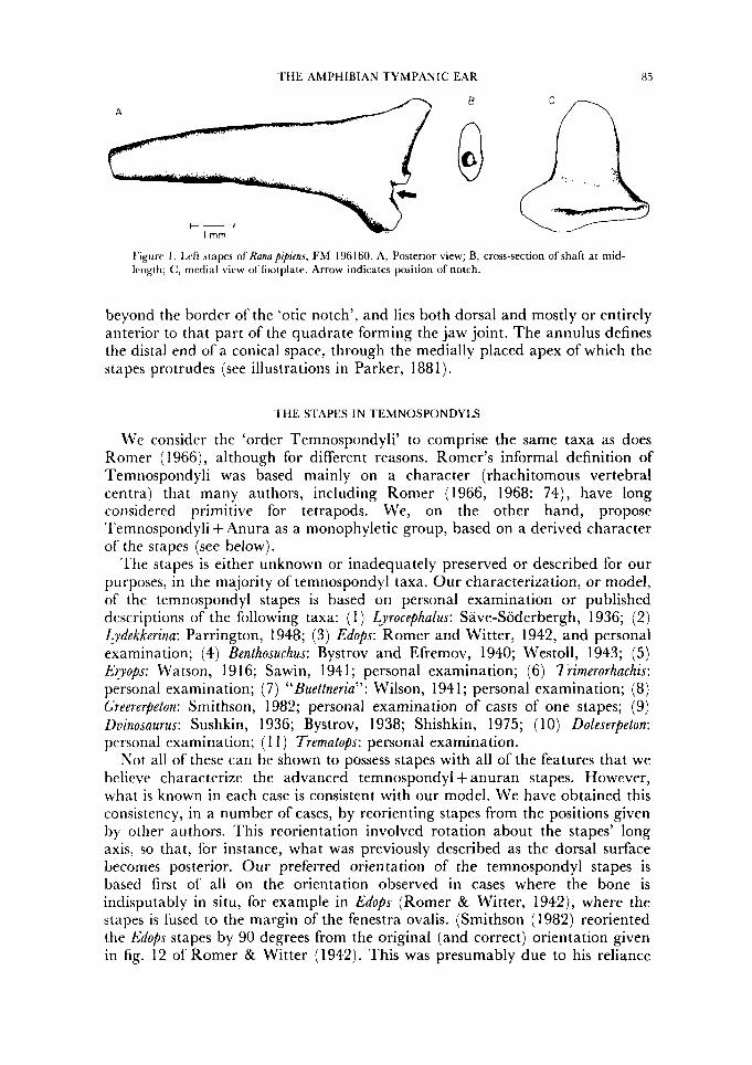
In adult frogs that have a middle-ear cavity closed externally by a tympanic membrane, the stapes nearly spans the distance between the otic capsule and the tympanum. Distally, the stapes is continued as a cartilaginous extrastapes inserted into the tympanum. The extrastapes in turn sometimes bears a process that attaches on or near the paroccipital process of the otic capsule. The bulk of the bony stapes (Fig. 1 ) consists of a distal shaft and proximal footplate, devoid of any ossified processes. The stapedial shaft is antero-posteriorly compressed, markedly so at mid-shaft. The footplate has a straight ventral edge that articulates with the otic capsule, and a cartilaginous continuation ( = pars interna plectri of Gaupp) that projects into a depression in the lateral wall of the otic capsule (most common) or into the fenestra ovalis. (The pars interna plectri is not shown in Fig. 1, in order to facilitate comparison with fossil stapes, but is well illustrated in Parker, 1881.) The posterior margin of the footplate has a complex articulation with the otic operculum, marked by a notch in the footplate circumference as seen in medial view (Fig. 1 ) . The operculum is a cartilaginous structure unique to frogs and salamanders, which arises ontogenetically from the otic capsule itself and lies within the fenestra ovalis (Kingsbury & Reed, 1909). Support for the tympanum is mainly provided by a cartilaginous tympanic annulus (Parker, 188 1 ; Sedra & Michael, 1959; Stadtmuller, 1936), although the dorsal margin of the tympanum may have a limited contact with the squamosal. The annulus (Fig. 4B) lies partly within an ‘otic notch’ formed mostly by the squamosal, with a variable contribution dorso- medially from the crista parotica (lateral end of the paroccipital process of the otic capsule). The tympanic annulus may extend anteriorly considerably
’L‘HE AMPHIBIAN TYMPANIC EAR H 5
A B
Figure 1. Left stapes of Hanapipiens, FM 196160. A, Posterior view; B, cross-section ofshaft at mid- Irngtli; C, medial vicw of footplate. Arrow indicates position of notch.
beyond the border of the ‘otic notch’, and lies both dorsal and mostly or entirely anterior to that part of the quadrate forming the jaw joint. The annulus defines the distal end of a conical space, through the medially placed apex of which the stapes protrudes (see illustrations in Parker, 188 1 ).
THE STAPES IN TEMNOSPONDYLS
We consider the ‘order Temnospondyli’ to comprise the same taxa as does Romer (1966), although for different reasons. Romer’s informal definition of Temnospondyli was based mainly on a character (rhachitomous vertebral centra) that many authors, including Romer (1966, 1968: 74), have long corisidered primitive for tetrapods. We, on the other hand, propose Temnospondyli + Anura as a monophyletic group, based on a derived character of the stapes (see below).
The stapes is either unknown or inadequately preserved or described for our purposes, in the majority of temnospondyl taxa. Our characterization, or model, of the temnospondyl stapes is based on personal examination or published descriptions of the following taxa: ( 1 ) Lyrocephalus: Save-Soderbergh, 1936; (2) Zgdekkerina: Parrington, 1948; (3) Edops: Romer and Witter, 1942, and personal examination; (4) Benlhosuchus: Bystrov and Efremov, 1940; Westoll, 1943; (5) Evops: Watson, 1 9 16; Sawin, 194 1 ; personal examination; (6) Trimerorhachis: personal examination; (7) “Buettneria”: Wilson, 194 1 ; personal examination; (8) Greererpelon: Smithson, 1982; personal examination of casts of one stapes; (9) Dvinosaurus: Sushkin, 1936; Bystrov, 1938; Shishkin, 1975; ( 10) Doleserpeton: personal examination; ( 1 1 ) Trematops: personal examination.
Not all of these can be shown to possess stapes with all of the features that we believe characterize the advanced temnospondyl + anuran stapes. However, what is known in each case is consistent with our model. We have obtained this consistency, in a number of cases, by reorienting stapes from the positions given by other authors. This reorientation involved rotation about the stapes’ long axis, so that, for instance, what was previously described as the dorsal surface becomes posterior. Our preferred orientation of the temnospondyl stapes is based first of all on the orientation observed in cases where the bone is indisputably in situ, for example in Edops (Romer & Witter, 1942), where the stapes is fused to the margin of the fenestra ovalis. (Smithson (1982) reoriented the Edops stapes by 90 degrees from the original (and correct) orientation given in fig. 12 of Romer & Witter (1942). This was presumably due to his reliance
86 J. R. BOLT AND R. E. LOMBARD
on their fig. 3 for stapes orientation. At first sight, that figure appears to show a dorsoventral orientation of the stapedial canal. But comparison with the Edops stapes, and with Romer and Witter’s fig. 12, shows that the apparent ‘canal’ in their fig. 3 likely represents only closely-spaced shading lines that were compressed into a ‘pseudocanal’ during the printing process.) We also relied on orientation of the stapedial canal (generally presumed to be for the stapedial artery). Whenever stapedial orientation can be reliably determined independently of the canal, the latter appears to run antero-posteriorly, and this is the orientation we have assumed in all cases. A stapedial canal is widespread among temnospondyls, and likely universal in Palaeozoic members of the group. It is surely primitive for dissorophoids, and appears in all taxa for which well- preserved stapes are known (contra Carroll, 1964: 204).
I t is not necessary to comment in detail on each reinterpretation. Reference to the cited descriptions, in combination with the foregoing rationale for determining orientation, will generally make clear what we have done and why. The question of stapedial processes, however, merits special comment because many authors have identified various processes (usually the dorsal process) on temnospondyl stapes. Despite the frequency of such reports, we find no evidence for an anatomical process in any temnospondyl except, possibly, Dvinosaurus. I n some cases the ‘process’ is simply one edge of a groove (for vena capitis lateralis?) in the dorsal edge of the shaft, or a nick in its distal extremity (possibly due to incomplete ossification). Several authors (for instance, Sushkin, 1927) identify as a process one end of the linear ventral articulating area of the footplate. If the stapes has been oriented incorrectly, this can easily be parlayed into a ‘dorsal process’. Tatarinov (1962) came to the same conclusion we have regarding the absence of processes on the temnospondyl stapes. He accepted the stapedial process in Dvinosaurus as the sole exception to the rule. (Curiously, Tatarinov cites the description of this stapes by Watson (1956), although it was actually described in more detail by other authors, not cited by Tatarinov.) The extent to which Dvinosaurus departs from our model of the ‘standard’ temnospondyl condition, if indeed it does, is difficult to determine. The three authors who have published accounts of this stapes present divergent accounts of its morphology, and we believe that all are mistaken in its orientation. Sushkin (1936) found at least two stapes ‘in situ’, but there is no mention in his or any other description of a sutural connection between the stapes and any other bone and the most recent description (Shishkin, 1975) also includes the most detailed figures of the Dvinosaurus stapes. From the orientation of the stapedial canal as depicted there, we believe that Shishkin’s ‘dorsal’, ‘ventral’ and ‘posterior’ views are respectively anterior, posterior and dorsal. Thus reoriented, the Dvinosaurus stapes appears to fit the standard pattern reasonably well, although the small ‘dorsal process’ still presents a problem. In the reoriented stapes Shiskin’s ‘dorsal process’ arises from the shaft just distal to the stapedial foramen, and is directed anteriorly. In this position it is an unlikely dorsal process. It may represent the attachment site of a stapedial muscle or ligament; but resolution of such problems must await re-examination of the original material.
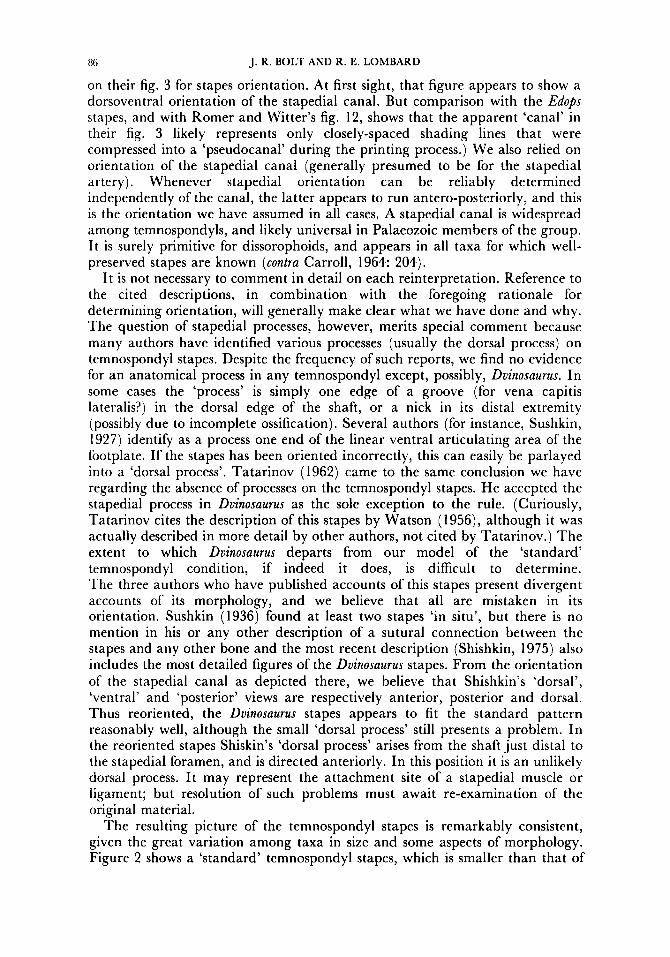
The resulting picture of the temnospondyl stapes is remarkably consistent, given the great variation among taxa in size and some aspects of morphology. Figure 2 shows a ‘standard’ temnospondyl stapes, which is smaller than that of
T H E AMPHIBIAN TYMPANIC EAR 87
A
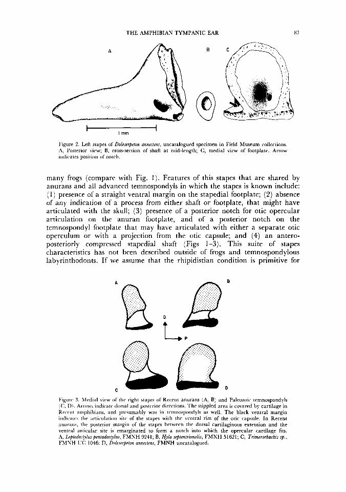
Figure 2. Left stapes of Doleserpeton annectens, uncatalogued specimen in Field Museum collections. A, Posterior view; B, cross-section of shaft at mid-length; C, medial view of footplate. Arrow indicates position of notch.
many frogs (compare with Fig. 1). Features of this stapes that are shared by anurans and all advanced temnospondyls in which the stapes is known include: ( 1 ) presence of a straight ventral margin on the stapedial footplate; (2) absence of any indication of a process from either shaft or footplate, that might have articulated with the skull; (3) presence of a posterior notch for otic opercular articulation on the anuran footplate, and of a posterior notch on the temnospondyl footplate that may have articulated with either a separate otic operculum or with a projection from the otic capsule; and (4) an antero- posteriorly compressed stapedial shaft (Figs 1-3). This suite of stapes characteristics has not been described outside of frogs and temnospondylous labyrinthodonts. If we assume that the rhipidistian condition is primitive for
B A
D
t
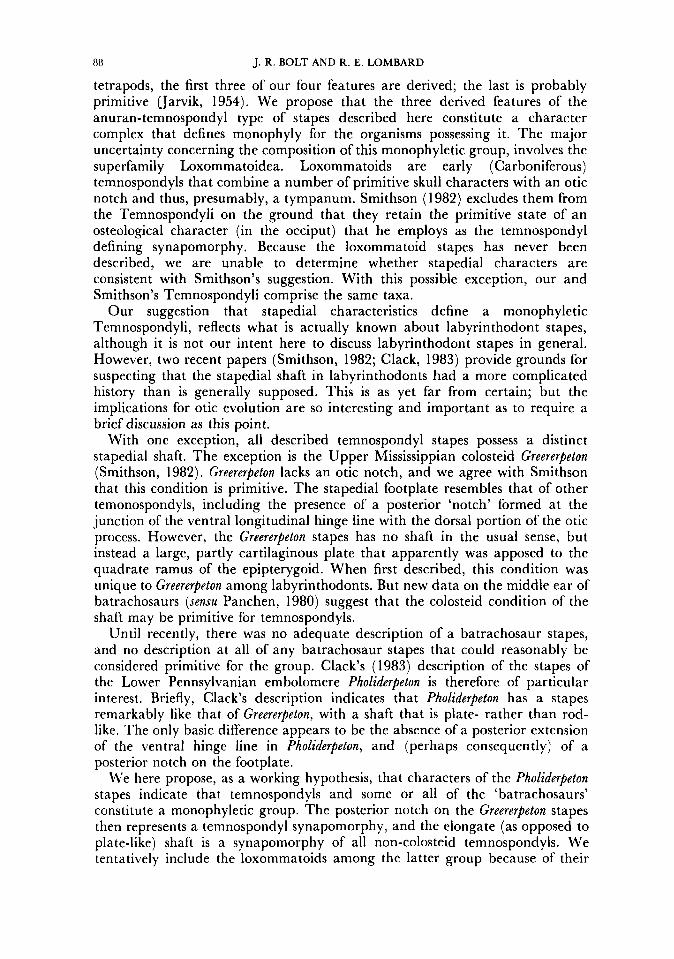
Figure 3. Medial view of the right stapes of Recent anurans (A, B) and Paleozoic temnospondyls (C, D). Arrows indicate dorsal and posterior directions. The stippled area is covered by cartilage in Rrrrnt amphibians, and presumably was in temnospondyls as well. The black ventral margin indicates the articulation site of the stapes with the ventral rim of the otic capsule. In Recent anurans, the posterior margin of the stapes between the dorsal cartilaginous extension and the ventral articular site is emarginated to form a notch into which the opercular cartilage fits. A, Leptodarplus pentadacplus, FMNH 9241; B, Hyla sepfentrionalis, FMNH 51621; C , Trimerorhachis sp., FMNH UC 1046; D, Doleserpeton annectens, FMNH uncatalogued.
138 J. R. BOLT AND R. E. LOMBARD
tetrapods, the first three of our four features are derived; the last is probably primitive Uarvik, 1954). We propose that the three derived features of the anuran-temnospondyl type of stapes described here constitute a character complex that defines monophyly for the organisms possessing it. The major uncertainty concerning the composition of this monophyletic group, involves the superfamily Loxomrnatoidea. Loxommatoids are early (Carboniferous) temnospondyls that combine a number of primitive skull characters with an otic notch and thus, presumably, a tympanum. Smithson (1982) excludes them from the Temnospondyli on the ground that they retain the primitive state of an osteological character (in the occiput) that he employs as the temnospondyl defining synapomorphy. Because the loxommatoid stapes has never been described, we are unable to determine whether stapedial characters are consistent with Smithson’s suggestion. With this possible exception, our and Smithson’s Temnospondyli comprise the same taxa.
Our suggestion that stapedial characteristics define a monophyletic Temnospondyli, reflects what is actually known about labyrinthodont stapes, although it is not our intent here to discuss labyrinthodont stapes in general. However, two recent papers (Smithson, 1982; Clack, 1983) provide grounds for suspecting that the stapedial shaft in labyrinthodonts had a more complicated history than is generally supposed. This is as yet far from certain; but the implications for otic evolution are so interesting and important as to require a brief discussion as this point.
With one exception, all described temnospondyl stapes possess a distinct stapedial shaft. The exception is the Upper Mississippian colosteid Greererpeton (Smithson, 1982). Greererpeton lacks an otic notch, and we agree with Smithson that this condition is primitive. The stapedial footplate resembles that of other temonospondyls, including the presence of a posterior ‘notch’ formed at the junction of the ventral longitudinal hinge line with the dorsal portion of the otic process. However, the Greererpeton stapes has no shaft in the usual sense, but instead a large, partly cartilaginous plate that apparently was apposed to the quadrate ramus of the epipterygoid. When first described, this condition was unique to Greererpeton among labyrinthodonts. But new data on the middle ear of batrachosaurs (sensu Panchen, 1980) suggest that the colosteid condition of the shaft may be primitive for temnospondyls.
Until recently, there was no adequate description of a batrachosaur stapes, and no description at all of any batrachosaur stapes that could reasonably be considered primitive for the group. Clack’s (1983) description of the stapes of the Lower Pennsylvanian embolomere Pholiderpeton is therefore of particular interest. Briefly, Clack’s description indicates that Pholiderpeton has a stapes remarkably like that of Greererpeton, with a shaft that is plate- rather than rod- like. The only basic difference appears to be the absence of a posterior extension of the ventral hinge line in Pholiderpelon, and (perhaps consequently) of a posterior notch on the footplate.
We here propose, as a working hypothesis, that characters of the Pholiderpeton stapes indicate that temnospondyls and some or all of the ‘batrachosaurs’ constitute a monophyletic group. The posterior notch on the Greererpeton stapes then represents a temnospondyl synapomorphy, and the elongate (as opposed to plate-like) shaft is a synapomorphy of all non-colosteid temnospondyls. We tentatively include the loxommatoids among the latter group because of their
THE AMPHIBIAN TYMPANIC EAR 89
apparently well-developed otic notch. These relationships are summarized in Fig. 7. Detailed exposition of the problems and implications of these proposals will be deferred to a later paper.
The foregoing list of three temnospondyl stapedial synapomorphies is conservatively short-additional characters could almost certainly be added- and deals strictly with morphological, rather than functional, aspects of the characters. Extensive discussion of these areas would be premature; but some comments are germane at this point:
(1) The stapedial footplate in temnospondyls appears to be larger than the fenestra ovalis, and does not lie within it. Rather, the footplate contacts the braincase (including the otic capsule) or dermal bones of the otic region (primarily the parasphenoid) external to the fenestra ovalis. We base this statement on our own observations, and on the numerous literature reports of contact between the footplate and extrafenestral bones (e.g. DeMar, 1968; Romer & Witter, 1942; Save-Soderbergh, 1936; Sawin, 1941). Tatarinov (1962) came to the same conclusion regarding the relatively large size of the temnospondyl footplate, and its relationship to the fenestra. He explicity-and appropriately- compared this to the situation in frogs, and contrasted it with that of amniotes. Tatarinov believed, however, that in both groups this constituted evidence for an immobile stapes, which was not and could not be actuated by the tympanum but instead served only as part of a bone-conduction hearing system. This notion is unequivocally disproved by manipulation of stapes in fresh specimens. The anuran stapes is actuated by the tympanum, and there is no reason to suppose that this was not the case in temnospondyls as well. In many anuran taxa, the stapedial footplate lies anterior to the fenestra ovalis, does not protrude into i t , and in fact is considerably larger than the fenestra (personal observation by R. E. L.). But rather than providing evidence of immobility, the ‘oversized’ anuran footplate reflects the nature of its contact with the periotic system. In frogs, the periotic cistern protrudes through the fenestra ovalis and comes into contact with the footplate in a more or less anatomically well-defined pericapsular space. This system is unique to frogs and salamanders among extant tetrapods (Lombard, 1977). There are enough indications of an ‘oversized’ footplate in temnospondyls, to suggest that this is part of their ‘standard’ stapedial morphology. If confirmed, this would suggest in turn a footplate-periotic system relationship like that of many frogs, and constitute an additional important synapomorphy between the two groups.
(2) Sawin (1941) notes that the ventral edge of the stapes is suturally attached to the parasphenoid in Eryops. This has been reported in other temnospondyls as well, and Save-Soderbergh (1936: 134) suggests that it is probably “the normal condition in the Labyrinthodontia, though overlooked in most forms’’. The contact between the base of the footplate and the otic capsule or parasphenoid has been variously reported as a suture, or complete fusion. Tatarinov (1962) holds the latter view, which is consistent with his belief that the stapes was immobile. We suspect that the junction is generally a sutural or some other type ofjoint, and seldom or never fused, for five reasons. First, a sutural junction has been reported in several cases. Secondly, fusion is often assumed in fossil material when a suture line cannot be demonstrated-but that is no proof of fusion. Thirdly, there are many documented instances of non- 6
90 J. R. BOLT AND R. E. LOMBARD
fusion, and in fact this is by far the commonest condition. Fourthly, the lower border of the ‘Buettneria’ footplate appears to be a definite joint surface, based on personal examination (cf. description of the same specimen by Wilson, 1941). Finally, a mobile stapes hinged at the bottom seems to agree well with the frog condition-not unexpected, given the synapomorphies of frogs and temnospondyls. Manipulation of fresh anuran specimens indicates not only that the stapes is mobile, as noted above, but also that the elongated ventral margin of the footplate is a hinge line, around which the stapes moves in an approximately vertical transverse plane (R. E. L., personal observation). Based on stapedial morphology, therefore, we propose that this area functions as a hinge in most or all anurans with a tympanic ear, and did so in ‘tympanic’ temnospondyls as well.
SUSPENSION OF THE TYMPANUM IN TEMNOSPONDYLS
We now turn to the problem of more precisely defining the phyletic relationship of anurans to temnospondyls. The stapes gives no guidance here, but the mode of suspension of the tympanic membrane does.
Temnospondyls (and labyrinthodonts in general) primitively lack both a tympanum and an otic notch (Lombard & Bolt, 1979; Smithson, 1982). As noted above only the colosteids, all of them Carboniferous, preserve this primitive condition. Some non-colosteid temnospondyls could be said to lack a notch, or at least to lack a clearly defined one as discussed by us in 1979. There is evidence that this condition is derived, although a detailed discussion is beyond the scope of this paper. The following discussion of tympanic membrane suspension excludes only the colosteids. I t thus applies to the great majority of known temnospondyl taxa.
In most temnospondyls the postero-lateral or postero-dorsal border of the skull is emarginated to form an otic notch, into which the distal end of the stapes projects in well preserved specimens. In most taxa, this notch is broadly open behind and hence, the tympanum would be attached to soft tissue posteriorly. Some groups, however, including (presumably as a result of convergence) capitosaurs and dissorophoids (Fig. 4A), partially or entirely close off the notch posteriorly with bone. Closure always involves a process of the tabular. In dissorophoids closure, whether partial or complete, always involves a dorsal process on the quadrate that is unique to this group. The dorsal quadrate process is best known in Doleserpeton and Cacops, which are represented by excellent material from the Lower Permian Fort Sill locality (see Bolt, 1969, 1977b). However, examination of specimens representing most of the named dissorophoid species confirms that, with differences in size and proportions, the following description applies to all species in which the otic region is well enough preserved to be described. A number of European dissorophoids have been described (most recently and completely by Boy, 1972, 1974, 1978), but middle-ear morphology of the European forms is still not well known, nor is that of Micropholis from the Lower Triassic of South Africa. For practical reasons, therefore, the (mostly Lower Permian) dissorophoids are treated here as though the group included only the North American representatives (families Dissorophidae and Trematopsidae) . These include the majority of described dissorophoid species, and all those in which the otic region is well enough known to be useful for our purposes.
T H E AMPHIBIAN TYMPANIC EAR
sq st
91
A
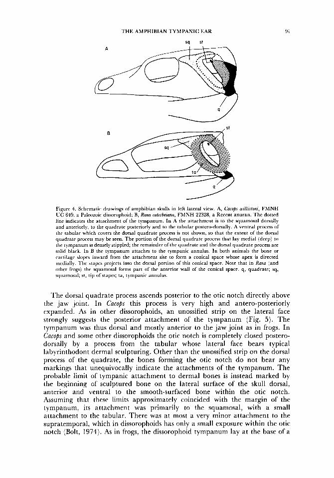
Figure 4. Schematic drawings of amphibian skulls in left lateral view. A, Camps willirfoni, FMNH UC 649, a Paleozoic dissorophoid; B, Rana catesbeiana, FMNH 22328, a Recent anuran. The dotted line indicates the attachment of the tympanum. In A the attachment is to the squamosal dorsally and anteriorly, to the quadrate posteriorly and to the tabular postero-dorsally. A ventral process of the tabular which covers the dorsal quadrate process is not shown, so that the extent of the dorsal quadrate process may be seen. The portion of the dorsal quadrate process that lay medial (deep) to the tympanum is densely stippled; the remainder of the quadrate and the dorsal quadrate process are solid black. In B the tympanum attaches to the tympanic annulus. In both animals the bone or rartilage slopes inward from the attachment site to form a conical space whose apex is directed medially. The stapes projects into the dorsal portion of this conical space. Note that in Rana (and other frogs) the squamosal forms part of the anterior wall of the conical space. q , quadrate; sq, squamosal; st, tip of stapes; ta, tympanic annulus.
The dorsal quadrate process ascends posterior to the otic notch directly above the jaw joint. In Cucops this process is very high and antero-posteriorly expanded. As in other dissorophoids, an unossified strip on the lateral face strongly suggests the posterior attachment of the tympanum (Fig. 5 ) . The tympanum was thus dorsal and mostly anterior to the jaw joint as in frogs. In C u c o ~ s and some other dissorophoids the otic notch is completely closed postero- dorsally by a process from the tabular whose lateral face bears typical labyrinthodont dermal sculpturing. Other than the unossified strip on the dorsal process of the quadrate, the bones forming the otic notch do not bear any markings that unequivocally indicate the attachments of the tympanum. The probable limit of tympanic attachment to dermal bones is instead marked by the beginning of sculptured bone on the lateral surface of the skull dorsal, anterior and ventral to the smooth-surfaced bone within the otic notch. Assuming that these limits approximately coincided with the margin of the tympanum, its attachment was primarily to the squamosal, with a small attachment to the tabular. There was at most a very minor attachment to the supratemporal, which in dissorophoids has only a small exposure within the otic notch (Bolt, 1974). As in frogs, the dissorophoid tympanum lay at the base of a
92 J. R. BOLT AND R. E. LOMBARD
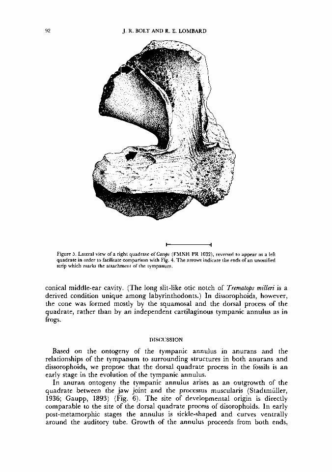
Figure 5. Lateral view of a right quadrate of Cncops (FMNH PR 1032), reversed to appear as a left quadrate in order to facilitate comparison with Fig. 4. The arrows indicate the ends of an unossified strip which marks the attachment of the tympanum.
conical middle-ear cavity. (The long slit-like otic notch of Trematops milleri is a derived condition unique among labyrinthodonts.) In dissorophoids, however, the cone was formed mostly by the squamosal and the dorsal process of the quadrate, rather than by an independent cartilaginous tympanic annulus as in frogs.
DISCUSSION
Based on the ontogeny of the tympanic annulus in anurans and the relationships of the tympanum to surrounding structures in both anurans and dissorophoids, we propose that the dorsal quadrate process in the fossils is an early stage in the evolution of the tympanic annulus.
In anuran ontogeny the tympanic annulus arises as an outgrowth of the quadrate between the jaw joint and the processus muscularis (Stadtmiiller, 1936; Gaupp, 1893) (Fig. 6). The site of developmental origin is directly comparable to the site of the dorsal quadrate process of disorophoids. In early post-metamorphic stages the annulus is sickle-shaped and curves ventrally around the auditory tube. Growth of the annulus proceeds from both ends,
T H E AMPHIBIAN TYMPANIC EAR
I t OD
B
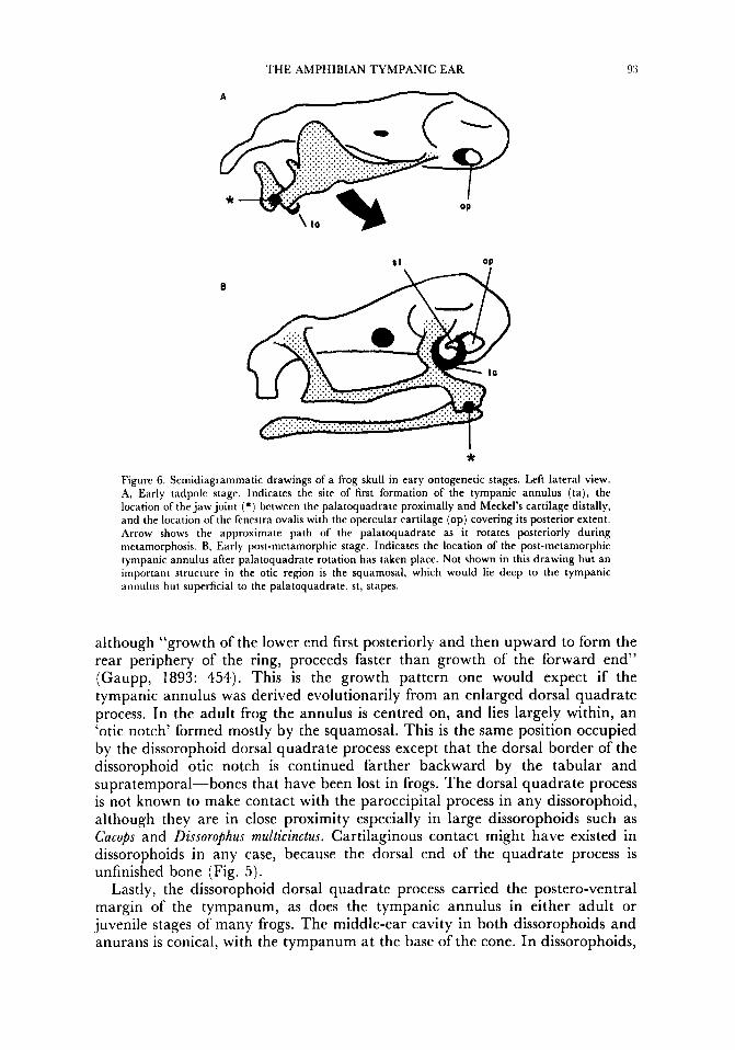
* Figure 6. Semidiagrammatic drawings of a frog skull in eary ontogenetic stages. Left lateral view. A, Early tadpole stage. Indicates the site of first formation of the tympanic annulus ( ta) , the location of the jaw joint ( * ) between the palatoquadrate proximally and Meckel’s cartilage distally, and the location of the fenestra ovalis with the opercular cartilage (op) covering its posterior extent. Arrow shows the approximate path of the palatoquadrate as it rotates posteriorly during metamorphosis. B, Early post-metamorphic stage. Indicates the location of the post-metamorphic tympanic annulus after palatoquadrate rotation has taken place. Not shown in this drawing but an important structure in the otic region is the squamosal, which would lie deep to the tympanic annulus but superficial to the palatoquadrate. st, stapes.
although “growth of the lower end first posteriorly and then upward to form the rear periphery of the ring, proceeds faster than growth of the forward end” (Gaupp, 1893: 454). This is the growth pattern one would expect if the tympanic annulus was derived evolutionarily from an enlarged dorsal quadrate process. In the adult frog the annulus is centred on, and lies largely within, an ‘otic notch’ formed mostly by the squamosal. This is the same position occupied by the dissorophoid dorsal quadrate process except that the dorsal border of the dissorophoid otic notch is continued farther backward by the tabular and supratemporal-bones that have been lost in frogs. The dorsal quadrate process is not known to make contact with the paroccipital process in any dissorophoid, although they are in close proximity especially in large dissorophoids such as Cacops and Dissorophus multicinctus. Cartilaginous contact might have existed in dissorophoids in any case, because the dorsal end of the quadrate process is unfinished bone (Fig. 5 ) .
Lastly, the dissorophoid dorsal quadrate process carried the postero-ventral margin of the tympanum, as does the tympanic annulus in either adult or juvenile stages of many frogs. The middle-ear cavity in both dissorophoids and anurans is conical, with the tympanum at the base of the cone. In dissorophoids,
9.1 J. R. BOLT AND R. E. LOMBARD
the cone is usually formed mainly by the squamosal, although occasionally (as in Cucops) with a major contribution from the dorsal quadrate process. The cone in anurans is formed mostly or entirely by the tympanic annulus itself, with a contribution from the squamosal (Fig. 4). If we accept the dissorophoid dorsal quadrate process as an intermediate step in the character transformation series:
no special process of quadrate for suspension of tympanum + process on quadrate for partial suspension of tympanum (dorsal quadrate process) +
complete quadrate suspension (tympanic annulus),
then anurans and dissorophoids might be hypothesized to be sister taxa. In fact, the sister group of anurans appears to be the family Dissorophidae.
Dissorophids alone among fossil amphibians are known to possess bicuspid, pedicellate teeth, a condition otherwise found only in lissamphibians. Such teeth appear to have been characteristic of juvenile stages in some or all of the dissorophids known to possess them, and at least some adults later developed non-pedicellate, single-cusped teeth (Bolt, 1977a, 1979). Presence of bicuspid, pedicellate teeth at some ontogenetic stage has been a principal basis for the suggestion that all three lissamphibian orders may be related to each other (Parsons & Williams, 1962, 1963) and to dissorophids (Bolt, 1969, 1979). The tympanic annulus, in combination with tooth morphology, provides the first evidence for a specific link between one of the lissamphibian orders and a fossil taxon-the Dissorophidae.
While the evidence for a close relationship of Anura and dissorophids thus seems strong, the position of the Urodela and Gymnophiona remains uncertain. If evidence for a monophyletic Lissamphibia comprising all three living orders is accepted (Parsons & Williams, 1962, 1963), then by implication the urodeles and caecilians have lost a once highly developed tympanic ear. If Lissamphibia on the other hand is not a monophyletic group, then its defining synapomorphies including bicuspid, pedicellate teeth represent either convergences, or primitive characters (i.e. synapomorphies a t a higher level). At present we know of no evidence that strongly favours either alternative.
The similarities in middle-ear morphology enumerated above suggest not only phylogenetic relationship, but functional continuity of a tympanic ear from temnospondyls to anurans; that is, a hearing mechanism derived within the early temnospondyls and preserved in anurans. The shape and attachments of the anuran-temnospondyl type of stapes, as well as manipulation in fresh anuran specimens, indicate that the ventral longitudinal junction with the otic capsule coincides with an axis of rotation, so that movement of the stapes is in an approximately vertical transverse plane. There is no process from the bony stapedial shaft to hinder this action, and the antero-posterior compression of the shaft is biomechanically consistent with this mode of movement. The stapes of anurans and temnospondyls thus moves and moved primarily as a ‘pump handle’ rather than primarily as a ‘piston’ as in amniotes.
A convincing otic operculum has not been described in any fossil amphibian. Eaton (1973) identified a remarkably large, ossified ‘operculum’ in a Middle Pennsylvanian dissorophoid. We suspect that this identification was incorrect, but unfortunately the unique specimen of Actiobates was not available for examination. Discovery in a fossil amphibian of a ‘proto-operculum’ either as an independent element or as a part of the otic capsule would constitute
THE AMPHIBIAN TYMPANIC EAR 95
important evidence of phylogenetic relationship and functional similarity. We have therefore mentioned the possible existence of a proto-operculum in temnospondyls even though the evidence is indirect (a posterior notch in the stapedial footplate). The exact nature of the functional continuity between temnospondyls and anurans is still poorly understood, perhaps largely because the anuran tympanic ear has traditionally been regarded as a primitive precursor of the amniote ear. There is increasing evidence that this is not so, and that anurans preserve a prior and separately evolved tympanic ear.
SUMMARY
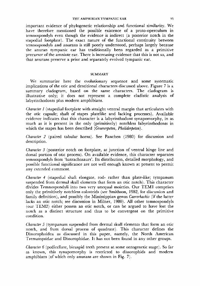
We summarize here the evolutionary sequence and some systematic implications of the otic and dentitional characters discussed above. Figure 7 is a summary cladogram, based on the same characters. The cladogram is illustrative only; i t does not represent a complete cladistic analysis of labyrinthodonts plus modern amphibians.
Character I (stapedial footplate with straight ventral margin that articulates with the otic capsule; shaft of stapes platelike and lacking processes). Available evidence indicates that this character is a labyrinthodont synapomorphy, in as much as it is present in the only (primitively) notchless labyrinthodonts in which the stapes has been described (Greererpeton, Pholiderpeton) .
Character 2 (paired tabular horns). See Panchen (1980) for discussion and description.
Character 3 (posterior notch on footplate, a t junction of ventral hinge line and dorsal portion of otic process). On available evidence, this character separates temnospondyls from ‘batrachosaurs’. Its distribution, detailed morphology, and possible functional significance are not well enough known at present to permit any extended comment.
Character 4 (stapedial shaft elongate, rod- rather than plate-like; tympanum suspended from dermal skull elements that form an otic notch). This character divides Temnospondyli into two very unequal moieties. Our T E M 1 comprises only the primitively notchless colosteids (see Smithson, 1982, for discussion and family definition), and possibly the Mississippian genus Caerorhachis (if the latter lacks an otic notch; see discussion in Milner, 1980). All other temnospondyls (our TEM2) either possess an otic notch, or can be argued to have lost the notch as a distinct structure and thus to be convergent on the primitive condition.
Character 5 (tympanum suspended from dermal skull elements that form an otic notch, and from dorsal process of quadrate). This character defines the Dissorophoidea as discussed in this paper, namely, the North American Trematopsidae and Dissorophidae. It has not been found in any other groups.
Character 6 (pedicellate, bicuspid teeth present at some ontogenetic stage). So far as known, this synapomorphy is restricted to dissorophids and modern amphibians (of which only anurans are shown in Fig. 7 ) .
96 J. R. BOLT AND R. E. LOMBARD
Dissorophoids
9AT TEM I TEM 2 TREM DIS ANU
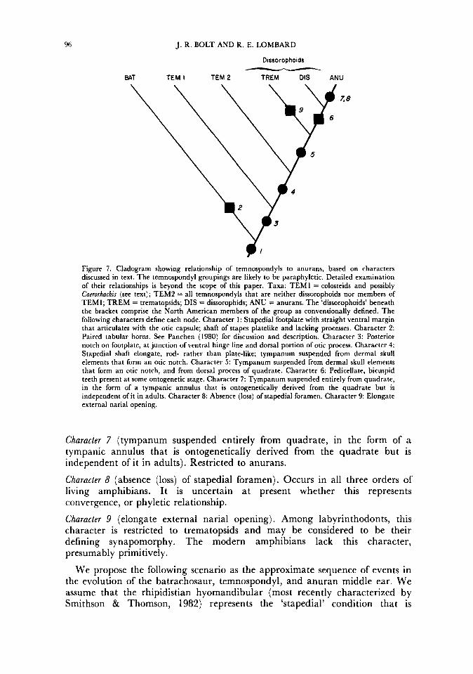
Figure 7. Cladogram showing relationship of temnospondyls to anurans, based on characters discussed in text. The temnospondyl groupings are likely to be paraphyletic. Detailed examination of their relationships is beyond the scope of this paper. Taxa: T E M I = colosteids and possibly Cacrorhachis (see text); TEM2 = all temnospondyls that are neither dissorophoids nor members of TEMI; TREM = trematopsids; DIS = dissorophids; ANU = anurans. The ‘dissorophoids’ beneath the bracket comprise the North American members of the group as conventionally defined. The following characters define each node. Character 1: Stapedial footplate with straight ventral margin that articulates with the otic capsule; shaft of stapes platelike and lacking processes. Character 2: Paired tabular horns. See Panchen (1980) for discussion and description. Character 3: Posterior notch on footplate, a t junction of ventral hinge line and dorsal portion of otic process. Character 4: Stapedial shaft elongate, rod- rather than plate-like; tympanum suspended from dermal skull elements that form an otic notch. Character 5 : Tympanum suspended from dermal skull elements that form an otic notch, and from dorsal process of quadrate. Character 6: Pedicellate, bicuspid teeth present at some ontogenetic stage. Character 7: Tympanum suspended entirely from quadrate, in the form of a tympanic annulus that is ontogenetically derived from the quadrate but is independent of it in adults. Character 8: Absence (loss) of stapedial foramen. Character 9: Elongate external narial opening.
Character 7 (tympanum suspended entirely from quadrate, in the form of a tympanic annulus that is ontogenetically derived from the quadrate but is independent of i t in adults). Restricted to anurans.
Character 8 (absence (loss) of stapedial foramen). Occurs in all three orders of living amphibians. It is uncertain at present whether this represents convergence, or phyletic relationship.
Character 9 (elongate external narial opening). Among labyrinthodonts, this character is restricted to trematopsids and may be considered to be their defining synapomorphy. The modern amphibians lack this character, presumably primitively.
We propose the following scenario as the approximate sequence of events in the evolution of the batrachosaur, temnospondyl, and anuran middle ear. We assume that the rhipidistian hyomandibular (most recently characterized by Smithson & Thomson, 1982) represents the ‘stapedial’ condition that is
THE AMPHIBIAN TYMPANIC EAR 97
primitive for all tetrapods. This hyomandibular articulated with the braincase via two heads (otic and dorsal). The hyomandibular swung anterio-posteriorly around an approximately vertical line passing through its two heads. The hyomandibular shaft was antero-posteriorly compressed, and elongate. There was no fenestra ovalis in rhipidistians:
( 1 ) The common ancestor of temnospondyls and (at least some) batrachosaurs possessed a stapes with a large footplate (otic process) that moved in ‘pump handle’ fashion around a ventral hinge line. There was a fenestra ovalis, but the stapedial footplate was considerably larger than the fenestra. The footplate presumably came into contact with the periotic system through the fenestra ovalis, but the periotic cistern did not protrude through the fenestra. The stapedial shaft was plate-like, and no ossified processes arose from either the shaft or footplate. The middle ear functioned as part of a relatively insensitive mechanism for perceiving airborne sound, but there was no tympanum, and no otic notch as defined either functionally or morphologically.
(2) The common ancestor of temnospondyls had a similar stapes, with the addition of a posterior extension of the ventral hinge line. One result of the latter was production of a posterior ‘notch’ in the footplate, or perhaps more accurately, of a salient from the otic capsule at this point. Such a salient might be moveable by muscles attaching at this point on the capsule; changes in muscle tonus could thereby affect the acoustic impedance of the ear.
(3 ) The ancestor of the ‘higher temnospondyls’ (our TEM2 in Fig. 7) had a tympanum, an otic notch, and a stapes with elongate shaft. The tympanum was suspended from dermal skull elements, with little or no contact with the quadrate.
(4) In the common ancestor of dissorophoids and anurans, the tympanum was suspended from dermal skull elements plus a dorsal process of the quadrate.
(5) Finally, in the anurans, the tympanum is suspended entirely or almost entirely from a derivative of the quadrate-the tympanic annulus. At some (unknown) point in this sequence, the periotic system has come to protrude through the fenestra ovalis, and makes contact with the stapedial footplate in an extracapsular space.
ACKNOWLEDGEMENTS
We thank James Hopson for his helpful advice and criticism, and Zbigniew Jastrzebski for drawing most of the figures. This work was supported in part by the Maurice Richardson Paleontological Fund at the Field Museum of Natural History, and by National Science Foundation grant DEB 8002619 to REL.
REFERENCES
BOLT. J. R., 1969. Lissamphibian origins: possible protolissamphibian from the Lower Permian of Oklahoma.
BOLT, J. R., 1974. A trematopsid skull from the Lower Permian, and analysis of some characters of the
BOLT, J . R., 1977a. Dissorophoid relationships and ontogeny, and the origin of the Lissamphibia. Journal of
Sctence, 166: 888-89 1 .
dissorophoid (Arnphibia: Labyrinthodontia) otic notch. Fieldiana: Geology, 30: 67-79.
Palaeontology, 51: 235-249.
98
BOLT, J. R., 1977b. Cacops (Amphibia: Labyrinthodontia) from the Fort Sill locality, Lower Permian of
BOLT, J. R., 1979. Amphibamus grandiceps as a juvenile dissorophid: evidence and implications. In M. H.
BOY, J. A,, 1972. Die Branchiosaurier (Amphibia) des saarpfalzischen Rotliegenden (Perm, SW-
BOY, J. A., 1974. Die Larven der rhachitomen Amphibien (Amphibia: Temnospondyli; Karbon-Trias).
BOY, J. A,, 1978. Die Tetrapodenfauna (Amphibia, Reptilia) des saarpfalzischen Rotliegenden (Unter-Perm;
BYSTROV, A. P., 1938. Dvinosaurus als neotenische Form der Stregocephalen. Acta (oologica, 19: 209-295. BYSTROV, A. P. & EFREMOV, I . A,, 1940. Benthosuchw swhkini Efr.-a labyrinthodont from the Eotriassic
of Sharzhenga River. Travaux de I'institut paleozoologique de I'Academie des sciences de I'URSS, 10: 1-152. CARROLL, R. L., 1964. Early evolution of the dissorophid amphibians. Bulletin of the Museum of Comparative
CLACK, J. A., 1983. The stapes of the Coal Measures embolomere Pholiderpeton scutigerum Huxley (Amphibia: Anthracosauria) and otic evolution in early tetrapods. <oological Journal of the Linnean Society, 79: 121-148.
DEMAR, R. E., 1968. The Permian labyrinthodont amphibian Dissorophus multicinctus, and adaptations and phylogeny of the family Dissorophidae. Journal of Paleontology, 42: 1210-1242.
EATON, T. H., JR., 1973. A Pennsylvanian dissorophid amphibian from Kansas. Occasional Papers of the Museum of Natural History, University of Kansas, 14: 1-8.
ESTES, R. & REIG, O., 1973. The early fossil record of frogs: a review of the evidence. In J. L. Vial (Ed.) , Evolutionary Biology of the Anurans: 1143. Columbia: University of Missouri Press.
CAUPP, E., 1893. Beitrage zur Morphologie des Schadels. I.: Primordial-Carnium und Kieferbogen von Rana fusca. In Schwalbe, G . (Ed.), Morphologische Arbeitm herausgegebcn von D r . G. Schwalbe, 2: 275481.
HECHT, M. K., 1962. A reevaluation of the early history of the frogs. Part I. $sternatic <ooIogy, 11: 39-44. JARVIK, E., 1954. O n the visceral skeleton in Eusthenopteron with a discussion of the parasphenoid and
KINGSBURY, B. F., & REED, H. D., 1909. The columella auris in Amphibia. Journal of Morphology, 20:
LOMBARD, R. E., 1977. Comparative morphology of the inner ear in salamanders (Caudata: Amphibia). In Hecht, M. K. & Szalay, F. S. (Eds), Contributions to Vertebrate Evolution, 2: 1-143. Basel: S. Karger.
LOMBARD, R. E., 1980. The structure of the amphibian auditory periphery: a unique experiment in terrestrial hearing. In A. N. Popper & R. R. Fay (Eds), Comparative Studies of Hearing in Vertebrates:
LOMBARD, R. E. & BOLT, J. R., 1979. Evolution of the tetrapcd ear: an analysis and reinterpretation. Biological Journal of the Linnean Society, 11: 19-76.
MILNER, A. R., 1980. The temnospondyl amphibian Dendrerpeton from the Upper Carboniferous of Ireland. Palaeontology, 23: 125- 14 1.
PANCHEN, A. L., 1980. The origin and relationships of the anthracosaur Amphibia from the Late Palaeozoic. In A. L. Panchen (Ed.), The Terrestrial Environment and the Origin of Land Vertebrates: 319-350. London and New York: Academic Press.
PARKER, W. K., 1881. O n the structure and development of the skull in the Batrachia. Philosophical Transactions of the R g a l Society of London ( B ) , 172: 1-266.
PARRINGTON, F. R., 1948. Labyrinthodonts from South Africa. Proceedings of the <oological Society of London, 118: 426-445.
PARSONS, T. S. & WILLIAMS, E. E., 1962. The teeth of Amphibia and their relation to amphibian phylogeny. Journal of Morphology, 110: 375-390.
PARSONS, T. S. & WILLIAMS, E. E., 1963. The relationships of the modern Amphibia: a re-examination. Quarterly Review of Biology, 38: 26-53.
ROMER, A. S., 1966. Vertebrate Paleontology. Chicago: University of Chicago Press. ROMER, A. S., 1968. Notes and Comments on Vertebrate Paleontology. Chicago: University of Chicago Press. ROMER, A. S. & WITTER, R. V., 1942. Edops, a primitive rhachitomous amphibian from the Texas red
beds. Journal of Geology, 50: 925-959. SAVE-SODERBERGH, G., 1936. O n the morphology of Triassic stegocephalians from Spitsbergen and the
interpretation of the endocranium in the Labyrinthodontia. Kungliga Svenska Vetenskapsakademiens Handlinear. 16 /21 : 1-181.
J. R. BOLT AND R. E. LOMBARD
Oklahoma. Fieldiana: Geology, 37: 61-73.
Nitecki (Ed.), Mazon Creek Fossils: 529-563. London and New York: Academic Press.
Deutschland). Abhandlungen des hessischen Landesamtes f u r Bodenforschung, 65: 1-137.
Palaontologisches <eitschrift, 48: 236-268.
SW-Deutschland). I . Branchiosaurus. Mainzer Geowissenschaftliche Mitteilungm, 7: 27-76.
< O O ~ O ~ V , 131: 161-250.
palatoquadrate in fishes. Kunglia Svenska Vetenskapsakademiens Handlingar, 5: 1-104.
449-628.
I2 1-138.
v , . I
SAWIN, H. J., 1941. The cranial anatomy of Eryops megacephalus. Bulletin of the Museum of Comparative <oology, xx- 407463. -. . . . . - - .
SEDRA. S. N. & MICHAEL. M. I.. 1959. The ontogenesis of the sound conducting apparatus of the I ..
Egyptian toad, BI& regularis'Reuss, with a review of t k apparatus in Salientia. Journal of Morphology, 104: 359-375.
SHISHKIN, M. A., 1975. Labyrinthodont middle ear and some problems of amniote evolution. Colloque
SMITHSON, T. R., 1982. The cranial morphology of Greererpeton burkemorani Romer (Amphibia: International C . N . R. S. 218: 337-348.
Temnospondyli). <oological Journal of the Linnean Society, 76: 2 5 9 0 .
T H E AMPHIBIAN TYMPANIC EAR 99
SMITHSON, T. R. & THOMSON, K. S., 1982. The hyomandibular of Eusthenopteron foordi Whiteaves (Pisces: Crossopterygii) and the early evolution of the tetrapod stapes. <oologicaI Journal of the Linnean Sociely, 74: 93-103.
SI'ADI'MULLER, F., 1936. Kranium und Visceralskelett der Stegocephalen und Amphibien. In Bolk, L. el al. (Eds), Handbuch der Vergleichenden Anatomie der Wirbeltiere, 4: 501498. Berlin and Vienna: Urban & Schwarzenberg.
SUSHKIN, P. P., 1927. O n the modifications of the mandibular and hyoid arches and their relations to the brain-rase in the early Tetrapoda. Pakaonlohgische Zeitschrifl, 8: 263-32 I .
SUSHKIN, P. P., 1936. Notes on the pre-Jurassic Tetrapods from USSR. 111. Dvinosaurccc Amalitzki, a pcrennibranrhiate stegocephalian from the Upper Permian of North Dvina. Trauaux de I'insfitut paleozoologique de I'Academie des sciences de I'URSS, 5: 43-9 I .
'IA'IARINOV, L. P., 1962. Functioning of the sound-conducting mechanisms of the labyrinthodonts. International Geology Reviews, 6: 1596-1603.
WA'I'SON, D. M. S., 1916. O n the structure of the brain-case in certain Lower Permian tetrapods. Bulletin of the American Museum OJ'Natural History, 35: 61 1-636.
WA'I'SON, D. M . S., 1956. The brachyopid labyrinthodonts. Bulletin of the British Museum (Natural History) Geology, 2: 3 15-392.
WES'I'OLL, T. S., 1943. The hyomandibular of Eusthenopteron and the tetrapod middle ear. Proceedings of the Royal Socieb of London ( B ) , 131: 393-414.
WILSON, J. A,, 1941, An interpretation of the skull of Buettneria, with special reference to the cartilages and soft parts. Contribulionr f r o m the Museum of Paleontology, Universily of Michigan, 6: 71-1 I I .