evidence that two pcl-like cyclins control cdk9 activity ... · evidence that two pcl-like cyclins...
TRANSCRIPT
Evidence that Two Pcl-Like Cyclins Control Cdk9 Activity during CellDifferentiation in Aspergillus nidulans Asexual Development
Claudia Kempf, Friederike Bathe, Reinhard Fischer
Karlsruhe Institute of Technology, Institute for Applied Biosciences, Department of Microbiology, Karlsruhe, Germany
Cyclin-dependent protein kinases (CDKs) are usually involved in cell cycle regulation. However, Cdk9 is an exception and pro-motes RNA synthesis through phosphorylation of the carboxy-terminal domain (CTD) of the largest subunit of RNA polymeraseII (RNAPII). The CTD is comprised of repeating heptapeptides, in which serine residues at positions 2, 5, and 7 are of crucialimportance. Ser5 phosphorylation causes transcription initiation and promoter escape. However, RNAPII pauses 20 to 50 bpdownstream from the transcription start site, until Cdk9 phosphorylates Ser2. This event relieves the checkpoint and promotesthe processivity of elongation. Here we present evidence that in the filamentous fungus Aspergillus nidulans, a Cdk9 homologue,PtkA, serves specific functions in conidiophore development. It was previously shown that PtkA interacts with two cyclins, PclAand the T cyclin PchA. Using yeast two-hybrid screens, we identified a third cyclin, PclB, and a kinase, PipABud32. Both proteinswere expressed in hyphae and in conidiophores, but interaction between each protein and PtkA was restricted to the conidio-phores. Deletion of pchA caused a severe growth defect, and deletion of pipA was lethal, suggesting basic functions in PtkA-de-pendent gene transcription. In contrast, deletion of pclB in combination with deletion of pclA essentially caused a block in sporeformation. We present evidence that the phosphorylation status of the CTD of RNA polymerase II in the conidiophore changesupon deletion of pclA or pclB. Our results suggest that tissue-specific modulation of Cdk9 activity by PclA and PclB is requiredfor proper differentiation.
Eukaryotic development and differentiation rely to a large ex-tent on differential gene expression, brought about by stage-
specific transcription factors, which control the expression of cer-tain sets of genes. In addition, epigenetic control is a commonprinciple for gene expression control. This phenomenon wasnamed the histone code, because posttranslational modificationsof histone proteins control the accessibility of certain genome re-gions for regulatory proteins (1). Another regulatory principle ismuch less well understood and named the carboxy-terminal do-main (CTD) code (2). This involves the cyclin-dependent kinase(CDK) Cdk9. Unlike other CDKs, Cdk9 does not regulate the cellcycle but, rather, promotes RNA synthesis (3). In higher eu-karyotes, it has been shown that members of the Cdk9 kinasefamily interact with T cyclins and that this complex is a subunit ofthe positive transcription elongation factor b (P-TEFb) (4, 5). Thiscomplex phosphorylates the CTD of the large subunit of RNApolymerase II (RNAPII) and thus promotes the elongation phaseduring transcription (6). The CTD contains 52 heptad repeats inhuman and 26 in Saccharomyces cerevisiae. Each heptad repeatcontains three serine residues, at positions 2, 5, and 7, and all threeare amenable to phosphorylation. First, Ser5 is phosphorylated byCDK7 (7). This leads to initiation of transcription but is not suf-ficient for transcription of the entire gene. RNAPII, rather, pauses20 to 50 bp downstream from the transcription initiation site untilCdk9 phosphorylates serine 2. With this event, the checkpoint canbe overcome and transcription continues. The pausing is probablyrequired for proper capping of the nascent mRNA. The highestSer2 phosphorylation levels are normally reached 600 to 1,000nucleotides downstream from the start site, and a dual-gradientmodel of increasing Ser2 and decreasing Ser5 phosphorylation hasbeen suggested (8, 9). The role of Ser7 is less clear, although itsphosphorylation has been shown in S. cerevisiae (7). Although thefunction of Cdk9 seems to be essential in regulation of global geneexpression in all cells, it was shown that expression is highest in
terminally differentiated cells, suggesting a role in specialized cel-lular functions. Not surprisingly, some diseases as well as cancerdevelopment were related to altered expression levels of Cdk9.Hence, human Cdk9 is regarded as a potential drug target in on-cology, virology, and cardiology (3). Furthermore, there is firstevidence for gene-specific modulation of gene expression by Cdk9kinase (10). Likewise, in Aspergillus nidulans we do have evidencethat a Cdk9 homologue specifically affects conidiophore develop-ment. A Cdk9 homologue (PtkA) was discovered in a targetedapproach to identify the interaction partners of a Pcl-like cyclin.This cyclin was named pclA and was the first Pcl-like cyclin de-scribed in A. nidulans (11). PclA is important for spore formationduring asexual development and interacts with the main cell cycleregulator nimX (cdk1) and may therefore have a second role in cellcycle regulation (12). The pclA-deletion mutant produced fewerspores, but vegetative growth was not affected. In contrast, dele-tion of the cdk9 kinase gene ptkA is lethal (13). These results sug-gest that PtkA conidiophore-specific functions depend on the in-teraction with PclA.
Here, we performed a screening for further PtkA-interactingproteins and identified another cyclin as well as a kinase. Theidentified cyclin shows sequence similarities to members of the Pclcyclin family of S. cerevisiae and was named PclB. The interactionis quite unusual, because the typical Cdk9-interacting proteins aremembers of the T-cyclin family. Deletion of pclB had only a minoreffect on asexual spore formation. However, in combination with
Received 5 July 2012 Accepted 22 October 2012
Published ahead of print 26 October 2012
Address correspondence to Reinhard Fischer, [email protected].
Copyright © 2013, American Society for Microbiology. All Rights Reserved.
doi:10.1128/EC.00181-12
January 2013 Volume 12 Number 1 Eukaryotic Cell p. 23–36 ec.asm.org 23
on April 19, 2019 by guest
http://ec.asm.org/
Dow
nloaded from

the deletion of pclA, asexual spore formation was almost com-pletely inhibited, despite normal vegetative growth. In addition tothe new cyclin, we identified a S. cerevisiae Bud32-related kinase,PipA (14, 15). An interaction between Bud32 and a Cdk9 kinasehas not been reported before in any other organism. In A. nidu-lans, PipA is essential for viability. Along with the already knowninteraction partners of PtkA, PclA and PipA are good future can-didates for deciphering the CTD code during A. nidulans devel-opment.
MATERIALS AND METHODSStrains, plasmids, and culture conditions. Supplemented minimal me-dium (MM) and complete medium (CM) for A. nidulans were prepared asdescribed by Hill and Käfer (16), and standard strain construction proce-dures are described by Hill and Käfer (16). A list of A. nidulans strains usedin this study is given in Table 1. Standard laboratory Escherichia coli strainTop 10 F= was used. Plasmids are listed in Table 2.
Molecular techniques. Standard DNA transformation procedureswere used for A. nidulans (19) and for E. coli and S. cerevisiae (20). ForPCR experiments, standard protocols were applied using a Biometra per-sonal cycler (Biometra, Göttingen, Germany) for the reaction cycles. DNAsequencing was done commercially (Eurofins MWG Operon, Ebersberg,Germany). Genomic DNA was extracted from A. nidulans with a DNeasyplant minikit (Qiagen, Hilden, Germany). DNA analyses (Southern hy-bridizations) were performed as described by Sambrook and Russell (20).
Deletion of pipA and pclB. pipA flanking regions were amplified byPCR using genomic DNA and the primers AN2513_P1_LB (5=-CGTCAGGCCATTGAGAACCAC-3=) and AN2513-P3-KO (5=-gaagagcattgtttgaggcgGAGAGAGCGGTGATAGTGAGG-3=, where lowercase nucleotides
indicate a linker) for the upstream region of pipA and AN2513-P4-KO(5=-atcagtgcctcctctcagacagGACAACGCCGCGCTCTTAGATG-3=) andBud32_P6_rev (5=-GCGTGCTGTGAACAGGCAATTAG-3=) for thedownstream region. The pyrG gene from plasmid pFNO3 (S. Osmani,OH) was amplified by PCR and used as the template together with pipAflanking regions for the fusion PCR. The deletion cassette was amplifiedwith the fusion PCR method (21) with the primers AN2513_P2nested(5=-CTAACCGTGCCATCATTCGTACC-3=) and Bud32_P5_rev (5=-GGCAGCCGTCAACATTCAAGTC-3=). The resulting PCR product wastransformed into pyrG89-auxotrophic A. nidulans strain TN02A3.
pclB flanking regions were amplified by PCR using genomic DNA andthe primers AN10741_P1_LB (5=-GGTGCCGAGAAATGTCGAGGAC-3=) and AN10741-P3-KO (5=-gaagagcattgtttgaggcgCAGGGCGGGATGAAGGATGAAG-3=) for the upstream region of pipA and AN10741-P4-KO(5=-atcagtgcctcctctcagacagGCAAGTGCAGAGGTTACGGATG-3=) andCyclin_P6_rev (5=-GCACGATATAATAGTGGCACCGC-3=) for thedownstream region. The pyrG gene from plasmid pFNO3 (S. Osmani,OH) was amplified by PCR and used as the template together with pipAflanking regions for the fusion PCR. The deletion cassette was amplifiedwith the fusion PCR method (21) with the primers AN10741_P2nested(5=-GCGATGGAGACGTCGATTTGACG-3=) and Cyclin_P5_rev (5=-CTTCGTCGGAGTAGTTCGCAGTG-3=). The resulting PCR product wastransformed into pyrG89-auxotrophic A. nidulans strain TN02A3.
Transformants were screened by PCR for the homologous integrationevent. Single integration of the construct was confirmed by Southern blot-ting. One diploid strain with the homologous integrated deletion cassetteand the remaining open reading frame (ORF) of pipA was selected andnamed SKC49. One pclB-deletion strain was selected from the transfor-mants and named SKC34.
TABLE 1 A. nidulans and S. cerevisiae strains used in this study
Strain Genotype Source or reference
FGSCA4 Glasgow wild type (veA�) Fungal Genetic Stock Centre, MOTN02A3 pyrG89 pyroA4 argB2 nkuA::argB veA1 Fungal Genetic Stock Centre, MOGR5 pyrG89 wA3 pyroA4 veA1 G. May, Houston, TXRMS011 pabaA1 yA2 �argB::trpC�B trpC801 veA1 17SSNI30 pyrG89 �argB::trpC�B pyroA4 �pclA::argB veA1 11SKC7 pclA-deletion strain (pyrG89 pyroA4 pclA::argB) crossed to RMS011 This studySKC9 TN02A3 transformed with pchA-deletion cassette This studySKC13 TN02A3 transformed with pKC24 [alcA(p)::GFP::pipA177::pyro] This studySKC19 GR5 transformed with pKC39 [alcA(p)::YFPN::pipA::pyro] and pFB13 [alcA(p)::YFPC::ptkA::pyr4] This studySKC20 GR5 transformed with pKC38 [alcA(p)::YFPN::pclB::pyro] and pFB13 [alcA(p)::YFPC::ptkA::pyr4] This studySKC27 TN02A3 transformed with pipA::GFP::pyrG-fusion construct This studySKC28 TN02A3 transformed with pclB::GFP::pyrG-fusion construct This studySKC34 TN02A3 transformed with pclB deletion cassette, pyrG89 pyroA4 argB2 �nkuA::argB veA1 �pclB::pyrG This studySKC40 SKC34 (pclB deletion) crossed to SKC9 (pchA deletion) This studySKC41 SKC34 (pclB deletion) crossed to SKC7 (pclA deletion) This studySKC44 GR5 transformed with pKC66 [alcA(p)::GFP::ptkA_D173A::pyro] This studySKC45 GR5 transformed with pKC71 [alcA(p)::GFP::ptkA_D155G::pyro] This studySKC46 GR5 transformed with pKC64 [alcA(p)::GFP::ptkA_PITALRE::pyro] This studySKC48 GR5 transformed with pKC87 [alcA(p)::3� HAa::CTD::pyr4] This studySKC49 TN02A3 transformed with pipA deletion cassette, pyrG89 pyroA4 argB2 �nkuA::argB veA1 �pipA::pyrG This studySKC50 GR5 transformed with pKC70 [alcA(p)::GFP::ptkA_GAGA::pyro] This studySKC51 GR5 transformed with pKC65 [alcA(p)::GFP::ptkA_K54Q::pyro] This studySKC72 GR5 transformed with pKC56 [alcA(p)::mRFP::pclB::pyr4] and pFB15 [alcA(p)::GFP::ptkA::pyro] This studySKC73 GR5 transformed with pKC55 [alcA(p)::mRFP::pipA::pyr4] and pFB15 [alcA(p)::GFPptkA::pyro] This studySKC77 SSNI17 transformed with pKC87 [alcA(p)::3� HA::CTD::pyr4] This studySKC79 SKC34 transformed with pKC87 [alcA(p)::3� HA::CTD::pyr4] and pNZ12 (pUMA::pyro) This studyAH109 MATa= trp1-901 leu2-3,112 ura3-52 his3-200 gal4� gal80� LYS2::GAL1UAS-GAL1TATA-His3 GAL2UAS-
GAL2TATA-Ade2 URA3::MEL1UAS-MEL1TATA-lacZ MEL1Clontech
Y187 MAT� ura3-52 his3-200 ade2-101 trp1-901 leu2-3,112, gal4� met� gal80� MEL1 URA3::GAL1UAS-GAL1TATA-lacZ MEL1
Clontech
a HA, hemagglutinin.
Kempf et al.
24 ec.asm.org Eukaryotic Cell
on April 19, 2019 by guest
http://ec.asm.org/
Dow
nloaded from
Tagging of proteins with mRFP and GFP under the control of thealcA or the natural promoter. To create an N-terminal monomeric redfluorescent protein (mRFP) fusion construct of PipA, the full-length pipA(starting from ATG) was amplified from genomic DNA, using the primersBud32_Asc_for (5=-ggcataggcgcgccaATGCCACCAACGAACCGC-3=)and Bud32_Pac_rev (5=-gctacgttaattaaCTACCCAATCATGCTCCTCTTCC-3=) and cloned via AscI/PacI (the restriction sites are underlined) intothe corresponding sites of pCMB17apx, yielding pKC55. Likewise, thetruncated pipA ORF was amplified with the primers Bud32_Asc_for andBud177_Bam_rev (5=-ggcataggatccCTACCCATGT ATAACACCCGC-3=) and cloned into pCMB17apx, yielding pKC24.
To create an mRFP fusion construct of PclB, the full-length pclB (start-ing from ATG) was amplified from genomic DNA, using the primers P7Cyclin_Asc_for (5=-ggcataggcgcgccaATGAACAGTGGAGTCGGC-3=)and P8 Cyclin_Pac_rev (5=-gctacgttaattaaCTACCCAATCATGCTCCTCTTCC-3=) and cloned via AscI/PacI (the restriction sites are underlined)into the corresponding sites of pCMB17apx, yielding pKC56.
The N-terminal green fluorescent protein (GFP) fusion construct ofPtkA in pCMB17apx used was described previously (13).
To create a C-terminal GFP fusion construct of PipA under the controlof the natural promoter, pipA flanking regions were amplified by PCRusing genomic DNA and the primers Bud32_P1_for (5=-GCATGGAATGATGTCCATGGCG-3=) and Bud32-P3-rev (5=-ctccagcgcctgcaccagctccCCCAATCATGCTCCTCTTCCTTC-3=) for the upstream region of pipAand Bud32-P4-for (5=-atcagtgcctcctctcagacagTAGTTACCTATATAACAGGTATCGAATGG-3=) and Bud32_P6_rev (5=-GCGTGCTGTGAACAGGCAATTAG-3=) for the downstream region. The gfp-pyrG cassette fromplasmid pFNO3 (S. Osmani, OH) was amplified by PCR and used asthe template together with pipA flanking regions for the fusion PCR. Thegfp-pyrG cassette was amplified with the fusion PCR method (21) with theprimers Bud32_P2_for (5=-CTTCCCCTCCTCTCTTCTTCTC-3=) and
Bud32_P5_rev (5=-GGCAGCCGTCAACATTCAAGTC-3=). The result-ing PCR product was transformed into pyrG89-auxotrophic A. nidulansstrain TN02A3.
To create a C-terminal GFP fusion construct of PclB under the controlof the natural promoter, pclB flanking regions were amplified by PCRusing genomic DNA and the primers Cyclin_P1_for (5=-GGATGGCTTGCCTAACAGCTCTTG-3=) and Cyclin_P3_rev (5=-ctccagcgctgcaccagctccCGTCGGGTGTTCCATGTACCAATG-3=) for the upstream region ofpclB and Cyclin-P4-for (5=-atcagtgcctcctctcagacagTGACTCTGGGTATGGTGACGTG-3=) and Cyclin-P6_rev (5=-GCACGATATAATAGTGGCACCGC-3=) for the downstream region. The gfp-pyrG cassette from plas-mid pFNO3 (S. Osmani, OH) was amplified by PCR and used as thetemplate together with pclB flanking regions for the fusion PCR. Thegfp-pyrG cassette was amplified with the fusion PCR method (21) with theprimers Cyclin_P2_for (5=-GTCCGACCATGCCCGTTTCATC-3=) andCyclin_P5_rev (5=-CTTCGTCGGAGTAGTTCGCAGTG-3=). The result-ing PCR product was transformed into pyrG89-auxotrophic A. nidulansstrain TN02A3.
Transformants were screened by PCR for the homologous integrationevent. Single integration of the construct was confirmed by Southern blot-ting. One pipA-gfp strain (SKC27) and one pclB-gfp strain (SKC28) wereselected from the transformants.
Tagging of proteins with YFPN/YFPC for BiFC analyses. For bimo-lecular fluorescence complementation (BiFC) analyses, the GFP inpCMB17apx was replaced with the N-terminal half of yellow fluorescentprotein (YFPN) or the C-terminal half of yellow fluorescent protein(YFPC), yielding pDV7 and pDV8, respectively, as described earlier (22).The YFPC fusion construct of PtkA in pDV8, yielding pFB13, was de-scribed previously (13). The YFPN fusion construct of PipA was generatedby amplification of the complete ORF from genomic DNA with the prim-ers Bud32_Asc_for and Bud32_Bam_rev (5=-ggcataggatccCTACCCAAT
TABLE 2 Plasmids used in this study
Plasmid Construction Source or reference
pMCB17apx alcA(p)::GFP, for N-terminal fusion of GFP to proteins of interest; contains N. crassa pyr4 V. Efimov (Piscataway, NJ)pGBKT7 Yeast two-hybrid bait vector, Gal4-BD ClontechpGADT7 Yeast two-hybrid prey vector, Gal4-AD ClontechpGBT9 Yeast two-hybrid bait vector, Gal4-BD ClontechpDV7 GFP replaced N-terminal half of YFP in pCMB17apx 18pDV8 GFP replaced C-terminal half of YFP in pCMB17apx 18pFNO3 ga5::GFP::pyrG Kanr Ampr Fungal Genetic Stock
Centre, MOpFB13 Full-length ptkA in pDV8 13pFB15 Full-length ptkA in pCMB17apx 13pKC3 Full-length ptkA in pGBT9 This studypKC5 Truncated ptkA_1/330 in pGBKT7 This studypKC6 Truncated ptkA_24/170 in pGBKT7 This studypKC8 Full-length pclA in pGADT7 This studypKC10 Full-length pchA in pGADT7 This studypKC19 Full-length pclB in pGADT7 This studypKC22 Full-length pipA in pGADT7 This studypKC24 Truncated pipA178 in pMCB17apx This studypKC38 Full-length pclB in pDV7 This studypKC39 Full-length pipA in pDV7 This studypKC55 Full-length pclB in pCMB17apx, with mRFP instead of GFP This studypKC56 Full-length pipA in pCMB17apx, with mRFP instead of GFP This studypKC64 Full-length PtkA with deletion of PITALRE in pCMB17apx, with pyroA instead of pyr4 This studypKC65 Full-length PtkA with point mutation K54Q in pCMB17apx, with pyroA instead of pyr4 This studypKC66 Full-length PtkA with point mutation D173A in pCMB17apx, with pyroA instead of pyr4 This studypKC70 Full-length PtkA with point mutation G32AG34A in pCMB17apx, with pyroA instead of
pyr4This study
pKC71 Full-length PtkA with point mutation D155G in pCMB17apx, with pyroA instead of pyr4 This studypKC87 CTD (1574–1746 aa) of the large subunit of RNA polymerase II in pSM14 This study
Cdk9 Kinase in A. nidulans
January 2013 Volume 12 Number 1 ec.asm.org 25
on April 19, 2019 by guest
http://ec.asm.org/
Dow
nloaded from
CATGCTCCTC-3=). The resulting PCR product was then cloned via AscI/BamHI into the corresponding sites of pDV7, yielding pKC39. Using thesame strategy, the YFPN fusion of PclB was generated, using the primersCyclin_Asc_for and Cyclin_Bam_rev (5=-ggcataggatccTCACGTCGGGTGTTCC-3=), yielding pKC38.
To create YFPN fusion constructs of PclB and PipA under the controlof the natural promoter, the same primers were used as described beforein tagging of proteins with gfp. A yfpN-pyro cassette was amplified by PCRand used as the template together with pclB or pipA flanking regions forthe fusion PCR.
To create a YFPC fusion construct of PtkA under the control of thenatural promoter, ptkA flanking regions were amplified by PCR usinggenomic DNA and the primers PtkA_P4 (5=-GAAGGACGTTCTTGGCTGACC-3=) and PtkA_P6 (5=-ctccagcgcctgcaccagctccCCGGCGATACGGACCCCTG-3=) for the upstream region of ptkA and PtkA-P5 (5=-atcagtgcctcctctcagacagTGAAGCTACCGTCTACCATAAAAC-3=) and PtkA-P7(5=-TGTGAAATCATCGCTCTTGCTC-3=) for the downstream region. AyfpC-pyrG-cassette was amplified by PCR and used as the templatetogether with ptkA flanking regions for the fusion PCR. The yfpC-pyrGcassette was amplified with the fusion PCR method (21) with the primersptkA_F1_neuer (5=-AAGGAAAGCCGATTCTTGCTGG-3=) and PtkA_KO_R1 (5=-GCGCGCCAAGTTTCGACCC-3=). The resulting PCR prod-uct of ptkA-yfpC-pyrG was transformed into A. nidulans strain TN02A3together with pipA-yfpN-pyro or pclB-yfpN-pyro.
Transformants were screened by PCR for the homologous integrationevent. One strain with ptkA-yfpC-pyrG and pipA-yfpN-pyro (SKC29) andone strain with ptkA-yfpC-pyrG and pipA-yfpN-pyro (SKC30) were se-lected from the transformants.
Insertion of mutations by site-directed mutagenesis. To introducemutations in an N-terminal GFP fusion construct of PtkA, a QuikChangeXL mutagenesis kit (Stratagene, Heidelberg, Germany) was used. To gen-erate the K54Q point mutation, the following primers were used: ptkA-KQ-F1 (5=-GATGGCTCCATCGTCGCGCTGCAAAAGATCCTCATGCATAATG-3=) and ptkA-KQ-R1 (5=-CATTATGCATGAGGATCTTTTGCAGCGCGACGATGGAGCCATC-3=) (the mutation sites are underlined),yielding pKC65. To delete the PITALRE domain, the primers PtkA_PITALRE_f (5=-PHO-CTACTGAAAATGTTGTCCCACACC-3=, wherePHO indicates phosphorylation of the primer) and PtkA_PITALRE_r(5=-PHO-CTTTGACGATCAGATAGCTGCC-3=) were used, yieldingpKC64. To generate the G32AG34A point mutations, the primersPtkA_GAGA_f (5=-GCAAACTGGCGGAGGCCACCTTTGG-3=) andPtkA_GAGA_r (5=-CCAAAGGTGGCCTCCGCCAGTTTGC-3=) wereused, yielding pKC70. For the D155G point mutation, the primersPtkA_D155G_f (5=-GTATCCTACACCGCGGCATGAAAGGCTAG-3=)and PtkA_D155G_r (5=-CTAGCCTTTCATGCCGCGGTGTAGGATAC-3=) were used, and for the mutation D173A, the primers PtkA_D173A_f(5=-CTGCAGATTGCCGCCTTCGGACTGGC-3=) and PtkA_D173A_r(5=-GCCAGTCCGAAGGCGGCAATCTGCAG-3=) were used, yieldingpKC71 and pKC66, respectively.
All plasmids were verified by sequencing and then transformed intostrain GR5.
Yeast two-hybrid analysis. The yeast two-hybrid screen was per-formed using a Matchmaker library construction and screening system(BD Clontech). For bait generation, the primers pGB-Y2H-F1 (5=-GGCATATGGGCATAGCGTCACTCGAACGG-3=) and pGB-Y2H-R1 (5=-GGATCCTCACCGGCGATACGGACCCCT-3=) were used to amplify afull-length ptkA cDNA fragment, the primers ptkA24/170_Nde (5=-GGCTACCATATGGAATTCGAGTTCTTAGGCAAACTGGG-3=) and ptkA24/170_Bam (5=-GGCTACGGATCCTCACTGCAGAATACCTTGGTTGCTTATAAG-3=) were used to amplify the truncated ptkA fragment fromresidues 24 to 170 (ptkA_24/170), and the primers ptkA_Bam_for (5=-GGATCCATGGGCATAGCGTCACTCG-3=) and ptkA995_Sal_rev (5=-GGGTCGACTCATGGCGGTGTCGAGAAATAA-3=) were used to amplifythe truncated ptkA_1/330. The full-length ptkA and the truncated ptkA_1/330 were cloned in the pGBT9 vector, and the truncated ptkA_24/170 was
cloned in the vector pGBKT7, which contains both the GAL4 DNA bind-ing domain and the TRP1 marker (BD Clontech). cDNA was isolatedfrom an A. nidulans wild-type strain, amplified, and cloned in thepGADT7-Rec vector, under the manufacturer’s instructions. ThepGADT7-Rec vector contains the GAL4 DNA-AD and the LEU2 marker(BD Clontech). pGBKT7- and pGBT9-associated plasmids were trans-formed in S. cerevisiae AH109 (mating type MATa), and pGADT7-asso-ciated plasmids were transformed into S. cerevisiae Y187 (mating typeMAT�).
For yeast two-hybrid analysis, a pipA cDNA fragment was amplifiedwith primers Bud32_Nde_for (5=-ggcatacatATGCCACCAACGAACCGC-3=) and Bud32_Bam_rev (5=-ggcataggatccCTACCCAATCATGCTCCTC-3=) and a pclB cDNA fragment was amplified with the primers Cyc-lin_Nde_for (5=-GGCATACATATGAACAGTGGAGTCGGCG-3=) andCyclin_Bam_rev (5=-GGCATAGGATCCTCACGTCGGGTGTTCC-3=),and the amplicons were cloned in pGADT7-Rec.
Light and fluorescence microscopy. For live-cell imaging of germ-lings and young hyphae, cells were grown on coverslips in 0.5 ml MM plus2% glycerol (derepression of the alcA promoter), MM plus 2% glucose(repression of the alcA promoter), or MM plus 2% threonine (activationof the alcA promoter). Cells were incubated at room temperature over-night. For pictures of conidiophores, spores were inoculated on glassslides coated with MM plus 2% glucose and 0.8% agarose and grown at37°C for 1 to 2 days. Images were captured at room temperature using anAxiophot microscope (Zeiss, Jena, Germany). Images were collected andanalyzed with an AxioVision system (Zeiss).
Gene expression analyses. Transferring development-competentvegetative hyphae from liquid medium to agar plates, thus inducing de-velopment by air exposure, synchronized asexual development of wild-type A. nidulans cells. At different time points, RNA was isolated. For that,the mycelium was harvested, dried, frozen in liquid N2, and ground to apowder. Total RNA was isolated by using TRIzol reagent (Invitrogen, NVLeek, Netherlands) according to the manufacturer’s instructions. For theexpression analyses, quantitative real-time PCR was performed by using100 ng of the isolated RNA for each reaction and an iScript one-stepreverse transcription-PCR (RT-PCR) kit with SYBR green and the iCyclerapparatus of Bio-Rad. For analysis of PclB, the primers Cyclin_for (5=-GGAGAGGGTGAACCTAGCCTTG-3=) and Cyclin_rev (5=-GAAGTTCCATGGCGTTCTCCG-3=) were used, and for analysis of PipA, the primersBud_for (5=-CCGCGTCCCTTCCTTCTTACTC-3=) and Bud_rev (5=-GGGTTTTGACGGGCGGATTTTG-3=) were used. As the housekeepinggene, �-tubulin (benA) was used.
Protein extracts and Western blotting. To prepare protein extractsfrom vegetative hyphae, A. nidulans strains were incubated in liquid MMfor 24 h at 37°C. For the isolation of proteins form asexual structures,hyphae were transferred to MM plates with cellophane and incubated foran additional 24 h at 37°C to induce asexual development. To induce thealcA promoter, the medium was supplemented with 0.2% glucose and 2%threonine. Mycelium was harvested by filtration through a Miracloth fil-ter (Calbiochem, Heidelberg, Germany), dried, and immediately groundin liquid nitrogen. Afterwards, the mycelial powder was resuspended inprotein extraction buffer (50 mM Tris-HCl [pH 8], 0.1% Triton X-100,250 mM NaCl) containing 1 mM protease inhibitor cocktail recom-mended for fungal and yeast extracts (Sigma-Aldrich, Munich, Germany)and 1 mM phosphatase inhibitor cocktail 3 (Sigma-Aldrich, Munich, Ger-many). Samples were rotated for 20 min in a head-over-tail incubator.Cell debris was pelleted by centrifugation (5403-R centrifuge; Eppendorf,Hamburg, Germany) at 13,000 rpm and 4°C for 12 min. The supernatantwas used for Western blotting. Equal amounts of protein extracts (75 �g)were loaded on 8% sodium dodecyl sulfate-polyacrylamide gels for detec-tion of GFP-fusion proteins or on 10% SDS-gels for detection of the CTD.Proteins were blotted onto polyvinylidene difluoride membranes (What-man; GE Healthcare, Little Chalfont, United Kingdom), and GFP-taggedproteins were analyzed with a monoclonal anti-GFP antibody (dilution,1:8,000; product G1544; Sigma-Aldrich, Munich, Germany). The
Kempf et al.
26 ec.asm.org Eukaryotic Cell
on April 19, 2019 by guest
http://ec.asm.org/
Dow
nloaded from
nontagged CTD was detected with the monoclonal antibody 8WG16 (di-lution, 1:1,000; Covance, Princeton, NJ). Two monoclonal anti-phospho-CTD antibodies were applied to quantify CTD specifically phosphory-lated at Ser2 (dilution, 1:1,000; antibody H5; Covance, Princeton, NJ) andCTD specifically phosphorylated at Ser5 (dilution, 1:1,000; antibody H14;Covance, Princeton, NJ).
RESULTSIdentification of novel interaction partners of the Cdk9 kinasePtkA. The cyclin-dependent kinase PtkA has been shown to inter-act with two cyclins, PclA (11) and PchA (13). Whereas pchAdeletion caused a severe growth defect, pclA deletion had no effecton vegetative growth but had an effect only on sporulation. Be-cause Cdk9 kinases are involved in several cellular pathways inhigher eukaryotes, we anticipated that PtkA specificities might bedetermined by even more interaction partners in A. nidulans. Toidentify such new interacting proteins, we screened a cDNA li-brary by using the Matchmaker library construction and screen-ing kit (Clontech). Full-length PtkA (PtkA_1/545) was cloned intothe standard yeast two-hybrid pGBKT7 vector, in which the con-structs are strongly expressed. Corresponding yeast transfor-mants, unfortunately, did not grow well and were unable to mate.This suggested an inhibitory function of the PtkA kinase. To avoidthe toxic effect, we used instead pGBT9 as the vector, in whichgenes are expressed at lower levels. In addition, we constructed atruncated version of PtkA (PtkA_24/170) lacking a short piece ofthe N terminus and the kinase domain, located in the C terminus.This construct could be screened in the overexpression vectorpGBKT7. In the screening experiment with PtkA_1/545, 120 yeastcolonies grew on the selection medium (SD-Leu-Trp-His, whereSD means synthetic dropout), from which 100 yeast colonies wereable to grow again on the same medium. The screening withPtkA_24/170 led to the isolation of about 450 yeast colonies, two-
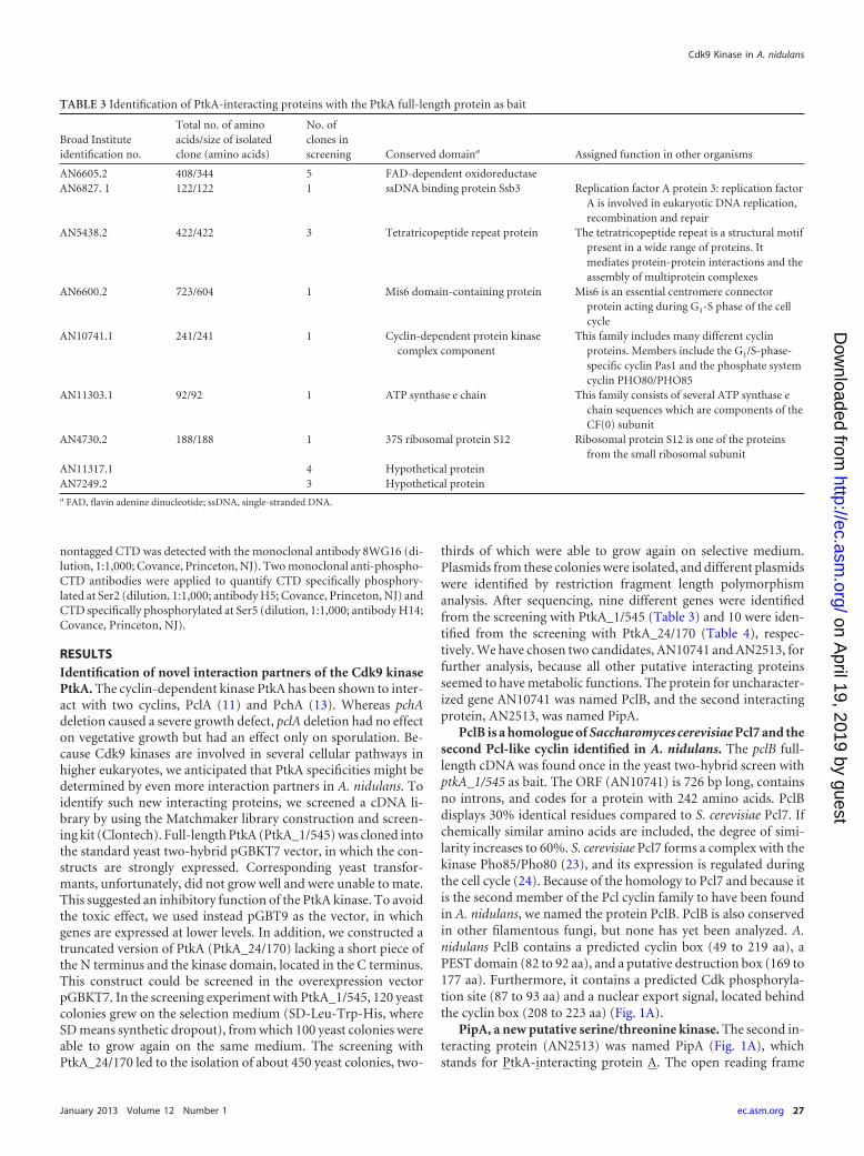
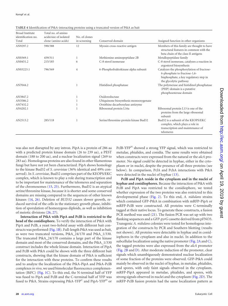
thirds of which were able to grow again on selective medium.Plasmids from these colonies were isolated, and different plasmidswere identified by restriction fragment length polymorphismanalysis. After sequencing, nine different genes were identifiedfrom the screening with PtkA_1/545 (Table 3) and 10 were iden-tified from the screening with PtkA_24/170 (Table 4), respec-tively. We have chosen two candidates, AN10741 and AN2513, forfurther analysis, because all other putative interacting proteinsseemed to have metabolic functions. The protein for uncharacter-ized gene AN10741 was named PclB, and the second interactingprotein, AN2513, was named PipA.
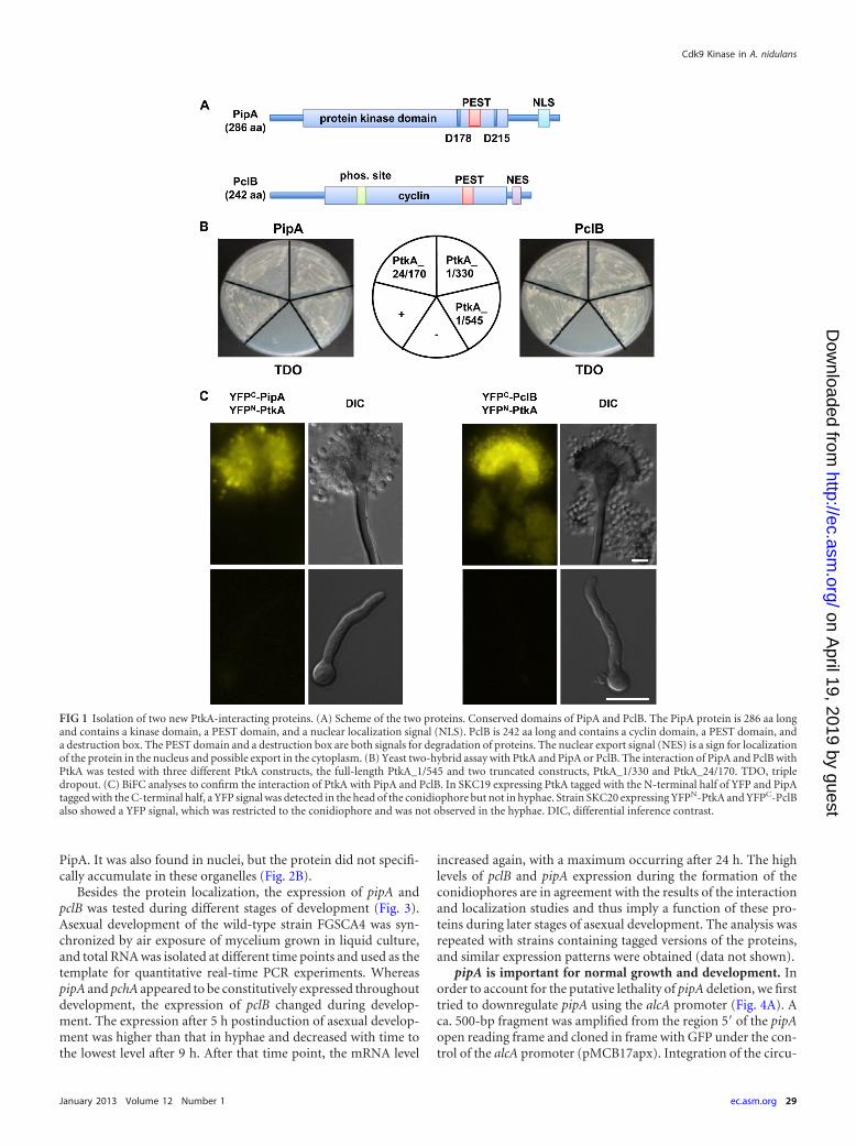
PclB is a homologue of Saccharomyces cerevisiae Pcl7 and thesecond Pcl-like cyclin identified in A. nidulans. The pclB full-length cDNA was found once in the yeast two-hybrid screen withptkA_1/545 as bait. The ORF (AN10741) is 726 bp long, containsno introns, and codes for a protein with 242 amino acids. PclBdisplays 30% identical residues compared to S. cerevisiae Pcl7. Ifchemically similar amino acids are included, the degree of simi-larity increases to 60%. S. cerevisiae Pcl7 forms a complex with thekinase Pho85/Pho80 (23), and its expression is regulated duringthe cell cycle (24). Because of the homology to Pcl7 and because itis the second member of the Pcl cyclin family to have been foundin A. nidulans, we named the protein PclB. PclB is also conservedin other filamentous fungi, but none has yet been analyzed. A.nidulans PclB contains a predicted cyclin box (49 to 219 aa), aPEST domain (82 to 92 aa), and a putative destruction box (169 to177 aa). Furthermore, it contains a predicted Cdk phosphoryla-tion site (87 to 93 aa) and a nuclear export signal, located behindthe cyclin box (208 to 223 aa) (Fig. 1A).
PipA, a new putative serine/threonine kinase. The second in-teracting protein (AN2513) was named PipA (Fig. 1A), whichstands for PtkA-interacting protein A. The open reading frame
TABLE 3 Identification of PtkA-interacting proteins with the PtkA full-length protein as bait
Broad Instituteidentification no.
Total no. of aminoacids/size of isolatedclone (amino acids)
No. ofclones inscreening Conserved domaina Assigned function in other organisms
AN6605.2 408/344 5 FAD-dependent oxidoreductaseAN6827. 1 122/122 1 ssDNA binding protein Ssb3 Replication factor A protein 3: replication factor
A is involved in eukaryotic DNA replication,recombination and repair
AN5438.2 422/422 3 Tetratricopeptide repeat protein The tetratricopeptide repeat is a structural motifpresent in a wide range of proteins. Itmediates protein-protein interactions and theassembly of multiprotein complexes
AN6600.2 723/604 1 Mis6 domain-containing protein Mis6 is an essential centromere connectorprotein acting during G1-S phase of the cellcycle
AN10741.1 241/241 1 Cyclin-dependent protein kinasecomplex component
This family includes many different cyclinproteins. Members include the G1/S-phase-specific cyclin Pas1 and the phosphate systemcyclin PHO80/PHO85
AN11303.1 92/92 1 ATP synthase e chain This family consists of several ATP synthase echain sequences which are components of theCF(0) subunit
AN4730.2 188/188 1 37S ribosomal protein S12 Ribosomal protein S12 is one of the proteinsfrom the small ribosomal subunit
AN11317.1 4 Hypothetical proteinAN7249.2 3 Hypothetical proteina FAD, flavin adenine dinucleotide; ssDNA, single-stranded DNA.
Cdk9 Kinase in A. nidulans
January 2013 Volume 12 Number 1 ec.asm.org 27
on April 19, 2019 by guest
http://ec.asm.org/
Dow
nloaded from
was also not disrupted by any intron. PipA is a protein of 286 aawith a predicted protein kinase domain (26 to 239 aa), a PESTdomain (188 to 200 aa), and a nuclear localization signal (269 to283 aa). Homologous proteins are also found in other filamentousfungi but have not yet been characterized. PipA shows homologyto the kinase Bud32 of S. cerevisiae (36% identical and 49% con-served). In S. cerevisiae, Bud32 comprises part of the KEOPS/EKCcomplex, which is known to play a role during transcription andto be important for maintenance of the telomeres and separationof the chromosomes (15, 25). Furthermore, Bud32 is an atypicalserine/threonine kinase, because it is shorter and some conserveddomains are missing compared to the sequences of other knownkinases (14, 26). Deletion of BUD32 causes slower growth, re-duced survival of the cells in the stationary growth phase, inhibi-tion of sporulation of homozygous diploids, as well as inhibitionof meiotic divisions (26, 27).
Interaction of PtkA with PipA and PclB is restricted to thehead of the conidiophore. To verify the interaction of PtkA withPipA and PclB, a yeast two-hybrid assay with different bait con-structs was performed (Fig. 1B). Full-length PtkA was used as bait,as were two truncated versions, PtkA_24/170 and PtkA_1/330.The truncated PtkA_24/170 contains a large part of the kinasedomain and most of the conserved domains, and the PtkA_1/330construct includes the whole kinase domain. Interaction of PipAand PclB with PtkA could be shown with the three different baitconstructs, showing that the kinase domain of PtkA is sufficientfor the interaction with these proteins. To confirm these resultsand to analyze the localization of the PtkA-PipA and PtkA-PclBcomplexes in vivo, we used bimolecular fluorescence complemen-tation (BiFC) (Fig. 1C). To this end, the N-terminal half of YFPwas fused to PipA and PclB and the C-terminal half of YFP wasfused to PtkA. Strains expressing PtkA-YFPC and PipA-YFPN or
PclB-YFPN showed a strong YFP signal, which was restricted tometulae, phialides, and conidia. The same results were obtainedwhen constructs were expressed from the natural or the alcA pro-moter. No signal could be detected in hyphae, either in the cyto-plasm or in nuclei, despite the presence of all three proteins (seebelow). In comparison, PclA and PchA interactions with PtkAwere detected in the nuclei of hyphae (13).
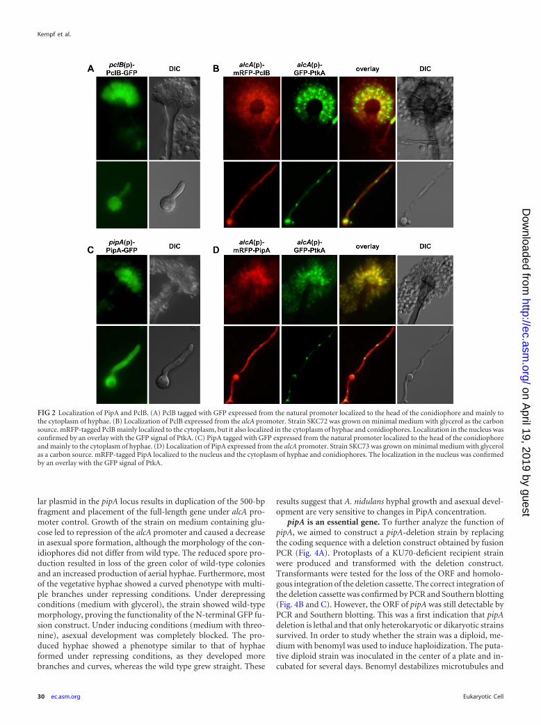
PclB and PipA reside in the cytoplasm and in the nuclei ofhyphae and conidiophores. Because the interaction of PtkA withPclB and PipA was restricted to the conidiophore, we testedwhether expression of the two proteins was also restricted to thisdevelopmental phase (Fig. 2). To this end, A. nidulans strainswhich contained GFP-PtkA in combination with mRFP-PipA ormRFP-PclB were constructed. All proteins were C-terminallytagged at their native locus. To generate these constructs, a fusionPCR method was used (21). The fusion PCR was set up with twoflanking sequences and a GFP-pyrG cassette derived from pFNO3.Transgenic A. nidulans colonies were tested for homologous inte-gration of the constructs by PCR and Southern blotting (resultsnot shown). All proteins were detectable in hyphae and in conid-iophores in the cytoplasm and also in nuclei. In addition to thesubcellular localization using the native promoter (Fig. 2A and C),the tagged proteins were also expressed from the alcA promoter(Fig. 2B and D). After moderate induction of the promoter, clearsignals which unambiguously demonstrated nuclear localizationof some fraction of the proteins were observed. GFP-PtkA couldmainly be observed in the nuclei of hyphae, in metulae, phialides,and spores, with only faint signals observed in the cytoplasm.mRFP-PipA appeared in metulae, phialides, and spores, withstrong signals observed in nuclei and the cytoplasm (Fig. 2D). ThemRFP-PclB fusion protein had the same localization pattern as
TABLE 4 Identification of PtkA-interacting proteins using a truncated version of PtkA as bait
Broad Instituteidentification no.Total
Total no. of aminoacids/size of isolatedclone (amino acids)
No. of clonesin screening Conserved domain Assigned function in other organisms
AN9297.2 590/388 12 Myosin cross-reactive antigen Members of this family are thought to havestructural features in common with thebeta chain of the class II antigens
AN0369.1 459/311 1 Methionine aminopeptidase 2B Metallopeptidase familyAN0451.2 215/185 6 C-8 sterol isomerase C-8 sterol isomerase, catalyzes a reaction in
ergosterol biosynthesisAN03223.1 796/569 4 6-Phosphofruktokinase alpha subunit Catalyzes the phosphorylation of fructose-
6-phosphate to fructose-1,6-bisphosphate, a key regulatory step inthe glycolytic pathway
AN7044.2 1 Histidinol phosphatase The polymerase and histidinol phosphatase(PHP) domain is a putativephosphoesterase domain
AN3847.2 1 OxidoreductaseAN3586.2 1 Ubiquinone biosynthesis monooxygenaseAN7432.2 1 Ornithine decarboxylase antizymeAN4202.1 1 Ribosomal protein L16a Ribosomal protein L13 is one of the
proteins from the large ribosomalsubunit
AN2513.2 285/118 1 Serine/threonine-protein kinase Bud32 Bud32 is a subunit of the KEOPS/EKCcomplex, which plays a role intranscription and maintenance oftelomeres
Kempf et al.
28 ec.asm.org Eukaryotic Cell
on April 19, 2019 by guest
http://ec.asm.org/
Dow
nloaded from
PipA. It was also found in nuclei, but the protein did not specifi-cally accumulate in these organelles (Fig. 2B).
Besides the protein localization, the expression of pipA andpclB was tested during different stages of development (Fig. 3).Asexual development of the wild-type strain FGSCA4 was syn-chronized by air exposure of mycelium grown in liquid culture,and total RNA was isolated at different time points and used as thetemplate for quantitative real-time PCR experiments. WhereaspipA and pchA appeared to be constitutively expressed throughoutdevelopment, the expression of pclB changed during develop-ment. The expression after 5 h postinduction of asexual develop-ment was higher than that in hyphae and decreased with time tothe lowest level after 9 h. After that time point, the mRNA level
increased again, with a maximum occurring after 24 h. The highlevels of pclB and pipA expression during the formation of theconidiophores are in agreement with the results of the interactionand localization studies and thus imply a function of these pro-teins during later stages of asexual development. The analysis wasrepeated with strains containing tagged versions of the proteins,and similar expression patterns were obtained (data not shown).
pipA is important for normal growth and development. Inorder to account for the putative lethality of pipA deletion, we firsttried to downregulate pipA using the alcA promoter (Fig. 4A). Aca. 500-bp fragment was amplified from the region 5= of the pipAopen reading frame and cloned in frame with GFP under the con-trol of the alcA promoter (pMCB17apx). Integration of the circu-
FIG 1 Isolation of two new PtkA-interacting proteins. (A) Scheme of the two proteins. Conserved domains of PipA and PclB. The PipA protein is 286 aa longand contains a kinase domain, a PEST domain, and a nuclear localization signal (NLS). PclB is 242 aa long and contains a cyclin domain, a PEST domain, anda destruction box. The PEST domain and a destruction box are both signals for degradation of proteins. The nuclear export signal (NES) is a sign for localizationof the protein in the nucleus and possible export in the cytoplasm. (B) Yeast two-hybrid assay with PtkA and PipA or PclB. The interaction of PipA and PclB withPtkA was tested with three different PtkA constructs, the full-length PtkA_1/545 and two truncated constructs, PtkA_1/330 and PtkA_24/170. TDO, tripledropout. (C) BiFC analyses to confirm the interaction of PtkA with PipA and PclB. In SKC19 expressing PtkA tagged with the N-terminal half of YFP and PipAtagged with the C-terminal half, a YFP signal was detected in the head of the conidiophore but not in hyphae. Strain SKC20 expressing YFPN-PtkA and YFPC-PclBalso showed a YFP signal, which was restricted to the conidiophore and was not observed in the hyphae. DIC, differential inference contrast.
Cdk9 Kinase in A. nidulans
January 2013 Volume 12 Number 1 ec.asm.org 29
on April 19, 2019 by guest
http://ec.asm.org/
Dow
nloaded from
lar plasmid in the pipA locus results in duplication of the 500-bpfragment and placement of the full-length gene under alcA pro-moter control. Growth of the strain on medium containing glu-cose led to repression of the alcA promoter and caused a decreasein asexual spore formation, although the morphology of the con-idiophores did not differ from wild type. The reduced spore pro-duction resulted in loss of the green color of wild-type coloniesand an increased production of aerial hyphae. Furthermore, mostof the vegetative hyphae showed a curved phenotype with multi-ple branches under repressing conditions. Under derepressingconditions (medium with glycerol), the strain showed wild-typemorphology, proving the functionality of the N-terminal GFP fu-sion construct. Under inducing conditions (medium with threo-nine), asexual development was completely blocked. The pro-duced hyphae showed a phenotype similar to that of hyphaeformed under repressing conditions, as they developed morebranches and curves, whereas the wild type grew straight. These
results suggest that A. nidulans hyphal growth and asexual devel-opment are very sensitive to changes in PipA concentration.
pipA is an essential gene. To further analyze the function ofpipA, we aimed to construct a pipA-deletion strain by replacingthe coding sequence with a deletion construct obtained by fusionPCR (Fig. 4A). Protoplasts of a KU70-deficient recipient strainwere produced and transformed with the deletion construct.Transformants were tested for the loss of the ORF and homolo-gous integration of the deletion cassette. The correct integration ofthe deletion cassette was confirmed by PCR and Southern blotting(Fig. 4B and C). However, the ORF of pipA was still detectable byPCR and Southern blotting. This was a first indication that pipAdeletion is lethal and that only heterokaryotic or dikaryotic strainssurvived. In order to study whether the strain was a diploid, me-dium with benomyl was used to induce haploidization. The puta-tive diploid strain was inoculated in the center of a plate and in-cubated for several days. Benomyl destabilizes microtubules and
FIG 2 Localization of PipA and PclB. (A) PclB tagged with GFP expressed from the natural promoter localized to the head of the conidiophore and mainly tothe cytoplasm of hyphae. (B) Localization of PclB expressed from the alcA promoter. Strain SKC72 was grown on minimal medium with glycerol as the carbonsource. mRFP-tagged PclB mainly localized to the cytoplasm, but it also localized in the cytoplasm of hyphae and conidiophores. Localization in the nucleus wasconfirmed by an overlay with the GFP signal of PtkA. (C) PipA tagged with GFP expressed from the natural promoter localized to the head of the conidiophoreand mainly to the cytoplasm of hyphae. (D) Localization of PipA expressed from the alcA promoter. Strain SKC73 was grown on minimal medium with glycerolas a carbon source. mRFP-tagged PipA localized to the nucleus and the cytoplasm of hyphae and conidiophores. The localization in the nucleus was confirmedby an overlay with the GFP signal of PtkA.
Kempf et al.
30 ec.asm.org Eukaryotic Cell
on April 19, 2019 by guest
http://ec.asm.org/
Dow
nloaded from
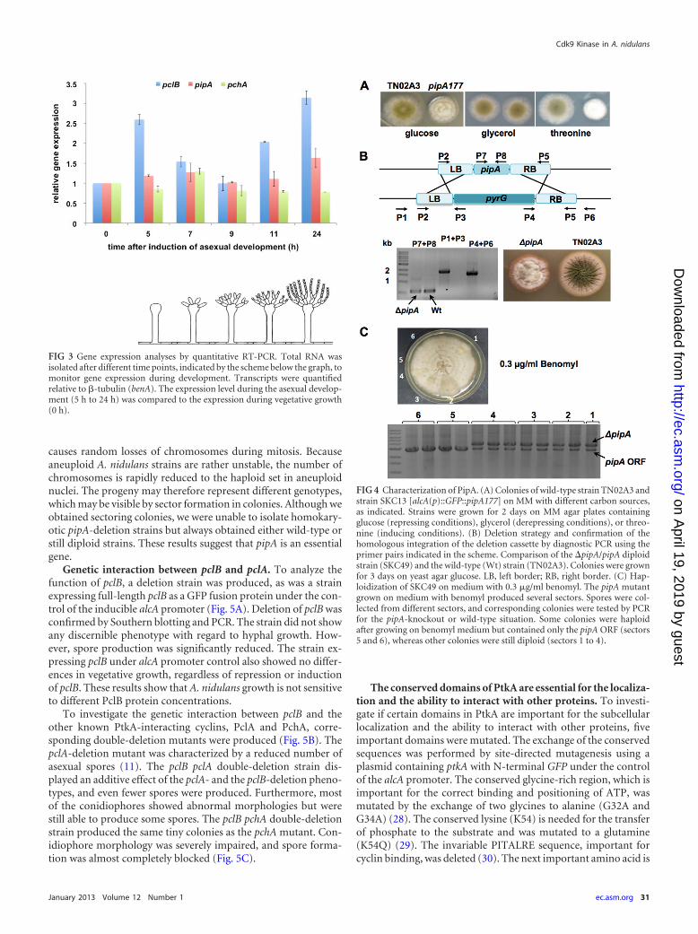
causes random losses of chromosomes during mitosis. Becauseaneuploid A. nidulans strains are rather unstable, the number ofchromosomes is rapidly reduced to the haploid set in aneuploidnuclei. The progeny may therefore represent different genotypes,which may be visible by sector formation in colonies. Although weobtained sectoring colonies, we were unable to isolate homokary-otic pipA-deletion strains but always obtained either wild-type orstill diploid strains. These results suggest that pipA is an essentialgene.
Genetic interaction between pclB and pclA. To analyze thefunction of pclB, a deletion strain was produced, as was a strainexpressing full-length pclB as a GFP fusion protein under the con-trol of the inducible alcA promoter (Fig. 5A). Deletion of pclB wasconfirmed by Southern blotting and PCR. The strain did not showany discernible phenotype with regard to hyphal growth. How-ever, spore production was significantly reduced. The strain ex-pressing pclB under alcA promoter control also showed no differ-ences in vegetative growth, regardless of repression or inductionof pclB. These results show that A. nidulans growth is not sensitiveto different PclB protein concentrations.
To investigate the genetic interaction between pclB and theother known PtkA-interacting cyclins, PclA and PchA, corre-sponding double-deletion mutants were produced (Fig. 5B). ThepclA-deletion mutant was characterized by a reduced number ofasexual spores (11). The pclB pclA double-deletion strain dis-played an additive effect of the pclA- and the pclB-deletion pheno-types, and even fewer spores were produced. Furthermore, mostof the conidiophores showed abnormal morphologies but werestill able to produce some spores. The pclB pchA double-deletionstrain produced the same tiny colonies as the pchA mutant. Con-idiophore morphology was severely impaired, and spore forma-tion was almost completely blocked (Fig. 5C).
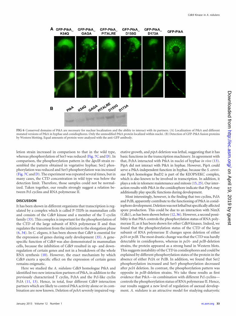
The conserved domains of PtkA are essential for the localiza-tion and the ability to interact with other proteins. To investi-gate if certain domains in PtkA are important for the subcellularlocalization and the ability to interact with other proteins, fiveimportant domains were mutated. The exchange of the conservedsequences was performed by site-directed mutagenesis using aplasmid containing ptkA with N-terminal GFP under the controlof the alcA promoter. The conserved glycine-rich region, which isimportant for the correct binding and positioning of ATP, wasmutated by the exchange of two glycines to alanine (G32A andG34A) (28). The conserved lysine (K54) is needed for the transferof phosphate to the substrate and was mutated to a glutamine(K54Q) (29). The invariable PITALRE sequence, important forcyclin binding, was deleted (30). The next important amino acid is
FIG 3 Gene expression analyses by quantitative RT-PCR. Total RNA wasisolated after different time points, indicated by the scheme below the graph, tomonitor gene expression during development. Transcripts were quantifiedrelative to �-tubulin (benA). The expression level during the asexual develop-ment (5 h to 24 h) was compared to the expression during vegetative growth(0 h).
FIG 4 Characterization of PipA. (A) Colonies of wild-type strain TN02A3 andstrain SKC13 [alcA(p)::GFP::pipA177] on MM with different carbon sources,as indicated. Strains were grown for 2 days on MM agar plates containingglucose (repressing conditions), glycerol (derepressing conditions), or threo-nine (inducing conditions). (B) Deletion strategy and confirmation of thehomologous integration of the deletion cassette by diagnostic PCR using theprimer pairs indicated in the scheme. Comparison of the �pipA/pipA diploidstrain (SKC49) and the wild-type (Wt) strain (TN02A3). Colonies were grownfor 3 days on yeast agar glucose. LB, left border; RB, right border. (C) Hap-loidization of SKC49 on medium with 0.3 �g/ml benomyl. The pipA mutantgrown on medium with benomyl produced several sectors. Spores were col-lected from different sectors, and corresponding colonies were tested by PCRfor the pipA-knockout or wild-type situation. Some colonies were haploidafter growing on benomyl medium but contained only the pipA ORF (sectors5 and 6), whereas other colonies were still diploid (sectors 1 to 4).
Cdk9 Kinase in A. nidulans
January 2013 Volume 12 Number 1 ec.asm.org 31
on April 19, 2019 by guest
http://ec.asm.org/
Dow
nloaded from

the aspartate of the DMKAAN domain, which facilitates the re-lease of the substrate after the phosphate transfer and was ex-changed to a glycine (D155G) (31). In the conserved DFG do-main, which is needed for the transfer of the phosphate residueand the binding of ATP, aspartate was changed to alanine(D173A) (32). PtkA-GFP was detected in the nuclei of hyphae andconidiophores, with faint GFP signals detected in the cytoplasm.All mutated PtkA-GFP fusion proteins mainly localized in thecytoplasm and only partly localized in the nuclei. To exclude lo-calization artifacts due to the degradation of the GFP-PtkA fusionproteins, protein integrity and abundance were analyzed in West-ern blots. All GFP-PtkA constructs were present in similar
amounts independent of the mutation of different domains ofPtkA (Fig. 6B). The localization results thus indicate an importantrole of all conserved domains for proper localization and activityof PtkA.
To test the relevance of the different domains for the interac-tion of PtkA with other interacting proteins (PclA, PclB, PchA,and PipA), yeast two-hybrid analyses were performed using thesame modified PtkAs. To select for the incorporation of the twoplasmids and for the interactions, different selection media wereused. Although the presence of both plasmids could be demon-strated, no interaction was detected (data not shown). In accor-dance with the loss of the normal localization pattern, the muta-tion of the conserved domains also inhibited the interaction withthe different interaction partners.
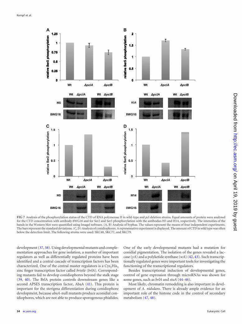
Evidence for a possible role of Pcl cyclins in controlling thephosphorylation status of the CTD of the large subunit of RNApolymerase II. Given that Cdk9 kinases are involved in the regu-lation of RNA polymerase II, we anticipated that A. nidulans PtkAfulfills a similar function and that the identified PtkA interactionpartners may control PtkA activity and thereby control RNA poly-merase II. The control of RNA polymerase II activity should in-volve the phosphorylation status of the CTD. Several approacheswere undertaken to test this hypothesis. Three antibodies (for hu-man and yeast) which can be used to analyze the phosphorylationstatus of RNA polymerase II are commercially available. The an-tibody 8WG16 recognizes the unphosphorylated form. The anti-body H5 is derived against phosphorylated serine 2 (Ser2) andH14 is derived against serine 5 (Ser5) in the heptad repeat of theCTD. One obvious experiment to analyze the phosphorylationstatus is, of course, to quantify the amounts of RNA polymeraseand the phosphorylated forms in cell extracts of A. nidulans. How-ever, the protein concentrations of RNA polymerase appeared tobe below our detection limit. Therefore, we aimed at the establish-ment of an in vitro phosphorylation assay. To this end, we success-fully expressed PclA, PclB, PtkA, and the CTD in E. coli. Unfortu-nately, most proteins were insoluble and thus could not be used inthe assay. Next, we overexpressed the CTD (pKC87) in A. nidulanswild type and pclA (SSNI30) and pclB (SKC34) mutant strains.Strains were incubated in liquid medium for 24 h at 37°C and theninduced for asexual development, and after an additional 24 h,protein extracts were prepared. The CTD could be detected in allprotein samples prepared from vegetative hyphae (Fig. 7A and B)or conidiophores (Fig. 7C and D). Interestingly, the concentrationof the CTD was very low in wild-type conidiophores, whereas theprotein was very abundant in pclA- or pclB-deletion strains, sug-gesting that the presence of either cyclin renders the CTD ratherunstable (Fig. 7C and D), indicating a connection between Pclcyclins and the CTD.
In order to determine the phosphorylation status of the CTD,phosphorylation of Ser2 and Ser5 of the CTD was compared be-tween wild type and the deletion mutants. The intensities of allbands in the Western blot were quantified and normalized usingthe signals obtained with the anti-CTD antibody (8WG16) in thewild-type strain. Afterwards, the values obtained with the phos-phorylation-specific antibodies were compared between the wild-type and the pcl-deletion strains. In hyphae, the phosphorylationof Ser2 decreased slightly in the absence of PclA or PclB, whereasSer5 phosphorylation increased almost two times in the case ofpclA deletion (Fig. 7A and B). The experiment was repeated fourtimes. In conidiophores, phosphorylation of Ser2 in the pclA-de-
FIG 5 Characterization of the cyclin PclB. (A) Deletion strategy and confir-mation of pclB deletion by diagnostic PCR using the primer pairs indicated inthe scheme. Comparison of the pclB-deletion strain (SKC34) and the wild type(TN02A3). Colonies were grown for 3 days on MM. LB, left border; RB, rightborder. (B) Characterization of double-deletion mutants of the different cy-clins interacting with PtkA. Conidiophore stalks are about 8 �m in diameter.(C) Quantification of asexual spores of wild-type (TN02A3), the pclA- andpclB-deletion strains, and the pclA pclB double-deletion strain. Strains weregrown on MM agar plates, and spores were counted after 24, 48, 72, and 96 h.
Kempf et al.
32 ec.asm.org Eukaryotic Cell
on April 19, 2019 by guest
http://ec.asm.org/
Dow
nloaded from
letion strain increased in comparison to that in the wild type,whereas phosphorylation of Ser5 was reduced (Fig. 7C and D). Incomparison, the phosphorylation pattern in the �pclB strain re-sembled the pattern obtained in vegetative hyphae; Ser2 phos-phorylation was reduced and Ser5 phosphorylation was increased(Fig. 7C and D). The experiment was repeated several times, but inmany cases, the CTD concentration in wild type was below thedetection limit. Therefore, those samples could not be normal-ized. Taken together, our results strongly suggest a relation be-tween Pcl cyclins and RNA polymerase II.
DISCUSSION
It has been shown in different organisms that transcription is reg-ulated by a complex which is called P-TEFb in mammalian cellsand consists of the Cdk9 kinase and a member of the T-cyclinfamily (33). This complex is important for the phosphorylation ofthe CTD of the large subunit of RNA polymerase II and thusregulates the transition from the initiation to the elongation phase(6, 34). In C. elegans, it has been shown that Cdk9 is essential forthe expression of genes during early development (35). A gene-specific function of Cdk9 was also demonstrated in mammaliancells, because the inhibition of Cdk9 resulted in up- and down-regulation of certain genes and not in a breakdown of the wholeRNA synthesis (10). However, the exact mechanism by whichCdk9 exerts a specific effect on the expression of certain genesremains enigmatic.
Here we studied the A. nidulans Cdk9 homologue PtkA andidentified two new interaction partners of PtkA, in addition to thepreviously characterized T cyclin, PchA and the Pcl-like cyclinPclA (11, 13). Hence, in total, four different Cdk9 interactionpartners which are likely to control PtkA activity alone or in com-bination are now known. Deletion of pchA severely impaired veg-
etative growth, and pipA deletion was lethal, suggesting that it hasbasic functions in the transcription machinery. In agreement withthat, PchA interacted with PtkA in nuclei of hyphae in vivo (13).PipA did not interact with PtkA in hyphae. However, PipA couldserve a PtkA-independent function in hyphae, because the S. cerevi-siae PipA homologue Bud32 is part of the KEOPS/EKC complex,which is also known to be involved in transcription. In addition, itplays a role in telomere maintenance and mitosis (15, 25). Our inter-action results with PtkA in the conidiophore indicate that PipA mayadditionally play specific functions during development.
Most interestingly, however, is the finding that two cyclins, PclAand PclB, apparently contribute to the functioning of PtkA in conid-iophore development. Deletion was not lethal but specifically affectedspore production. This could be due to an interaction with NimX(Cdk1), as has been shown before (12, 36). However, a second possi-bility is that PtkA controls the phosphorylation status of RNA poly-merase II, as it has been shown for other Cdk9 kinases. Indeed, wefound that the phosphorylation status of the CTD of the largesubunit of RNA polymerase II changes upon deletion of eitherpclA or pclB. The most drastic change was that the CTD was hardlydetectable in conidiophores, whereas in pclA- and pclB-deletionstrains, the protein appeared as a strong band in Western blots.This suggests instability of the CTD in conidiophores and could beexplained by different phosphorylation states of the protein in theabsence of either PclA or PclB. In addition, we found that Ser2phosphorylation increased and Ser5 phosphorylation decreasedafter pclA deletion. In contrast, the phosphorylation pattern wasopposite in pclB-deletion strains. We take these results as firstevidence that PtkA—in combination with different Pcl cyclins—controls the phosphorylation status of RNA polymerase II. Hence,our results suggest a new level of regulation of asexual develop-ment. A. nidulans is an attractive model for studying eukaryotic
FIG 6 Conserved domains of PtkA are necessary for nuclear localization and the ability to interact with its partners. (A) Localization of PtkA and differentmutated versions of PtkA in hyphae and conidiophores. Only the unmodified PtkA protein localized within nuclei. (B) Detection of GFP-PtkA fusion proteinsby Western blotting. Equal amounts of protein were analyzed with the anti-GFP antibody.
Cdk9 Kinase in A. nidulans
January 2013 Volume 12 Number 1 ec.asm.org 33
on April 19, 2019 by guest
http://ec.asm.org/
Dow
nloaded from
development (37, 38). Using developmental mutants and comple-mentation approaches for gene isolation, a number of importantregulators as well as differentially regulated proteins have beenidentified and a central cascade of transcription factors has beencharacterized. One of the central master regulators is a Cys2His2
zinc finger transcription factor called bristle (brlA). Correspond-ing mutants fail to develop conidiophores beyond the stalk stage(39, 40). The BrlA protein controls downstream genes like asecond APSES transcription factor, AbaA (41). This protein isimportant for the sterigma differentiation during conidiophoredevelopment, because abaA-null mutants produce aconidial con-idiophores, which are not able to produce sporogenous phialides.
One of the early developmental mutants had a mutation forconidial pigmentation. The isolation of the genes revealed a lac-case (yA) and a polyketide synthase (wA) (42, 43). Such transcrip-tionally regulated genes were important tools for investigating thefunctioning of the transcriptional regulators.
Besides transcriptional induction of developmental genes,control of gene expression through microRNAs was shown forsome genes, such as brlA and stuA (44–46).
Most likely, chromatin remodeling is also important in devel-opment of A. nidulans. There is already ample evidence for animportant role of the histone code in the control of secondarymetabolism (47, 48).
FIG 7 Analysis of the phosphorylation status of the CTD of RNA polymerase II in wild-type and pcl-deletion strains. Equal amounts of protein were analyzedfor the CTD concentration with antibody 8WG16 and for Ser2 and Ser5 phosphorylation with the antibodies H5 and H14, respectively. The intensities of thebands in the Western blot were quantified using ImageJ software. (A, B) Analysis of hyphae. The values represent the means of four independent experiments.The bars represent the standard deviations. (C, D) Analysis of conidiophores. A representative experiment is displayed. The amount of CTD in wild type was oftenbelow the detection limit. The following strains were used: SKC48, SKC77, and SKC79.
Kempf et al.
34 ec.asm.org Eukaryotic Cell
on April 19, 2019 by guest
http://ec.asm.org/
Dow
nloaded from
Here, we present evidence that modulation of RNA polymer-ase II through the Cdk9 kinase PtkA is involved in developmentalcontrol. Of course, one of the major challenges for future researchwill be the identification of target genes whose activity is con-trolled in a PtkA-dependent manner. A. nidulans appears to be anice model system with many possibilities to unravel the CTDcode and its interplay with the histone code.
ACKNOWLEDGMENTS
This work was funded through the Baden-Württemberg Stiftung and theGerman Science Foundation (DFG FOR1334). C.K. was a fellow of theLandesgraduiertenförderung of the Karlsruhe Institute of Technology(KIT).
REFERENCES1. Jenuwein T, Allis CD. 2001. Translating the histone code. Science 293:
1074 –1080.2. Zhang DW, Rodriguez-Molina JB, Tietjen JR, Nemec CM, Ansari AZ.
2012. Emerging views on the CTD code. Genet. Res. Int. 2012:347214.doi:10.1155/2012/347214.
3. Wang S, Fischer PM. 2008. Cyclin-dependent kinase 9: a key transcrip-tional regulator and potential drug target in oncology, virology and car-diology. Trends Pharmacol. Sci. 29:302–313.
4. Peng J, Marshall NF, Price DH. 1998. Identification of a cyclin subunitrequired for the function of Drosophila P-TEFb. J. Biol. Chem. 273:13855–13860.
5. Peng J, Zhu Y, Milton JT, Price DH. 1998. Identification of multiplecyclin subunits of human P-TEFb. Genes Dev. 12:755–762.
6. Marshall NF, Peng J, Xie Z, Price DH. 1996. Control of RNA polymeraseII elongation potential by a novel carboxyl-terminal domain kinase. J.Biol. Chem. 271:27176 –27183.
7. Czudnochowski N, Bösken CA, Geyer M. 2011. Serine-7 but not serine-5phosphorylation primes RNA polymerase II CTD for P-TEFb recognition.Nat. Commun. 3:842. doi:10.1038/ncomms1846.
8. Buratowski S. 2009. Progression through the RNA polymerase II CTDcycle. Mol. Cell 36:541–546.
9. Egloff S, Murphy S. 2008. Cracking the RNA polymerase II CTD code.Trends Genet. 24:280 –288.
10. Garriga J, Xie H, Obradovic Z, Grana X. 2010. Selective control of geneexpression by CDK9 in human cells. J. Cell. Physiol. 222:200 –208.
11. Schier N, Liese R, Fischer R. 2001. A Pcl-like cyclin of Aspergillus nidulansis transcriptionally activated by developmental regulators and is involvedin sporulation. Mol. Cell. Biol. 21:4075– 4088.
12. Schier N, Fischer R. 2002. The Aspergillus nidulans cyclin PclA accumu-lates in the nucleus and interacts with the central cell cycle regulatorNimX(Cdc2). FEBS Lett. 523:143–146.
13. Bathe F, Kempf C, Osmani A, Osmani AH, Hettinger S, Wohlmann E,Fischer R. 2010. Functional characterization of a new member of theCdk9 family in Aspergillus nidulans. Eukaryot. Cell 9:1901–1912.
14. Facchin S, Lopreiato R, Stocchetto S, Arrigoni G, Cesaro L, Marin O,Carignani G, Pinna LA. 2002. Structure-function analysis of yeastpiD261/Bud32, an atypical protein kinase essential for normal cell life.Biochem. J. 364:457– 463.
15. Srinivasan M, Mehta PYY, Prugar E, Koonin EV, Karzai AW,Sternglanz R. 2011. The highly conserved KEOPS/EKC complex is essen-tial for a universal tRNA modification, t6A. EMBO J. 30:873– 881.
16. Hill TW, Käfer E. 2001. Improved protocols for Aspergillus minimalmedium: trace element and minimal medium salt stock solutions. FungalGenet. Newsl. 48:20 –21.
17. Stringer MA, Dean RA, Sewall TC, Timberlake WE. 1991. Rodletless, anew Aspergillus developmental mutant induced by directed gene inactiva-tion. Genes Dev. 5:1161–1171.
18. Takeshita N, Higashitsuji Y, Konzack S, Fischer R. 2008. Apical sterol-rich membranes are essential for localizing cell end markers that deter-mine growth directionality in the filamentous fungus Aspergillus nidulans.Mol. Biol. Cell 19:339 –351.
19. Yelton MM, Hamer JE, Timberlake WE. 1984. Transformation of Asper-gillus nidulans by using a trpC plasmid. Proc. Natl. Acad. Sci. U. S. A.81:1470 –1474.
20. Sambrook J, Russell DW. 1999. Molecular cloning: a laboratory manual,3rd ed. Cold Spring Harbor Laboratory Press, Cold Spring Harbor, NY.
21. Szewczyk E, Nayak T, Oakley CE, Edgerton H, Xiong Y, Taheri-TaleshN, Osmani SA, Oakley BR. 2006. Fusion PCR and gene targeting inAspergillus nidulans. Nat. Protoc. 1:3111–3120.
22. Zekert N, Veith D, Fischer R. 2010. Interaction of the Aspergillus nidulansmicrotubule-organizing center (MTOC) component ApsB with gamma-tubulin and evidence for a role of a subclass of peroxisomes in the forma-tion of septal MTOCs. Eukaryot. Cell 9:795– 805.
23. Measday V, Moore L, Retnakaran R, Lee J, Donoviel M, Neiman AM,Andrews B. 1997. A family of cyclin-like proteins that interact with thePho85 cyclin-dependent kinase. Mol. Cell. Biol. 17:1212–1223.
24. Lee M, O’Regan S, Moreau JL, Johnson AL, Johnston LH, Goding CR.2000. Regulation o the Pcl7-Pho85 cyclin-cdk complex by Pho81. Mol.Microbiol. 38:411– 422.
25. Hecker A, Lopreiato R, Graille M, Collinet B, Forterre P, Libri D, vanTilbeurgh H. 2008. Structure of the archaeal Kae1/Bud32 fusion proteinMJ1130: a model for the eukaryotic EKC/KEOPS subcomplex. EMBO J.27:2340 –2351.
26. Briza P, Bogengruber E, Thur A, Rutzler M, Munsterkotter M, DowesIW, Breitenbach M. 2002. Systematic analysis of sporulation phenotypesin 624 non-lethal homozygous deletion strains of Saccharomyces cerevi-siae. Yeast 19:403– 422.
27. Sartori G, Mazzotta G, Stocchetto S, Pavanello A, Carignani G. 2000.Inactivation of six genes from chromosomes VII and XIV of Saccharomy-ces cerevisiae and basic phenotypic analysis of the mutant strains. Yeast16:255–265.
28. Wierenga RK, Hol WG. 1983. Predicted nucleotide-binding properties ofp21 protein and its cancer-associated variant. Nature 302:842– 844.
29. Carrera AC, Alexandrov K, Roberts TM. 1993. The conserved lysine ofthe catalytic domain of protein kinases is actively involved in the phos-photransfer reaction and not required for anchoring ATP. Proc. Natl.Acad. Sci. U. S. A. 90:442– 446.
30. Grana X, De Luca A, Sang N, Fu Y, Claudio PP, Rosenblatt J, MorganDO, Giordano A. 1994. PITALRE, a nuclear CDC2-related protein kinasethat phosphorylates the retinoblastoma protein in vitro. Proc. Natl. Acad.Sci. U. S. A. 91:3834 –3838.
31. Gibbs CS, Zoller MJ. 1991. Rational scanning mutagenesis of a proteinkinase identifies functional regions involved in catalysis and substrate in-teractions. J. Biol. Chem. 266:8923– 8931.
32. Taylor SS, Knighton DR, Zheng J, Ten Eyck LF, Sowadski JM. 1992.Structural framework for the protein kinase family. Annu. Rev. Cell Dev.Biol. 8:429 – 462.
33. Zhu Y, Pe’ery T, Peng J, Ramanathan Y, Marshall NF, Marshall T,Amendt B, Mathews MB, Price DH. 1997. Transcription elongationfactor P-TEFb is required for HIV-1 tat transactivation in vitro. GenesDev. 11:2622–2632.
34. Prelich G. 2002. RNA polymerase II carboxy-terminal domain kinases:emerging clues to their function. Eukaryot. Cell 1:153–162.
35. Shim EY, Walker AK, Shi Y, Blackwell TK. 2002. CDK-9/cyclin T(P-TEFb) is required in two postinitiation pathways for transcription inthe C. elegans embryo. Genes Dev. 16:2135–2146.
36. Ye XS, Lee S-L, Wolkow TD, McGuire S-L, Hamer JE, Wood GC,Osmani SA. 1999. Interaction between developmental and cell cycle reg-ulators is required for morphogenesis in Aspergillus nidulans. EMBO J.18:6994 –7001.
37. Adams TH, Wieser JK, Yu J-H. 1998. Asexual sporulation in Aspergillusnidulans. Microbiol. Mol. Biol. Rev. 62:35–54.
38. Etxebeste O, Garzia A, Espeso EA, Ugalde U. 2010. Aspergillus nidulansasexual development: making the most of cellular modules. Trends Mi-crobiol. 18:569 –576.
39. Aguirre J. 1993. Spatial and temporal controls of the Aspergillus brlAdevelopmental regulatory gene. Mol. Microbiol. 8:211–218.
40. Prade R, Timberlake WE. 1993. The Aspergillus nidulans brlA regulatorylocus consists of two overlapping transcription units that are individuallyrequired for conidiophore development. EMBO J. 12:2439 –2447.
41. Andrianopoulos A, Timberlake WE. 1994. The Aspergillus nidulans abaAgene encodes a transcriptional activator that acts as a genetic switch tocontrol development. Mol. Cell. Biol. 14:2503–2515.
42. Aramayo R, Timberlake WE. 1993. The Aspergillus nidulans yA gene isregulated by abaA. EMBO J. 12:2039 –2048.
43. Mayorga ME, Timberlake WE. 1990. Isolation and molecular character-ization of the Aspergillus nidulans wA gene. Genetics 126:73–79.
44. Han S, Adams TH. 2001. Complex control of the developmental regula-
Cdk9 Kinase in A. nidulans
January 2013 Volume 12 Number 1 ec.asm.org 35
on April 19, 2019 by guest
http://ec.asm.org/
Dow
nloaded from

tory locus brlA in Aspergillus nidulans. Mol. Genet. Genomics 266:260 –270.
45. Han S, Navarro J, Greve RA, Adams TH. 1993. Translational repressionof brlA expression prevents premature development in Aspergillus. EMBOJ. 12:2449 –2457.
46. Wu J, Miller BL. 1997. Aspergillus asexual reproduction and sexual re-production are differentially affected by transcriptional and translationalmechanisms regulating stunted gene expression. Mol. Cell. Biol. 17:6191–6201.
47. Bok JW, Chiang YM, Szewczyk E, Reyes-Dominguez Y, Davidson AD,Sachez JF, Lo HC, Watanabe K, Strauss J, Oakley BR, Wang CC, KellerNP. 2009. Chromatin-level regulation of biosynthetic gene clusters. Nat.Chem. Biol. 5:462– 464.
48. Nützmann HW, Reyes-Dominguez Y, Scherlach K, Schoeckh V, HornF, Gacek A, Schümann J, Hertweck C, Strauss J, Brakhage AA. 2011.Bacteria-induced natural product formation in the fungus Aspergillus ni-dulans requires Saga/Ada-mediated histone acetylation. Proc. Natl. Acad.Sci. U. S. A. 108:14282–14287.
Kempf et al.
36 ec.asm.org Eukaryotic Cell
on April 19, 2019 by guest
http://ec.asm.org/
Dow
nloaded from