evaluation of children with ventricular...
TRANSCRIPT
Archives of Disease in Childhood, 1977, 52, 345-353
Evaluation of children with ventricular arrhythmiasDOROTHY J. RADFORD, T. IZUKAWA, AND R. D. ROWE
From the Department ofPaediatrics, Division of Cardiology, Hospital for Sick Children, Toronto, Canada
SUMMARY Ventricular arrhythmias are rare in childhood but may be associated with syncope andsudden death. This report describes 8 children with ventricular arrhythmias, 6 of whom sufferedsyncopal episodes. Ventricular tachycardia was documented in 5. One boy died suddenly. Completecardiac investigation was carried out with exercise testing, Holter monitoring, echocardiography,cardiac catheterization, angiography, and electrophysiological studies. The spectrum of abnor-malities related to the arrhythmias included prolapsing mitral valve, prolonged QT syndrome, sicksinus syndrome, congenital heart disease, cardiomyopathy, and idiopathic ventricular tachycardia.Exercise testing and Holter monitoring were particularly useful in documenting the arrhythmias andmonitoring response to therapy.
Cardiac syncope may occur in children as a resultof outflow obstruction (e.g. aortic stenosis) or fromconduction defects and arrhythmias. Complete heartblock resulting in Stokes-Adams attacks is infrequentin children (Molthan et al., 1962; Nakamura andNadas, 1964) but the sick sinus syndrome, a recentlyrecognized cause of syncopal episodes may occurmore frequently (Radford and Izukawa, 1975;Scott et al., 1976). Ventricular tachycardia is a rarearrhythmia causing syncope in childhood (Videbaeket al., 1973; Hernandez et al., 1975).Within a period of 12 months our Cardiology
Division was presented with the problems of 6children with syncope and ventricular arrhythmias,of whom 3 had documented ventricular tachycardia.Another 2 had ventricular tachycardia withoutsyncope. Such children carry the risk of suddendeath and the clinician must therefore make carefulinvestigations to determine aetiology, precipitatingfactors, and suitable treatment. This report reviewsthe evaluation of these 8 patients, the wide spectrumof aetiologies, and methods of assessment oftherapeutic regimens.
Methods
All 8 children were assessed clinically by electro-cardiograms (ECG), exercise stress testing, echo-cardiography, and cardiac catheterization withangiography. Electrophysiological studies were donein 6 and Holter monitoring in 5.
Exercise testing was conducted on a treadmill with10% elevation. The protocol was based on that of
Received 2 September 1976
Ellestad et al. (1969). The child exercised for 10minutes-3 min at 1 * 7 mph, 2 min at 3 mph, 2 minat 4 mph, and finally 3 min at 5 mph. A physicianwatched a continuous ECG oscilloscope monitorand recorded arrhythmias as well as routine record-ings at 1-minute intervals during the test and for8 minutes after.
Holter monitoring (Holter, 1961) or dynamicECG was performed with the child wearing a cassettetape recorder for 16 hours. Playback was made on arapid scanner (Medcraft CDC 70) and arrhythmiaswere recorded by direct write out. Echocardiographywas performed using an Ekoline 20 ultrasonoscopeand a Cambridge strip chart recorder. Cardiaccatheterization was done from a percutaneousapproach to the femoral vessels. Routine pressureand 02-saturation data were obtained. Angiographyincluded a selective contrast injection into theaortic root to delineate the coronary arteries, andother views to outline ventricular anatomy andfunction.
His bundle recordings by intracardiac ECG wereobtained by the standard method of Scherlag et al.(1969). Normal values for children were taken fromthe tables of Abella et al. (1972). Sinus node re-covery times were recorded after high atrial pacingfor 1 minute and timing the interval from the lastpaced beat to the first spontaneous sinus beat. Anormal time was <150% resting RR interval.
Ventricular tachycardia was defined as a sequenceof three or more wide, abnormal QRS complexes ata rate between 150 and 250/min. In order to excludesupraventricular tachycardia with aberrant conduc-tion it was necessary to observe other criteria,namely (i) a dissociated atrial rhythm slower than
345
on 27 June 2018 by guest. Protected by copyright.
http://adc.bmj.com
/A
rch Dis C
hild: first published as 10.1136/adc.52.5.345 on 1 May 1977. D
ownloaded from
346 Radford, Izukawa, and Rowe
the ventricular rate, (ii) capture and fusion beats,(iii) ventricular ectopic beats occurring during sinusrhythm with the same configuration as the complexesof the tachycardia (Lown et al., 1973).
Case reports
Case 1. At age 11 years this girl had a syncopalepisode on a ski slope and was promptly resuscitated.Her past history included the occurrence of cardiacarrhythmias during general anaesthesia for dentaland orthopaedic procedures at the age of 6 years,and a report of 'concussion' after falling from a
bicycle at age 8. Her parents had observed deteriora-tion of her motor and mental skills.
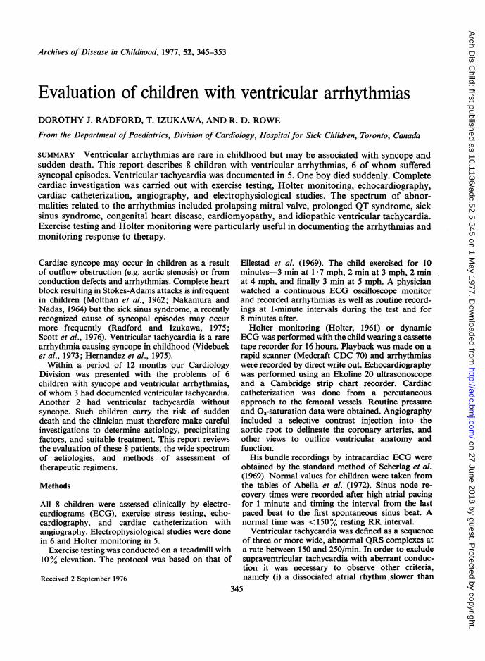
Detailed assessments were made. Intelligencetesting confirmed retardation. There was no definiteneurological abnormality, but the electroencephalo-gram showed poorly organized rhythms. At theinitial examination auscultatory findings werenormal and the ECG showed normal QRS com-plexes and ST-T wave segments, with a wanderingatrial pacemaker. An exercise ECG precipitatedmultifocal ventricular ectopic beats and runs ofventricular tachycardia (Fig. 1). The echocardio-
RESTING
AFTER. EXERCISE
Fig. I Case 1. ECG after exercise. Ventricular
tachycardia and multifocal ventricular ectopic beats.
gram showed prolapse of the posterior leaflet of themitral valve. At cardiac catheterization no otheranatomical or haemodynamic abnormalities were
found and the sinus node recovery time and Hisbundle intervals were normal Supraventriculartachycardia confirmed by His spike recordings was
induced by an isoprenaline infusion. Otherwise,only occasional supraventricular and ventricularectopic beats occurred.
Treatment with oral propranolol was started andserial exercise testing was done to monitor thistherapy. Initially multifocal ventricular ectopics andventricular tachycardia continued to occur withexercise, but with adequate doses only occasional
unifocal ventricular ectopic beats were precipitatedby this stress testing. Also, a midsystolic click andlate systolic murmur of mitral valve prolapse becameaudible. She now remains symptom free.
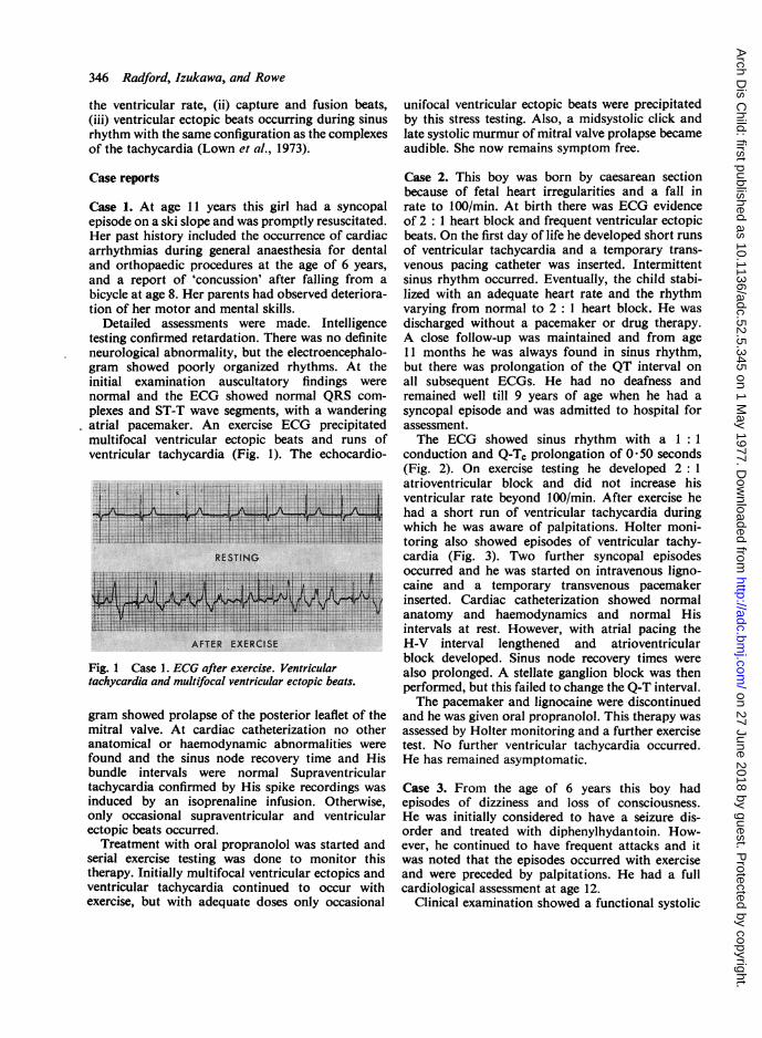
Case 2. This boy was born by caesarean sectionbecause of fetal heart irregularities and a fall inrate to 100/min. At birth there was ECG evidenceof 2: 1 heart block and frequent ventricular ectopicbeats. On the first day of life he developed short runsof ventricular tachycardia and a temporary trans-venous pacing catheter was inserted. Intermittentsinus rhythm occurred. Eventually, the child stabi-lized with an adequate heart rate and the rhythmvarying from normal to 2 :1 heart block. He wasdischarged without a pacemaker or drug therapy.A close follow-up was maintained and from age11 months he was always found in sinus rhythm,but there was prolongation of the QT interval onall subsequent ECGs. He had no deafness andremained well till 9 years of age when he had asyncopal episode and was admitted to hospital forassessment.The ECG showed sinus rhythm with a 1 : 1
conduction and Q-T, prolongation of 0 50 seconds(Fig. 2). On exercise testing he developed 2 :1atrioventricular block and did not increase hisventricular rate beyond 100/min. After exercise hehad a short run of ventricular tachycardia duringwhich he was aware of palpitations. Holter moni-toring also showed episodes of ventricular tachy-cardia (Fig. 3). Two further syncopal episodesoccurred and he was started on intravenous ligno-caine and a temporary transvenous pacemakerinserted. Cardiac catheterization showed normalanatomy and haemodynamics and normal Hisintervals at rest. However, with atrial pacing theH-V interval lengthened and atrioventricularblock developed. Sinus node recovery times werealso prolonged. A stellate ganglion block was thenperformed, but this failed to change the Q-T interval.The pacemaker and lignocaine were discontinued
and he was given oral propranolol. This therapy wasassessed by Holter monitoring and a further exercisetest. No further ventricular tachycardia occurred.He has remained asymptomatic.
Case 3. From the age of 6 years this boy hadepisodes of dizziness and loss of consciousness.He was initially considered to have a seizure dis-order and treated with diphenylhydantoin. How-ever, he continued to have frequent attacks and itwas noted that the episodes occurred with exerciseand were preceded by palpitations. He had a fullcardiological assessment at age 12.
Clinical examination showed a functional systolic
on 27 June 2018 by guest. Protected by copyright.
http://adc.bmj.com
/A
rch Dis C
hild: first published as 10.1136/adc.52.5.345 on 1 May 1977. D
ownloaded from
Evaluation of children with ventricular arrhythmias 347
Fig. 2 Case 2. ECG shows QT interval ofO50 seconds.
Fig. 3 Case 2. Recording from the Holter monitor shows ventricular tachycardia.
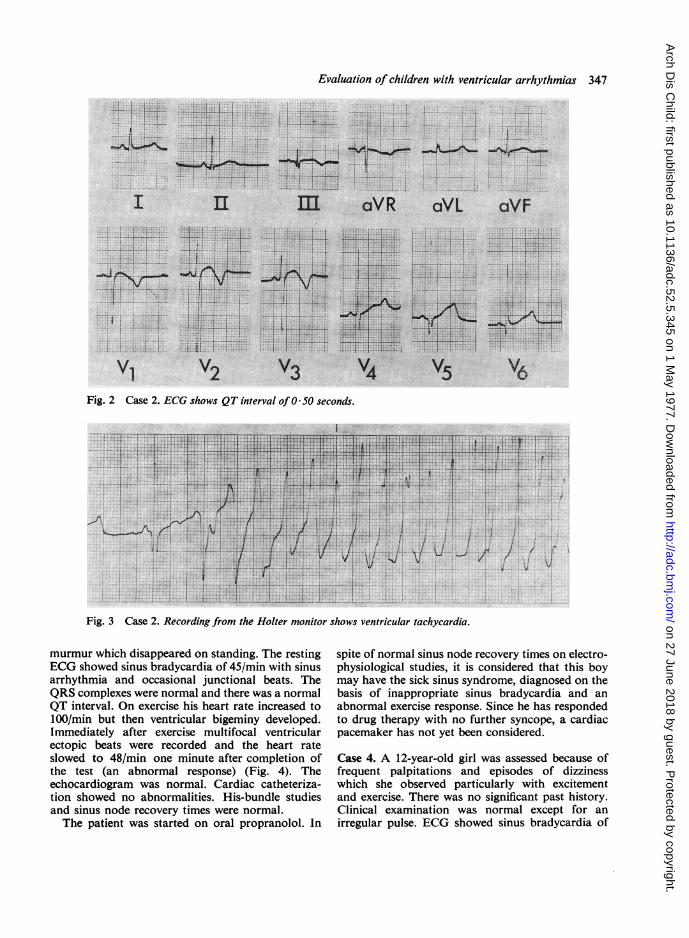
murmur which disappeared on standing. The restingECG showed sinus bradycardia of 45/min with sinusarrhythmia and occasional junctional beats. TheQRS complexes were normal and there was a normalQT interval. On exercise his heart rate increased to100/min but then ventricular bigeminy developed.Immediately after exercise multifocal ventricularectopic beats were recorded and the heart rateslowed to 48/min one minute after completion ofthe test (an abnormal response) (Fig. 4). Theechocardiogram was normal. Cardiac catheteriza-tion showed no abnormalities. His-bundle studiesand sinus node recovery times were normal.The patient was started on oral propranolol. In
spite of normal sinus node recovery times on electro-physiological studies, it is considered that this boymay have the sick sinus syndrome, diagnosed on thebasis of inappropriate sinus bradycardia and anabnormal exercise response. Since he has respondedto drug therapy with no further syncope, a cardiacpacemaker has not yet been considered.
Case 4. A 12-year-old girl was assessed because offrequent palpitations and episodes of dizzinesswhich she observed particularly with excitementand exercise. There was no significant past history.Clinical examination was normal except for anirregular pulse. ECG showed sinus bradycardia of
on 27 June 2018 by guest. Protected by copyright.
http://adc.bmj.com
/A
rch Dis C
hild: first published as 10.1136/adc.52.5.345 on 1 May 1977. D
ownloaded from
348 Radford, Izukawa, and Rowe
I.
4,IbtJINL ArITtRK IMINUTE AFTEFig. 4 Case 3. Exercise test. Ventricular ectopicsdeveloped and the heart rate slowed to 48/min, one
minute after completion of the test.
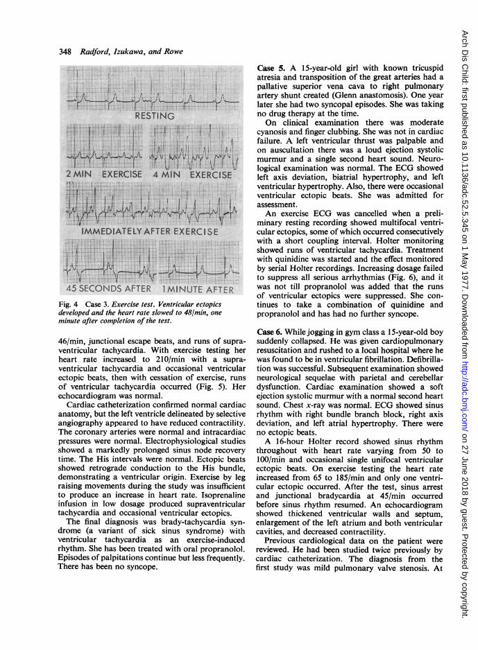
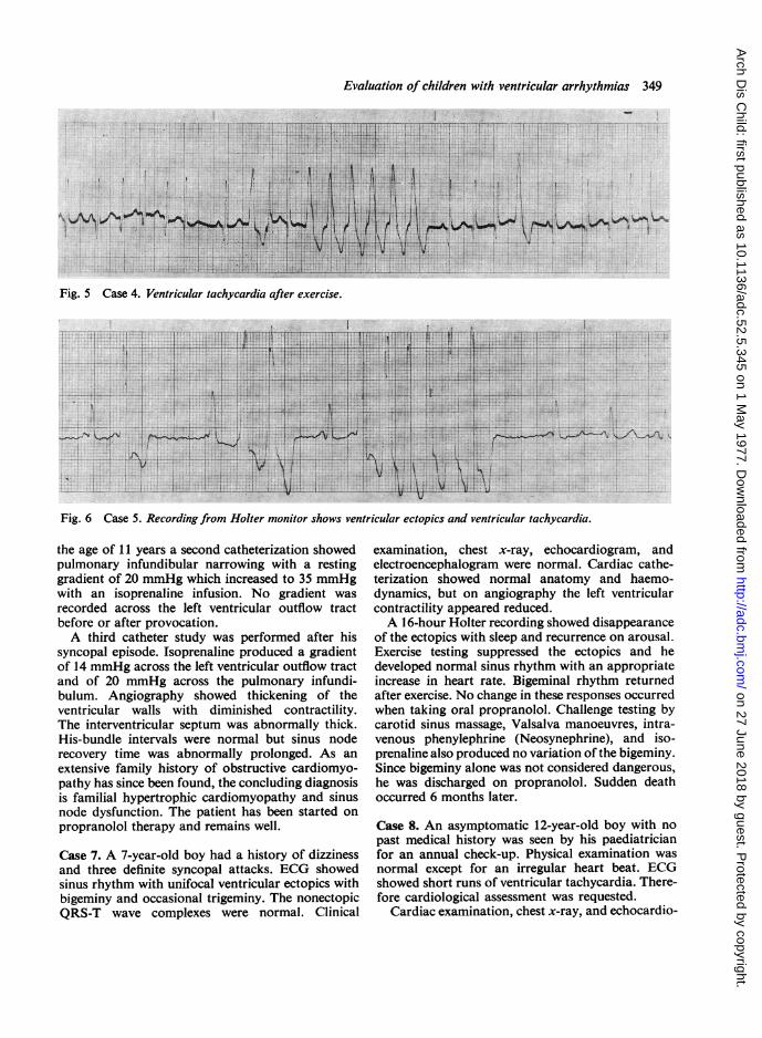
46/min, junctional escape beats, and runs of supra-ventricular tachycardia. With exercise testing herheart rate increased to 210/min with a supra-ventricular tachycardia and occasional ventricularectopic beats, then with cessation of exercise, runsof ventricular tachycardia occurred (Fig. 5). Herechocardiogram was normal.
Cardiac catheterization confirmed normal cardiacanatomy, but the left ventricle delineated by selectiveangiography appeared to have reduced contractility.The coronary arteries were normal and intracardiacpressures were normal. Electrophysiological studiesshowed a markedly prolonged sinus node recoverytime. The His intervals were normal. Ectopic beatsshowed retrograde conduction to the His bundle,demonstrating a ventricular origin. Exercise by legraising movements during the study was insufficientto produce an increase in heart rate. Isoprenalineinfusion in low dosage produced supraventriculartachycardia and occasional ventricular ectopics.The final diagnosis was brady-tachycardia syn-
drome (a variant of sick sinus syndrome) withventricular tachycardia as an exercise-inducedrhythm. She has been treated with oral propranolol.Episodes of palpitations continue but less frequently.There has been no syncope.
Case 5. A 15-year-old girl with known tricuspidatresia and transposition of the great arteries had apallative superior vena cava to right pulmonaryartery shunt created (Glenn anastomosis). One yearlater she had two syncopal episodes. She was takingno drug therapy at the time.On clinical examination there was moderate
cyanosis and finger clubbing. She was not in cardiacfailure. A left ventricular thrust was palpable andon auscultation there was a loud ejection systolicmurmur and a single second heart sound. Neuro-logical examination was normal. The ECG showedleft axis deviation, biatrial hypertrophy, and leftventricular hypertrophy. Also, there were occasionalventricular ectopic beats. She was admitted forassessment.An exercise ECG was cancelled when a preli-
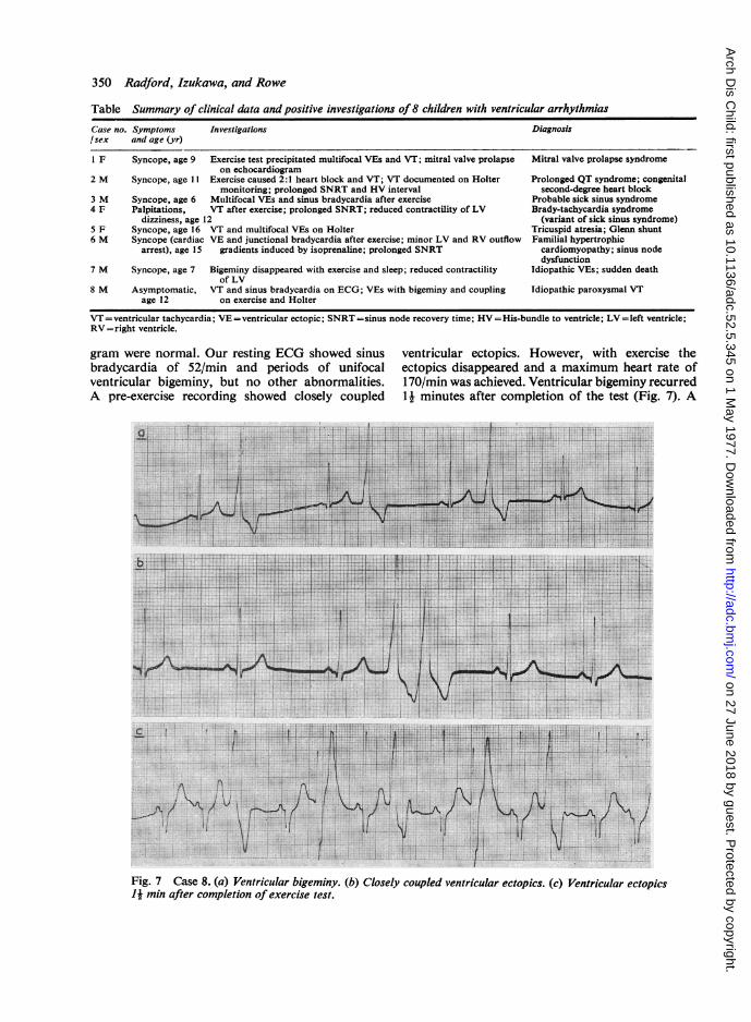
minary resting recording showed multifocal ventri-cular ectopics, some of which occurred consecutivelywith a short coupling interval. Holter monitoringshowed runs of ventricular tachycardia. Treatmentwith quinidine was started and the effect monitoredby serial Holter recordings. Increasing dosage failedto suppress all serious arrhythmias (Fig. 6), and itwas not till propranolol was added that the runsof ventricular ectopics were suppressed. She con-tinues to take a combination of quinidine andpropranolol and has had no further syncope.
Case 6. While jogging in gym class a 15-year-old boysuddenly collapsed. He was given cardiopulmonaryresuscitation and rushed to a local hospital where hewas found to be in ventricular fibrillation. Defibrilla-tion was successful. Subsequent examination showedneurological sequelae with parietal and cerebellardysfunction. Cardiac examination showed a softejection systolic murmur with a normal second heartsound. Chest x-ray was normal. ECG showed sinusrhythm with right bundle branch block, right axisdeviation, and left atrial hypertrophy. There wereno ectopic beats.A 16-hour Holter record showed sinus rhythm
throughout with heart rate varying from 50 to100/min and occasional single unifocal ventricularectopic beats. On exercise testing the heart rateincreased from 65 to 185/min and only one ventri-cular ectopic occurred. After the test, sinus arrestand junctional bradycardia at 45/min occurredbefore sinus rhythm resumed. An echocardiogramshowed thickened ventricular walls and septum,enlargement of the left atrium and both ventricularcavities, and decreased contractility.
Previous cardiological data on the patient werereviewed. He had been studied twice previously bycardiac catheterization. The diagnosis from thefirst study was mild pulmonary valve stenosis. At
.... EU,
A..:91.:L:. 4V
on 27 June 2018 by guest. Protected by copyright.
http://adc.bmj.com
/A
rch Dis C
hild: first published as 10.1136/adc.52.5.345 on 1 May 1977. D
ownloaded from
Evaluation of children with ventricular arrhythmias 349
Fig. 5 Case 4. Ventricular tachycardia after exercise.
Fig. 6 Case 5. Recording from Holter monitor shows ventricular ectopics and ventricular tachycardia.
the age of 11 years a second catheterization showedpulmonary infundibular narrowing with a restinggradient of 20 mmHg which increased to 35 mmHgwith an isoprenaline infusion. No gradient wasrecorded across the left ventricular outflow tractbefore or after provocation.A third catheter study was performed after his
syncopal episode. Isoprenaline produced a gradientof 14 mmHg across the left ventricular outflow tractand of 20 mmHg across the pulmonary infundi-bulum. Angiography showed thickening of theventricular walls with diminished contractility.The interventricular septum was abnormally thick.His-bundle intervals were normal but sinus noderecovery time was abnormally prolonged. As anextensive family history of obstructive cardiomyo-pathy has since been found, the concluding diagnosisis familial hypertrophic cardiomyopathy and sinusnode dysfunction. The patient has been started onpropranolol therapy and remains well.
Case 7. A 7-year-old boy had a history of dizzinessand three definite syncopal attacks. ECG showedsinus rhythm with unifocal ventricular ectopics withbigeminy and occasional trigeminy. The nonectopicQRS-T wave complexes were normal. Clinical
examination, chest x-ray, echocardiogram, andelectroencephalogram were normal. Cardiac cathe-terization showed normal anatomy and haemo-dynamics, but on angiography the left ventricularcontractility appeared reduced.A 16-hour Holter recording showed disappearance
of the ectopics with sleep and recurrence on arousal.Exercise testing suppressed the ectopics and hedeveloped normal sinus rhythm with an appropriateincrease in heart rate. Bigeminal rhythm returnedafter exercise. No change in these responses occurredwhen taking oral propranolol. Challenge testing bycarotid sinus massage, Valsalva manoeuvres, intra-venous phenylephrine (Neosynephrine), and iso-prenaline also produced no variation of the bigeminy.Since bigeminy alone was not considered dangerous,he was discharged on propranolol. Sudden deathoccurred 6 months later.
Case 8. An asymptomatic 12-year-old boy with nopast medical history was seen by his paediatricianfor an annual check-up. Physical examination wasnormal except for an irregular heart beat. ECGshowed short runs of ventricular tachycardia. There-fore cardiological assessment was requested.
Cardiac examination, chest x-ray, and echocardio-
on 27 June 2018 by guest. Protected by copyright.
http://adc.bmj.com
/A
rch Dis C
hild: first published as 10.1136/adc.52.5.345 on 1 May 1977. D
ownloaded from
350 Radford, Izukawa, and Rowe
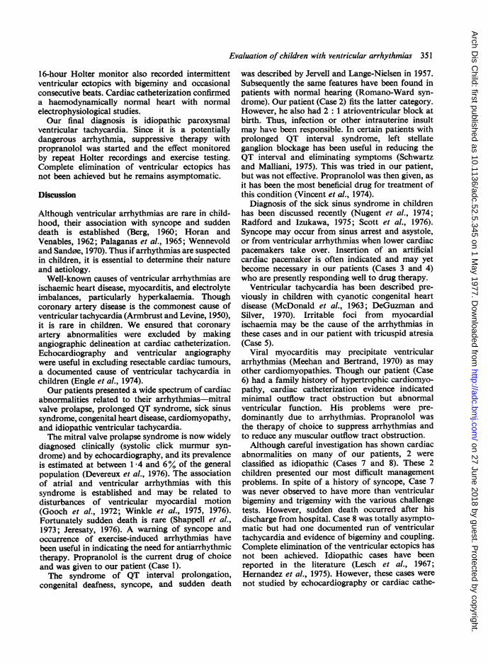
Table Summary of clinical data and positive investigations of8 children with ventricular arrhythmiasCase no. Symptoms Investigations DiagnosisIsex and age (yr)
1 F Syncope, age 9 Exercise test precipitated multifocal VEs and VT; mitral valve prolapse Mitral valve prolapse syndromeon echocardiogram
2 M Syncope, age 1 Exercise caused 2:1 heart block and VT; VT documented on Holter Prolonged QT syndrome; congenitalmonitoring; prolonged SNRT and HV interval second-degree heart block
3 M Syncope, age 6 Multifocal VEs and sinus bradycardia after exercise Probable sick sinus syndrome4 F Palpitations, VT after exercise; prolonged SNRT; reduced contractility of LV Brady-tachycardia syndrome
dizziness, age 12 (variant of sick sinus syndrome)5 F Syncope, age 16 VT and multifocal VEs on Holter Tricuspid atresia; Glenn shunt6 M Syncope (cardiac VE and junctional bradycardia after exercise; minor LV and RV outflow Familial hypertrophic
arrest), age 15 gradients induced by isoprenaline; prolonged SNRT cardiomyopathy; sinus nodedysfunction
7 M Syncope, age 7 Bigeminy disappeared with exercise and sleep; reduced contractility Idiopathic VEs; sudden deathof LV
8 M Asymptomatic, VT and sinus bradycardia on ECG; VEs with bigeminy and coupling Idiopathic paroxysmal VTage 12 on exercise and Holter
VT= ventricular tachycardia; VE =ventricular ectopic; SNRT =sinus node recovery time; HV =His-bundle to ventricle; LV =left ventricle;RV= right ventricle.
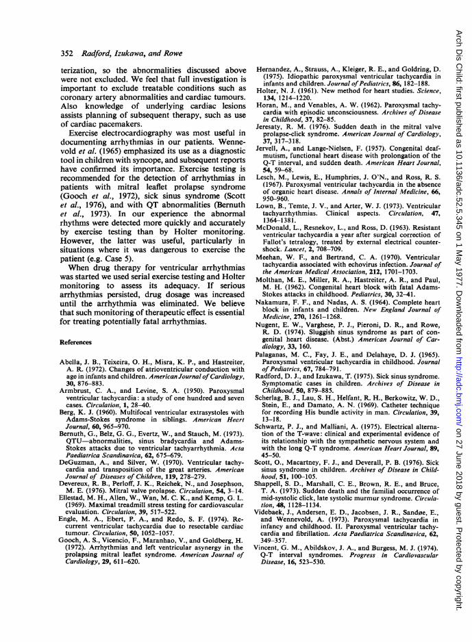
gram were normal. Our resting ECG showed sinusbradycardia of 52/min and periods of unifocalventricular bigeminy, but no other abnormalities.A pre-exercise recording showed closely coupled
ventricular ectopics. However, with exercise theectopics disappeared and a maximum heart rate of170/min was achieved. Ventricular bigeminy recurredIj minutes after completion of the test (Fig. 7). A
Fig. 7 Case 8. (a) Ventricular bigeminy. (b) Closely coupled ventricular ectopics. (c) Ventricular ectopicsJj min after completion of exercise test.
.:Jl: -li ...'J.- ;I. ;..Il. :..: .....
on 27 June 2018 by guest. Protected by copyright.
http://adc.bmj.com
/A
rch Dis C
hild: first published as 10.1136/adc.52.5.345 on 1 May 1977. D
ownloaded from
Evaluation of children with ventricular arrhythmias 351
16-hour Holter monitor also recorded intermittentventricular ectopics with bigeminy and occasionalconsecutive beats. Cardiac catheterization confirmeda haemodynamically normal heart with normalelectrophysiological studies.Our final diagnosis is idiopathic paroxysmal
ventricular tachycardia. Since it is a potentiallydangerous arrhythmia, suppressive therapy withpropranolol was started and the effect monitoredby repeat Holter recordings and exercise testing.Complete elimination of ventricular ectopics hasnot been achieved but he remains asymptomatic.
Discussion
Although ventricular arrhythmias are rare in child-hood, their association with syncope and suddendeath is established (Berg, 1960; Horan andVenables, 1962; Palaganas et al., 1965; Wennevoldand Sand0e, 1970). Thus if arrhythmias are suspectedin children, it is essential to determine their natureand aetiology.Well-known causes of ventricular arrhythmias are
ischaemic heart disease, myocarditis, and electrolyteimbalances, particularly hyperkalaemia. Thoughcoronary artery disease is the commonest cause ofventricular tachycardia (Armbrust and Levine, 1950),it is rare in children. We ensured that coronaryartery. abnormalities were excluded by makingangiographic delineation at cardiac catheterization.Echocardiography and ventricular angiographywere useful in excluding resectable cardiac tumours,a documented cause of ventricular tachycardia inchildren (Engle et al., 1974).Our patients presented a wide spectrum of cardiac
abnormalities related to their arrhythmias-mitralvalve prolapse, prolonged QT syndrome, sick sinussyndrome, congenital heart disease, cardiomyopathy,and idiopathic ventricular tachycardia.The mitral valve prolapse syndrome is now widely
diagnosed clinically (systolic click murmur syn-drome) and by echocardiography, and its prevalenceis estimated at between 1I4 and 6% of the generalpopulation (Devereux et al., 1976). The associationof atrial and ventricular arrhythmias with thissyndrome is established and may be related todisturbances of ventricular myocardial motion(Gooch et al., 1972; Winkle et al., 1975, 1976).Fortunately sudden death is rare (Shappell et al.,1973; Jeresaty, 1976). A warning of syncope andoccurrence of exercise-induced arrhythmias havebeen useful in indicating the need for antiarrhythmictherapy. Propranolol is the current drug of choiceand was given to our patient (Case 1).The syndrome of QT interval prolongation,
congenital deafness, syncope, and sudden death
was described by Jervell and Lange-Nielsen in 1957.Subsequently the same features have been found inpatients with normal hearing (Romano-Ward syn-drome). Our patient (Case 2) fits the latter category.However, he also had 2: 1 atrioventricular block atbirth. Thus, infection or other intrauterine insultmay have been responsible. In certain patients withprolonged QT interval syndrome, left stellateganglion blockage has been useful in reducing theQT interval and eliminating symptoms (Schwartzand Malliani, 1975). This was tried in our patient,but was not effective. Propranolol was then given, asit has been the most beneficial drug for treatment ofthis condition (Vincent et al., 1974).
Diagnosis of the sick sinus syndrome in childrenhas been discussed recently (Nugent et al., 1974;Radford and Izukawa, 1975; Scott et al., 1976).Syncope may occur from sinus arrest and asystole,or from ventricular arrhythmias when lower cardiacpacemakers take over. Insertion of an artificialcardiac pacemaker is often indicated and may yetbecome necessary in our patients (Cases 3 and 4)who are presently responding well to drug therapy.
Ventricular tachycardia has been described pre-viously in children with cyanotic congenital heartdisease (McDonald et al., 1963; DeGuzman andSilver, 1970). Irritable foci from myocardialischaemia may be the cause of the arrhythmias inthese cases and in our patient with tricuspid atresia(Case 5).
Viral myocarditis may precipitate ventriculararrhythmias (Meehan and Bertrand, 1970) as mayother cardiomyopathies. Though our patient (Case6) had a family history of hypertrophic cardiomyo-pathy, cardiac catheterization evidence indicatedminimal outflow tract obstruction but abnormalventricular function. His problems were pre-dominantly due to arrhythmias. Propranolol wasthe therapy of choice to suppress arrhythmias andto reduce any muscular outflow tract obstruction.Although careful investigation has shown cardiac
abnormalities on many of our patients, 2 wereclassified as idiopathic (Cases 7 and 8). These 2children presented our most difficult managementproblems. In spite of a history of syncope, Case 7was never observed to have more than ventricularbigeminy and trigeminy with the various challengetests. However, sudden death occurred after hisdischarge from hospital. Case 8 was totally asympto-matic but had one documented run of ventriculartachycardia and evidence of bigeminy and coupling.Complete elimination of the ventricular ectopics hasnot been achieved. Idiopathic cases have beenreported in the literature (Lesch et al., 1967;Hernandez et al., 1975). However, these cases werenot studied by echocardiography or cardiac cathe-
on 27 June 2018 by guest. Protected by copyright.
http://adc.bmj.com
/A
rch Dis C
hild: first published as 10.1136/adc.52.5.345 on 1 May 1977. D
ownloaded from
352 Radford, Izukawa, and Rowe
terization, so the abnormalities discussed abovewere not excluded. We feel that full investigation isimportant to exclude treatable conditions such ascoronary artery abnormalities and cardiac tumours.Also knowledge of underlying cardiac lesionsassists planning of subsequent therapy, such as useof cardiac pacemakers.
Exercise electrocardiography was most useful indocumenting arrhythmias in our patients. Wenne-vold et al. (1965) emphasized its use as a diagnostictool in children with syncope, and subsequent reportshave confirmed its importance. Exercise testing isrecommended for the detection of arrhythmias inpatients with mitral leaflet prolapse syndrome(Gooch et al., 1972), sick sinus syndrome (Scottet al., 1976), and with QT abnormalities (Bernuthet al., 1973). In our experience the abnormalrhythms were detected more quickly and accuratelyby exercise testing than by Holter monitoring.However, the latter was useful, particularly insituations where it was dangerous to exercise thepatient (e.g. Case 5).When drug therapy for ventricular arrhythmias
was started we used serial exercise testing and Holtermonitoring to assess its adequacy. If seriousarrhythmias persisted, drug dosage was increaseduntil the arrhythmia was eliminated. We believethat such monitoring of therapeutic effect is essentialfor treating potentially fatal arrhythmias.
References
Abella, J. B., Teixeira, 0. H., Misra, K. P., and Hastreiter,A. R. (1972). Changes of atrioventricular conduction withage in infants and children. American Journal ofCardiology,30, 876-883.
Armbrust, C. A., and Levine, S. A. (1950). Paroxysmalventricular tachycardia: a study of one hundred and sevencases. Circulation, 1, 28-40.
Berg, K. J. (1960). Multifocal ventricular extrasystoles withAdams-Stokes syndrome in siblings. American HeartJournal, 60, 965-970.
Bernuth, G., Belz, G. G., Evertz, W., and Stauch, M. (1973).QTU-abnormalities, sinus bradycardia and Adams-Stokes attacks due to ventricular tachyarrhythmia. ActaPaediatrica Scandinavica, 62, 675-679.
DeGuzman, A., and Silver, W. (1970). Ventricular tachy-cardia and transposition of the great arteries. AmericanJournal of Diseases of Children, 119, 278-279.
Devereux, R. B., Perloff, J. K., Reichek, N., and Josephson,M. E. (1976). Mitral valve prolapse. Circulation, 54, 3-14.
Ellestad, M. H., Allen, W., Wan, M. C. K., and Kemp, G. L.(1969). Maximal treadmill stress testing for cardiovascularevaluation. Circulation, 39, 517-522.
Engle, M. A., Ebert, P. A., and Redo, S. F. (1974). Re-current ventricular tachycardia due to resectable cardiactumour. Circulation, 50, 1052-1057.
Gooch, A. S., Vicencio, F., Maranhao, V., and Goldberg, H.(1972). Arrhythmias and left ventricular asynergy in theprolapsing mitral leaflet syndrome. American Journal ofCardiology, 29, 611-620.
Hernandez, A., Strauss, A., Kleiger, R. E., and Goldring, D.(1975). Idiopathic paroxysmal ventricular tachycardia ininfants and children. Journal of Pediatrics, 86, 182-188.
Holter, N. J. (1961). New method for heart studies. Science,134, 1214-1220.
Horan, M., and Venables, A. W. (1962). Paroxysmal tachy-cardia with episodic unconsciousness. Archives of Diseasein Childhood, 37, 82-85.
Jeresaty, R. M. (1976). Sudden death in the mitral valveprolapse-click syndrome. American Journal of Cardiology,37, 317-318.
Jervell, A., and Lange-Nielsen, F. (1957). Congenital deaf-mutism, functional heart disease with prolongation of theQ-T interval, and sudden death. American Heart Journal,54, 59-68.
Lesch, M., Lewis, E., Humphries, J. O'N., and Ross, R. S.(1967). Paroxysmal ventricular tachycardia in the absenceof organic heart disease. Annals of Internal Medicine, 66,950-960.
Lown, B., Temte, J. V., and Arter, W. J. (1973). Ventriculartachyarrhythmias. Clinical aspects. Circulation, 47,1364-1381.
McDonald, L., Resnekov, L., and Ross, D. (1963). Resistantventricular tachycardia a year after surgical correction ofFallot's tetralogy, treated by external electrical counter-shock. Lancet, 2, 708-709.
Meehan, W. F., and Bertrand, C. A. (1970). Ventriculartachycardia associated with echovirus infection. Journal ofthe American Medical Association, 212, 1701-1703.
Molthan, M. E., Miller, R. A., Hastreiter, A. R., and Paul,M. H. (1962). Congenital heart block with fatal Adams-Stokes attacks in childhood. Pediatrics, 30, 32-41.
Nakamura, F. F., and Nadas, A. S. (1964). Complete heartblock in infants and children. New England Journal ofMedicine, 270, 1261-1268.
Nugent, E. W., Varghese, P. J., Pieroni, D. R., and Rowe,R. D. (1974). Sluggish sinus syndrome as part of con-genital heart disease. (Abst.) American Journal of Car-diology, 33, 160.
Palaganas, M. C., Fay, J. E., and Delahaye, D. J. (1965).Paroxysmal ventricular tachycardia in childhood. Journalof Pediatrics, 67, 784-791.
Radford, D. J., and Izukawa, T. (1975). Sick sinus syndrome.Symptomatic cases in children. Archives of Disease inChildhood, 50, 879-885.
Scherlag, B. J., Lau, S. H., Helfant, R. H., Berkowitz, W. D.,Stein, E., and Damato, A. N. (1969). Catheter techniquefor recording His bundle activity in man. Circulation, 39,13-18.
Schwartz, P. J., and Malliani, A. (1975). Electrical alterna-tion of the T-wave: clinical and experimental evidence ofits relationship with the sympathetic nervous system andwith the long Q-T syndrome. American Heart Journal, 89,45-50.
Scott, O., Macartney, F. J., and Deverall, P. B. (1976). Sicksinus syndrome in children. Archives of Disease in Child-hood, 51, 100-105.
Shappell, S. D., Marshall, C. E., Brown, R. E., and Bruce,T. A. (1973). Sudden death and the familial occurrence ofmid-systolic click, late systolic murmur syndrome. Circula-tion, 48, 1128-1134.
Videbaek, J., Andersen, E. D., Jacobsen, J. R., Sand0e, E.,and Wennevold, A. (1973). Paroxysmal tachycardia ininfancy and childhood. II. Paroxysmal ventricular tachy-cardia and fibrillation. Acta Paediatrica Scandinavica, 62,349-357.
Vincent, G. M., Abildskov, J. A., and Burgess, M. J. (1974).Q-T interval syndromes. Progress in CardiovascularDisease, 16, 523-530.
on 27 June 2018 by guest. Protected by copyright.
http://adc.bmj.com
/A
rch Dis C
hild: first published as 10.1136/adc.52.5.345 on 1 May 1977. D
ownloaded from
Evaluation of children with ventricular arrhythmias 353
Wennevold, A., and Sand0e, E. (1970). Paroxysmal malignantarrhythmias in childhood and adolescence. Symposium onCardiac Arrhythmias, p. 767. Ed. by E. Sand0e, E. Flensted-Jensen and K. H. Olesen. Astra, S6dertalje.
Wennevold, A., Melchior, J. C., and Sand0e, E. (1965).Adams-Stokes syndrome in children without organic heartdisease. Electrocardiogram after exercise as a diagnostictool. Acta Medica Scandinavica, 177, 557-563.
Winkle, R. A., Lopes, M. G., Fitzgerald, J. W., Goodman,D. J., Schroeder, J. S., and Harrison, D. C. (1975).
Arrhythmias in patients with mitral valve prolapse.Circulation, 52, 73-81.
Winkle, R. A., Lopes, M. C., Popp, R. L., and Hancock,E. W. (1976). Life-threatening arrhythmias in the mitralvalve prolapse syndrome. American Journal of Medicine,60, 961-967.
Correspondence to Dr. T. Izukawa, Hospital forSick Children, 555 University Avenue, Toronto,Ontario M5G 1X8, Canada.
on 27 June 2018 by guest. Protected by copyright.
http://adc.bmj.com
/A
rch Dis C
hild: first published as 10.1136/adc.52.5.345 on 1 May 1977. D
ownloaded from