estrogen receptors in glucose homeostasiscdn.intechweb.org/pdfs/22058.pdf · estrogen receptors in...
TRANSCRIPT
5
Estrogen Receptors in
Glucose Homeostasis
Malin Hedengran Faulds and Karin Dahlman-Wright Karolinska Institutet
Sweden
1. Introduction
Metabolic diseases affect more than 230 million people worldwide with an expectancy to
increase to around 350 million in the coming 25 years. It is currently the fourth leading cause
of death by disease. The metabolic syndrome refers to a group of interrelated metabolic
abnormalities that include disturbed glucose homeostasis, insulin resistance (IR), increased
body weight and abdominal fat accumulation, mild dyslipidemia and hypertension.
Individuals with the metabolic syndrome have an increased risk of cardiovascular disease
(CVD) and Type 2 diabetes (T2D).
Estrogens have traditionally been connected with female reproduction, however, the
importance of these hormones in tissues outside of the reproductive system including the
liver, bone, the cardiovascular system and brain have since been established (Gruber et al.,
2002). Estrogen and the estrogen receptors (ERs) are well-known regulators of glucose
homeostasis and several epidemiological and prospective studies associate estrogen to
various aspects of the metabolic syndrome (Louet et al., 2004). Postmenopausal women
develop visceral obesity, IR and are at high risk for T2D. Treatment of healthy
postmenopausal women with estrogen has been shown to improve insulin sensitivity and to
lower blood glucose (Crespo et al., 2002). Furthermore, hormone replacement therapy (HRT)
in postmenopausal women with coronary artery disease was associated with a 35%
reduction in the incidence of T2D (Kanaya et al., 2003). Male aromatase-deficient patients, as
well as a male patient with loss of ER┙ function, display impaired glucose metabolism, IR
and hyperinsulinemia (Zirilli, et al. 2008). In addition, the aromatase deficient patients
showed impaired liver functions, hepatic steatosis, and altered lipid profile (Maffei, et al.
2004). Additional observations in rodents support the notion that estrogen mediates anti-
diabetic effects. For example, female rodents are protected against hyperglycemia, unless
they are ovariectomized, in spontaneous rodent models of T2D.
Studies in knock-out mouse models have shed light on the role of estrogen and its receptors
in rodent obesity and glucose tolerance. Mice with functional knock-out of the aromatase
enzyme (ArKO mice) are unable to synthesize endogenous estrogen and display an obese
and insulin resistant phenotype (Fisher et al., 1998). A similar phenotype was observed in
mice lacking ER┙ (ER┙KO) but not in mice lacking ER┚ (ER┚KO), indicating that ER┙ is the
major mediator for the estrogenic effects on insulin sensitivity and body weight (Heine et
www.intechopen.com
Update on Mechanisms of Hormone Action – Focus on Metabolism, Growth and Reproduction 70
al., 2000). Although ERα appears to be more important in relation to body weight and
insulin sensitivity, it remains possible that ERβ also contributes in this context, particularly
in older mice and under specific metabolic conditions (Foryst-Ludwig et al., 2008).
2. Carbohydrate metabolism
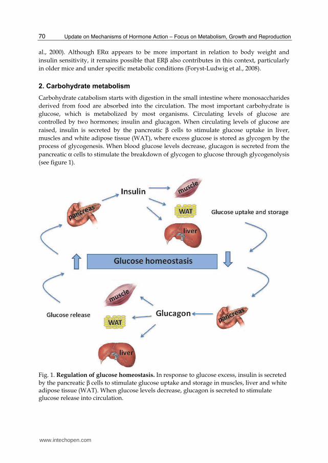
Carbohydrate catabolism starts with digestion in the small intestine where monosaccharides
derived from food are absorbed into the circulation. The most important carbohydrate is
glucose, which is metabolized by most organisms. Circulating levels of glucose are
controlled by two hormones; insulin and glucagon. When circulating levels of glucose are
raised, insulin is secreted by the pancreatic β cells to stimulate glucose uptake in liver,
muscles and white adipose tissue (WAT), where excess glucose is stored as glycogen by the
process of glycogenesis. When blood glucose levels decrease, glucagon is secreted from the
pancreatic α cells to stimulate the breakdown of glycogen to glucose through glycogenolysis
(see figure 1).
Fig. 1. Regulation of glucose homeostasis. In response to glucose excess, insulin is secreted
by the pancreatic β cells to stimulate glucose uptake and storage in muscles, liver and white adipose tissue (WAT). When glucose levels decrease, glucagon is secreted to stimulate glucose release into circulation.
www.intechopen.com
Estrogen Receptors in Glucose Homeostasis 71
The β cells in the pancreatic islets of Langerhans release insulin in two phases. The first is a rapid response to increase blood glucose levels and the second phase is a slow release that is triggered independently of glucose. There are several substances apart from glucose known to stimulate insulin release, including amino acids from dietary proteins, acetylcholine released from vagus nerve endings, gastrointestinal hormones and glucose-dependent insulinotropic peptides (reviewed by (Kieffer et al., 1996)). The hormone glucagon is secreted from the α cells in the pancreatic islets of Langerhans and promotes conversion of hepatic glycogen into glucose, which is subsequently released into the blood. The output of glucagon is triggered by low levels of circulating glucose (Nussey & Whitehead, 2001). IR is a physiological condition where insulin is less effective in lowering circulating glucose. IR in muscle and WAT reduces glucose uptake whereas hepatic IR results in reduced glycogen synthesis and storage and a failure of insulin to suppress glucose production and subsequent release into the blood (reviewed by (Benito, 2011)). IR commonly refers to the reduced glucose lowering effects of insulin as described above. However, other functions of insulin are also affected. For example, IR in adipocytes results in reduced uptake of circulating lipids and increased hydrolysis of stored triglycerides, which leads to elevated levels of circulating free fatty acids (Savage et al., 2007). High plasma levels of insulin, glucose and lipids due to IR are major components of the metabolic syndrome, which could develop into T2D.
3. Estrogen signaling and estrogen receptors
Estrogens are sex steroids, which stem from the common pre-cursor cholesterol. The last step in the synthesis of estrogen from androgens is catalyzed by the P450 enzyme aromatase. The three major physiological estrogens include 17β-estradiol (E2), estrone (E1) and estriol. The major physiological estrogen in fertile females is E2, which has a similar affinity for both ERs. In addition, ERs are activated by a range of synthetic ligands including selective estrogen receptor modulators (SERMs) such as raloxifen and tamoxifen, the ER┙ selective agonist propyl-pyrazole-triol (PPT) and the ER┚-selective agonist diarylpropionitrile (DPN) (Heldring et al., 2007). Estrogens exert their physiological effects through the two ER subtypes, ER┙ and ER┚, which are members of the superfamily of nuclear receptors. The human ESR1 gene, which is encoding for ERα, is located on the chromosome 6, at 6q25.1, and includes 8 exons. The ERβ encoding gene, ESR2, is located on chromosome 14 at 14q22–24. ERα is mainly expressed in reproductive tissues, kidney, bone, WAT and liver, while ERβ is expressed in the ovary, prostate, lung, gastrointestinal tract, bladder and the central nervous systems (CNS) (Matthews & Gustafsson, 2003). Genetic associations have been described for polymorphisms of the ESR1 gene and several pathological conditions related to metabolism in general, including cardiovascular diseases, T2D, myocardial infarction, hypertension and venous thromboembolism (Schuit et al., 2004; Shearman et al., 2003; Yoshihara et al., 2009). Polymorphisms of the ESR1 gene can also affect lipoprotein metabolism (Lamon-Fava et al., 2010). ESR2 polymorphisms have been associated with anorexia nervosa, bulimic disease and premature coronary artery disease (Eastwood et al., 2002; Nilsson et al., 2004; Peter et al., 2005). ERs share a common structure with the other members of the nuclear receptor family. The N-terminal A/B domain is the most variable region with less than 20% amino acid identity between the two ERs and confers subtype specific actions on target genes. This region
www.intechopen.com
Update on Mechanisms of Hormone Action – Focus on Metabolism, Growth and Reproduction 72
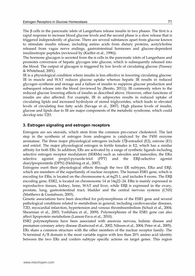
harbors the activation function-1 (AF-1), which is ligand-independent and shows promoter- and cell-specific activities. The centrally located C-domain harbors the DNA binding domain (DBD), which is involved in DNA binding and receptor dimerization. This domain
is highly conserved between ERα and ERβ with 97% amino acid identity. The D-domain is
referred to as the hinge domain and displays low conservation between ERα and ERβ (30%). This domain has been shown to contain a nuclear localization signal. The C-terminal E-domain contains the ligand-binding domain (LBD) and the two subtypes display 56% conservation in this region. The LBD contains a hormone-dependent activation function (AF-2) and also includes functions responsible for ligand binding and receptor dimerization. The F-domain has less than 20% amino acid identity between the two ER subtypes and the functions of this domain remain undefined (Zhao et al., 2008).
Fig. 2. Structure and homology between human ERα and ERβ. The A/B domain is referring to the ligand independent transcription activation function-1 (AF-1). The C domain is mediating DNA binding and the D domain represents the hinge domain, which harbors nuclear localization signals. The E domain is involved in ligand binding and contains the ligand dependent AF-2 function, which is involved in ligand binding. Depicted is also the homology in percent between the various domains between the two subtypes.
Like other nuclear receptors, ligand-bound ERs act as dimers to regulate transcriptional
activation. Full transcriptional activity of the ERs is mediated through a synergistic action
between the two activation domains, AF-1 and AF-2. Both ERα and ERβ contain a potent
AF-2 function, but unlike ERα, ERβ seems to have a weaker corresponding AF-1 function
and depends more on the ligand-dependent AF-2 for its transcriptional activation function
(Dahlman-Wright et al., 2006). In their unliganded state, ERs are associated with protein
complexes of heat shock proteins, which inhibit their functions.
The classical estrogen signaling occurs through a direct binding of ligand activated ER
dimers to estrogen-responsive elements (EREs) in the regulatory regions of estrogen target
genes followed by activation of the transcriptional machinery at the transcription start sites
of regulated genes. Estrogen also modulates gene expression by a second mechanism in
which ERs interact with other DNA bound transcription factors, such as activating protein-1
(AP-1) and stimulating protein-1 (Sp-1) to regulate gene expression, through a process
referred to as transcription factor cross-talk. Estrogen may also elicit effects through non-
genomic mechanisms, which involve the activation of downstream signaling cascades like
protein kinase A (PKA), protein kinase C (PKC) and mitogen-activated protein (MAP)
kinase via membrane-localized ERs.
www.intechopen.com
Estrogen Receptors in Glucose Homeostasis 73
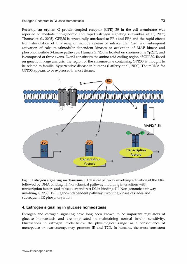
Recently, an orphan G protein-coupled receptor (GPR) 30 in the cell membrane was
reported to mediate non-genomic and rapid estrogen signaling (Revankar et al., 2005;
Thomas et al., 2005). GPR30 is structurally unrelated to ERα and ERβ and the rapid effects
from stimulation of this receptor include release of intracellular Ca2+ and subsequent
activation of calcium-calmodulin-dependent kinases or activation of MAP kinase and
phosphoinositide 3-kinase pathways. Human GPR30 is located on chromosome 7p22.3, and
is composed of three exons. Exon3 constitutes the amino acid coding region of GPR30. Based
on genetic linkage analysis, the region of the chromosome containing GPR30 is thought to
be related to familial hypertensive disease in humans (Lafferty et al., 2000). The mRNA for
GPR30 appears to be expressed in most tissues.
Fig. 3. Estrogen signaling mechanisms. I. Classical pathway involving activation of the ERs followed by DNA binding. II. Non-classical pathway involving interactions with transcription factors and subsequent indirect DNA binding. III. Non-genomic pathway involving GPR30. IV. Ligand-independent pathway involving kinase cascades and subsequent ER phosphorylation.
4. Estrogen signaling in glucose homeostasis
Estrogen and estrogen signaling have long been known to be important regulators of
glucose homeostasis and are implicated in maintaining normal insulin sensitivity.
Fluctuations in estrogen levels below the physiological range, as a consequence of
menopause or ovariectomy, may promote IR and T2D. In humans, the most consistent
www.intechopen.com
Update on Mechanisms of Hormone Action – Focus on Metabolism, Growth and Reproduction 74
effects of oral contraceptives or HRT are decreased levels of fasting plasma glucose and
improved glucose tolerance. Absence of estrogen signaling in men, due to deficiency of the
aromatase enzyme or ER┙, results in impaired glucose metabolism. It has also been shown
that polymorphisms in the ER┙ gene are associated with development of the metabolic
syndrome and T2D (Yoshihara et al., 2009).
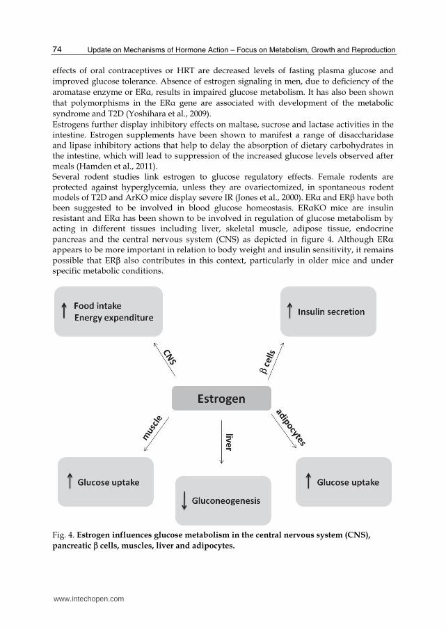
Estrogens further display inhibitory effects on maltase, sucrose and lactase activities in the intestine. Estrogen supplements have been shown to manifest a range of disaccharidase and lipase inhibitory actions that help to delay the absorption of dietary carbohydrates in the intestine, which will lead to suppression of the increased glucose levels observed after meals (Hamden et al., 2011). Several rodent studies link estrogen to glucose regulatory effects. Female rodents are protected against hyperglycemia, unless they are ovariectomized, in spontaneous rodent models of T2D and ArKO mice display severe IR (Jones et al., 2000). ER┙ and ER┚ have both been suggested to be involved in blood glucose homeostasis. ER┙KO mice are insulin resistant and ER┙ has been shown to be involved in regulation of glucose metabolism by acting in different tissues including liver, skeletal muscle, adipose tissue, endocrine
pancreas and the central nervous system (CNS) as depicted in figure 4. Although ERα appears to be more important in relation to body weight and insulin sensitivity, it remains
possible that ERβ also contributes in this context, particularly in older mice and under specific metabolic conditions.
Fig. 4. Estrogen influences glucose metabolism in the central nervous system (CNS),
pancreatic β cells, muscles, liver and adipocytes.
www.intechopen.com
Estrogen Receptors in Glucose Homeostasis 75
4.1 ERs and the role of the central nervous system in glucose homeostasis
The first finding supporting that the central nervous system (CNS) was involved in the regulation of glucose homeostasis was that ruptures in the fourth ventricle resulted in glucosuria. This initial study was followed by numerous other studies and it is now firmly established that the CNS regulates glucose homeostasis through the hormones insulin, leptin and glucagon-like peptide (GLP)-1, as well as by glucose and fatty acids (FA). A series of complex systems regulate energy homeostasis in order to keep energy levels and body weight stable (Miller, 1982). Glucose is the vital energy source for the brain. There are several glucose sensing neurons in the hypothalamus, which have been established to be essential components in the regulation of feeding behavior and hypoglycemic counter-regulatory responses (reviewed in (Marty et al., 2007)). The hypothalamus is subdivided into interconnecting nuclei, including the arcuate nucleus (ARC), paraventricular nucleus (PVN), ventromedial nucleus (VMN), dorsomedial nucleus (DMN) and lateral hypothalamic area (LHA) (Simpson et al., 2009). These central brain circuits receive signals from the periphery, which indicate satiety, energy levels and energy stores (Morton et al, 2006) and process these afferent signals to modulate food intake and energy expenditure. The actions of insulin have also been shown to play a direct role in the CNS since neuron-specific insulin receptor deficient (NIRKO) mice develop mild IR and display elevated circulating insulin levels (Bruning et al., 2000). Injections of insulin directly into the third cerebral ventricle have been shown to suppress hepatic glucose production without effecting body weight or circulating levels of insulin (Obici et al., 2002). Further, inhibition of insulin or its downstream signaling pathway in the CNS, i.e. the insulin receptor and phosphatidylinositol-3 kinase (PI3K), impaired the ability of increased levels of insulin to suppress gluconeogenesis. Targeted deletion of insulin receptor expression selectively in the hypothalamus elicited IR in rats, which is in accordance with the results in NIRKO mice. These studies show that the CNS regulates glucose homeostasis through the action of insulin and requires intact insulin signaling pathways involving the binding of insulin to its receptor and subsequent activation of down-stream mediators. Estrogen is known to be highly relevant for the regulation of satiety, energy expenditure and body weight. Ovariectomy and menopause are associated with increased food intake, which can be reversed with estrogen replacement therapy (Eckel, 2004; Tchernof et al., 2000). The anorectic effects of estrogen are partially mediated through actions in the hypothalamus as demonstrated by studies showing that direct E2 injections into the PVN area or the ARC/VMN of the hypothalamus effectively reduced food intake (Butera & Beikirch, 1989; Nunez et al., 1980). The same study also showed that the hypothalamic neurons, which regulate energy homeostasis, were affected by E2 administration. Energy homeostasis and feeding behavior controlled by the hypothalamus also follow the menstrual cycle and food intake in women varies across the cycle with the lowest daily food intake during the peri-ovulatory period when estrogen levels are peaking (Asarian & Geary, 2006). ER┙ and ER┚ are both expressed in the different areas of the hypothalamus (Gillies & McArthur, 2010). ER┙ appears to be the major mediator of the estrogenic effects on central regulation of body weight by estrogens but whether this is regulated by food intake or actions on energy expenditure is controversial. Total ER┙ knockout mice are obese with increased fat accumulation in the absence of increased food intake. Targeted disruption of ER┙ in the VMN areas in the hypothalamus of female mice leads to weight gain, increased visceral adiposity, hyperphagia, hyperglycemia and impaired energy expenditure (Musatov
www.intechopen.com
Update on Mechanisms of Hormone Action – Focus on Metabolism, Growth and Reproduction 76
et al., 2007). ER┚ knockout mice, on the other hand, display similar food consumption patterns as wild-type mice (Foryst-Ludwig et al., 2008).
4.2 ERs and the role of pancreatic β cells in glucose homeostasis
The endocrine pancreas is an adapting tissue with the capacity to quickly respond to variations in the metabolic status of the organism. The ┚ cells in the islets of Langerhans readily adapt to peripheral IR by increasing their secretory response, as well as their cell mass. If ┚ cells fail to compensate, blood glucose concentration will rise to pathological levels and frank T2D will develop. Estrogenic effects on various physiological aspects of the islet of Langerhans have been
known for a long time and estrogens are established regulators of pancreatic β cell functions. In humans, E2 reverses the effect of menopause on glucose and insulin metabolism, resulting in increased pancreatic insulin secretion, as well as improved insulin sensitivity (Brussaard et al., 1997; Stevenson et al., 1994). Plasma insulin levels are increased in pregnant rats in response to the increased levels of estrogen. Studies in mice have suggested that long-term exposure to E2 increased insulin content, insulin gene expression
and insulin release without changing β cell mass. E2 has also been shown to acutely enhance glucose stimulated insulin secretion at physiological concentrations through the action of ER┙ both in vitro and in vivo (Alonso-Magdalena et al., 2008; Nadal et al., 1998). ER┙ has been identified as the functional predominant receptor isoform in the murine pancreas. E2-dependent insulin release in cultured pancreatic islets was reduced in ER┙-deficient mice, when compared to islets derived from either ER┚-deficient or wild-type mice (Alonso-Magdalena et al., 2008). Also, E2, acting mainly through ER┙, has been shown to protect pancreatic ┚ cells from apoptosis induced by oxidative stress in mice.
Even though ERα seems to be the dominant subtype to convey the estrogenic response in
the pancreas, the role of ERβ might also be of importance. ER┚-deficient mice have been shown to display a mild islet hyperplasia and delayed first phase insulin release (Barros et al., 2009). The membrane bound estrogen-responsive GPR30 is also expressed in rodent pancreas. Studies using adult female GPR30-deficient mice reveal that these mice do not exhibit E2-induced release of insulin, which is consistent with experiments using isolated pancreatic islet cells in vitro (Martensson et al., 2009). There are not any differences in expression of glucose-related genes, such as the glucose transporter (GLUT) 2 and glucokinase, in GPR30 knockout mice when compared with wild-type mice (Martensson et al., 2009). Thus, GPR30 may act as a regulator of insulin release after E2 stimulation. Consistent with this, GPR30 mRNA is expressed in secretory gland cells, which may indicate that GPR30 is involved in insulin secretion pathways (Levin & Weissman, 2009).
4.3 ERs and the role of the liver in glucose homeostasis
The liver is the largest organ in the body and possesses purifying and metabolizing functions. One of its most important tasks is to store glucose in the form of glycogen. The liver is capable of containing up to 10% of its volume as glycogen. The liver releases glycogen when nutrients are scarce. Liver glycogen is converted into circulating glucose in response to pancreatic signals; in hypoglycemic conditions glucagon is released to stimulate a release of hepatic glycogen. In a hyperglycemic state, the pancreas releases insulin to stimulate the liver to release less glucose. The maintenance of glucose homeostasis is
www.intechopen.com
Estrogen Receptors in Glucose Homeostasis 77
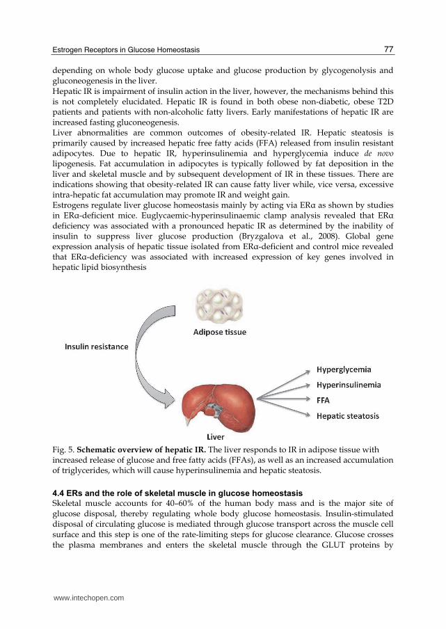
depending on whole body glucose uptake and glucose production by glycogenolysis and gluconeogenesis in the liver. Hepatic IR is impairment of insulin action in the liver, however, the mechanisms behind this is not completely elucidated. Hepatic IR is found in both obese non-diabetic, obese T2D patients and patients with non-alcoholic fatty livers. Early manifestations of hepatic IR are increased fasting gluconeogenesis. Liver abnormalities are common outcomes of obesity-related IR. Hepatic steatosis is primarily caused by increased hepatic free fatty acids (FFA) released from insulin resistant adipocytes. Due to hepatic IR, hyperinsulinemia and hyperglycemia induce de novo lipogenesis. Fat accumulation in adipocytes is typically followed by fat deposition in the liver and skeletal muscle and by subsequent development of IR in these tissues. There are indications showing that obesity-related IR can cause fatty liver while, vice versa, excessive intra-hepatic fat accumulation may promote IR and weight gain. Estrogens regulate liver glucose homeostasis mainly by acting via ER┙ as shown by studies in ER┙-deficient mice. Euglycaemic-hyperinsulinaemic clamp analysis revealed that ER┙ deficiency was associated with a pronounced hepatic IR as determined by the inability of insulin to suppress liver glucose production (Bryzgalova et al., 2008). Global gene expression analysis of hepatic tissue isolated from ER┙-deficient and control mice revealed that ER┙-deficiency was associated with increased expression of key genes involved in hepatic lipid biosynthesis
Fig. 5. Schematic overview of hepatic IR. The liver responds to IR in adipose tissue with increased release of glucose and free fatty acids (FFAs), as well as an increased accumulation of triglycerides, which will cause hyperinsulinemia and hepatic steatosis.
4.4 ERs and the role of skeletal muscle in glucose homeostasis
Skeletal muscle accounts for 40–60% of the human body mass and is the major site of glucose disposal, thereby regulating whole body glucose homeostasis. Insulin-stimulated disposal of circulating glucose is mediated through glucose transport across the muscle cell surface and this step is one of the rate-limiting steps for glucose clearance. Glucose crosses the plasma membranes and enters the skeletal muscle through the GLUT proteins by
www.intechopen.com
Update on Mechanisms of Hormone Action – Focus on Metabolism, Growth and Reproduction 78
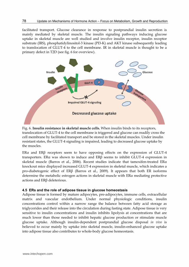
facilitated transport. Glucose clearance in response to postprandial insulin secretion is mainly mediated by skeletal muscle. The insulin signaling pathways inducing glucose uptake in skeletal muscle are well studied and involve insulin receptor, insulin receptor substrate (IRS), phosphatidylinositol-3 kinase (PI3-K) and AKT kinase subsequently leading to translocation of GLUT-4 to the cell membrane. IR in skeletal muscle is thought to be a primary defect in T2D (see fig. 6 for overview).
Fig. 6. Insulin resistance in skeletal muscle cells. When insulin binds to its receptors, translocation of GLUT-4 to the cell membrane is triggered and glucose can readily cross the cell membrane by facilitated transport and be stored in the skeletal muscles. Under insulin resistant states, the GLUT-4 signaling is impaired, leading to decreased glucose uptake by the muscles.
ER┙ and ER┚ receptors seem to have opposing effects on the expression of GLUT-4
transporters. ER┙ was shown to induce and ER┚ seems to inhibit GLUT-4 expression in
skeletal muscle (Barros et al., 2006). Recent studies indicate that tamoxifen-treated ER┙
knockout mice displayed increased GLUT-4 expression in skeletal muscle, which indicates a
pro-diabetogenic effect of ER┚ (Barros et al., 2009). It appears that both ER isoforms
determine the metabolic estrogen actions in skeletal muscle with ER┙ mediating protective
actions and ER┚ deleterious.
4.5 ERs and the role of adipose tissue in glucose homeostasis
Adipose tissue is formed by mature adipocytes, pre-adipocytes, immune cells, extracellular
matrix and vascular endothelium. Under normal physiologic conditions, insulin
concentrations control within a narrow range the balance between fatty acid storage as
triglycerides and their release into the circulation during fasting state. Adipose tissue is very
sensitive to insulin concentrations and insulin inhibits lipolysis at concentrations that are
much lower than those needed to inhibit hepatic glucose production or stimulate muscle
glucose uptake. Although insulin-dependent postprandial glucose disposal in vivo is
believed to occur mainly by uptake into skeletal muscle, insulin-enhanced glucose uptake
into adipose tissue also contributes to whole-body glucose homeostasis.
www.intechopen.com
Estrogen Receptors in Glucose Homeostasis 79
There are well-documented sex differences in the pathophysiology of obesity and metabolic disorders. Women tend to accumulate more subcutaneous fat whereas men accumulate more visceral fat (Bonds et al., 2006; Crespo et al., 2002; Nuutila et al., 1995). The prevalence of early IR and impaired glucose tolerance seem to be higher in men than in women. E2 is considered an important regulator of adipose tissue development and lipid deposition
in humans, rodents and other species. The deficiency of estrogen hormones after menopause
or in experimental models, causes an increase in body mass and intra-abdominal adipose
tissue leading to an android feature. The effects observed as a consequence of deficiency of
sex steroids can be reversed by hormone replacement therapy.
Changes in adipose tissue distribution in women have been associated with increased risk
of diseases and metabolic disturbances, including coronary artery disease, IR and glucose
intolerance (Bonds et al., 2006). In obesity and T2D, there is a marked adipocyte resistance to
the anti-lipolytic effects of insulin and the circulating FFA concentrations are typically
elevated (Kissebah et al., 1976). Chronic over feeding induces metabolic stress and the
adipocytes become hypertrophic and fail to proliferate and differentiate in a sufficient
manner.
Sex hormone programming in animals has also been shown to affect adipose tissue. A single
postnatal injection of estradiol benzoate resulted in the development of IR, increased
adipose tissue mass and adipocyte size in adult female rats, suggesting that postnatal ER
activation exerts strong programming effects on metabolic processes (Alexanderson et al.,
2010).
Stromal cells from adipose tissue have been shown to locally produce estrogens (Simpson et
al., 2009). It is well confirmed that there is a decrease in steroid hormone-binding globulins
in obese states in both pre- and post-menopausal women. In post-menopausal women, there
is a direct association between estrogen levels and body mass index (BMI) (Cleary &
Grossmann, 2009; Lukanova et al., 2004). E2 is mainly produced by the adipocytes after
menopause through conversion of androgens or estrone and this production is not regulated
by feedback mechanisms (Siiteri, 1987). It is suggested that this adipose-derived E2 may
participate in the ER┙-mediated enrichment of insulin biosynthesis (Alonso-Magdalena et
al., 2008) and in the induction of glucose stimulated insulin secretion to help the pancreatic
┚–cells adapt to the higher demand of insulin during obesity.
5. Concluding remarks
Lifestyle evolution and the higher intake of high calorie diets largely contribute to the
worldwide growing incidence of metabolic diseases. In addition to the most common
preventive strategy based on physical activity and reduced calorie intake, identification of
new molecular targets able to limit the development of metabolic disturbances represents
one of the most important public health challenges.
Estrogens have emerged as important regulators of glucose homeostasis during the last
decades, corroborating data from clinical and experimental studies. Insulin sensitivity has
been demonstrated to be higher in women before menopause than in age-matched men and
postmenopausal women, which supports a beneficial effect of estrogens on insulin action
and glucose homeostasis. It is also highly recognized that menopause promotes visceral fat
accumulation and IR, which will ultimately lead to significantly higher risk of developing
T2D. In addition, HRT has been reported to reverse the symptoms and to dampen the
www.intechopen.com
Update on Mechanisms of Hormone Action – Focus on Metabolism, Growth and Reproduction 80
incidence of T2D in postmenopausal women by 21–35% compared to women not given
HRT.
Effects of estrogen signaling on glucose homeostasis have been further demonstrated by
studies showing that patients bearing genetic mutations and, thus, lack either ERα or
aromatase expression, develop obesity, IR and impaired glucose tolerance. Genetically
engineered mice models have confirmed these clinical observations, as ERα or aromatase
gene deficiency similarly promotes several features of the metabolic syndrome. Taken
together, ERα seems to play a protective role in insulin and glucose metabolism, with
actions on the liver, adipose tissue, muscle and pancreatic β cells. In addition, ERα regulates
food intake and energy expenditures through actions on the CNS. ERβ seems to have an
opposing role with the potential to negatively influence insulin and glucose metabolism by
impairing the function of adipose tissue and inhibiting the expression of GLUT4 in the
muscle.
Established and novel ER subtype selective ligands are valuable tools for deciphering the
specific roles of ER┙ and ER┚ in physiology and disease. The development of novel
treatment regimes for metabolic disease targeting ER┙ is hampered by the uterotrophic and
mammotrophic effects of ER┙ with the major concern being the risk of developing hormone-
dependent cancer. Further studies are needed to identify and develop novel compounds
that target estrogen signaling in selective metabolic tissues but lack the mitogenic effects in
others, like ovaries and the breast.
6. References
Alexanderson, C., Stener-Victorin, E., Kullberg, J., Nilsson, S., Levin, M., Cajander, S., et al. (2010) A single early postnatal estradiol injection affects morphology and gene expression of the ovary and parametrial adipose tissue in adult female rats. J Steroid Biochem Mol Biol, 122(1-3), 82-90.
Alonso-Magdalena, P., Ropero, A. B., Carrera, M. P., Cederroth, C. R., Baquie, M., Gauthier, B. R., et al. (2008). Pancreatic insulin content regulation by the estrogen receptor ER alpha. PLoS One, 3(4), e2069.
Asarian, L., & Geary, N. (2006). Modulation of appetite by gonadal steroid hormones. Philos Trans R Soc Lond B Biol Sci, 361(1471), 1251-1263.
Barros, R. P., Gabbi, C., Morani, A., Warner, M., & Gustafsson, J. A. (2009). Participation of ERalpha and ERbeta in glucose homeostasis in skeletal muscle and white adipose tissue. Am J Physiol Endocrinol Metab, 297(1), E124-133.
Barros, R. P., Machado, U. F., Warner, M., & Gustafsson, J. A. (2006). Muscle GLUT4 regulation by estrogen receptors ERbeta and ERalpha. Proc Natl Acad Sci U S A, 103(5), 1605-1608.
Benito, M. Tissue specificity on insulin action and resistance: past to recent mechanisms. (2011) Acta Physiol (Oxf), 201(3), 297-312.
Bonds, D. E., Lasser, N., Qi, L., Brzyski, R., Caan, B., Heiss, G., et al. (2006). The effect of conjugated equine oestrogen on diabetes incidence: the Women's Health Initiative randomised trial. Diabetologia, 49(3), 459-468.
Bruning, J. C., Gautam, D., Burks, D. J., Gillette, J., Schubert, M., Orban, P. C., et al. (2000). Role of brain insulin receptor in control of body weight and reproduction. Science, 289(5487), 2122-2125.
www.intechopen.com
Estrogen Receptors in Glucose Homeostasis 81
Brussaard, H. E., Gevers Leuven, J. A., Frolich, M., Kluft, C., & Krans, H. M. (1997). Short-term oestrogen replacement therapy improves insulin resistance, lipids and fibrinolysis in postmenopausal women with NIDDM. Diabetologia, 40(7), 843-849.
Bryzgalova, G., Lundholm, L., Portwood, N., Gustafsson, J. A., Khan, A., Efendic, S., et al. (2008). Mechanisms of antidiabetogenic and body weight-lowering effects of estrogen in high-fat diet-fed mice. Am J Physiol Endocrinol Metab, 295(4), E904-912.
Butera, P. C., & Beikirch, R. J. (1989). Central implants of diluted estradiol: independent effects on ingestive and reproductive behaviors of ovariectomized rats. Brain Res, 491(2), 266-273.
Cleary, M. P., & Grossmann, M. E. (2009). Minireview: Obesity and breast cancer: the estrogen connection. Endocrinology, 150(6), 2537-2542.
Crespo, C. J., Smit, E., Snelling, A., Sempos, C. T., & Andersen, R. E. (2002). Hormone replacement therapy and its relationship to lipid and glucose metabolism in diabetic and nondiabetic postmenopausal women: results from the Third National Health and Nutrition Examination Survey (NHANES III). Diabetes Care, 25(10), 1675-1680.
Dahlman-Wright, K., Cavailles, V., Fuqua, S. A., Jordan, V. C., Katzenellenbogen, J. A., Korach, K. S., et al. (2006). International Union of Pharmacology. LXIV. Estrogen receptors. Pharmacol Rev, 58(4), 773-781.
Eastwood, H., Brown, K. M., Markovic, D., & Pieri, L. F. (2002). Variation in the ESR1 and ESR2 genes and genetic susceptibility to anorexia nervosa. Mol Psychiatry, 7(1), 86-89.
Eckel, L. A. (2004). Estradiol: a rhythmic, inhibitory, indirect control of meal size. Physiol Behav, 82(1), 35-41.
Fisher, C. R., Graves, K. H., Parlow, A. F., & Simpson, E. R. (1998). Characterization of mice deficient in aromatase (ArKO) because of targeted disruption of the cyp19 gene. Proc Natl Acad Sci U S A, 95(12), 6965-6970.
Foryst-Ludwig, A., Clemenz, M., Hohmann, S., Hartge, M., Sprang, C., Frost, N., et al. (2008). Metabolic actions of estrogen receptor beta (ERbeta) are mediated by a negative cross-talk with PPARgamma. PLoS Genet, 4(6), e1000108.
Gillies, G. E., & McArthur, S. (2010) Estrogen actions in the brain and the basis for differential action in men and women: a case for sex-specific medicines. Pharmacol Rev, 62(2), 155-198.
Gruber, C. J., Tschugguel, W., Schneeberger, C., & Huber, J. C. (2002). Production and actions of estrogens. N Engl J Med, 346(5), 340-352.
Hamden, K., Jaouadi, B., Zarai, N., Rebai, T., Carreau, S., & Elfeki, A. (2011). Inhibitory effects of estrogens on digestive enzymes, insulin deficiency, and pancreas toxicity in diabetic rats. J Physiol Biochem, 67(1), 121-128.
Heine, P. A., Taylor, J. A., Iwamoto, G. A., Lubahn, D. B., & Cooke, P. S. (2000). Increased adipose tissue in male and female estrogen receptor-alpha knockout mice. Proc Natl Acad Sci U S A, 97(23), 12729-12734.
Heldring, N., Pike, A., Andersson, S., Matthews, J., Cheng, G., Hartman, J., et al. (2007). Estrogen receptors: how do they signal and what are their targets. Physiol Rev, 87(3), 905-931.
www.intechopen.com
Update on Mechanisms of Hormone Action – Focus on Metabolism, Growth and Reproduction 82
Jones, M. E., Thorburn, A. W., Britt, K. L., Hewitt, K. N., Wreford, N. G., Proietto, J., et al. (2000). Aromatase-deficient (ArKO) mice have a phenotype of increased adiposity. Proc Natl Acad Sci U S A, 97(23), 12735-12740.
Kanaya, A. M., Vittinghoff, E., Shlipak, M. G., Resnick, H. E., Visser, M., Grady, D., et al. (2003). Association of total and central obesity with mortality in postmenopausal women with coronary heart disease. Am J Epidemiol, 158(12), 1161-1170.
Kieffer, T. J., Heller, R. S., Unson, C. G., Weir, G. C., & Habener, J. F. (1996). Distribution of glucagon receptors on hormone-specific endocrine cells of rat pancreatic islets. Endocrinology, 137(11), 5119-5125.
Kissebah, A. H., Alfarsi, S., Adams, P. W., & Wynn, V. (1976). Role of insulin resistance in adipose tissue and liver in the pathogenesis of endogenous hypertriglyceridaemia in man. Diabetologia, 12(6), 563-571.
Lafferty, A. R., Torpy, D. J., Stowasser, M., Taymans, S. E., Lin, J. P., Huggard, P., et al. (2000). A novel genetic locus for low renin hypertension: familial hyperaldosteronism type II maps to chromosome 7 (7p22). J Med Genet, 37(11), 831-835.
Lamon-Fava, S., Asztalos, B. F., Howard, T. D., Reboussin, D. M., Horvath, K. V., Schaefer, E. J., et al. (2010). Association of polymorphisms in genes involved in lipoprotein metabolism with plasma concentrations of remnant lipoproteins and HDL subpopulations before and after hormone therapy in postmenopausal women. Clin Endocrinol (Oxf), 72(2), 169-175.
Levin, P. D., & Weissman, C. (2009). Obesity, metabolic syndrome, and the surgical patient. Anesthesiol Clin, 27(4), 705-719.
Louet, J. F., LeMay, C., & Mauvais-Jarvis, F. (2004). Antidiabetic actions of estrogen: insight from human and genetic mouse models. Curr Atheroscler Rep, 6(3), 180-185.
Lukanova, A., Lundin, E., Zeleniuch-Jacquotte, A., Muti, P., Mure, A., Rinaldi, S., et al. (2004). Body mass index, circulating levels of sex-steroid hormones, IGF-I and IGF-binding protein-3: a cross-sectional study in healthy women. Eur J Endocrinol, 150(2), 161-171.
Maffei, L., Murata, Y., Rochira, V., Tubert, G., Aranda, C., Vazquez, M., Clyne, C.D., Davis, S., Simpson, E.R. & Carani, C. (2004) Dysmetabolic syndrome in a man with a novel mutation of the aromatase gene: effects of testosterone, alendronate, and estradiol treatment. J Clin Endocrinol Metab, 89, 61-70.
Martensson, U. E., Salehi, S. A., Windahl, S., Gomez, M. F., Sward, K., Daszkiewicz-Nilsson, J., et al. (2009). Deletion of the G protein-coupled receptor 30 impairs glucose tolerance, reduces bone growth, increases blood pressure, and eliminates estradiol-stimulated insulin release in female mice. Endocrinology, 150(2), 687-698.
Marty, N., Dallaporta, M., & Thorens, B. (2007). Brain glucose sensing, counterregulation, and energy homeostasis. Physiology (Bethesda), 22, 241-251.
Matthews, J., & Gustafsson, J. A. (2003). Estrogen signaling: a subtle balance between ER alpha and ER beta. Mol Interv, 3(5), 281-292.
Miller, D. S. (1982). Factors affecting energy expenditure. Proc Nutr Soc, 41(2), 193-202. Morton, G. J., Cummings, D. E., Baskin, D. G., Barsh, G. S., & Schwartz, M. W. (2006).
Central nervous system control of food intake and body weight. Nature, 443(7109), 289-295.
www.intechopen.com
Estrogen Receptors in Glucose Homeostasis 83
Musatov, S., Chen, W., Pfaff, D. W., Mobbs, C. V., Yang, X. J., Clegg, D. J., et al. (2007). Silencing of estrogen receptor alpha in the ventromedial nucleus of hypothalamus leads to metabolic syndrome. Proc Natl Acad Sci U S A, 104(7), 2501-2506.
Nadal, A., Rovira, J. M., Laribi, O., Leon-quinto, T., Andreu, E., Ripoll, C., et al. (1998). Rapid insulinotropic effect of 17beta-estradiol via a plasma membrane receptor. Faseb J, 12(13), 1341-1348.
Nilsson, M., Naessen, S., Dahlman, I., Linden Hirschberg, A., Gustafsson, J. A., & Dahlman-Wright, K. (2004). Association of estrogen receptor beta gene polymorphisms with bulimic disease in women. Mol Psychiatry, 9(1), 28-34.
Nunez, A. A., Gray, J. M., & Wade, G. N. (1980). Food intake and adipose tissue lipoprotein lipase activity after hypothalamic estradiol benzoate implants in rats. Physiol Behav, 25(4), 595-598.
Nussey, S., & Whitehead, S. (2001). Nuutila, P., Maki, M., Laine, H., Knuuti, M. J., Ruotsalainen, U., Luotolahti, M., et al. (1995).
Insulin action on heart and skeletal muscle glucose uptake in essential hypertension. J Clin Invest, 96(2), 1003-1009.
Obici, S., Zhang, B. B., Karkanias, G., & Rossetti, L. (2002). Hypothalamic insulin signaling is required for inhibition of glucose production. Nat Med, 8(12), 1376-1382.
Peter, I., Shearman, A. M., Vasan, R. S., Zucker, D. R., Schmid, C. H., Demissie, S., et al. (2005). Association of estrogen receptor beta gene polymorphisms with left ventricular mass and wall thickness in women. Am J Hypertens, 18(11), 1388-1395.
Revankar, C. M., Cimino, D. F., Sklar, L. A., Arterburn, J. B., & Prossnitz, E. R. (2005). A transmembrane intracellular estrogen receptor mediates rapid cell signaling. Science, 307(5715), 1625-1630.
Savage, D. B., Petersen, K. F., & Shulman, G. I. (2007). Disordered lipid metabolism and the pathogenesis of insulin resistance. Physiol Rev, 87(2), 507-520.
Schuit, S. C., Oei, H. H., Witteman, J. C., Geurts van Kessel, C. H., van Meurs, J. B., Nijhuis, R. L., et al. (2004). Estrogen receptor alpha gene polymorphisms and risk of myocardial infarction. Jama, 291(24), 2969-2977.
Shearman, A. M., Cupples, L. A., Demissie, S., Peter, I., Schmid, C. H., Karas, R. H., et al. (2003). Association between estrogen receptor alpha gene variation and cardiovascular disease. Jama, 290(17), 2263-2270.
Siiteri, P. K. (1987). Adipose tissue as a source of hormones. Am J Clin Nutr, 45(1 Suppl), 277-282.
Simpson, K. A., Martin, N. M., & Bloom, S. R. (2009). Hypothalamic regulation of food intake and clinical therapeutic applications. Arq Bras Endocrinol Metabol, 53(2), 120-128.
Stevenson, J. C., Crook, D., Godsland, I. F., Collins, P., & Whitehead, M. I. (1994). Hormone replacement therapy and the cardiovascular system. Nonlipid effects. Drugs, 47 Suppl 2, 35-41.
Tchernof, A., Poehlman, E. T., & Despres, J. P. (2000). Body fat distribution, the menopause transition, and hormone replacement therapy. Diabetes Metab, 26(1), 12-20.
Thomas, P., Pang, Y., Filardo, E. J., & Dong, J. (2005). Identity of an estrogen membrane receptor coupled to a G protein in human breast cancer cells. Endocrinology, 146(2), 624-632.
www.intechopen.com

Update on Mechanisms of Hormone Action – Focus on Metabolism, Growth and Reproduction 84
Yoshihara, R., Utsunomiya, K., Gojo, A., Ishizawa, S., Kanazawa, Y., Matoba, K., et al. (2009). Association of polymorphism of estrogen receptor-alpha gene with circulating levels of adiponectin in postmenopausal women with type 2 diabetes. J Atheroscler Thromb, 16(3), 250-255.
Zhao, C., Dahlman-Wright, K., & Gustafsson, J. A. (2008). Estrogen receptor beta: an overview and update. Nucl Recept Signal, 6, e003.
Zirilli, L., Rochira, V., Diazzi, C., Caffagni, G., & Carani, C. (2008) Human models of aromatase deficiency. J Steroid Biochem Mol Biol, 109, 212-218.
www.intechopen.com
Update on Mechanisms of Hormone Action - Focus on Metabolism,Growth and ReproductionEdited by Prof. Gianluca Aimaretti
ISBN 978-953-307-341-5Hard cover, 470 pagesPublisher InTechPublished online 26, October, 2011Published in print edition October, 2011
InTech EuropeUniversity Campus STeP Ri Slavka Krautzeka 83/A 51000 Rijeka, Croatia Phone: +385 (51) 770 447 Fax: +385 (51) 686 166www.intechopen.com
InTech ChinaUnit 405, Office Block, Hotel Equatorial Shanghai No.65, Yan An Road (West), Shanghai, 200040, China
Phone: +86-21-62489820 Fax: +86-21-62489821
The purpose of the present volume is to focus on more recent aspects of the complex regulation of hormonalaction, in particular in 3 different hot fields: metabolism, growth and reproduction. Modern approaches to thephysiology and pathology of endocrine glands are based on cellular and molecular investigation of genes,peptide, hormones, protein cascade at different levels. In all of the chapters in the book all, or at least some, ofthese aspects are described in order to increase the endocrine knowledge.
How to referenceIn order to correctly reference this scholarly work, feel free to copy and paste the following:
Malin Hedengran Faulds and Karin Dahlman-Wright (2011). Estrogen Receptors in Glucose Homeostasis,Update on Mechanisms of Hormone Action - Focus on Metabolism, Growth and Reproduction, Prof. GianlucaAimaretti (Ed.), ISBN: 978-953-307-341-5, InTech, Available from: http://www.intechopen.com/books/update-on-mechanisms-of-hormone-action-focus-on-metabolism-growth-and-reproduction/estrogen-receptors-in-glucose-homeostasis

© 2011 The Author(s). Licensee IntechOpen. This is an open access articledistributed under the terms of the Creative Commons Attribution 3.0License, which permits unrestricted use, distribution, and reproduction inany medium, provided the original work is properly cited.