erythropoiesis and conversion of rbcs and hemoglobins from larval to adult type during amphibian...
TRANSCRIPT

BioOne sees sustainable scholarly publishing as an inherently collaborative enterprise connecting authors, nonprofit publishers, academic institutions,research libraries, and research funders in the common goal of maximizing access to critical research.
Erythropoiesis and Conversion of RBCs and Hemoglobins from Larval to AdultType during Amphibian DevelopmentAuthor(s): Masami Wakahara and Masahiro YamaguchiSource: Zoological Science, 18(7):891-904. 2001.Published By: Zoological Society of JapanDOI: http://dx.doi.org/10.2108/zsj.18.891URL: http://www.bioone.org/doi/full/10.2108/zsj.18.891
BioOne (www.bioone.org) is a nonprofit, online aggregation of core research in the biological, ecological,and environmental sciences. BioOne provides a sustainable online platform for over 170 journals and bookspublished by nonprofit societies, associations, museums, institutions, and presses.
Your use of this PDF, the BioOne Web site, and all posted and associated content indicates your acceptance ofBioOne’s Terms of Use, available at www.bioone.org/page/terms_of_use.
Usage of BioOne content is strictly limited to personal, educational, and non-commercial use. Commercialinquiries or rights and permissions requests should be directed to the individual publisher as copyright holder.
ZOOLOGICAL SCIENCE 18: 891–904 (2001) © 2001 Zoological Society of Japan
* Corresponding author: Tel. +81-11-706-4455;FAX. +81-11-706-4455.E-mail: [email protected]
† Present Address: Daiichi Pharmaceutical Co., Ltd., Tokyo R&D Cen-ter, 16-13, Kita-Kasai 1-Chome, Edogawa-ku, Tokyo 134-8630, Japan.
[REVIEW]
Erythropoiesis and Conversion of RBCs and Hemoglobins fromLarval to Adult Type during Amphibian Development
Masami Wakahara* and Masahiro Yamaguchi†
Division of Biological Sciences, Graduate School of Science, Hokkaido University,Sapporo 060-0810, Japan
ABSTRACT—In anuran amphibians transitions of hemoglobins (Hbs) and red blood cells (RBCs) from thelarval to the adult type have been reported to occur at metamorphosis, depending on certain influence ofthyroid hormones (THs). Contrary to this, transition of RBCs/Hbs from the larval to the adult type during themetamorphosis in a urodele, Hynobius retardatus occurs almost independently of thyroid activity, but depen-dent on certain pituitary factor(s). All findings reported so far support the idea that the Hb switching in H.retardatus occurs in a single RBC population (“Hb switching” model), rather than the concept that larvalRBCs are replaced by new, adult RBCs (“RBC replacement” model) as is known to occur in many anurans.Erythropoiesis in vertebrates occurs with two distinct phases, termed primitive and definitive. Primitive eryth-ropoiesis generally provides embryonic/larval erythroids, and definitive hematopoiesis contributes to adultRBCs. Primitive erythropoiesis in Xenopus laevis occurs in the ventral blood island (VBI), and the dorsolat-eral plate (DLP) cells remain undifferentiated until later for definitive hematopoiesis. H. retardatus embryosalso have two distinct hematopoietic sites, the VBI and DLP. The DLP cells of H. retardatus, however,differentiate in situ to RBCs containing larval globin, suggesting that both the VBI and DLP contribute to“primitive” erythropoiesis. Some DLP cells may be set aside in an undifferentiated state during embryogen-esis for future “definitive” erythropoiesis coming to express only adult globin during metamorphosis. A tenta-tive model was proposed to explain similarities and dissimilarities in erythropoiesis and conversion of RBCs/Hbs between anurans and urodeles.Key words: erythropoiesis, RBCs, hemoglobin transition, metamorphosis, amphibians.
INTRODUCTION
Hemoglobin (Hb) switching has long been one of the lead-ing models for investigating the regulation of gene expressionduring animal development. In most species of vertebratesglobin genes are organized in clusters in which different globinsequences are closely spaced. The expression of these genesis typically regulated both at a tissue-specific and at a stage-specific level (see Gilbert, 1994). The Hb transition may bephysiologically important for inducing the change in oxygenaffinity required for the adaptation from an embryonic or fetalenvironment to outdoor atmosphere in mammals and birds,or from aquatic environment to terrestrial life in amphibians(Hourdry, 1993a). In amphibians, Hb switching from the larvalto the adult type has been investigated with special interest in
metamorphosis (Cardellini and Sala, 1979; Ducibella, 1974a,b; Hosbach et al., 1982; Hourdry, 1993b; MacLean and Jurd,1972; Weber, 1996), which is a complete reconstruction ofthe body at the biochemical as well as the morphological leveltriggered by thyroid hormones (THs) (Weber, 1967; Yoshizato,1989, 1992). The TH-dependent globin gene expression dur-ing amphibian metamorphosis is a useful model to investi-gate hormone-dependent gene expression (Widmer et al.,1981; Hosbach et al., 1982; Banville and Williams, 1985;Weber et al., 1991).
We have investigated several phenotypic transitions fromthe larval to the adult type during the metamorphosis of a sala-mander Hynobius retardatus (Arai and Wakahara, 1993; Kankiand Wakahara, 1999, 2000; Kanki et al., 2001; Ohmura andWakahara, 1998; Satoh and Wakahara, 1997, 1999;Wakahara et al., 1994; Wakahara and Yamaguchi, 1996;Yamaguchi et al., 1996; Yamaguchi and Wakahara, 1997).Among these, Hb transition from the larval to the adult type inH. retardatus has been reported to be very unique, and some-what different from other amphibians: 1) the Hb transition
M. Wakahara and M. Yamaguchi892
occurs on almost the same time schedule in normally meta-morphosing animals and in metamorphosis-arrested (goitro-gen-treated) larvae (Arai and Wakahara, 1993; Wakahara andYamaguchi, 1996). 2) The Hb transition is extraordinarilyretarded in metamorphosis-arrested larvae whose pituitarygland has been surgically removed, whereas the transition inthyroidectomized larvae occurs on the same time scheduleas normally metamorphosed controls (Satoh and Wakahara,1997, 1999). These observations suggest that the Hb switch-ing depends on the activity of pituitary gland, but not on thatof the thyroid gland in this species. 3) Larval and adult Hbsare coexpressed in one RBC, suggesting that the Hb transi-tion occurs within a single erythroid population (Yamaguchiand Wakahara, 1997), whereas that in other amphibiansinvolves replacement of the larval red blood cells (RBCs) byadult ones (Hollyfield, 1966; Dorn and Broyles, 1982; Weberet al., 1989; Just and Klaus-Just, 1996).
Here we describe recent progress in studies on erythro-poiesis and conversion of RBCs/Hbs from the larval to theadult type during the metamorphosis in amphibians, at thelevel of molecular biology as well as cell biology, and discusspossible differences in the erythropoiesis and the conversionbetween anurans and urodeles. In this review, the erythro-poiesis and conversion of RBCs/Hbs in Xenopus and Ranaas representatives of anurans, and in Hynobius as of urode-les are reviewed, whereas we are not so confident at present,whether H. retardatus is eligible for a representative of urode-les.
AMPHIBIANS AS EXPERIMENTAL ANIMALS
Anurans vs. UrodelesModern amphibians can be grouped into three orders,
such as Anura (Salientia), Urodela (Caudata) and Caecilia(Apoda and Gymnophiona) (Shi, 2000). Recent molecularstudies indicate that the anuran group may have branchedrelatively early from the urodele/caecilian group, perhaps dur-ing the beginning of the Mesozoic period (240 million yearsago), while the urodele and caecilian groups probablybranched relatively late, in the late Mesozoic period (160-190million years ago) (Feller and Hedges, 1998). It seems thusreasonable to assume that anurans and urodeles/caeciliansare quite different animals, even though they constitute a Phy-lum Amphibia. Indeed, pattern of primordial gem cells forma-tion in urodeles is completely different from that in anurans(Wakahara, 1996b). The urodele pattern is closer to mam-mals than to anurans. Furthermore, it has been recentlyreviewed that mechanisms of egg activation and polyspermyblock at fertilization are considerably different between anuransand urodeles (Iwao, 2000).
Anuran metamorphosis is the most studied and the mostdramatic metamorphosis. Almost all experiments on erythro-poiesis and conversion of RBCs/Hbs from the larval to theadult type during metamorphosis are substantially limited toonly three species of anurans: Xenopus laevis, Ranacatesbeiana and R. pipiens. Contrary to the dramatic mor-
phological changes in anuran metamorphosis, urodelesundergo fairly subtle morphological changes during the larva-to-adult transition. The most noticeable morphological changesare the resorption of the three sets of external gills and the tailfin at the final stage of metamorphosis (Weber, 1967). In spiteof the subtle morphological changes in the urodele metamor-phosis, it is controlled by THs, as in anurans. Experimentalstudies on conversion of RBCs/Hbs are very few in urodelesexcept for several neotenic salamanders such as the axolotl,a neotenic form of Ambystoma mexicanum and Hynobiusretardatus, a Japanese salamander that has been recentlyused in our laboratory. Caecilian metamorphosis is the leaststudied among the three classes of amphibians, and thus eryth-ropoiesis and possible conversion of RBCs/Hbs from larval toadult type in caecilians are entirely unknown.
Neoteny vs. Direct DevelopmentChanges in developmental timing (heterochrony) are con-
sidered to be important in producing morphological changesduring evolution (Gould, 1977; Akam et al., 1994; Richardson,1995). The heterochrony is also important in analyzing ontog-eny of chronological expression of several phenotypes suchas Hb transition. The heterochrony is conventionally catego-rized into neoteny (retardation in somatic development, andthus resulting in reproduction in larval form), progenesis(acceleration in germ cell development, and also resulting insexual maturity in larval form), and direct development(acceleration in somatic development, and resulting in lack oflarval stages).
In neoteny, the reproductive system (and germ cells)mature, while the rest of the body remains its juvenile formthroughout its life (Lynn, 1961; Dent, 1968; Gould, 1977;Armstrong and Malacinski, 1989; Wakahara, 1996a). Theneotenic urodeles have been divided into three categoriesaccording to their ability of metamorphosis; permanent orobligate neoteny which cannot metamorphose at all in bothnatural and experimental conditions (Necturus, Proteus,Siren), “inducible” obligate neoteny which cannot metamor-phose in nature but can metamorphose after treatment withTHs (axolotl), and facultative neoteny which metamorphosesdepending on the environmental conditions (Ambystomatigrinum, A. gracile) (Frieden, 1981).
A particular population of H. retardatus has been reportedto show neoteny in a specific environment of Lake Kuttara, asmall volcanic lake in Hokkaido, Japan (Sasaki, 1924; Sasakiand Nakamura, 1937). Unfortunately, however, the neotenicpopulation in Lake Kuttara is believed to be extinct at present.Since it was reported that the neotenic individuals of H.retardatus that had been captured at Lake Kuttara metamor-phosed under the laboratory condition (Sakai and Nakamura,1937), it is reasonable to assume that this neoteny in H.retardatus must be a facultative neoteny, in which animalsmetamorphose depending on the environments (Wakahara,1996a). Because adaptation from aquatic environment to ter-restrial life is not necessary in neotenic forms, the RBCs/Hbsconversion in neotenic urodeles is expected to be different
Development of RBCs in Amphibians 893
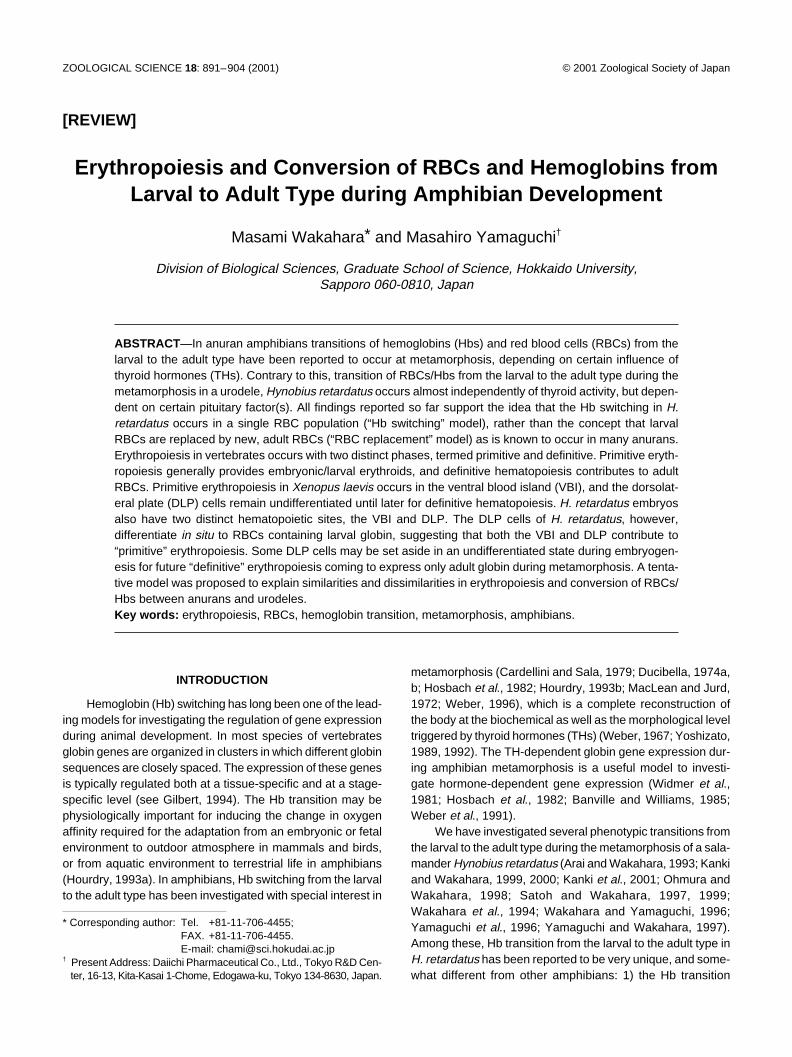
Fig. 1. RBCs double-stained with TUNEL and Hb immunohistochemistry. A section of the spleen from metamorphosing Xenopus was stainedwith antibody to larval Hb conjugated with Cy3 (red) and with TUNEL (fluorescein, green). The section was observed with different excitationfilters for (A) Cy3 (larval Hb), (B) for fluorescein (TUNEL), and (C) for Cy3 and fluorescein (double-stained with larval Hb and TUNEL). A double-positive RBC is clearly shown, emitting bright yellow fluorescence mixed with red and green (C, arrowhead). From Tamori and Wakahara (2000).
from that in non-neotenic urodeles and anurans, and thus mayprovide unique experimental system.
The direct development is a widespread alternate repro-ductive mode in living amphibians that is characterized byevolutionary loss of the free-living, aquatic larval stage (Lynn,1961; Dent, 1968; Wake and Hanken, 1996). The directdeveloping larvae have no gill slits because respiration in wateris not required. Instead, they specialize other organs/tissuesfor respiration, abdominal respiratory folds in Cornufer anda balloon-like structure of a highly vascularized tail inEleutherodactylus (Dent, 1968). The direct development inamphibians is quite similar to the oviparous development typi-cal of birds and reptiles which allows the animal to withdrawfrom water (Shi, 2000). A few studies have been carried outto determine the hormonal requirements during the develop-mental process that leads to the formation of the miniatureadult (Jennings and Hanken, 1996), and to know several phe-notypic transitions from the larval to the adult type (Calleryand Elinson, 1996). Despite their radically altered ontogeny,direct developers still undergo a TH-dependent metamorpho-sis, which occurs before hatching (Callery and Elinson, 2000).
CONVERSION OF RBC/HB DURING AMPHIBIANMETAMORPHOSIS
Conversion of RBCs/Hbs in AnuransIn an anuran amphibian, Xenopus laevis, a switch in Hb
synthesis occurs at metamorphosis resulting from the replace-ment of the larval globin subunits by a set of distinct adultones (Hosbach et al., 1982; Sadmeyer et al., 1988). The tran-sition of Hbs during metamorphosis in Xenopus has beenreported to involve replacement of the larval RBCs (i. e., RBCscontaining only larval Hb) by adult RBCs (RBCs containingonly adult Hb) (Weber et al., 1989). Similar replacement ofthe larval RBCs by adult ones has been reported in Ranapipiens (Hollyfield, 1966) and R. catesbeiana (Dorn andBroyles, 1982; Just and Klaus-Just, 1996; Moss and Ingram,1968). In X. laevis, however, Jurd and MacLean (1970)reported that approximately 25% of the RBCs contained bothlarval and adult globins using larval- and adult-specific anti-bodies, suggesting that the Hb switching occurs within a single
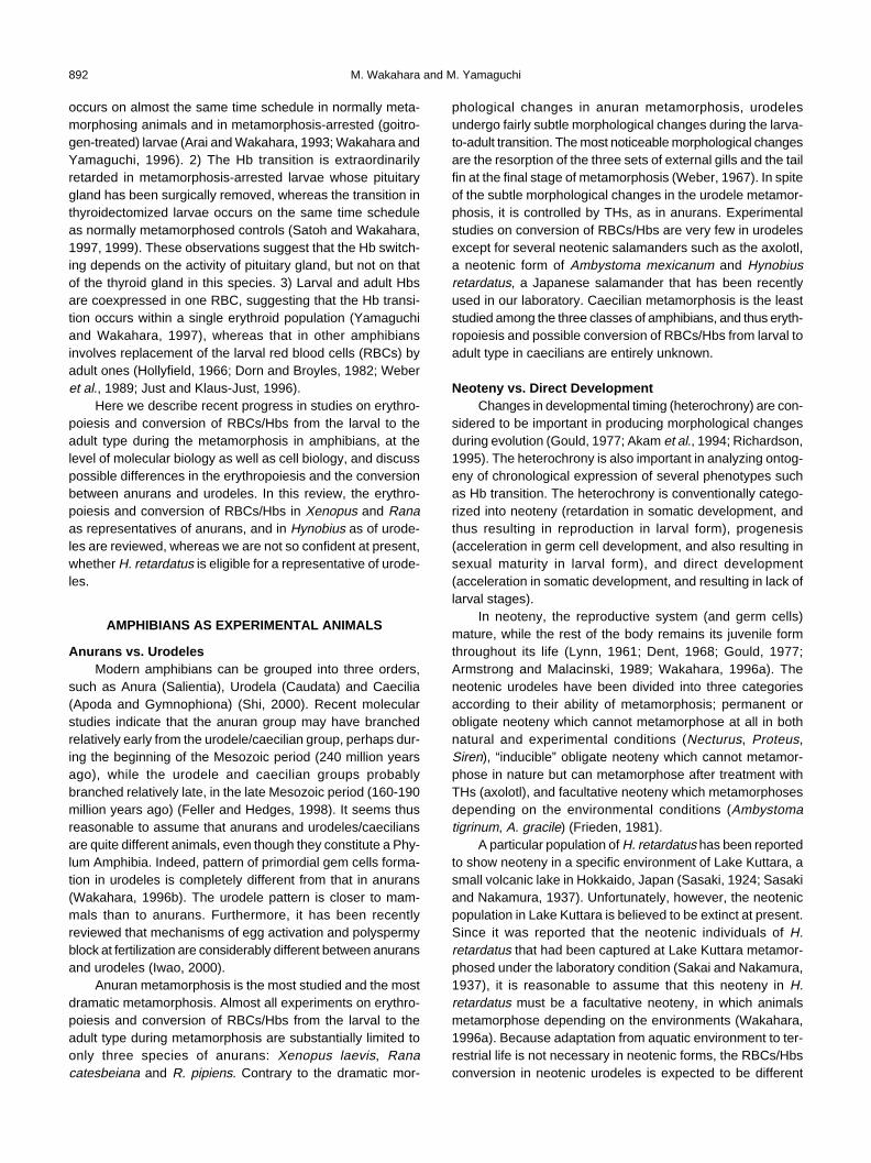
RBC population. Recently, Tamori and Wakahara (2000) haveconvincingly demonstrated the replacement of larval RBCsby adult ones using Hb immunostain in X. laevis. The larvalRBCs in circulation gradually decreased in number duringnormal metamorphosis, and that adult ones converselyincreased. Because the sum of the percentages of larval andadult RBCs relative to total RBCs did not exceed 100% at anytime during the period of RBC conversion, it was concludedthat no RBCs expressed both larval and adult Hbs concur-rently.
To examine mechanisms of the conversion of RBCs inXenopus apoptotic cell death in larval or adult RBCs wasdetected by means of double-staining with in situ DNA nick-end labeling (TUNEL) (Gabrieli et al., 1992) and Hbimmunostain (Fig. 1). Using different fluorescent dyes conju-gated with secondary antibodies, it was possible to identifythe origins of the RBCs that underwent apoptosis (Tamori andWakahara, 2000). Fig. 2 shows chronological changes in theproportions of RBCs of the larval and adult types and in theproportion of TUNEL-positive RBCs to total RBCs in the spleenof X. laevis. The conversion of RBCs was at the halfway pointat stage 59 in the spleen. Even at the end of the metamorpho-sis (stage 66), 30% of the RBCs showed larval Hbs, basicallyidentical to the case of RBCs in circulation (Tamori andWakahara, 2000). Although larval RBCs that also stained withTUNEL were not observed before stage 57, a prometamorphicstage, they began to be observed at and after stage 58. Dur-ing the stages 60 to 62, the climax stage of metamorphosis,5% to 7% of larval RBCs showed TUNEL-positive staining,suggesting that a certain proportion of the larval RBCs under-went apoptosis. The proportion of double-stained larval RBCsgradually decreased thereafter until the end of metamorpho-sis. In contrast, no adult RBCs showing the TUNEL reactionwere observed during any developmental stages examinedso far, suggesting that adult RBCs were not subjected toapoptotic cell death during the conversion of RBCs in X. laevis.
Selective Removal of Larval RBCs during MetamorphosisRecently, Hasebe et al. (1999) demonstrated that the lar-
val RBCs were selectively removed from the systemic circu-lation at the time of the metamorphic climax in R. catesbeiana,
M. Wakahara and M. Yamaguchi894
by means of in vitro fluorescence labeling of RBCs and injec-tion of the labeled cells into animals at various metamorphicstages. The labeled larval RBCs were ingested by hepaticand splenic macrophages, indicating that macrophages areinvolved in the splenic elimination of larval cells. In thisrespect, Nishikawa and Hayashi (1999) showed that larvalerythroblasts decreased through the apoptotic process in X.laevis, by means of double-staining experiments with TUNELand Hb immunostaining. Their results indicated that the eryth-ropoietic system is converted during metamorphosis effec-tively by two distinct hormonal mechanisms, T3-hydrocortisone(HC) synergism on adult erythroblast proliferation and T3-me-diated programmed death of larval erythroblasts.
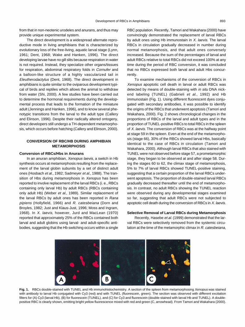
Selective removal of larval RBCs from circulation of meta-morphosing and metamorphosed animals was thus examinedmore simply by using histocompatible J strain of Xenopus(Nakamura et al., 1985; 1987) than using wild animals, with-out considering factors such as immunological rejection oftransferred cells (Tamori and Wakahara, 2000). The resultsof our experiments with this strain showed that transferredlarval RBCs cannot survive in T3-treated adults while thosecan survive in control adults (Fig. 3), suggesting that the ma-ture, larval RBCs are selectively removed from circulationunder the influences of THs. Double-staining experiments withTUNEL and Hb immunostain convincingly demonstrate thatmature, larval RBCs of X. laevis are subjected to the apoptosisin the spleen and liver of the recipients under the influence ofTHs (Tamori and Wakahara, 2000). It is thus concluded thatthe larval RBCs are specifically removed by apoptotic cell death
Fig. 2. Chronological changes in the proportion of larval and adultRBCs to total RBCs in the spleen and in TUNEL-positive RBCs dur-ing Xenopus metamorphosis. Sections of the spleen were double-stained with TUNEL and Hb immunohistochemistry. Numbers of RBCsstained with antibodies (open symbols) to larval (circle) and adult(square) Hbs, and with TUNEL (closed symbols) were counted.Double-positive RBCs were detected during and after the metamor-phic climax, although adult Hb-positive and TUNEL-positive RBCs(closed square) were not detected at all throughout metamorphosis.Open triangles indicate the percentage of adult RBCs in goitrogen-treated, metamorphosis-arrested larvae. From Tamori and Wakahara(2000).
Fig. 3. Fates of transferred, larval RBCs in adult recipients of thehistocompatible J strain of Xenopus laevis. Larval RBCs were injecteddirectly into the heart of either control or T3-treated adults, and thenRBCs were collected from recipients every other day. Although thetransferred larval RBCs survived for a long time in control adults, andthus did not decrease in number (closed circle), they gradually de-creased in number in T3-treated adults (closed triangle, 10–7 M T3;closed square, 10–9 M T3). When adult recipients were treated with T3
21 days after the transfer (arrow), the population of the transferredlarval RBCs to total RBCs drastically decreased. From Tamori andWakahara (2000).
from the circulation during the metamorphic climax. Nishikawaand Hayashi (1999) have shown that larval-type erythroblastsare subjected to the apoptosis in the liver, suggesting the lar-val-adult conversion of RBCs is conducted by T3-mediatedprogrammed death of larval precursor cells. In this model,however, it is difficult to explain how the mature, larval RBCscirculating in metamorphosing tadpoles are selectivelyremoved from the circulation. Because selective apoptosis ofmature, larval RBCs in the spleen was demonstrated in vivoat the metamorphosis climax, and in recipient adults treatedwith T3 in RBC-transfer experiments (Tamori and Wakahara,2000), it can be concluded the larval-adult conversion of RBCsin X. laevis is conducted by replacement of RBC populations,which is similar to the mechanism known in general transfor-mation in anuran metamorphosis, selective removal ofmature, larval specific cells (Hasebe et al., 1999; Izutsu et al.,1996; Nishikawa and Hayashi, 1995; Ohmura and Wakahara,1998; Yoshizato, 1992).
Conversion of RBCs/Hbs in UrodelesAdult RBCs in H. retardatus were readily distinguished
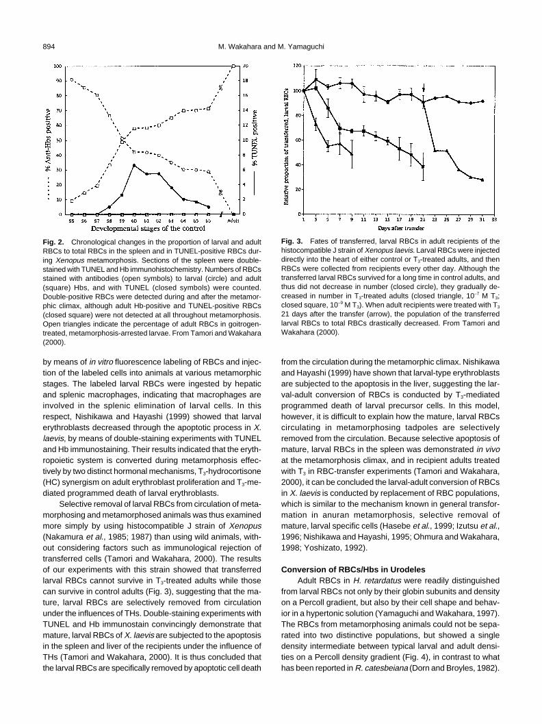
from larval RBCs not only by their globin subunits and densityon a Percoll gradient, but also by their cell shape and behav-ior in a hypertonic solution (Yamaguchi and Wakahara, 1997).The RBCs from metamorphosing animals could not be sepa-rated into two distinctive populations, but showed a singledensity intermediate between typical larval and adult densi-ties on a Percoll density gradient (Fig. 4), in contrast to whathas been reported in R. catesbeiana (Dorn and Broyles, 1982).
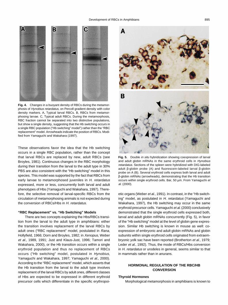
Development of RBCs in Amphibians 895
Fig. 4. Changes in a buoyant density of RBCs during the metamor-phosis of Hynobius retardatus, on Percoll gradient density with colordensity markers. A, Typical larval RBCs. B, RBCs from metamor-phosing larvae. C, Typical adult RBCs. During the metamorphosis,RBC fraction cannot be separated into two distinctive populations,but show a single density, suggesting that the Hb switching occurs ina single RBC population (“Hb switching” model”) rather than the “RBCreplacement” model. Arrowheads indicate the position of RBCs. Modi-fied from Yamaguchi and Wakahara (1997).
These observations favor the idea that the Hb switchingoccurs in a single RBC population, rather than the conceptthat larval RBCs are replaced by new, adult RBCs (seeBroyles, 1981). Continuous changes in the RBC morphologyduring their transition from the larval to the adult type in 30%PBS are also consistent with the “Hb switching” model in thisspecies. This model was supported by the fact that RBCs fromearly larvae to metamorphosed juveniles in H. retardatusexpressed, more or less, concurrently both larval and adultphenotypes of Hbs (Yamaguchi and Wakahara, 1997). There-fore, the selective removal of larval-specific RBCs from thecirculation of metamorphosing animals is not expected duringthe conversion of RBCs/Hbs in H. retardatus.
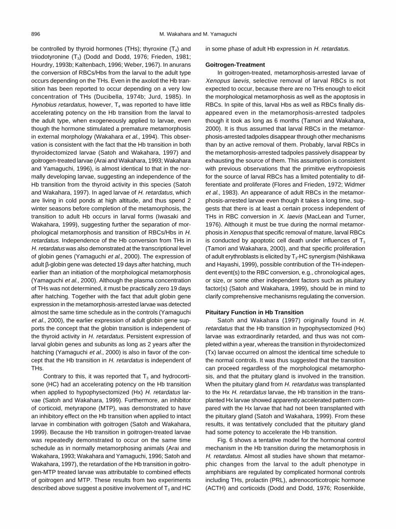
“RBC Replacement” vs. “Hb Switching” ModelsThere are two concepts explaining the Hbs/RBCs transi-
tion from the larval to the adult type in amphibians: eitherthe transition involves replacement of the larval RBCs byadult ones (“RBC replacement” model, postulated in Rana,Hollyfield, 1966; Dorn and Broyles, 1982; in Xenopus, Weberet al., 1989, 1991; Just and Klaus-Just, 1996; Tamori andWakahara, 2000), or the Hb transition occurs within a singleerythroid population and thus no replacement of RBCsoccurs (“Hb switching” model, postulated in Hynobius,Yamaguchi and Wakahara, 1997; Yamaguchi et al., 2000).According to the “RBC replacement” model, which asserts thatthe Hb transition from the larval to the adult type involvesreplacement of the larval RBCs by adult ones, different classesof Hbs are expected to be expressed in different erythroidprecursor cells which differentiate in the specific erythropoi-
Fig. 5. Double in situ hybridization showing coexpression of larvaland adult globin mRNAs in the same erythroid cells in Hynobiusretardatus. Sections of the spleen were hybridized with DIG-labeledadult β-globin probe (A) and fluorescein-labeled larval β-globinprobe on A (B). Several erythroid cells express both larval and adultβ-globin mRNAs (arrowheads), demonstrating that the Hb transitionoccurs within single erythroid cells. Bar, 50 µm. From Yamaguchi etal. (2000).
etic organs (Weber et al., 1991). In contrast, in the “Hb switch-ing” model, as postulated in H. retardatus (Yamaguchi andWakahara, 1997), the Hb switching may occur in the sameerythroid precursor cells. Yamaguchi et al. (2000) conclusivelydemonstrated that the single erythroid cells expressed both,larval and adult globin mRNAs concurrently (Fig. 5), in favorof the “Hb switching” model at the level of globin gene expres-sion. Similar Hb switching is known in mouse as well: co-expression of embryonic and adult globin mRNAs and globinsubunits within single erythroid cells originated from extraem-bryonic yolk sac have been reported (Brotherton et al., 1979;Leder et al., 1992). Thus, the mode of RBCs/Hbs conversionin H. retardatus or urodeles in general, seems similar to thatin mammals rather than in anurans.
HORMONAL REGULATION OF THE RBC/HBCONVERSION
Thyroid HormonesMorphological metamorphosis in amphibians is known to
M. Wakahara and M. Yamaguchi896
be controlled by thyroid hormones (THs); thyroxine (T4) andtriiodotyronine (T3) (Dodd and Dodd, 1976; Frieden, 1981;Hourdry, 1993b; Kaltenbach, 1996; Weber, 1967). In anuransthe conversion of RBCs/Hbs from the larval to the adult typeoccurs depending on the THs. Even in the axolotl the Hb tran-sition has been reported to occur depending on a very lowconcentration of THs (Ducibella, 1974b; Jurd, 1985). InHynobius retardatus, however, T4 was reported to have littleaccelerating potency on the Hb transition from the larval tothe adult type, when exogeneously applied to larvae, eventhough the hormone stimulated a premature metamorphosisin external morphology (Wakahara et al., 1994). This obser-vation is consistent with the fact that the Hb transition in boththyroidectomized larvae (Satoh and Wakahara, 1997) andgoitrogen-treated larvae (Arai and Wakahara, 1993; Wakaharaand Yamaguchi, 1996), is almost identical to that in the nor-mally developing larvae, suggesting an independence of theHb transition from the thyroid activity in this species (Satohand Wakahara, 1997). In aged larvae of H. retardatus, whichare living in cold ponds at high altitude, and thus spend 2winter seasons before completion of the metamorphosis, thetransition to adult Hb occurs in larval forms (Iwasaki andWakahara, 1999), suggesting further the separation of mor-phological metamorphosis and transition of RBCs/Hbs in H.retardatus. Independence of the Hb conversion from THs inH. retardatus was also demonstrated at the transcriptional levelof globin genes (Yamaguchi et al., 2000). The expression ofadult β-globin gene was detected 19 days after hatching, muchearlier than an initiation of the morphological metamorphosis(Yamaguchi et al., 2000). Although the plasma concentrationof THs was not determined, it must be practically zero 19 daysafter hatching. Together with the fact that adult globin geneexpression in the metamorphosis-arrested larvae was detectedalmost the same time schedule as in the controls (Yamaguchiet al., 2000), the earlier expression of adult globin gene sup-ports the concept that the globin transition is independent ofthe thyroid activity in H. retardatus. Persistent expression oflarval globin genes and subunits as long as 2 years after thehatching (Yamaguchi et al., 2000) is also in favor of the con-cept that the Hb transition in H. retardatus is independent ofTHs.
Contrary to this, it was reported that T3 and hydrocorti-sone (HC) had an accelerating potency on the Hb transitionwhen applied to hypophysectomized (Hx) H. retardatus lar-vae (Satoh and Wakahara, 1999). Furthermore, an inhibitorof corticoid, metyrapone (MTP), was demonstrated to havean inhibitory effect on the Hb transition when applied to intactlarvae in combination with goitrogen (Satoh and Wakahara,1999). Because the Hb transition in goitrogen-treated larvaewas repeatedly demonstrated to occur on the same timeschedule as in normally metamorphosing animals (Arai andWakahara, 1993; Wakahara and Yamaguchi, 1996; Satoh andWakahara, 1997), the retardation of the Hb transition in goitro-gen-MTP treated larvae was attributable to combined effectsof goitrogen and MTP. These results from two experimentsdescribed above suggest a positive involvement of T3 and HC
in some phase of adult Hb expression in H. retardatus.
Goitrogen-TreatmentIn goitrogen-treated, metamorphosis-arrested larvae of
Xenopus laevis, selective removal of larval RBCs is notexpected to occur, because there are no THs enough to elicitthe morphological metamorphosis as well as the apoptosis inRBCs. In spite of this, larval Hbs as well as RBCs finally dis-appeared even in the metamorphosis-arrested tadpolesthough it took as long as 6 months (Tamori and Wakahara,2000). It is thus assumed that larval RBCs in the metamor-phosis-arrested tadpoles disappear through other mechanismsthan by an active removal of them. Probably, larval RBCs inthe metamorphosis-arrested tadpoles passively disappear byexhausting the source of them. This assumption is consistentwith previous observations that the primitive erythropoiesisfor the source of larval RBCs has a limited potentiality to dif-ferentiate and proliferate (Flores and Frieden, 1972; Widmeret al., 1983). An appearance of adult RBCs in the metamor-phosis-arrested larvae even though it takes a long time, sug-gests that there is at least a certain process independent ofTHs in RBC conversion in X. laevis (MacLean and Turner,1976). Although it must be true during the normal metamor-phosis in Xenopus that specific removal of mature, larval RBCsis conducted by apoptotic cell death under influences of T3
(Tamori and Wakahara, 2000), and that specific proliferationof adult erythroblasts is elicited by T3-HC synergism (Nishikawaand Hayashi, 1999), possible contribution of the TH-indepen-dent event(s) to the RBC conversion, e.g., chronological ages,or size, or some other independent factors such as pituitaryfactor(s) (Satoh and Wakahara, 1999), should be in mind toclarify comprehensive mechanisms regulating the conversion.
Pituitary Function in Hb TransitionSatoh and Wakahara (1997) originally found in H.
retardatus that the Hb transition in hypophysectomized (Hx)larvae was extraordinarily retarded, and thus was not com-pleted within a year, whereas the transition in thyroidectomized(Tx) larvae occurred on almost the identical time schedule tothe normal controls. It was thus suggested that the transitioncan proceed regardless of the morphological metamorpho-sis, and that the pituitary gland is involved in the transition.When the pituitary gland from H. retardatus was transplantedto the Hx H. retardatus larvae, the Hb transition in the trans-planted Hx larvae showed apparently accelerated pattern com-pared with the Hx larvae that had not been transplanted withthe pituitary gland (Satoh and Wakahara, 1999). From theseresults, it was tentatively concluded that the pituitary glandhad some potency to accelerate the Hb transition.
Fig. 6 shows a tentative model for the hormonal controlmechanism in the Hb transition during the metamorphosis inH. retardatus. Almost all studies have shown that metamor-phic changes from the larval to the adult phenotype inamphibians are regulated by complicated hormonal controlsincluding THs, prolactin (PRL), adrenocorticotropic hormone(ACTH) and corticoids (Dodd and Dodd, 1976; Rosenkilde,
Development of RBCs in Amphibians 897
Fig. 6. Tentative model for hormonal control mechanisms of the Hb transition during the metamorphosis in Hynobius retardatus. A, Generalscheme for hormonal control mechanism of the metamorphosis in amphibians. B, Specific scheme for hormonal control mechanism of Hbtransition during the metamorphosis in H. retardatus. The Hb transition proceeds autonomously within each erythroid cell at very low speed. BothT3 and unknown pituitary factor(s) are assumed to have an accelerating effect on Hb transition. See text for detail. From Satoh and Wakahara(1999).
1985). Major hormones involved in the amphibian metamor-phosis are (1) T3, an active metamorphic hormone, which isconverted from T4 secreted from the thyroid gland that is stimu-lated by TSH secreted from the pituitary gland, (2) PRLsecreted from the pituitary gland, and (3) corticoids which aresecreted from the adrenal cortex following stimulation by ACTHsecreted from the pituitary gland (Fig. 6A). Corticoids stimu-late the conversion of T4 to T3 by inducing the conversion en-zyme, and T3 elicits metamorphic changes in somatic tissues,whereas PRL inhibits the metamorphic changes. In the Hbtransition in H. retardatus, however, no inhibitory factor(s) aredemonstrated, such as PRL in the general metamorphosis inamphibians. Both T3 and unknown pituitary factor(s) (Satohand Wakahara, 1999) are assumed to have acceleratingpotency on the Hb transition in H. retardatus (Fig. 6B).
ERYTHROPOIESIS IN AMPHIBIANS
Primitive and Definitive ErythropoiesisHematopoiesis in vertebrates occurs with two distinct
phases, termed primitive and definitive, based on the timingand site of their development, morphology, and their potenti-alities to differentiate (Zon, 1995). In general the primitive eryth-ropoiesis provides embryonic and/or larval erythroids, and thedefinitive hematopoiesis supplies adult RBCs. In mammalsand birds, primitive erythropoiesis occurs in the extraembry-onic yolk sac and consists primarily of erythrocytes. On theother hand, definitive hematopoietic cells are derived from theintraembryonic region such as dorsal mesentery, para-aorticfoci, anterior region of the mesonephros in association withthe post cardinal vein, and dorsal aorta in birds (Martin et al.,1978; Dieterlen-Lievre and Martin, 1981; Dieterlen-Lievre,1993) and aorta-gonads-mesonephros (AGM) region in mam-mals (Medvinsky et al., 1993; Medvinsky and Dzierzak, 1996),
although there are contrary reports claiming that yolk sac stemcells provide all lineages of blood cells (Yoder et al., 1997a,b). In amphibians, the hematopoiesis also occurs in two waves,primitive and definitive similarly to the mammals and birds asdescribed below. The erythropoietic organs change dramati-cally during ontogeny, from embryonic organ (the ventral bloodisland, VBI), which produces the primitive blood cells to thedorsolateral plate (DLP) mainly contributing to the definitivecells. In many fishes, however, both primitive and definitiveblood cells arise from intraembryonic intermediate cell mass(ICM) (Detrich et al., 1995).
Erythropoietic Organs in AnuransIn Xenopus transplantation experiments demonstrate that
hematopoiesis occurs in two waves: primitive RBCs whichexclusively produce larval Hb are derived from the VBI, whiledefinitive ones (expressing adult Hb) originate mainly fromthe DLP (Turpen et al., 1997), although cells of the VBI con-tribute to adult erythropoiesis to some extent (Maeno et al.,1985a, b: Rollins-Smith and Blair, 1990). Primitive erythro-poiesis in X. laevis occurs in the VBI during early embryogen-esis, and the DLP cells remain undifferentiated until later fordefinitive hematopoiesis. A switch from larval to adult globingene expression in Xenopus has been reported to be medi-ated by erythroid cells from distinct, two compartments (We-ber et al., 1991), in agreement with the “RBC replacement”model. The erythropoietic transition from the VBI to the DLPis dependent on the THs (Rollins-Smith and Blair, 1990). Theexpressions of globin and GATA-1 genes, terminal differen-tiation markers of erythroid cells, are detected exclusively inthe VBI of tailbud embryos (Kelley et al., 1994; Bertwistle etal., 1996), suggesting further that the VBI is the sole source ofprimitive erythroid cells in X. laevis. The different responsesin larval and adult RBCs to THs, apoptotic cell death in larval
M. Wakahara and M. Yamaguchi898
Fig. 7. In situ hybridization of an embryo of Hynobius retardatus atstage 37/38 with antisense larva β-globin probe, performed to thewhole mount embryo (A) or sectioned specimens (B-D) made of dif-ferent embryos from A. (A) Ventral view of the whole mount embryo.V-shaped VBI is stained clearly (arrowheads). (B) Sectioned speci-mens of the VBI, at the level corresponding to the dashed line b in A.Erythroid cells expressing larval β-globin gene are nested in the VBI.(C) Anterior region of the DLP, at the level corresponding to the dashedline c in A. Erythroid cells stained positively with larval β-globin probeare detected (arrowheads). (D) Posterior region of the DLP, at the levelcorresponding to the dashed line d in A. Erythroid cells expressinglarval β-globin gene are observed, but only a few (arrowhead). Bars,1mm (A), 100 µm (B). Modified from Yamaguchi and Wakahara (2001).
but not in adult RBCs in X. laevis (Figs. 2, 3), might reflect thedifferent origins of these two RBC populations. It has also beenreported in Rana pipiens that primitive erythroid cells origi-nate in the VBI and definitive cells in the DLP (Zon, 1995). InR. catesbeiana, however, it has been suggested that the DLP(mesonephric kidney) has both primitive and definitive hemato-poietic cells (Broyles et al., 1981; Maples et al., 1983).
The exact erythropoietic organ(s) in larvae and adults inanurans have been controversial: the kidney (Turner, 1988),liver (Ohinata and Enami, 1991), or peripheral to the liver(Weber et al., 1991) in Xenopus, and the liver and kidney inRana (Broyles et al., 1981).
Erythropoietic Organs in UrodelesThe transition in the erythropoietic organs in Hynobius
retardatus has been studied on the basis of immunohis-tochemical studies using specific antibodies to larval and adultHbs (Yamaguchi and Wakahara, 1997). Thereafter, thescheme of the transition was substantially confirmed by
molecular biological studies using cDNA probes for globingenes (Yamaguchi et al., 2000; Yamaguchi and Wakahara,2001). In H. retardatus, the VBI at a tailbud stage (stage 34,according to the developmental stages by Iwasawa andYamashita, 1991) is intensely stained in situ hybridization withthe larval β-globin RNA probe. At stage 37/38, the VBI is stillrecognizable with the larval globin antibody and β-globin RNAprobe. In addition, cells of the DLP also became intenselystained with the larval antibody as well as with the larval RNAprobe (Fig. 7), suggesting that cells of the VBI as well as theDLP differentiate in situ to erythroid cells that contain larvalglobin subunit and larval globin mRNA. In other words, boththe VBI and DLP contribute to primitive erythropoiesis (Yama-guchi et al., 2000). The contribution of the DLP to the primi-tive erythropoiesis probably characterizes the erythropoiesisin Hynobius or in urodeles, in general. There are two, pos-sible explanations for the contribution of the VBI and DLP tothe primitive and/or definitive erythropoiesis: both the VBI andDLP supply common erythroid precursor cells which differen-tiate to every type of RBCs, such as larval, larval-adult andadult RBCs, or the VBI gives rise to only larval erythroid cellsbut the DLP supplies common erythroid cells which differenti-ate to every type of RBCs (Yamaguchi et al., 2000). Unfortu-nately, however, it was difficult to determine conclusively thedevelopmental fates of the erythroid stem cells from the VBIand the DLP by examining only the expression of globin genes.To obtain molecular probes such as GATA genes, whichsequentially regulate vertebrate hematopoiesis (Kulessa et al.,1995; Visvader et al., 1995), GATA-3 was cloned to elucidatefurther mechanisms controlling the hematopoiesis in H.retardatus (Yamaguchi and Wakahara, 2001).
GATA-3 Expression during ErythropoiesisThe GATA-3 clone possessed highly conserved zinc-fin-
ger domains, suggesting that the Hynobius GATA-3 geneshave similar regulatory roles in hematopoiesis as do those ofother vertebrates. We tentatively regarded GATA-3 as amarker of definitive hematopoietic cells for the reasondescribed below. First, GATA-3 knock-out mice have a defectaffecting definitive hematopoiesis, although primitive hemato-poiesis occurs normally even in such mice (Pandolfi et al.,1995; Ting et al., 1996). Second, GATA-3 gene expression inRBCs increases at the time of the Hb transition in chicken(Leonard et al., 1993). Third, GATA-3 is expressed in the DLP,but not in the VBI, at tailbud stage in X. laevis (Bertwistle etal., 1996). In H. retardatus, the GATA-3 gene was expressedas early as stage 39 embryos (Fig. 8), but it was not expressedin RBCs of larvae at 20 days after the hatching (Yamaguchiand Wakahara, 2001). These findings suggest that the GATA-3 gene works at the early stages of development but not atlater stages. In situ hybridization convincingly demonstratedthat the GATA-3 gene was expressed in the DLP (Fig. 8C)but not in the VBI (Fig. 8D) or peripheral blood at stage 39.These facts may imply that some DLP cells contribute to eryth-ropoiesis at early stages of development, while others are setaside in an undifferentiated state for hematopoiesis at later

Development of RBCs in Amphibians 899
Fig. 8. In situ hybridization of Hynobius retardatus embryos at stage39 treated with GATA-3 probe. A, Lateral view of the whole embryotreated with antisense probe. The DLP region is clearly stained (ar-rowheads), but not the VBI. B, Lateral view of the whole embryo treatedwith sense GATA-3 probe. No signals are detected at all. C, Crosssection of the specimen shown in A at the level indicated by a dashedline c. Cells of the DLP are positively stained. D, Cross section throughthe VBI region of the specimen shown in A at the level indicated bydashed line d. Cells of the ventral region are not stained. Bars, 1 mm(A, B); 100 µm (C, D). Modified from Yamaguchi and Wakahara (2001).
stages. In other words, the DLP cells set aside in an undiffer-entiated state may have more “definitive” characteristics.These explanations are consistent with the concept that primi-tive erythropoiesis occurs in cells of the VBI and in some cellsof the DLP, but that other DLP cells are reserved during em-bryogenesis for future definitive erythropoiesis.
During early developmental stages, the spleen and livermust be major erythropoietic organs in H. retardatus. In theliver, however, erythropoietic activity ceases and thus RBC-precursor cells are hardly observed in metamorphosing lar-vae and metamorphosed juveniles (Yamaguchi andWakahara, 1997). This suggests that the liver functions as anerythropoietic organ only at early larval stages, from whichRBCs containing only larval globins differentiate (Yamaguchiet al., 2000). The spleen becomes the major erythropoieticorgan before the Hb transition in H. retardatus like in Triturus(Tournefier, 1973). It is thus suggested that the spleen func-tions as a major erythropoietic organ throughout life in thisspecies, in contrast to Rana and Xenopus (Broyles, 1981;Weber et al., 1991).
Induction of AnemiaInduction of anemia by phenylhydrazine (PHZ), resulting
in a rapid regeneration of the circulating RBCs, has been fre-quently used to explore the origin of the developmental prop-
erties of the larval RBCs (Flores and Frieden, 1972; Widmeret al., 1983). These authors have shown a precociousappearance of adult Hbs or adult globin mRNAs in tadpolesrecovering from anemia of R. catesbeiana and X. laevis. Thesefindings suggest that the larval erythropoietic system has alimited capacity for self-renewal, and by its decline may trig-ger the outgrowth of the adult erythroid cell population, andsupport the “RBC replacement” model. Contrary to this, in pre-and prometamorphic larvae of H. retardatus recovering fromanemia induced by PHZ-treatment, a precocious Hb transi-tion did not occur (Yamaguchi et al., 2000), suggesting thatthe RBC replacement during ontogeny could not be expectedin this species.
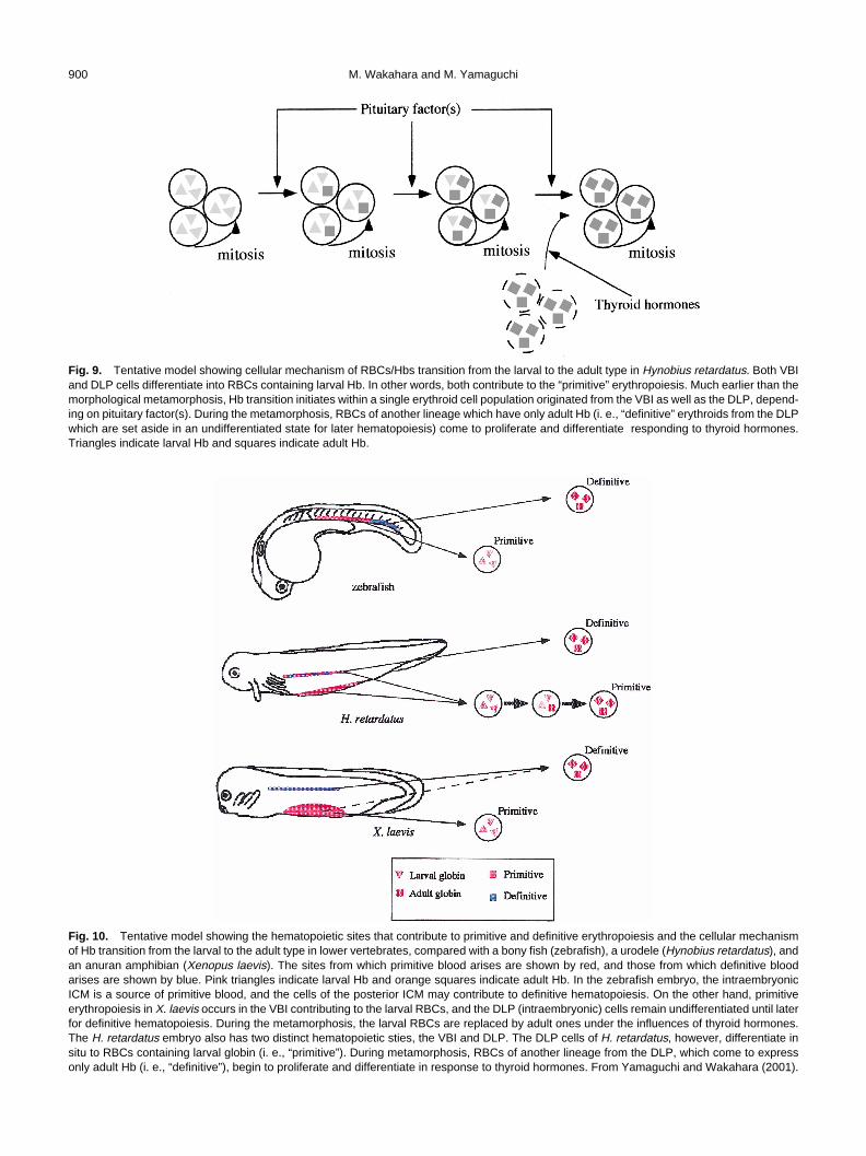
On the other hand, when metamorphosing larvae of H.retardatus were treated with PHZ to induce anemia and thenbled at a postmetamorphic stage after they recovered fromthe anemia, a precocious Hb transition was observed in theseanimals (Yamaguchi and Wakahara, 2001). This finding sug-gests that RBCs expressing only adult Hb, i. e., “definitive”RBCs, proliferate and differentiate during and after metamor-phosis. We consider that PHZ treatment reduces the numberof mature “primitive” RBCs (expressing larval and adult globins)in metamorphosing larvae, and that after the treatment, only“definitive” but not “primitive” erythroid cells proliferate inresponse to THs. Contrary to this, the number of “primitive”RBCs in the controls does not decrease. As a result, the Hbtransition in PHZ-treated animals might progress faster thanin the controls. If blood is collected before metamorphosis,“definitive” RBCs will not have emerged yet, and no preco-cious transition occurs (Yamaguchi et al., 2000). Theseobservations suggest that there are two distinctive RBC popu-lations which express and/or come to express adult Hb inH. retardatus: the “primitive” and “definitive” erythroids(Yamaguchi and Wakahara, 2001). The former initially ex-presses larval Hb and comes to express finally adult Hb byswitching globin genes within every single cell, and the latterwhich is reserved for later ontogenesis proliferates and differ-entiates to express only adult Hb during metamorphosis inresponse to THs (Fig. 9). This model can explain the facts (1)that exogeneously applied T3 accelerate the Hb transition inH. retardatus (Satoh and Wakahara, 1999) even though thetransition is not affected by goitrogen-treatment (Arai andWakahara, 1993; Wakahara and Yamaguchi, 1996), (2) thatthe precocious Hb transition was not observed in H. retardatusrecovering from anemia before metamorphosis (Yamaguchiet al., 2000), but was observed in H. retardatus recoveringfrom anemia after the metamorphosis (Yamaguchi andWakahara 2001), and (3) that the number of RBCs express-ing only adult Hb, which must be definitive, was significantlylarger in controls than in the goitrogen-treated, metamorpho-sis-arrested animals (Yamaguchi and Wakahara, 2001), eventhough there were no differences in the degree of completionof the Hb transition between controls and goitrogen-treatedanimals (Arai and Wakahara, 1993; Wakahara and Yama-guchi, 1996).
In mammals, it has generally been accepted that the primi-
M. Wakahara and M. Yamaguchi900
Fig. 9. Tentative model showing cellular mechanism of RBCs/Hbs transition from the larval to the adult type in Hynobius retardatus. Both VBIand DLP cells differentiate into RBCs containing larval Hb. In other words, both contribute to the “primitive” erythropoiesis. Much earlier than themorphological metamorphosis, Hb transition initiates within a single erythroid cell population originated from the VBI as well as the DLP, depend-ing on pituitary factor(s). During the metamorphosis, RBCs of another lineage which have only adult Hb (i. e., “definitive” erythroids from the DLPwhich are set aside in an undifferentiated state for later hematopoiesis) come to proliferate and differentiate responding to thyroid hormones.Triangles indicate larval Hb and squares indicate adult Hb.
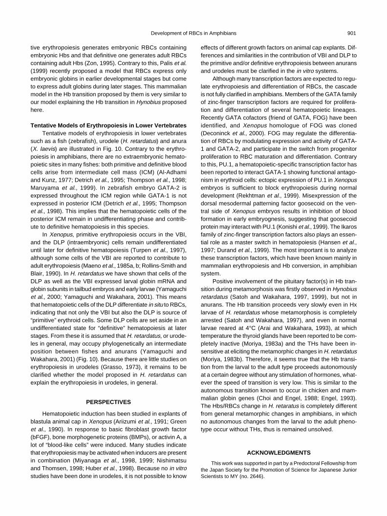
Fig. 10. Tentative model showing the hematopoietic sites that contribute to primitive and definitive erythropoiesis and the cellular mechanismof Hb transition from the larval to the adult type in lower vertebrates, compared with a bony fish (zebrafish), a urodele (Hynobius retardatus), andan anuran amphibian (Xenopus laevis). The sites from which primitive blood arises are shown by red, and those from which definitive bloodarises are shown by blue. Pink triangles indicate larval Hb and orange squares indicate adult Hb. In the zebrafish embryo, the intraembryonicICM is a source of primitive blood, and the cells of the posterior ICM may contribute to definitive hematopoiesis. On the other hand, primitiveerythropoiesis in X. laevis occurs in the VBI contributing to the larval RBCs, and the DLP (intraembryonic) cells remain undifferentiated until laterfor definitive hematopoiesis. During the metamorphosis, the larval RBCs are replaced by adult ones under the influences of thyroid hormones.The H. retardatus embryo also has two distinct hematopoietic sties, the VBI and DLP. The DLP cells of H. retardatus, however, differentiate insitu to RBCs containing larval globin (i. e., “primitive”). During metamorphosis, RBCs of another lineage from the DLP, which come to expressonly adult Hb (i. e., “definitive”), begin to proliferate and differentiate in response to thyroid hormones. From Yamaguchi and Wakahara (2001).
Development of RBCs in Amphibians 901
tive erythropoiesis generates embryonic RBCs containingembryonic Hbs and that definitive one generates adult RBCscontaining adult Hbs (Zon, 1995). Contrary to this, Palis et al.(1999) recently proposed a model that RBCs express onlyembryonic globins in earlier developmental stages but cometo express adult globins during later stages. This mammalianmodel in the Hb transition proposed by them is very similar toour model explaining the Hb transition in Hynobius proposedhere.
Tentative Models of Erythropoiesis in Lower VertebratesTentative models of erythropoiesis in lower vertebrates
such as a fish (zebrafish), urodele (H. retardatus) and anura(X. laevis) are illustrated in Fig. 10. Contrary to the erythro-poiesis in amphibians, there are no extraembryonic hemato-poietic sites in many fishes: both primitive and definitive bloodcells arise from intermediate cell mass (ICM) (Al-Adhamiand Kunz, 1977; Detrich et al., 1995; Thompson et al., 1998;Maruyama et al., 1999). In zebrafish embryo GATA-2 isexpressed throughout the ICM region while GATA-1 is notexpressed in posterior ICM (Detrich et al., 1995; Thompsonet al., 1998). This implies that the hematopoietic cells of theposterior ICM remain in undifferentiating phase and contrib-ute to definitive hematopoiesis in this species.
In Xenopus, primitive erythropoiesis occurs in the VBI,and the DLP (intraembryonic) cells remain undifferentiateduntil later for definitive hematopoiesis (Turpen et al., 1997),although some cells of the VBI are reported to contribute toadult erythropoiesis (Maeno et al., 1985a, b; Rollins-Smith andBlair, 1990). In H. retardatus we have shown that cells of theDLP as well as the VBI expressed larval globin mRNA andglobin subunits in tailbud embryos and early larvae (Yamaguchiet al., 2000; Yamaguchi and Wakahara, 2001). This meansthat hematopoietic cells of the DLP differentiate in situ to RBCs,indicating that not only the VBI but also the DLP is source of“primitive” erythroid cells. Some DLP cells are set aside in anundifferentiated state for “definitive” hematopoiesis at laterstages. From these it is assumed that H. retardatus, or urode-les in general, may occupy phylogenetically an intermediateposition between fishes and anurans (Yamaguchi andWakahara, 2001) (Fig. 10). Because there are little studies onerythropoiesis in urodeles (Grasso, 1973), it remains to beclarified whether the model proposed in H. retardatus canexplain the erythropoiesis in urodeles, in general.
PERSPECTIVES
Hematopoietic induction has been studied in explants ofblastula animal cap in Xenopus (Ariizumi et al., 1991; Greenet al., 1990). In response to basic fibroblast growth factor(bFGF), bone morphogenetic proteins (BMPs), or activin A, alot of “blood-like cells” were induced. Many studies indicatethat erythropoiesis may be activated when inducers are presentin combination (Miyanaga et al., 1998, 1999; Nishimatsuand Thomsen, 1998; Huber et al., 1998). Because no in vitrostudies have been done in urodeles, it is not possible to know
effects of different growth factors on animal cap explants. Dif-ferences and similarities in the contribution of VBI and DLP tothe primitive and/or definitive erythropoiesis between anuransand urodeles must be clarified in the in vitro systems.
Although many transcription factors are expected to regu-late erythropoiesis and differentiation of RBCs, the cascadeis not fully clarified in amphibians. Members of the GATA familyof zinc-finger transcription factors are required for prolifera-tion and differentiation of several hematopoietic lineages.Recently GATA cofactors (friend of GATA, FOG) have beenidentified, and Xenopus homologue of FOG was cloned(Deconinck et al., 2000). FOG may regulate the differentia-tion of RBCs by modulating expression and activity of GATA-1 and GATA-2, and participate in the switch from progenitorproliferation to RBC maturation and differentiation. Contraryto this, PU.1, a hematopoietic-specific transcription factor hasbeen reported to interact GATA-1 showing functional antago-nism in erythroid cells: ectopic expression of PU.1 in Xenopusembryos is sufficient to block erythropoiesis during normaldevelopment (Rekhtman et al., 1999). Misexpression of thedorsal mesodermal patterning factor goosecoid on the ven-tral side of Xenopus embryos results in inhibition of bloodformation in early embryognesis, suggesting that goosecoidprotein may interact with PU.1 (Konishi et al., 1999). The Ikarosfamily of zinc-finger transcription factors also plays an essen-tial role as a master switch in hematopoiesis (Hansen et al.,1997; Durand et al., 1999). The most important is to analyzethese transcription factors, which have been known mainly inmammalian erythropoiesis and Hb conversion, in amphibiansystem.
Positive involvement of the pituitary factor(s) in Hb tran-sition during metamorphosis was firstly observed in Hynobiusretardatus (Satoh and Wakahara, 1997, 1999), but not inanurans. The Hb transition proceeds very slowly even in Hxlarvae of H. retardatus whose metamorphosis is completelyarrested (Satoh and Wakahara, 1997), and even in normallarvae reared at 4°C (Arai and Wakahara, 1993), at whichtemperature the thyroid glands have been reported to be com-pletely inactive (Moriya, 1983a) and the THs have been in-sensitive at eliciting the metamorphic changes in H. retardatus(Moriya, 1983b). Therefore, it seems true that the Hb transi-tion from the larval to the adult type proceeds autonomouslyat a certain degree without any stimulation of hormones, what-ever the speed of transition is very low. This is similar to theautonomous transition known to occur in chicken and mam-malian globin genes (Choi and Engel, 1988; Engel, 1993).The Hbs/RBCs change in H. retaratus is completely differentfrom general metamorphic changes in amphibians, in whichno autonomous changes from the larval to the adult pheno-type occur without THs, thus is remained unsolved.
ACKNOWLEDGMENTS
This work was supported in part by a Predoctoral Fellowship fromthe Japan Society for the Promotion of Science for Japanese JuniorScientists to MY (no. 2646).
M. Wakahara and M. Yamaguchi902
REFERENCES
Akam M, Holland P, Ingham P, Wray G (1994)(Eds) The Evolution ofDevelopmental Mechanisms. The Company of Biologists Ltd,Cambridge
Al-Adhami MA, Kunz YW (1977) Ontogenesis of hematopoietic sitesin Brachydanio rerio (Hamilton-Buchanan) (tereostei). Dev GrowthDiffer 19: 171–179
Arai T, Wakahara M (1993) Hemoglobin transition from larval to adulttypes in normally metamorphosing, metamorphosed and meta-morphosis-arrested Hynobius retardatus. Zool Sci 10: 637–644
Ariizumi T, Moriya N, Uchiyama H, Asashima M (1991) Concentra-tion-dependent inducing activity of activin A. Roux’s Arch DevBiol 200: 230–233
Armstrong JB, Malacinski GM (1989) Developmental Biology of theAxolotl. Oxford Univ Press, Oxford
Banville D, Williams JC (1985) Developmental changes in the patternof larval β-globin gene expression in Xenopus laevis. J Mol Biol84: 611–620
Bertwistle D, Walmsley ME, Read EM, Pizzey JA, Patient RK (1996)GATA factors and the origin of adult and embryonic blood inXenopus: responses to retinoic acid. Mech Dev 57: 199–214
Brotherton TW, Chui DH, Garldie J, Patterson M (1979) Hemoglobinontogeny during normal mouse fetal development. Proc Natl AcadSci USA 76: 2853–2857
Broyles RH (1981) Changes in the blood during amphibian metamor-phosis. In “Metamorphosis - a Problem in Developmental Biol-ogy” Ed by Gilbert LI and Frieden E, Plenum Press, New York,pp 461–490
Broyles RH, Johnson GM, Maples PB, Kindell GR (1981) Two eryth-ropoietic microenvironments and two larval red cell lines in bull-frog tadpoles. Dev Biol 81: 299–314
Callery EM, Elinson RP (1996) Developmental regulation of theurea-cycle enzyme arginase in the direct-developing frog,Eleutherodactylus coqui. J Exp Zool 275: 61–66
Callery EM, Elinson RP (2000) Thyroid hormone-dependent meta-morphosis in a direct developing frog. Proc Natl Acad Sci USA97: 2615–2620
Cardellini P, Sala M (1979) Metamorphic variations in the hemoglo-bins of Bombina variegata (L.). Comp Biochem Physiol 64B:113–116
Choi O-R, Engel JD (1988) Developmental regulation of β-globin geneswitching. Cell 55: 17–26
Deconinck AE, Mead PE, Tevosian SG, Crispino JD, Katz SG, ZonLI, Orkin SH (2000) FOG acts as a repressor of red blood celldevelopment in Xenopus. Development 127: 2031–2040
Dent JN (1968) Survey of amphibian metamorphosis. In “Metamor-phosis” Ed by Etkin W, Gilbert LI, North-Holland Publ Co,Amsterdam, pp 271–311
Detrich HW, Kieran MW, Chan FY, Barone LM, Yee K, RundstadlerJA, Pratt S, Ransom D, Zon LI (1995) Intraembryonic hemato-poietic cell migration during vertebrate development. Proc NatlAcad Sci USA 92:10713–10717
Dieterlen-Lievre F (1993) Developmental rules in the hematopoieticand immune systems of birds: how general are they? Semin DevBiol 4: 325–332
Dieterlen-Lievre F, Martin C (1981) Diffuse intraembryonic hemopoie-sis in normal and chimeric avian development. Dev Biol 88: 180–191
Dodd MHI, Dodd JM (1976) The biology of metamorphosis. In “ThePhysiology of the Amphibia, vol III” Ed by Lofts B, Academic Press,New York, pp 467–599
Dorn AR, Broyles RH (1982) Erythrocyte differentiation during themetamorphic hemoglobin switch of Rana catesbeiana. Proc NatlAcad Sci USA 9: 5592–5596
Ducibella T (1974a). The occurrence of biochemical metamorphic
events without anatomical metamorphosis in the axolotl. Dev.Biol. 38: 175–186
Ducibella T (1974b) The influence of L-thyroxine on the change in redblood cell type in the axolotl. Dev Biol 38: 187–194
Durand C, Charlemagne J, Fellah JS (1999) Structure and develop-mental expression of Ikaros in the Mexican axolotl. Immunoge-netics 50: 336–343
Engel JD (1993) Developmental regulation of human β-globin genetranscription: a switch of loyalties? Trends Gen 9: 304–309
Feller AE, Hedges SB (1998) Molecular evidence for the early historyof living amphibians. Mol Phylogenet Evol 9: 509–516
Flores G, Frieden E (1972) Hemolytic effect of phenylhydrazine dur-ing amphibian metamorphosis. Dev Biol 27: 406–418
Frieden E (1981) The dual role of thyroid hormones in vertebratedevelopment and calorigenesis. In “Metamorphosis: A Problemin Developmental Biology” Ed by LI Gilbert, E Frieden, PlenumPress, New York, pp. 545–564
Gabrieli Y, Sherman Y, Ben-Sasson SA (1992) Identification of pro-grammed cell death in situ via specific labeling of nuclear DNAfragmentation. J Cell Biol 119: 493–501
Gilbert SF (1994) Transcriptional regulation of gene expression. In“Developmental Biology (4th Ed)”, Sinauer Assoc Inc Publ,Sunderland, pp 411–437
Gould SJ (1977) Ontogeny and Phylogeny. Harvard Univ Press, Cam-bridge
Grasso JA (1973) Erythropoiesis in the newt, Triturus cristatus Laur.Identification of the ‘erythroid precursor cells’. J Cell Sci 12: 469–489
Green JBA, Howes G, Symes K, Cooke J, Smith JC (1990) The bio-logical effects of XTC-MIF: quntitative comparison with XenopusbFGF. Development 108: 173–183
Hansen JD, Strassburger P, Du Pasquier L (1997) Conservation of amaster hematopoietic switch gene during vertebrate evolution:isolation and characterization of Ikaros from teleost and amphib-ian species. Eur J Immunol 27: 3049–3058
Hasebe T, Oshima H, Kawamura K, Kikuyama S (1999) Rapid andselective removal of larval erythrocytes from systemic circulationduring metamorphosis of the bullfrog, Rana catesbeiana. DevelGrowth Differ 41: 639–643
Hollyfield JG (1966) Erythrocyte replacement at metamorphosis inthe frog, Rana pipiens. J Morphol 119: 1–5
Hosbach HA, Widmer HJ, Andres A-C, Weber R (1982) Expressionand organization of the globin genes in Xenopus laevis. In “Em-bryonic Development, part A: Genetic aspects” Ed by Burger MM,Weber R, Alan R Liss, NY, pp 115–125
Hourdry J (1993a) Passage to the terrestrial life in amphibians: Eventsaccompanying this ecological transition. Zool Sci 10: 715–731
Hourdry J (1993b) Passage to the terrestrial life in amphibians II.Endocrine determinism. Zool Sci 10: 887–902
Huber TL, Zhou Y, Mead PE, Zon LI (1998) Cooperative effects ofgrowth factors involved in the induction of hematopoietic meso-derm. Blood 92: 4128–4137
Iwao Y (2000) Mechanisms of egg activation and polyspermy block inamphibians and comparative aspects with fertilization in othervertebrates. Zool Sci 17: 699–709
Iwasaki F, Wakahara M (1999) Adaptable larval life histories in differ-ent populations of the salamander, Hynobius retardatus, living invarious habitats. Zool Sci 16: 667–674
Iwasawa H, Yamashita K (1991) Normal stages of development of ahynobiid salamander, Hynobius nigrescens Stejneger. Jpn JHerpetol 14: 39–62
Izutsu Y, Yoshizato K, Tochinai S (1996) Adult-type splenocytes ofXenopus induce apoptosis of histocompatible larval tail cells invitro. Differentiation 60: 277–286
Jennings DH, Hanken J (1996) Mechanistic basis of life history evo-lution in anuran amphibians: thyroid gland development in thedirect-developing frog, Eleutherodactylus coqui. Gen Comp
Development of RBCs in Amphibians 903
Endocrinol 111: 225–232Jurd RD (1985) Haematological and immunological ‘metamorphosis’
in neotenous urodeles. In “Metamorphosis” Ed by Balls M,Bownes M, Clarendon Press, Oxford, pp 313–331
Jurd RD, MacLean N, (1970) An immunofluorescent study of thehaemoglobins in metamorphosing Xenopus laevis. J Embryol ExpMorph 23: 299–309
Just JJ, Klaus-Just J (1996) Controls of thyroid hormones and theirinvolvement in hemoglobin transition during Xenopus and Ranametamorphosis. In “Biology of Xenopus” Ed by Tinsley RC, KobelHR, Oxford Univ Press, London, pp 213–229
Kaltenbach JC (1996) Endocrinology of amphibian metamorphosis.In “Metamorphosis” Ed by Gilbert LI, Tata JR, Atkinson BG, Aca-demic Press, New York, pp 403–431
Kanki K, Wakahara M (1999) Precocious testicular growth in meta-morphosis-arrested larvae of a salamander Hynobius retardatus:role of thyroid-stimulating hormone. J Exp Zool 283: 548–558
Kanki K, Wakahara M (2000) Spatio-temporal expression of TSHβand FSHβ genes in normally metamorphosing, metamorphosed,and metamorphosis-arrested Hynobius retardatus. Gen CompEndocrinol 119: 276–286
Kanki K, Takaguchi Y, Wakahara M (2001) Heterochronic develop-ment of gonads and external morphology in overwintered larvaeof the salamander Hynobius retardatus: possible contribution ofpituitary hormones to this. Int J Dev Biol in press
Kelley C, Yee K, Harland R, Zon LI (1994) Ventral expression of GATA-1 and GATA-2 in the Xenopus embryo defines induction of he-matopoietic mesoderm. Dev Biol 165: 193–205
Konishi Y, Tominaga M, Watanabe Y, Imamura F, Goldfarb A, MakiR, Blum M, DeRobertis EM, Tominaga A (1999) Goosecoid in-hibits erythrocyte differentiation by competing with Rb for PU.1binding in murine cells. Oncogene 18: 6795–6805
Kulessa H, Frampton J, Graf T (1995) GATA-1 reprograms avianmyelomonocytic cell lines into eosinophils, thromboblasts, anderythroblasts. Genes Dev 9: 1250–1262
Leder A, Kuo A, Shen MM, Leder P (1992) In situ hybridization re-veals co-expression of embryonic and adult a globin genes inthe earliest murine erythrocyte progenitors. Development 116:1041–1049
Leonard MW, Lim K-C, Engel JD (1993) Expression of the chickenGATA family during early erythroid development and differentia-tion. Development 119: 519–531
Lynn WG (1961) Types of amphibian metamorphosis. Am Zool 1:151–161
MacLean N, Jurd RD (1972) The control of haemoglobin synthesis.Biol Rev 47: 393–437
MacLean N, Turner S (1976) Adult haemoglobin in developmentallyretarded tadpoles of Xenopus laevis. J Embryol Exp Morph 35:261–266
Maeno M, Tochinai S, Katagiri Ch (1985a) Differential participation ofventral and dorsolateral mesoderms in the hemopoiesis of Xe-nopus, as revealed in diploid-triploid or interspecific chimeras.Dev Biol 110: 503–508
Maeno M, Todate A, Katagiri Ch (1985b) The localization of precur-sor cells for larval and adult hemopoietic cells in Xenopus laevisin two regions of embryos. Dev Growth Differ 27: 137–148
Maples PB, Dorn AR, Broyles RH (1983) Embryonic and larvalhemoglobins during early development of the bullfrog, Ranacatesbeiana. Dev Biol 96: 515–519
Martin C, Beaupainm D, Dieterlen-Lievre F (1978) Developmentalrelationships between vitelline and intraembryonic haemopoiesisstudied in avian “yolk sac chimeras”. Cell Differ 7: 115–130
Maruyama K, Yasumasu S, Iuchi I (1999) Characterization andepxression of embryonic globin in the rainbow trout, Oncor-hynchus mykiss: intra-embryonic initiation of erythropoiesis. DevGrowth Differ 41: 589–599
Medvinsky AL, Samoylina NL, Muller AM, Dzierzak EA (1993) An
early pre-liver intraembryonic source of CFU-S in the developingmouse. Nature 364: 64–67
Medvinsky A, Dzierzak E (1996) Definitive hematopoiesis is autono-mously initiated by the AGM region. Cell 86: 897–906
Miyanaga Y, Shiurba R, Asashima M (1999) Blood cell induction inXenopus animal cap explants: effects of fibroblast growth factor,bone morphogenetic proteins, and activin. Dev Genes Evol 209:69–76
Miyanaga Y, Shiurba R, Nagata S, Pfeiffer CJ, Asashima M (1998)Induction of blood cells in Xenopus embryo explants. Dev GenesEvol 207: 417–426
Moriya T (1983a) The effect of temperature on the action of thyroidhormone and prolactin in larvae of the salamander, Hynobiusretardatus. Gen Com Endocrinol 49: 1–7
Moriya T (1983b) Cytological changes induced by low temperature inthe thyroid gland of larvae of the salamander Hynobius retardatus.Gen Comp Endocrinol 49: 8–14
Moss B, Ingram VM (1968) Hemoglobin synthesis during amphibianmetamorphosis. I. Chemical studies on the hemoglobins fromthe larval and adult stages of Rana catesbeiana. J Mol Biol 32:481–492
Nakamura T, Kawahara H, Katagiri Ch (1985) Rapid production of ahistocompatible colony of Xenopus laevis by gynogenic proce-dure. Zool Sci 2: 71–79
Nakamura T, Maeno M, Tochinai S, Katagiri Ch (1987) Toleranceinduced by grafting semi-allogeneic adult skin to larval Xenopuslaevis: Possible involvement of specific suppressor cell activity.Differentiation 35: 108–114
Nishikawa A, Hayashi H (1995) Spacial, temporal and hormonal regu-lation of programmed muscle cell death during metamorphosisof the frog, Xenopus leavis. Differentiation 59: 207–214
Nishikawa A, Hayashi H (1999) T3-hydrocortisone synergism on adulttype erythroblast proliferation and T3-mediated apoptosis of lar-val-type erythroblasts during erythropoietic conversion in Xeno-pus laevis. Histochem Cell Biol 111: 325–334
Nishimatsu S, Thomsen GH (1998) Ventral mesoderm induction andpatterning by bone morphogenic protein heterodimers in Xeno-pus embryos. Mech Dev 74: 75–88
Ohinata H, Enami T (1991) Contribution of ventral blood island (VBI)-derived cells to postembryonic liver erythropoiesis in Xenopuslaevis. Devel Growth, Differ 33: 299–306
Ohmura H, Wakahara M (1998) Transformation of skin from larval toadult types in normally metamorphosing and metamorphosis-ar-rested salamander, Hynobius retardatus. Differentiation 63: 237–246
Palis J, Robertson S, Kennedy M, Wall C and Keller G (1999) Devel-opment of erythroid and myeloid progenitors in the yolk sac andembryo proper of the mouse. Development 126: 5073–5084
Pandolfi PP, Roth ME, Karis A, Leonard MW, Dzierzak E, GrosveldFG, Engel JD, Lindenbaum MH (1995) Targeted disruption ofthe GATA-3 gene cause severe abnormalities in the nervoussystem and in fetal haematopoiesis. Nature Genet 11: 40–44
Rekhtman N, Radparvar F, Evans T, Skoultchi AI (1999) Direct inter-action of hematopoietic transcription factors PU.1 and GATA-1:functional antagonism in erythroid cells. Genes Dev 13: 1398–1411
Richardson MK (1995) Heterochrony and the phylotypic period. DevBiol 172: 412–421
Rollins-Smith LA, Blair P (1990) Contribution of ventral blood islandmesoderm to hematopoiesis in postmetamorphic and metamor-phosis-inhibited Xenopus laevis. Dev Biol 142: 178–183
Rosenkilde P (1985) The role of hormones in the regulation of am-phibian metamorphosis. In “Metamorphosis” Ed by Balls M,Bownes M, Clarendon, Oxford, pp 211–259
Sadmeyer E, Gydi D, Wyler T, Nyffenger U, Weber R (1988) Devel-opmental pattern and molecular identification of globin changesin Xenopus laevis. Roux’s Arch Dev Biol 197: 406–412
M. Wakahara and M. Yamaguchi904
Sasaki M (1924) On a Japanese salamander, in Lake Kuttarush, whichpropagates like the axolotl. J Coll Agr Hokkaido Imp Univ 15: 1–36
Sasaki M, Nakamura H (1937) Relation of endocrine system to neo-teny and skin pigmentation in a salamander, Hynobius lichenatusBoulenger. Annot Zool Japon 16: 81–97
Satoh SJ, Wakahara M (1997) Hemoglobin transition from larval toadult types in a salamander (Hynobius retardatus) depends onactivity of the pituitary gland, but not that of the thyroid gland. JExp Zool 278: 87–92
Satoh SJ, Wakahara M (1999) Humoral regulation of hemoglobin tran-sition from larval to adult types in a salamander, Hynobiusretardatus. Gen Comp Endocrinol 114: 225–234
Shi Y-B (2000) Amphibian Metamorphosis. From Morphology to Mo-lecular Biology. Wiley-Liss, New York
Tamori Y, Wakahara M (2000) Conversion of red blood cells (RBCs)from the larval to the adult type during metamorphosis in Xeno-pus: specific removal of mature larval-type RBCs by apoptosis.Int J Dev Biol 44: 373–380
Thompson MA, Ransom DG, Pratt SJ, MacLennan H, Kieran MW,Detrich HW, Vail B, Huber TL, Paw B, Brownlie AJ, Oates AC,Fritz A, Gates MA, Amores A, Bahary N, Talbot WS, Her H, BeierDR, Postlethwait JH, Zon LI (1998) The cloche and spadetailgenes differentially affect hematopoiesis and vasculogenesis. DevBiol 197: 248–269
Ting N-C, Olson MC, Barton KP, Leiden JM (1996) Transcription fac-tor GATA-3 is required for development of the T-cell lineage.Nature 384: 474–478
Tournefier A (1973) Development des organs lymphoides chezl’amphibien urodele Triturus alpestris Laur: Tolerance desallograffes apres la thymectomie larvaire. J Embryol Exp Morph29: 389–396
Turner RJ (1988) Chapter 3, Amphibians. In “Vertebrate Blood Cells”Ed by F Rowley, Flajnik NA) Cambridge Univ Press, Cambridge,pp 129–209
Turpen JB, Kelly CM, Mead PE, Zon LI (1997) Bipotential primitive-definitive hematopoietic progenitors in the vertebrate embryo.Immunity 7: 325–334
Visvader JE, Crossley M, Hill J, Orkin SH, Adams JM (1995) The C-terminal zinc finger of GATA-1 or GATA-2 is sufficient to inducemegakaryocytic differentiation of an early myeloid cell line. MolCell Biol 15: 634–641
Wakahara M (1994) Spermatogenesis is extraordinarily acceleratedin metamorphosis-arrested larvae of a salamander, Hynobiusretardatus. Experientia 50: 94–98
Wakahara M (1996a) Heterochrony and neotenic salamanders: pos-sible clues for understanding the animal development and evo-lution. Zool Sci 13: 756–766
Wakahara M (1996b) Primordial germ cell developments: is the urodelepattern closer to mammals than to anurans? Int J Dev Biol 40:653–659
Wakahara M, Miyashita N, Sakamoto A, Arai T (1994) Several bio-chemical alterations from larval to adult types are independenton morphological metamorphosis in a salamander, Hynobiusretardatus. Zool Sci 11: 583–588
Wakahara M, Yamaguchi M (1996) Heterochronic expression of sev-eral adult phenotypes in normally metamorphosing and meta-morphosis-arrested larvae of a salamander Hynobius retardatus.Zool Sci 13: 483–488
Wake DB, Hanken J (1996) Direct development in the lungless sala-manders: what are the consequences for developmental biol-ogy, evolution and phylogenesis? Int J Dev Biol 40: 859–869
Weber R (1967) Biochemistry of amphibian metamorphosis. In “TheBiochemistry of Animal Development, vol. 2" Ed by R Weber,Academic Press, New York, pp 227–301
Weber R (1996) Switching of globin genes during anuran metamor-phosis. In “Metamorphosis” Ed by Gilbert LI, Tata JR, AtkinsonBC, Academic Press, New York, pp 567–597
Weber R, Geizer M, Muller P, Sadmeyer E, Wyler T (1989) The meta-morphic switch in hemoglobin phenotype of Xenopus laevisinvolves erythroid cell replacement. Roux’s Arch Dev Biol 198:57–64
Weber R, Blum B, Muller PR (1991) The switch from larval to adultglobin gene expression in Xenopus laevis is mediated by eryth-roid cells from distinct compartments. Development 112: 1021–1029
Widmer HJ, Andres A-C, Niessing J, Hosbach HA and Weber R (1981)Comparative analysis of cloned larval and adult globin cDNAsequences of Xenopus laevis. Dev Biol 88: 325–332
Widmer HJ, Hosbach HA, Weber R (1983) Globin gene expression inXenopus laevis : Anemia induces precocious globin transition andappearance of adult erythroblasts during metamorphosis. DevBiol 99: 50–60
Yamaguchi M, Tanaka S, Wakahara M (1996) Immunohisto- andimmunocytochemical studies on the dynamics of TSH and GTHcells in normally metamorphosing, metamorphosed, and meta-morphosis-arrested Hynobius retardatus. Gen Comp Endocrinol104: 273–283
Yamaguchi M, Wakahara M (1997) Hemoglobin transition from larvalto adult types occurs within a single erythroid cell population dur-ing metamorphosis of the salamander Hynobius retardatus. Int JDev Biol 41: 581–589
Yamaguchi M, Takahashi H, Wakahara M (2000) Erythropoiesis andunexpected expression pattern of globin genes in the salamanderHynobius retardatus. Dev Gene Evol 210: 180–189
Yamaguchi M, Wakahara M (2001) Contribution of ventral and dorsalmesoderm to primitive and definitive erythropoiesis in the sala-mander Hynobius retardatus. Dev Biol 230: 204–216
Yoder MC, Hiatt K, Dutt P, Mukherjee P, Bodine DM, Orlic D (1997a)Characterization of definitive lymphohematopoietic stem cells inthe day 9 murine yolk sac. Immunity 7: 335–344
Yoder MC, Hiatt K, Mukherjee P (1997b) In vivo repopulatinghematopoietic stem cells are present in the murine yolk sac atday 9.0 postcoitus. Proc Natl Acad Sci USA 94: 6776–6780
Yoshizato K (1989) Biochemistry and cell biology of amphibian meta-morphosis with special emphasis on the mechanism of removalof larval organs. Int Rev Cytol 119: 97–149
Yoshizato K (1992) Death and transformation of larval cells duringmetamorphosis of anura. Devel Growth Differ 34: 607–612
Zon LI (1995) Developmental biology of hematopoiesis. Blood 86:2876–2891
(Received April 12, 2001 / Invited Review)