epitope analysis of anti-myeloperoxidase antibodies in propylthiouracil-induced antineutrophil...
TRANSCRIPT

RESEARCH ARTICLE Open Access
Epitope analysis of anti-myeloperoxidaseantibodies in propylthiouracil-inducedantineutrophil cytoplasmic antibody-associatedvasculitisChen Wang1,2,3,4, Shen-ju Gou1,2,3,4, Peng-cheng Xu1,2,3,4, Ming-hui Zhao1,2,3,4 and Min Chen1,2,3,4*
Abstract
Introduction: Increasing evidence has suggested that linear epitopes of antineutrophil cytoplasmic antibody (ANCA)directed to myeloperoxidase (MPO) might provide clues to the pathogenesis of propylthiouracil (PTU)-inducedANCA-associated vasculitis (AAV). This study mapped epitopes of MPO-ANCA in sera from patients with PTU-inducedMPO-ANCA (with or without vasculitis) and primary AAV, aiming to analyze certain epitopes associated with thedevelopment of PTU-induced AAV.
Methods: Six recombinant linear fragments, covering the whole amino acid sequence of a single chain of MPO, wereproduced from Escherichia coli. Sera from 17 patients with PTU-induced AAV, 17 patients with PTU-induced MPO-ANCAbut without clinical evidence of vasculitis, and 64 patients with primary AAV were collected at presentation. Of the 17patients with PTU-induced AAV, 12 also had sera at remission. The epitope specificities were detected byenzyme-linked immunosorbent assay by using the recombinant fragments as solid-phase ligands.
Results: Compared with patients with PTU-induced MPO-ANCA but without clinical vasculitis, sera from PTU-inducedAAV patients showed significantly higher reactivity against the H1 fragment of MPO (optical density values: 0.17 (0.10 to0.35) versus 0.10 (0.04 to 0.21), P = 0.038) and could recognize a significantly higher number of fragments (two (none tofour) versus one (none to two), P = 0.026). Compared with sera from primary AAV patients, sera from PTU-induced AAVpatients had significantly higher reactivity to the P fragment and the H4 fragment (47.1% versus 14.1% P < 0.001; 41.2%versus 14.1%, P = 0.034, respectively), and could recognize a significantly higher number of fragments (two (none tofour) versus one (none to two), P = 0.013]. Among the 12 PTU-induced AAV patients with sequential samples, thenumber of fragments recognized in remission was significantly less than that in initial onset (two (none to four) versusnone (none to 0.75), P < 0.001].
Conclusions: Linear epitopes of MPO molecules might be associated closely with PTU-induced AAV. In particular, the Pand H4 fragments may be important epitopes in PTU-induced AAV.
IntroductionAntineutrophil cytoplasmic antibody (ANCA)-associatedvasculitis (AAV) includes granulomatosis with polyangiitis(GPA), microscopic polyangiitis (MPA), and eosinophilicgranulomatosis with polyangiitis (EGPA). ANCAs areserologic hallmarks for the previously mentioned primary
small-vessel vasculitis. Proteinase 3 (PR3) and myeloper-oxidase (MPO) are the two most important target antigensof ANCA [1,2]. ANCAs are also involved in the pathogen-esis of AAV [3-5]. One of the most important develop-ments in the ANCA field is the increasing recognition of anumber of drugs that could induce AAV. Among thesedrugs, the most often implicated one is propylthiouracil(PTU) [6,7], a common antithyroid agent. It was reportedthat most patients with PTU-induced AAV are MPO-ANCA positive [8]. The immunologic characteristics of
* Correspondence: [email protected] Division, Department of Medicine, Peking University First Hospital,Beijing 100034, China2Peking University Institute of Nephrology, Beijing 100034, ChinaFull list of author information is available at the end of the article
© 2013 Wang et al.; licensee BioMed Central Ltd. This is an open access article distributed under the terms of the CreativeCommons Attribution License (http://creativecommons.org/licenses/by/2.0), which permits unrestricted use, distribution, andreproduction in any medium, provided the original work is properly cited.
Wang et al. Arthritis Research & Therapy 2013, 15:R196http://arthritis-research.com/content/15/6/R196
MPO-ANCA, including IgG subclasses, titers, avidity, andepitopes, are contributors to PTU-induced AAV [9-13].Many similarities in clinical manifestations are present
between PTU-induced AAV and primary AAV. However,it has been suggested that the mechanism involved in thesynthesis of PTU-induced MPO-ANCA might be differentfrom that in patients with primary AAV [12]. For example,our previous study preliminarily suggested that epitopesrecognized by PTU-induced MPO-ANCA were differentfrom those recognized by MPO-ANCA from patients withprimary AAV [12]. By investigating the association be-tween epitope profiles and clinical manifestations of PTU-induced AAV in children, Fujieda et al. [13] speculatedthat the clonality of MPO-ANCA might be a risk factorfor developing vasculitis. These findings implied that epi-tope mapping of MPO, especially linear epitopes, mightdraw some distinction between PTU-induced AAV andprimary AAV, and provide some clues for exploring thepathogenesis of PTU-induced AAV.In patients with PTU-induced AAV, after discontinu-
ation of the offending drug and initiation of immuno-suppressive treatment, patients often achieve remissionquickly, with ANCA avidity and titers declining [11]. In-vestigating epitopes of MPO in sequential serum samples(that is, in active stage and remission of PTU-inducedAAV) may help us to find the epitope(s) associated withactive diseases.Moreover, in our previous cross-sectional study, it was
found that among patients with PTU-induced ANCA,only about one in five developed clinically evident vascu-litis [14]; even in those who were MPO-ANCA positive,not all developed clinically evident vasculitis [15]. It is rea-sonable to investigate whether the difference of epitope(s)of MPO contribute to the development of these two differ-ent phenotypes (that is, with or without vasculitis).With these previously mentioned questions in mind,
we produced six linear recombinant deletion mutants ofthe MPO molecule and mapped the epitopes of MPO-ANCA in sera from both patients with PTU-inducedMPO-ANCA (with or without vasculitis) and patientswith primary MPO-AAV.
MethodsPatients and seraSeventeen patients with PTU-induced AAV, diagnosedat Peking University First Hospital from October 1999to October 2005, were recruited in this study. All the pa-tients met the criteria of the Chapel Hill ConsensusConference definition of AAV [16]. PTU-induced AAVwas defined as follows: (a) the signs and symptoms ofvasculitis were temporally related to using PTU, andregressed with its discontinuation; (b) serum ANCA waspositive, especially in those with multi-antigenicity; and(c) medical conditions that mimicked vasculitis were
excluded, especially infections and malignancies, andother definable types of vasculitis [17]. At the time ofdiagnosis, all the patients were positive for MPO-ANCA.Serum samples were collected from the 17 patients ondiagnosis before the initiation of immunosuppressivetreatments. Serum samples from 12 of these 17 patientswho achieved remission were collected at their regularambulatory visits. Serum MPO-ANCA in all these 12patients remained positive despite clinical remission. Re-mission was defined as “absence of disease activity at-tributable to active disease qualified by the need forongoing stable maintenance immunosuppressive ther-apy”, as described previously [18]. Serum samples werealso collected from the following participants: (a) 17 pa-tients with PTU-induced serum MPO-ANCA but with-out clinical evidence of vasculitis; (b) 64 patients withprimary AAV that were MPO-ANCA positive; (c) threepatients with PTU-induced lupus. PTU-induced lupuswas defined as previously described [19]; and (d) 35healthy blood donors were collected as normal controls.Sera from all subjects were obtained and kept at −70°Cuntil use.The research was in compliance of the Declaration of
Helsinki and approved by the ethics committee ofPeking University First Hospital. Written informed con-sent was obtained from each participant.
Detection of MPO-ANCAAll sera were screened for ANCA with indirect im-munofluorescence by using precooled ethanol-fixednormal peripheral neutrophils as substrate, according tothe manufacturer (Euroimmun, Lübeck, Germany), andMPO-ANCAs were measured with enzyme-linked im-munosorbent assay (ELISA), as described previously [20].
Preparation of recombinant MPO fragmentsSix recombinant linear fragments, covering the whole-length amino acid sequence of a single chain of MPO (thatis, P, L, H1, H2, H3, and H4) were prepared as deletionmutants of MPO from Escherichia coli, as described in ourprevious study [21]. The amino acid sequences of the sixfragments were as follows: 49 to 164 for propeptide (P),165 to 272 for light chain (L), 279 to 409 for the N terminalof the heavy chain (H1), 399 to 519 for the second part ofthe heavy chain (H2); similarly, 510 to 631 for H3 and 622to 745 for H4. All the six recombinant MPO fragmentswere highly purified as proteins tagged with histidines with>80% purity by Ni-NTA column chromatography (an add-itional figure shows this in more detail (see Additionalfile 1)). The mature MPO, which is produced by two heavy-light protomer units interacting, is a symmetric homodi-mer of approximately 150 kDa, with each half linked by adisulfide bond between C319 residues of the heavy subunit
Wang et al. Arthritis Research & Therapy 2013, 15:R196 Page 2 of 7http://arthritis-research.com/content/15/6/R196
(additional figures show this in more detail [see Additionalfiles 2 and 3].
Determination of the reactivity of recombinant fragmentof MPO by ELISAHighly purified recombinant MPO fragment were recon-stituted to 10 μg/ml with coating buffer (0.05M bicar-bonate buffer, pH 9.6). A 100-μl portion of the mixturewas then plated to a well of a polystyrene microtiterplate (Nunc Immunoplate; Nunc, Roskilde, Denmark)and kept overnight at 4°C. Every plate contained nativeMPO (2 μg/well) as a positive antigen control. The platewas washed 3 times with PBS containing 0.1% Tween-20(PBST) (Chemical Reagents, Beijing, China). Then 2%BSA diluted by PBS was used to block the nonspecificbinding sites. The sera of subjects were diluted to 1:100by PBST/0.5M NaCl (NaCl 0.5M, KCl 2.7 mM,Na2HPO4 10 mM, KH2PO4 2 mM, pH 7.4), and wereadded in duplication. Every plate contained positive,negative, and blank controls. The plate was incubated at37°C for 1 hour and then washed 3 times with PBST, andthe binding was detected with alkaline phosphatase-conjugated goat anti-human IgG (Fc specific; Sigma, St.Louis, MO, USA) at a dilution of 1:5,000. The plate waswashed 3 times with washing buffer and the P-nitrophenylphosphate (pNPP, 1 mg/ml; Sigma) was used in substratebuffer [1M diethanolamine and 0.5 mM MgCl2 (pH 9.8)].The results were recorded as the absorbance at 405 nm (A405 nm), and samples were considered positive if the A405 nm exceeded mean + 2 SD of the A 405 nm of thesera from 35 normal blood donors.
Detection of anti-endothelial cell antibodies andautoantibodies directed to specific ANCA antigens otherthan MPOAECA as well as ANCA directed to six specific targetantigens, including proteinase 3, cathepsin G, lactoferrin,human leukocyte elastase (HLE), azurocidin, and bac-tericidal/permeability-increasing protein (BPI) were ex-amined. AECA was detected by Western blot analysis,and ANCA directed to the previously mentioned sixspecific target antigens were detected with ELISA, as de-scribed in our previous study [22,23].
Statistical analysisDifferences of quantitative parameters between groupswere assessed by using the t test (for data that were nor-mally distributed) or nonparametric test (for data thatwere not normally distributed). Differences in qualitativedata were compared by using χ2 tests. The differencewas considered significant if a P value was <0.05. Ana-lysis was performed with SPSS statistical software pack-age (version 18.0, Chicago, IL, USA).
ResultsGeneral data of the patientsAmong the 17 PTU-induced AAV patients, 15 were fe-male and two were male patients, with an age of 30.8 ±15.2 (range, 11–58) years at diagnosis. The BirminghamVasculitis Activity Scores (BVASs) were 17.1 ± 5.5 (range,7 to 31 years). The level of initial serum creatinine was75.62 ± 40.64 μM. Among the 64 primary AAV patients,32 were male and 32 were female patients, with an ageof 60.5 ± 15.1 (range, 15 to 83) years at diagnosis. TheBVASs were 20.27 ± 5.18 years (range, 13 to 36 years)(Table 1).Among the 12 PTU-induced AAV patients at remis-
sion, 10 were female and two were male patients, withan age of 34.7 ± 20.0 (range, 11 to 76) years at diagnosis.The BVAS levels were all zero (Table 2).Among the patients with PTU-induced MPO-ANCA,
except for only one with clinical vasculitis had positiveANA, all the patients with and without clinical vasculitiswere negative for ANA, anti-dsDNA, anti-histone, andanti-Sm antibodies.Among the three patients with PTU-induced lupus, all
were women, with age of 17, 30 and 57 years at diagno-sis, respectively. All these three patients were positiveserum ANA.
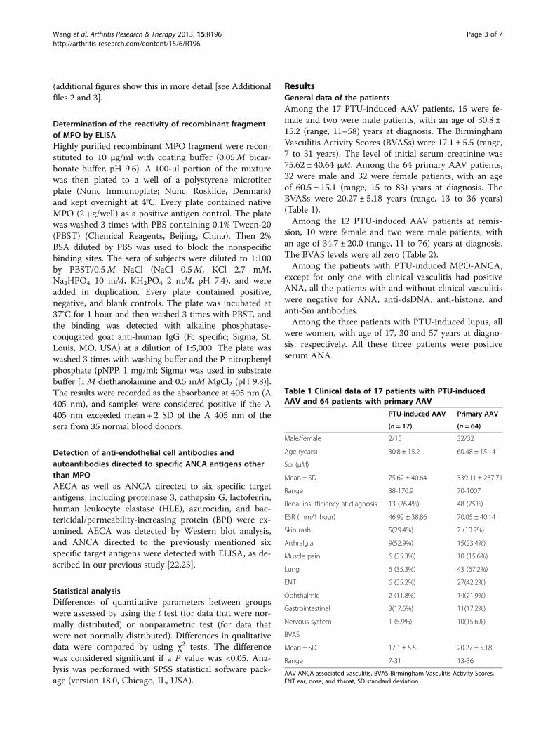
Table 1 Clinical data of 17 patients with PTU-inducedAAV and 64 patients with primary AAV
PTU-induced AAV Primary AAV
(n = 17) (n = 64)
Male/female 2/15 32/32
Age (years) 30.8 ± 15.2 60.48 ± 15.14
Scr (μM)
Mean ± SD 75.62 ± 40.64 339.11 ± 237.71
Range 38-176.9 70-1007
Renal insufficiency at diagnosis 13 (76.4%) 48 (75%)
ESR (mm/1 hour) 46.92 ± 38.86 70.05 ± 40.14
Skin rash 5(29.4%) 7 (10.9%)
Arthralgia 9(52.9%) 15(23.4%)
Muscle pain 6 (35.3%) 10 (15.6%)
Lung 6 (35.3%) 43 (67.2%)
ENT 6 (35.2%) 27(42.2%)
Ophthalmic 2 (11.8%) 14(21.9%)
Gastrointestinal 3(17.6%) 11(17.2%)
Nervous system 1 (5.9%) 10(15.6%)
BVAS
Mean ± SD 17.1 ± 5.5 20.27 ± 5.18
Range 7-31 13-36
AAV ANCA-associated vasculitis, BVAS Birmingham Vasculitis Activity Scores,ENT ear, nose, and throat, SD standard deviation.
Wang et al. Arthritis Research & Therapy 2013, 15:R196 Page 3 of 7http://arthritis-research.com/content/15/6/R196
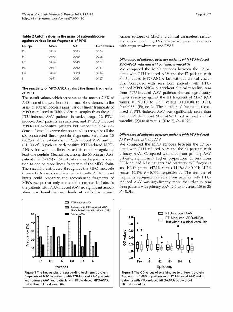
The reactivity of MPO-ANCA against the linear fragmentsof MPOThe cutoff values, which were set as the mean + 2 SD ofA405 nm of the sera from 35 normal blood donors, in theassay of autoantibodies against various linear fragments ofMPO were listed in Table 2. Serum samples from these 17PTU-induced AAV patients in active stage, 12 PTU-induced AAV patients in remission, and 17 PTU-inducedMPO-ANCA-positive patients but without clinical evi-dence of vasculitis were demonstrated to recognize all thesix constructed linear protein fragments. Sera from 15(88.2%) of 17 patients with PTU-induced AAV and 11(61.1%) of 18 patients with positive PTU-induced MPO-ANCA but without clinical vasculitis could recognize atleast one peptide. Meanwhile, among the 64 primary AAVpatients, 37 (57.8%) of 64 patients showed a positive reac-tion to one or more linear fragments of the MPO chain.The reactivity distributed throughout the MPO molecule(Figure 1). None of sera from patients with PTU-inducedlupus could recognize the recombinant fragments ofMPO, except that only one could recognize L chain. Inthe patients with PTU-induced AAV, no significant associ-ation was found between levels of antibodies against
various epitopes of MPO and clinical parameters, includ-ing serum creatinine, ESR, C-reactive protein, numberswith organ involvement and BVAS.
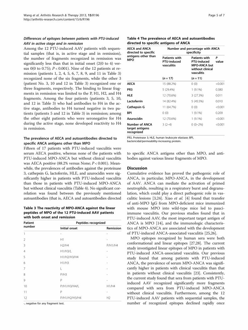
Differences of epitopes between patients with PTU-inducedMPO-ANCA with and without clinical vasculitisWe compared the MPO epitopes between the 17 pa-tients with PTU-induced AAV and the 17 patients withPTU-induced MPO-ANCA but without clinical vascu-litis. Compared with sera from patients with PTU-induced MPO-ANCA but without clinical vasculitis, serafrom PTU-induced AAV patients showed significantlyhigher reactivity against the H1 fragment of MPO [ODvalues: 0.17(0.10 to 0.35) versus 0.10(0.04 to 0.21),P = 0.038] (Figure 2). The number of fragments recog-nized in PTU-induced AAV was significantly more thanthat in PTU-induced MPO-ANCA but without clinicalvasculitis (2(0 to 4) versus 1(0 to 2), P = 0.026).
Differences of epitopes between patients with PTU-inducedAAV and with primary AAVWe compared the MPO epitopes between the 17 pa-tients with PTU-induced AAV and the 64 patients withprimary AAV. Compared with that from primary AAVpatients, significantly higher proportions of sera fromPTU-induced AAV patients had reactivity to P fragmentand H4 fragment. (47.1% versus 14.1%; P < 0.001; 41.2%versus 14.1%; P = 0.034, respectively). The number offragments recognized in sera from patients with PTU-induced AAV was significantly more than that in serafrom patients with primary AAV (2(0 to 4) versus. 1(0 to 2);P = 0.013].
Table 2 Cutoff values in the assay of autoantibodiesagainst various linear fragments of MPO
Epitope Mean SD Cutoff values
Pre 0.058 0.033 0.124
H1 0.076 0.066 0.208
H2 0.074 0.049 0.172
H3 0.061 0.040 0.141
H4 0.094 0.070 0.234
L 0.051 0.043 0.137
Figure 1 The frequencies of sera binding to different proteinfragments of MPO in patients with PTU-induced AAV, patientswith primary AAV, and patients with PTU-induced MPO-ANCAbut without clinical vasculitis.
Figure 2 The OD values of sera binding to different proteinfragments of MPO in patients with PTU-induced AAV and inpatients with PTU-induced MPO-ANCA but withoutclinical vasculitis.
Wang et al. Arthritis Research & Therapy 2013, 15:R196 Page 4 of 7http://arthritis-research.com/content/15/6/R196
Differences of epitopes between patients with PTU-inducedAAV in active stage and in remissionAmong the 12 PTU-induced AAV patients with sequen-tial samples (that is, in active stage and in remission),the number of fragments recognized in remission wassignificantly less than that in initial onset (2(0 to 4) ver-sus 0(0 to 0.75); P < 0.001). Nine of the 12 patients at re-mission (patients 1, 2, 4, 5, 6, 7, 8, 9, and 11 in Table 3)recognized none of the six fragments, while the other 3(patient No. 3, 10 and 12 in Table 3) recognized one orthree fragments, respectively. The binding to linear frag-ments in remission was limited to the P, H1, H2, and H4fragments. Among the four patients (patients 3, 5, 10,and 12 in Table 3) who had antibodies to H4 in the ac-tive stage, antibodies to H4 turned negative in two pa-tients (patients 5 and 12 in Table 3) in remission; amongthe other eight patients who were seronegative for H4during the active stage, none developed reactivity to H4in remission.
The prevalence of AECA and autoantibodies directed tospecific ANCA antigens other than MPOFifteen of 17 patients with PTU-induced vasculitis wereserum AECA positive, whereas none of the patients withPTU-induced MPO-ANCA but without clinical vasculitiswas AECA positive (88.2% versus None; P < 0.001). Mean-while, the prevalences of antibodies against the proteinase3, cathepsin G, lactoferrin, HLE, and azurocidin were sig-nificantly higher in patients with PTU-induced vasculitisthan those in patients with PTU-induced MPO-ANCAbut without clinical vasculitis (Table 4). No significant cor-relation was found between the previously mentionedautoantibodies (that is, AECA and autoantibodies directed
to specific ANCA antigens other than MPO, and anti-bodies against various linear fragments of MPO.
DiscussionCumulative evidence has proved the pathogenic role ofANCA, in particular, MPO-ANCA, in the developmentof AAV. ANCA can mediate the activation of primedneutrophils, resulting in a respiratory burst and degranu-lation, which could play a direct pathogenic role in vas-culitic lesions [3,24]. Xiao et al. [4] found that transferof anti-MPO IgG from MPO-deficient mice immunizedwith mouse MPO into wild-type mice led to pauci-immune vasculitis. Our previous studies found that inPTU-induced AAV, the most important target antigen ofANCA is MPO [14], and the immunologic characteris-tics of MPO-ANCA are associated with the developmentof PTU-induced ANCA-associated vasculitis [25,26].MPO epitopes recognized by human sera were both
conformational and linear epitopes [27,28]. The currentstudy investigated linear epitopes of MPO in patients withPTU-induced ANCA-associated vasculitis. Our previousstudy found that among patients with PTU-inducedANCA, the prevalence of serum MPO-ANCA was signifi-cantly higher in patients with clinical vasculitis than thatin patients without clinical vasculitis [23]. Consistently,the current study found that sera from patients with PTU-induced AAV recognized significantly more fragmentscompared with sera from PTU-induced MPO-ANCAwithout clinical vasculitis. Furthermore, among the 12PTU-induced AAV patients with sequential samples, thenumber of recognized epitopes declined rapidly once
Table 3 The reactivity of MPO-ANCA against the linearpeptides of MPO of the 12 PTU-induced AAV patientswith both onset and remission
Patientnumber
Peptides recognized
Initial onset Remission
1 - -
2 H1 -
3 H2/H4 P/H1/H4
4 H1/H3/L -
5 H1/H2/H3/H4 -
6 H1/H3 -
7 L -
8 P/H3 -
9 P -
10 P/H1/H3/H4/L H1/H4
11 P -
12 P/H1/H2/H3/H4 H2
-, negative for any fragment test.
Table 4 The prevalence of AECA and autoantibodiesdirected to specific antigens of ANCA
AECA and ANCAdirected to specificantigens other thanMPO
Number and percentage with ANCAspecificity
Patients withPTU-inducedvasculitis
Patients withPTU-inducedMPO-ANCA butwithout clinicalvasculitis
Pvalue
(n = 17) (n = 11)
AECA 15 (88.2%) 0 (0) <0.001
PR3 5 (29.4%) 1 (9.1%) 0.380
HLE 12 (70.6%) 3 (27.3%) 0.011
Lactoferrin 14 (82.4%) 5 (45.5%) 0.010
Cathepsin G 11 (64.7%) 0 (0) <0.001
BPI 0 (0) 1 (9.1%) 0.209
Azurocidin 12 (70.6%) 1 (9.1%) <0.001
Number of ANCAtarget antigensrecognized
3 (2–4) 0 (0–2%) <0.001
PR3, Proteinase 3; HLE, human leukocyte elastase; BPI,bactericidal/permeability-increasing protein.
Wang et al. Arthritis Research & Therapy 2013, 15:R196 Page 5 of 7http://arthritis-research.com/content/15/6/R196
remission was achieved, whereas the levels of MPO-ANCA were persistently positive from active stage to re-mission. All these findings suggest that the linear epitopes,compared with conformational ones, might be associatedmore closely with PTU-induced AAV.Compared with sera from primary AAV patients, sera
from PTU-induced AAV patients could recognize signifi-cantly higher numbers of fragments, and had significantlyhigher reactivity to P fragment and H4 fragment. More-over, among the four patients who had antibodies to H4 inthe active stage, antibodies to H4 turned negative in twopatients in remission; among the other eight patients whowere seronegative for H4 during the active stage, no onedeveloped reactivity to H4 in remission. These findings in-dicate that the linear epitope might be of more closely as-sociated with PTU-induced AAV than that in primaryAAV patients. However, the detailed role of antibody di-rected to the P and H4 fragment in the development ofPTU-induced vasculitis demands further investigation.We also found that PTU-induced AAV patients had
higher reactivity against the H1 fragment compared withpatients with PTU-induced MPO-ANCA but withoutclinical vasculitis. However, one patient with PTU-inducedAAV was negative for H1 during the active stage but de-veloped reactivity to H1 in remission. Therefore, the sig-nificance of the H1 fragment in PTU-induced AAVremains more uncertain.Some limitations existed in our study. First, patients
with PTU-induced AAV and patients with primary AAVwere not age- or gender-matched because of the charac-teristics of these two diseases per se. Second, the samplesize was relatively limited because PTU-induced AAV isan uncommon disease.
ConclusionsThe current study provided evidence that PTU-inducedMPO-ANCA could recognize linear epitopes throughoutthe corresponding antigen molecule MPO. Linear epi-topes of the MPO molecule, compared with conform-ational ones, might be associated more closely withPTU-induced AAV. In particular, the P and H4 frag-ments may be important epitopes in PTU-induced AAV.
Additional files
Additional file 1: Figure S1. Schema of linear epitopes of MPO-ANCA.Description: Six recombinant linear fragments, P, L, H1, H2, H3, and H4, wereproduced by using Escherichia coli. P represents propeptide part, amino acids(aa) 49 to 164; L represents light chain, aa165 to 272; H1 to H4 represent fourfragments of the heavy chain, H1 for aa279 to 409, H2 for aa399 to 519, H3for aa510 to 631, and H4 for 622 to 745. About 10 amino acids overlappedbetween the two adjacent fragments on the heavy chain.
Additional file 2: Figure S2. Structure of mature MPO. Description: Themature MPO is produced by two heavy-light protomer units interacting,
which then formed a symmetric homodimer of approximately 150 kDa,with each half linked by a disulfide bond (MMDB ID 48480).
Additional file 3: Figure S3. Location of L, H1, H2, H3, and H4fragments on MPO. Description: L, H1, H2, H3, and H4 fragments weremarked in different colors in one heavy-light protomer unit (MMDB ID75307) as follows: L by purple; H1, H2, H3, and H4 by yellow. The secondrow was viewed from the opposite side of the first row.
AbbreviationsAAV: ANCA-associated vasculitis; AECA: Anti-endothelial cellantibodies; ANCA: Antineutrophil cytoplasmic antibody;BPI: Bactericidal/permeability-increasing protein; BVAS: BirminghamVasculitis Activity Score; EGPA: Eosinophilic granulomatosis withpolyangiitis; GPA: Granulomatosis with polyangiitis; HLE: Human leukocyteelastase; MPA: Microscopic polyangiitis; MPO: Myeloperoxidase;PR3: proteinase 3; PTU: Propylthiouracil.
Competing interestsThe authors declare that they have no competing interests.
Authors’ contributionsCW carried out the experiments, analyzed the data, and wrote and revisedthe manuscript. SJG and PCX participated in the design of the study andcontributed reagents/materials/analysis tools. MHZ and MC designed,coordinated, and directed the study, and helped to write and revise themanuscript. All authors read and approved the final manuscript.
AcknowledgementsThis study is supported by a grant of Chinese 973 project (no.2012CB517702), two grants of the National Natural Science Fund (no.81370829 and no. 81021004), and the Research Fund for the DoctoralProgram of Higher Education of China (no. 20120001110018).
Author details1Renal Division, Department of Medicine, Peking University First Hospital,Beijing 100034, China. 2Peking University Institute of Nephrology, Beijing100034, China. 3Key Laboratory of Renal Disease, Ministry of Health of China,Beijing 100034, China. 4Key Laboratory of Chronic Kidney Disease Preventionand Treatment (Peking University), Ministry of Education, No 8, Xishiku Street,Xicheng District, Beijing 100034, China.
Received: 29 March 2013 Accepted: 12 November 2013Published: 20 November 2013
References1. Falk RJ, Jennette JC: ANCA small-vessel vasculitis. J Am Soc Nephrol 1997,
8:314–322.2. Segelmark M, Wieslander J: IgG subclasses of antineutrophil cytoplasm
autoantibodies (ANCA). Nephrol Dial Transplant 1993, 8:696–702.3. Falk RJ, Terrell RS, Charles LA, Jennette JC: Anti-neutrophil cytoplasmic
autoantibodies induce neutrophils to degranulate and produce oxygenradicals in vitro. Proc Natl Acad Sci USA 1990, 87:4115–4119.
4. Xiao H, Heeringa P, Hu P, Liu Z, Zhao M, Aratani Y, Maeda N, Falk RJ, Jennette JC:Antineutrophil cytoplasmic autoantibodies specific for myeloperoxidase causeglomerulonephritis and vasculitis in mice. J Clin Invest 2002, 110:955–963.
5. Schlieben DJ, Korbet SM, Kimura RE, Schwartz MM, Lewis EJ: Pulmonary-renalsyndrome in a newborn with placental transmission of ANCAs. Am J KidneyDis 2005, 45:758–761.
6. Dolman KM, Gans RO, Vervaat TJ, Zevenbergen G, Maingay D, Nikkels RE,Donker AJ, von dem Borne AE, Goldschmeding R: Vasculitis andantineutrophil cytoplasmic autoantibodies associated withpropylthiouracil therapy. Lancet 1993, 342:651–652.
7. Slot MC, Links TP, Stegeman CA, Tervaert JW: Occurrence of antineutrophilcytoplasmic antibodies and associated vasculitis in patients withhyperthyroidism treated with antithyroid drugs: a long-term followupstudy. Arthritis Rheum 2005, 53:108–113.
8. Zhao MH, Chen M, Gao Y, Wang HY: Propylthiouracil-inducedanti-neutrophil cytoplasmic antibody-associated vasculitis. Kidney Int2006, 69:1477–1481.
Wang et al. Arthritis Research & Therapy 2013, 15:R196 Page 6 of 7http://arthritis-research.com/content/15/6/R196
9. Gao Y, Ye H, Yu F, Guo XH, Zhao MH: Anti-myeloperoxidase IgG subclassdistribution and avidity in sera from patients with propylthiouracil-inducedantineutrophil cytoplasmic antibodies associated vasculitis. Clin Immunol2005, 117:87–93.
10. Aalberse RC, van der Gaag R, van Leeuwen J: Serologic aspects of IgG4antibodies. I. Prolonged immunization results in an IgG4-restrictedresponse. J Immunol 1983, 130:722–726.
11. Gao Y, Chen M, Ye H, Guo XH, Zhao MH, Wang HY: Follow-up of avidityand titre of antimyeloperoxidase antibodies in sera from patients withpropylthiouracil-induced vasculitis. Clin Endocrinol (Oxf ) 2007,66:543–547.
12. Ye H, Zhao MH, Gao Y, Guo XH, Wang HY: Antimyeloperoxidaseantibodies in sera from patients with propylthiouracil-induced vasculitismight recognize restricted epitopes on myeloperoxidase molecule.Clin Exp Immunol 2004, 138:179–182.
13. Fujieda M, Suzuki K, Sato H, Hattori M, Wada N, Tsuchiya M, Okamoto N,Murata T, Matsudaira M, Shimizu M, Ohta K, Naruse K, Sugihara S,Wakiguchi H: Epitope analysis of myeloperoxidase-specific antineutrophilcytoplasmic autoantibodies (MPO-ANCA) in childhood onset Graves’disease treated with propylthiouracil. Clin Nephrol 2005, 63:437–445.
14. Gao Y, Zhao MH, Guo XH, Xin G, Gao Y, Wang HY: The prevalence andtarget antigens of antithyroid drugs induced antineutrophil cytoplasmicantibodies (ANCA) in Chinese patients with hyperthyroidism. Endocr Res2004, 30:205–213.
15. Ye H, Gao Y, Guo XH, Zhao MH: Titre and affinity of propylthiouracil-inducedanti-myeloperoxidase antibodies are closely associated with thedevelopment of clinical vasculitis. Clin Exp Immunol 2005, 142:116–119.
16. Jennette JC, Falk RJ, Bacon PA, Basu N, Cid MC, Ferrario F, Flores-Suarez LF,Gross WL, Guillevin L, Hagen EC, et al: Please list all authors throughout.2012 revised International Chapel Hill Consensus ConferenceNomenclature of Vasculitides. Arthritis Rheum 2013, 65:1–11.
17. Merkel PA: Drug-induced vasculitis. Rheum Dis Clin North Am 2001, 27:849–862.18. Hellmich B, Flossmann O, Gross WL, Bacon P, Cohen-Tervaert JW, Guillevin L,
Jayne D, Mahr A, Merkel PA, Raspe H, Scott DG, Witter J, Yazici H, Luqmani RA:EULAR recommendations for conducting clinical studies and/or clinicaltrials in systemic vasculitis: focus on anti-neutrophil cytoplasmantibody-associated vasculitis. Ann Rheum Dis 2007, 66:605–617.
19. Aloush V, Litinsky I, Caspi D, Elkayam O: Seminar on propylthiouracil-inducedautoimmune syndromes: two distinct clinical presentations with differentcourse and management. Semin Arthritis Rheum 2006, 36:4–9.
20. Zhao MH, Lockwood CM: A comprehensive method to purify three majorANCA antigens: proteinase 3, myeloperoxidase and bactericidal/permeability-increasing protein from human neutrophil granule acidextract. J Immunol Methods 1996, 197:121–130.
21. Gou SJ, Xu PC, Chen M, Zhao MH: Epitope analysis of anti-myeloperoxidaseantibodies in patients with ANCA-associated vasculitis. PLoS One2013, 8:e60530.
22. Yu F, Zhao MH, Zhang YK, Zhang Y, Wang HY: Anti-endothelial cellantibodies (AECA) in patients with propylthiouracil (PTU)-induced ANCApositive vasculitis are associated with disease activity. Clin Exp Immunol2005, 139:569–574.
23. Gao Y, Chen M, Ye H, Guo XH, Zhao MH, Wang HY: The target antigens ofantineutrophil cytoplasmic antibodies (ANCA) induced bypropylthiouracil. Int Immunopharmacol 2007, 7:55–60.
24. Kettritz R, Jennette JC, Falk RJ: Crosslinking of ANCA-antigens stimulatessuperoxide release by human neutrophils. J Am Soc Nephrol 1997, 8:386–394.
25. Chen M, Gao Y, Guo XH, Zhao MH: Propylthiouracil-induced antineutrophilcytoplasmic antibody-associated vasculitis. Nat Rev Nephrol 2012, 8:476–483.
26. Gao Y, Chen M, Ye H, Yu F, Guo XH, Zhao MH: Long-term outcomes ofpatients with propylthiouracil-induced antineutrophil cytoplasmicautoantibody-associated vasculitis. Rheumatology (Oxford) 2008,47:1515–1520.
27. Falk RJ, Becker M, Terrell R, Jennette JC: Anti-myeloperoxidase autoantibodiesreact with native but not denatured myeloperoxidase. Clin Exp Immunol1992, 89:274–278.
28. Tomizawa K, Mine E, Fujii A, Ohashi YY, Yamagoe S, Hashimoto Y,Ishida-Okawara A, Ito M, Tanokura M, Yamamoto T, Arimura Y, Nagasawa T,Mizuno S, Suzuki K: A panel set for epitope analysis of myeloperoxidase(MPO)-specific antineutrophil cytoplasmic antibody MPO–ANCA usingrecombinant hexamer histidine-tagged MPO deletion mutants. J ClinImmunol 1998, 18:142–152.
doi:10.1186/ar4386Cite this article as: Wang et al.: Epitope analysis of anti-myeloperoxidaseantibodies in propylthiouracil-induced antineutrophil cytoplasmicantibody-associated vasculitis. Arthritis Research & Therapy 2013 15:R196.
Submit your next manuscript to BioMed Centraland take full advantage of:
• Convenient online submission
• Thorough peer review
• No space constraints or color figure charges
• Immediate publication on acceptance
• Inclusion in PubMed, CAS, Scopus and Google Scholar
• Research which is freely available for redistribution
Submit your manuscript at www.biomedcentral.com/submit
Wang et al. Arthritis Research & Therapy 2013, 15:R196 Page 7 of 7http://arthritis-research.com/content/15/6/R196