epidermal growth factor and fibroblast growth factor-2 ... · epidermal growth factor and...
TRANSCRIPT
Epidermal Growth Factor and Fibroblast Growth Factor-2 HaveDifferent Effects on Neural Progenitors in the Adult Rat Brain
H. Georg Kuhn,1 Jurgen Winkler,2,3 Gerd Kempermann,1 Leon J. Thal,2,3 and Fred H. Gage1
1Laboratory of Genetics, The Salk Institute, La Jolla, California 92186, 2Department of Neurosciences, University ofCalifornia San Diego, La Jolla, California 92093, and 3Neurology Service (127), Veterans Affairs Medical Center, La Jolla,California 92161
Neurons and glia are generated throughout adulthood fromproliferating cells in two regions of the rat brain, the subven-tricular zone (SVZ) and the hippocampus. This study shows thatexogenous basic fibroblast growth factor (FGF-2) and epider-mal growth factor (EGF) have differential and site-specific ef-fects on progenitor cells in vivo. Both growth factors expandedthe SVZ progenitor population after 2 weeks of intracerebro-ventricular administration, but only FGF-2 induced an increasein the number of newborn cells, most prominently neurons, inthe olfactory bulb, the normal destination for neuronal progen-itors migrating from the SVZ. EGF, on the other hand, reducedthe total number of newborn neurons reaching the olfactorybulb and substantially enhanced the generation of astrocytes inthe olfactory bulb. Moreover, EGF increased the number ofnewborn cells in the striatum either by migration of SVZ cells or
by stimulation of local progenitor cells. No evidence of neuronaldifferentiation of newborn striatal cells was found by three-dimensional confocal analysis, although many of these new-born cells were associated closely with striatal neurons. Theproliferation of hippocampal progenitors was not affected byeither growth factor. However, EGF increased the number ofnewborn glia and reduced the number of newborn neurons,similar to the effects seen in the olfactory bulb. These findingsmay be useful for elucidating the in vivo role of growth factorsin neurogenesis in the adult CNS and may aid development ofneuronal replacement strategies after brain damage.
Key words: subventricular zone; hippocampus; epidermalgrowth factor; basic fibroblast growth factor; intracerebroven-tricular administration; progenitor cells; stem cells; proliferation;neurogenesis; gliogenesis
The adult CNS appears to have only limited potential to generatenew neurons, making it vulnerable to injury and disease. How-ever, certain areas of the brain retain the capacity for neurogen-esis well into adulthood (Altman and Das, 1965; Kuhn et al.,1996). In the adult rodent a rapidly dividing population of stemcells in the subventricular zone (SVZ) of the lateral ventriclegenerates all neural cell types: neurons, astrocytes, and oligoden-drocytes (Lewis, 1968; Privat and Leblond, 1972; Corotto et al.,1993; Lois and Alvarez-Buylla, 1993; Morshead et al., 1994;Goldman, 1995; Hauke et al., 1995). From the SVZ neuronalprogenitors migrate tangentially (sagittally) along the rostral mi-gratory stream (RMS) into the olfactory bulb (OB), where theydifferentiate into granule and periglomerular neurons (Corotto etal., 1993; Lois and Alvarez-Buylla, 1993, 1994; Luskin, 1993;Goldman, 1995; Lois et al., 1996). In contrast, glial progenitorcells migrate radially into neighboring brain structures such asstriatum, corpus callosum, and neocortex (Levison and Goldman,1993; Levison et al., 1993; Luskin and McDermott, 1994). In thehippocampal dentate gyrus of adult rats, neural precursor cellscontinue to proliferate and differentiate into granule cells
(Kaplan and Hinds, 1977; Kaplan and Bell, 1983; Cameron et al.,1993; Kuhn et al., 1996).
To be able to manipulate the endogenous adult progenitors, webelieve it is crucial to determine the extracellular signals that canstimulate cell division and regulate the fate of these neural stemand progenitor cells (Cattaneo and McKay, 1991; Gage, 1994).Recently, several groups successfully have isolated and propa-gated adult neural progenitor cells in vitro (Reynolds and Weiss,1992; Richards et al., 1992; Lois and Alvarez-Buylla, 1993;Vescovi et al., 1993; Morshead et al., 1994; Gage et al., 1995a;Palmer et al., 1995; Gritti et al., 1996). Dissociated cells from theSVZ and the hippocampus required basic fibroblast growth factor(FGF-2) or epidermal growth factor (EGF) for proliferation andlong-term survival in vitro. Some of these cells retain the ability togenerate both neurons and glia, suggesting that newborn cells ofthe adult brain may originate from stem cell-like progenitors(Morshead et al., 1994; Gage et al., 1995a; Palmer et al., 1995;Gritti et al., 1996; Suhonen et al., 1996).
The possibility that growth factors also may influence neuralprogenitors in vivo has been supported by findings that intrace-rebroventricular administration of EGF expanded proliferativeprogenitors in the SVZ of adult mice (Craig et al., 1996). Numer-ous newborn cells were found in the adjacent striatum, septum,and cortex, and a small portion of these cells expressed neuronalantigens.
The goal of the present study was to explore systematically theeffects of FGF-2 or EGF on the proliferation and differentiationof neural progenitor cells in the SVZ/OB system and the dentategyrus of adult rats. FGF-2, EGF, or artificial CSF (aCSF) werechronically infused into the lateral ventricle. The proliferative
Received Dec. 11, 1996; revised March 27, 1997; accepted May 9, 1997.This work was supported by the National Institute on Aging, National Institutes of
Health, Veterans Affairs Research Service, and Sam and Rose Stein Institute forResearch on Aging. H.G.K. is a fellow of the Hereditary Disease Foundation. J.W.is a fellow of the National Brookdale Foundation. G.K. is a fellow of the DeutscheForschungsgemeinschaft. We thank Gilbert Ramirez for his excellent technicalassistance and Theo D. Palmer, Lisa J. Fisher, Mireya Nadal-Vicens, and MaryLynn Gage for their critical review of this manuscript.
H.G.K. and J.W. have contributed equally to this manuscript.Correspondence should be addressed to Dr. Fred H. Gage, The Salk Institute,
Laboratory of Genetics, P.O. Box 85800, San Diego, CA 92186-5800.Copyright © 1997 Society for Neuroscience 0270-6474/97/175820-10$05.00/0
The Journal of Neuroscience, August 1, 1997, 17(15):5820–5829
zones, migratory paths, and areas of neuronal differentiation wereanalyzed quantitatively for the number and phenotype of new-born cells.
MATERIALS AND METHODSAnimals and surgeryMale Fischer-344 albino rats (n 5 30; Harlan Sprague Dawley, India-napolis, IN) were used in this experiment. The animals were 13–14weeks old and weighed between 260 and 300 gm at the start of theexperiment. Anesthesia was induced by an intramuscular injection con-sisting of 62.5 mg/kg ketamine (Ketaset, 100 mg/ml, Bristol Laborato-ries, Syracuse, NY), 3.175 mg/kg xylazine (Rompun, 20 mg/ml, MilesLaboratories, Shawnee, KS), and 0.625 mg/kg acepromazine maleate(10 mg/ml, TechAmerica Group, Elwood, KS) dissolved in 0.9% sterilesaline. Rats were mounted in a small animal stereotaxic apparatus(David Kopf, Tujunga, CA) with bregma and lambda in the samehorizontal plane. A stainless steel cannula (28 gauge, Plastic Products,Roanoke, VA) was implanted in the lateral ventricle [anteroposterior(AP) 18.5 mm, lateral 11.5 mm from the center of the interaural line inflat skull position; cannula length, 5 mm] and connected by 3.5 cm vinyltubing (size V/4, Bolab, Lake Havasu City, AZ) to an osmotic minipump(model 2002, Alza, Palo Alto, CA). Human recombinant EGF (30 mg/ml,Promega, Madison, WI) or FGF-2 (30 mg/ml, A. Baird, Prizm Pharma-ceuticals, San Diego, CA) was dissolved in aCSF [(in mM): 148 NaCl, 3KCl, 1.4 CaCl2 , 0.8 MgCl2 , 1.5 Na2HPO4 , and 0.2 NaH2PO4 , pH 7.4]containing 100 mg/ml rat serum albumin (Sigma, St. Louis, MO). Anantibiotic (gentamycin, 50 mg/ml, Sigma) was included in the infusate.The animals received EGF (n 5 10), FGF-2 (n 5 10), or aCSF (n 5 10)at a flow rate of 0.50 ml /hr, resulting in a delivery of 360 ng of growthfactor per day for 14 d. During the last 12 d of the pump period animalsreceived daily intraperitoneal injections of bromodeoxyuridine (BrdU,50 mg/kg, Sigma). At the end of the treatment one-half of the animals(n 5 5 per group) were anesthetized deeply and perfused intracardiallywith 4% paraformaldehyde in 100 mM phosphate buffer, pH 7.4. Brainswere removed, post-fixed overnight in 4% paraformaldehyde, and trans-ferred to 0.32 M sucrose. In the remaining one-half of the animals thepumps were removed under methoxyflurane anesthesia. The vinyl tubingwas ligated with sterile nonabsorbable black monofilament nylon (3–0Dermalon, American Cyanamid, Danbury, CT). These animals wereperfused after an additional 4 week period without growth factorinfusion.
HistologyThe brains were cut in three parts, providing material for (1) coronalsections of the SVZ and hippocampus and sagittal sections of the (2) OBand (3) cerebellum. Sections (40 mm) were cut with a sliding microtomeand stored at 220°C in a cryoprotectant solution (glycerol, ethyleneglycol, and 0.1 M phosphate buffer, pH 7.4, 3:3:4 by volume).
Antibodies and immunochemicals. The following antibodies and finaldilutions were used: mouse (mo) a-BrdU (1:400, Boehringer Mannheim,Indianapolis, IN), rat a-BrdU (1:100, Accurate, Westbury, NY), moa-PSA-NCAM (1:2500, clone MenB kindly provided by Dr. G. Rougon,University of Marseille, Marseille, France), mo a-NeuN (1:20, clone A60kindly provided by Dr. R. Mullen, University of Utah, Salt Lake City,UT), rabbit a-S100b (1:5000, Swant, Bellinzona, Switzerland), biotiny-lated horse a-mouse IgG (1:160, Vector Laboratories, Burlingame, CA),avidin–biotin–peroxidase complex (1:100, Vectastain Elite, Vector Lab-oratories), and donkey a-rat–FITC, a-mouse–Texas Red, and a-rabbit–CY5 (all 1:300, Jackson ImmunoResearch, West Grove, PA).
Immunoperoxidase. Free-floating sections were treated with 0.6%H2O2 in TBS (0.15 M NaCl and 0.1 M Tris-HCl, pH 7.5) for 30 min toblock endogenous peroxidase. For DNA denaturation, sections wereincubated for 2 hr in 50% formamide/23 SSC (0.3 M NaCl and 0.03 Msodium citrate) at 65°C, rinsed for 5 min in 23 SSC, incubated for 30 minin 2N HCl at 37°C, and rinsed for 10 min in 0.1 M boric acid, pH 8.5.Several rinses in TBS were followed by incubation in TBS/0.1% TritonX-100/3% normal horse serum (TBS–Ths) for 30 min and incubationwith mo a-BrdU antibody in TBS–Ths overnight at 14°C. After beingrinsed in TBS–Ths, sections were incubated for 1 hr with biotinylatedhorse a-mouse antibody. With intermittent rinses in TBS, avidin–biotin–peroxidase complex was applied for 1 hr, followed by peroxidase detec-tion for 5 min (0.25 mg/ml DAB, 0.01% H2O2 , 0.04% NiCl).
Immunofluorescence. Sections were treated for DNA denaturation asdescribed above, followed by several rinses in TBS and incubation in
TBS/0.1% Triton X-100/3% normal donkey serum (TBS–Tds) for 30min. Primary antibodies were applied in TBS–Tds for 48 hr at 14°C,rinsed in TBS three times for 10 min, and blocked in TBS–Tds for 10min. Antibodies were detected with donkey a-rat, mouse, or rabbitcoupled to FITC, Texas Red, or CY5 for 2 hr. Fluorescent signals weredetected and processed by a confocal scanning laser microscope (Bio-Rad MRC1024, Hercules, CA) and Adobe Photoshop (Adobe Systems,Mountainview, CA).
QuantificationQuantification of BrdU-positive cells was accomplished with unbiasedcounting methods. The optical disector procedure (Sterio, 1984) wasused to determine the three-dimensional numerical density of BrdU-positive cells, which is expressed as cells/mm 3. Structures were sampledeither by selecting predetermined areas on each section (Fig. 1; SVZ,striatum, RMS, and OB) or by analyzing entire structures on each section(dentate gyrus and cerebellum). In the latter case we used a point-counting grid for determination of the sampling volume via the Cavalierimethod (Michel and Cruz-Orive, 1988).
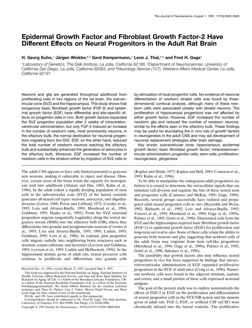
Lateral ventricle and striatum. Every 12th section from a coronal seriesof the striatum was selected between AP 110.6 mm—genu corpuscallosum—and AP 18.74 mm—anterior commissure crossing (Paxinosand Watson, 1986). As illustrated in Figure 1 B, BrdU-positive cells werecounted in three predetermined areas (50 3 50 mm) of the lateralventricle wall on all selected sections. A rectangular area of the striatum(300 3 600 mm) was selected at a 50 mm distance from the lateralventricle wall and analyzed on each section. All BrdU-positive nuclei inthese selected areas were counted and presented as the number of cells(in thousands)/mm 3 (Table 1).
RMS and OB. Every sixth section (40 mm) from sagittal series of theOB/frontal cortex was selected and stained for BrdU immunohistochem-
Figure 1. Analysis of the subventricular zone (SVZ) and olfactory bulb.A, Sagittal view of the rat brain illustrating the anatomical sites ofprogenitor proliferation in the SVZ, migration along the rostral migratorystream (RMS), and differentiation in the olfactory bulb (OB). Hatched barindicates position of coronal view in B. B, Coronal plane of the lateralventricle with the corpus callosum (CC), medial septum (MS), and stria-tum (Str). Three areas in the SVZ (ventral, lateral, and dorsal squares,50 3 50 mm) and one area in the striatum (large rectangle, 300 3 600 mm)were analyzed for BrdU-positive cells on each section. C, Parasagittalplane of frontal cortex and olfactory bulb. Two areas of the RMS (smallsquares, 50 3 50 mm) and four areas of the OB granule cell layer (largesquares, 100 3 100 mm) were analyzed for BrdU-positive cells andcolabeling with NeuN or S100b.
Kuhn et al. • EGF and FGF-2 Effects on Adult Neural Progenitors In Vivo J. Neurosci., August 1, 1997, 17(15):5820–5829 5821
istry. As depicted in Figure 1C, two predetermined areas (50 3 50 mm)in the RMS and four areas (100 3 100 mm) in the granule cell layer(GCL) of the OB were analyzed on each section. All BrdU-positivenuclei in these selected areas were counted and presented as the numberof cells (in thousands)/mm 3 (Table 1).
Dentate gyrus. Every 12th section (40 mm) from a coronal series wasselected from each animal and processed for immunoperoxidase. Sixsections from the dorsal hippocampus (AP 15.86 to 12.96 mm) wereanalyzed entirely for BrdU-positive cells in the molecular layer, theGCL, and the hilus. The subgranular zone, defined as a two-cell body-wide zone along the border of the GCL and the hilus, always wascombined with the GCL for quantification. All BrdU-positive nuclei inthese selected areas were counted and presented as cells (in thousands)/mm 3 (Table 2).
Cerebellum. On two sagittal sections of the cerebellum the fourthlobulus was analyzed entirely for the density of BrdU-positive cells in themolecular layer, GCL, and white matter. Cell numbers are expressed asBrdU-positive cells (in thousands)/mm 3 (Table 2).
Statistical analysis was performed with one-way ANOVA, followed bypost hoc comparison with the Tukey post hoc test.
RESULTSTo study the effect of growth factors on the proliferation of adultneural progenitor cells in vivo, we chronically infused EGF,FGF-2, or aCSF for 2 weeks into the lateral ventricle of adult rats,using osmotic minipumps. BrdU was administered intraperitone-ally during the period of growth factor infusion to label dividingcells. Animals either were killed on the last day of growth factorinfusion to analyze the mitotic effect of the growth factors or werekept for an additional 4 weeks after terminating growth factorinfusion to study the differentiation of the newborn cells. Bothsystems of adult neurogenesis, the SVZ/OB and dentate gyrus,were analyzed for BrdU-positive cells. The cerebellum served asa control area in the adult rat brain because it lacks detectableneurogenesis. To quantify the proliferation of neural progenitorcells and the survival of newborn cells after growth factor infu-sion, we determined the density of BrdU-positive cells by stereo-
logical counting techniques. To characterize cell fate, we com-bined BrdU labeling with the astroglial marker S100b, whichlabels astrocytic cell bodies (Boyes et al., 1986), and the neuronalmarker NeuN, which recognizes neuronal cell bodies and nuclei(Mullen et al., 1992). The percentage of BrdU-positive cellscolabeled either with NeuN or S100b was determined by tripleimmunofluorescence and confocal laser scanning microscopy andwas multiplied by the overall density of BrdU-positive cells todetermine the density of newborn neurons and astrocytes.
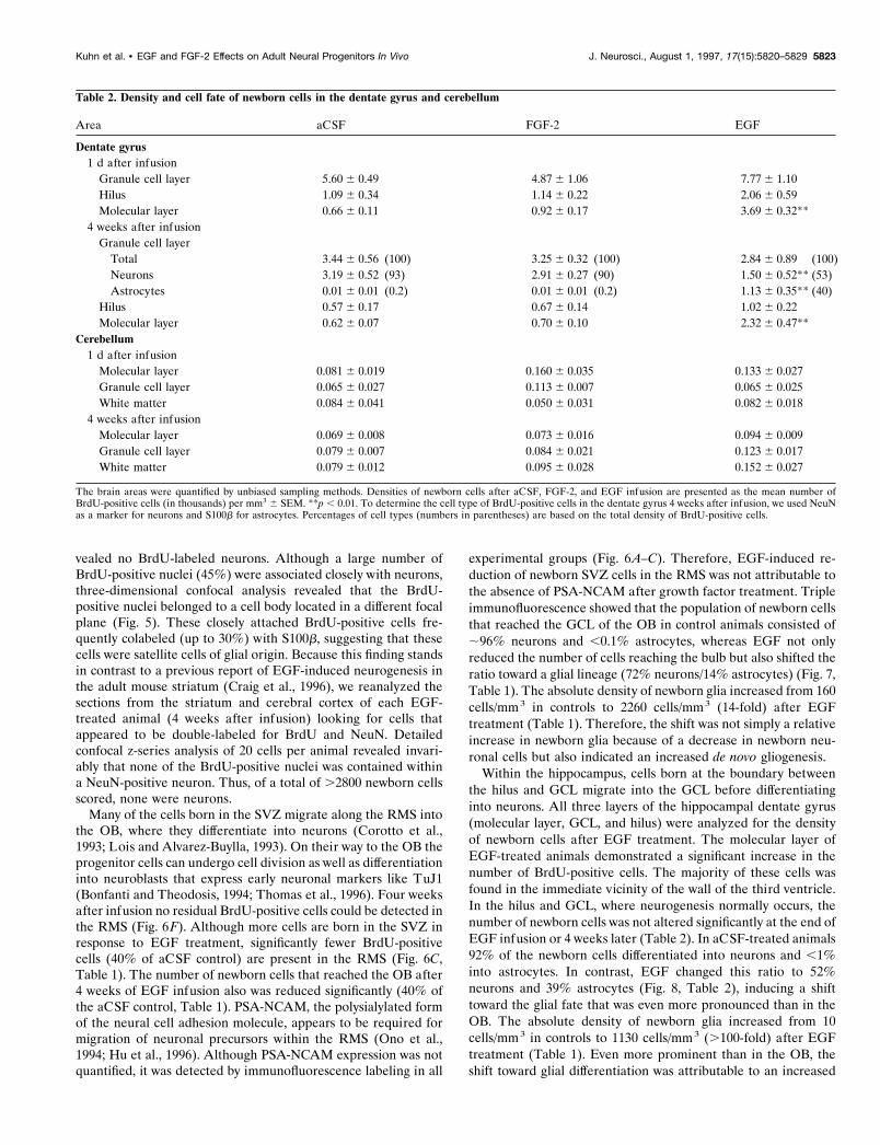
EGF effects on proliferation and differentiation ofneural progenitorsInfusion of EGF induced a striking proliferation of the SVZprecursor population. Expansion of BrdU-positive cells was mostpronounced in the lateral wall of the lateral ventricle (Fig. 2C). Inaddition, newborn cells were found in the medial and posteriorcircumference of the lateral ventricle, suggesting that progenitorsalso were recruited to divide in “quiescent” areas of the SVZ.Infusion of EGF resulted in “polyp-like” hyperplasias of theventricle wall, which consisted of BrdU-positive cells that wereimmunonegative for either S100b or NeuN (Fig. 3). These EGF-induced hyperplasias had regressed completely after 4 weeks(Fig. 3C).
Quantification of the SVZ revealed a ninefold increase in thedensity of newborn cells over aCSF controls immediately afterEGF infusion. The number of labeled cells present after 4 weeksremained increased relative to controls (Fig. 2, Table 1). Therewas also an increase in the number of labeled cells observed inadjacent areas, particularly in the striatum (Fig. 4C, Table 1), butalso in cortex and septum (Fig. 4F,I), where the majority ofnewborn cortical cells was detected around the cannula tract (Fig.4F). Interestingly, in the striatum triple labeling of BrdU-positiveEGF-generated striatal cells with BrdU, NeuN, and S100b re-
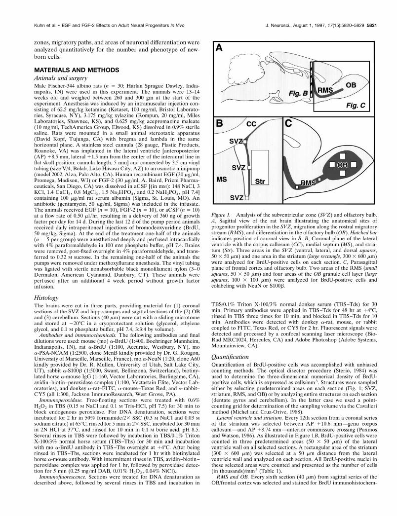
Table 1. Density and cell fate of newborn cells in the subventricular zone, olfactory bulb, and striatum
Area aCSF FGF-2 EGF
Subventricular zone1 d after infusion
Cannula side 62.2 6 9.8 208.5 6 62.2** 595.5 6 133.1**Contralateral side 37.1 6 8.1 32.6 6 4.4 70.3 6 9.9**
4 weeks after infusionCannula side 30.6 6 10.7 104.8 6 27.7** 153.6 6 27.8**Contralateral side 13.3 6 3.9 13.5 6 2.7 88.0 6 11.8**
Rostral migratory str.1 d after infusion 915.5 6 50.0 714.4 6 38.0* 368.8 6 25.0**
Olfactory bulb4 weeks after infusion
Total 40.9 6 2.0 (100) 46.9 6 1.2* (100) 16.3 6 1.6** (100)Neurons 39.3 6 1.6 (96) 45.2 6 0.9* (96) 11.8 6 1.5** (72)Astrocytes 0.16 6 0.11 (,0.1) 0.26 6 0.10 (,0.1) 2.26 6 0.54** (14)
Striatum4 weeks after infusion
Total 2.8 6 0.4 (100) 6.1 6 0.8** (100) 12.3 6 3.5** (100)Neurons 0 (0) 0 (0) 0 (0)Astrocytes 0.2 6 0.1 (6) 1.4 6 0.4** (22) 4.5 6 1.4** (39)BrdU1/satellite cells 1.1 6 0.1 (40) 3.2 6 0.5* (53) 5.2 6 1.1* (46)BrdU1/sat./astrocytes 0.05 6 0.03 (1.8) 0.6 6 0.2* (10) 1.7 6 0.7* (14)
The brain areas were selected for unbiased quantification, as shown in Figure 1. Densities of newborn cells after aCSF, FGF-2, and EGF infusion are presented as the meannumber of BrdU-positive cells (in thousands) per mm3 6 SEM. *p , 0.05; **p , 0.01. To determine the cell type of BrdU-positive cells 4 weeks after infusion, we used NeuNas a marker for neurons and S100b for astrocytes. Percentages of cell types (numbers in parentheses) are based on the total density of BrdU-positive cells.
5822 J. Neurosci., August 1, 1997, 17(15):5820–5829 Kuhn et al. • EGF and FGF-2 Effects on Adult Neural Progenitors In Vivo
vealed no BrdU-labeled neurons. Although a large number ofBrdU-positive nuclei (45%) were associated closely with neurons,three-dimensional confocal analysis revealed that the BrdU-positive nuclei belonged to a cell body located in a different focalplane (Fig. 5). These closely attached BrdU-positive cells fre-quently colabeled (up to 30%) with S100b, suggesting that thesecells were satellite cells of glial origin. Because this finding standsin contrast to a previous report of EGF-induced neurogenesis inthe adult mouse striatum (Craig et al., 1996), we reanalyzed thesections from the striatum and cerebral cortex of each EGF-treated animal (4 weeks after infusion) looking for cells thatappeared to be double-labeled for BrdU and NeuN. Detailedconfocal z-series analysis of 20 cells per animal revealed invari-ably that none of the BrdU-positive nuclei was contained withina NeuN-positive neuron. Thus, of a total of .2800 newborn cellsscored, none were neurons.
Many of the cells born in the SVZ migrate along the RMS intothe OB, where they differentiate into neurons (Corotto et al.,1993; Lois and Alvarez-Buylla, 1993). On their way to the OB theprogenitor cells can undergo cell division as well as differentiationinto neuroblasts that express early neuronal markers like TuJ1(Bonfanti and Theodosis, 1994; Thomas et al., 1996). Four weeksafter infusion no residual BrdU-positive cells could be detected inthe RMS (Fig. 6F). Although more cells are born in the SVZ inresponse to EGF treatment, significantly fewer BrdU-positivecells (40% of aCSF control) are present in the RMS (Fig. 6C,Table 1). The number of newborn cells that reached the OB after4 weeks of EGF infusion also was reduced significantly (40% ofthe aCSF control, Table 1). PSA-NCAM, the polysialylated formof the neural cell adhesion molecule, appears to be required formigration of neuronal precursors within the RMS (Ono et al.,1994; Hu et al., 1996). Although PSA-NCAM expression was notquantified, it was detected by immunofluorescence labeling in all
experimental groups (Fig. 6A–C). Therefore, EGF-induced re-duction of newborn SVZ cells in the RMS was not attributable tothe absence of PSA-NCAM after growth factor treatment. Tripleimmunofluorescence showed that the population of newborn cellsthat reached the GCL of the OB in control animals consisted of;96% neurons and ,0.1% astrocytes, whereas EGF not onlyreduced the number of cells reaching the bulb but also shifted theratio toward a glial lineage (72% neurons/14% astrocytes) (Fig. 7,Table 1). The absolute density of newborn glia increased from 160cells/mm3 in controls to 2260 cells/mm3 (14-fold) after EGFtreatment (Table 1). Therefore, the shift was not simply a relativeincrease in newborn glia because of a decrease in newborn neu-ronal cells but also indicated an increased de novo gliogenesis.
Within the hippocampus, cells born at the boundary betweenthe hilus and GCL migrate into the GCL before differentiatinginto neurons. All three layers of the hippocampal dentate gyrus(molecular layer, GCL, and hilus) were analyzed for the densityof newborn cells after EGF treatment. The molecular layer ofEGF-treated animals demonstrated a significant increase in thenumber of BrdU-positive cells. The majority of these cells wasfound in the immediate vicinity of the wall of the third ventricle.In the hilus and GCL, where neurogenesis normally occurs, thenumber of newborn cells was not altered significantly at the end ofEGF infusion or 4 weeks later (Table 2). In aCSF-treated animals92% of the newborn cells differentiated into neurons and ,1%into astrocytes. In contrast, EGF changed this ratio to 52%neurons and 39% astrocytes (Fig. 8, Table 2), inducing a shifttoward the glial fate that was even more pronounced than in theOB. The absolute density of newborn glia increased from 10cells/mm3 in controls to 1130 cells/mm3 (.100-fold) after EGFtreatment (Table 1). Even more prominent than in the OB, theshift toward glial differentiation was attributable to an increased
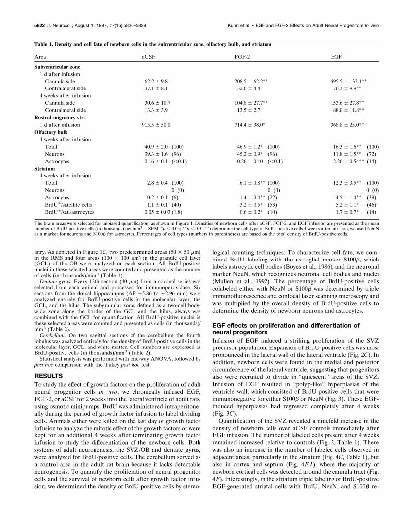
Table 2. Density and cell fate of newborn cells in the dentate gyrus and cerebellum
Area aCSF FGF-2 EGF
Dentate gyrus1 d after infusion
Granule cell layer 5.60 6 0.49 4.87 6 1.06 7.77 6 1.10Hilus 1.09 6 0.34 1.14 6 0.22 2.06 6 0.59Molecular layer 0.66 6 0.11 0.92 6 0.17 3.69 6 0.32**
4 weeks after infusionGranule cell layer
Total 3.44 6 0.56 (100) 3.25 6 0.32 (100) 2.84 6 0.89 (100)Neurons 3.19 6 0.52 (93) 2.91 6 0.27 (90) 1.50 6 0.52** (53)Astrocytes 0.01 6 0.01 (0.2) 0.01 6 0.01 (0.2) 1.13 6 0.35** (40)
Hilus 0.57 6 0.17 0.67 6 0.14 1.02 6 0.22Molecular layer 0.62 6 0.07 0.70 6 0.10 2.32 6 0.47**
Cerebellum1 d after infusion
Molecular layer 0.081 6 0.019 0.160 6 0.035 0.133 6 0.027Granule cell layer 0.065 6 0.027 0.113 6 0.007 0.065 6 0.025White matter 0.084 6 0.041 0.050 6 0.031 0.082 6 0.018
4 weeks after infusionMolecular layer 0.069 6 0.008 0.073 6 0.016 0.094 6 0.009Granule cell layer 0.079 6 0.007 0.084 6 0.021 0.123 6 0.017White matter 0.079 6 0.012 0.095 6 0.028 0.152 6 0.027
The brain areas were quantified by unbiased sampling methods. Densities of newborn cells after aCSF, FGF-2, and EGF infusion are presented as the mean number ofBrdU-positive cells (in thousands) per mm3 6 SEM. **p , 0.01. To determine the cell type of BrdU-positive cells in the dentate gyrus 4 weeks after infusion, we used NeuNas a marker for neurons and S100b for astrocytes. Percentages of cell types (numbers in parentheses) are based on the total density of BrdU-positive cells.
Kuhn et al. • EGF and FGF-2 Effects on Adult Neural Progenitors In Vivo J. Neurosci., August 1, 1997, 17(15):5820–5829 5823
de novo gliogenesis and not merely a relative increase because ofa decrease in newborn neuronal cells.
FGF-2 effects on proliferation and differentiation ofneural progenitorsThe density of newborn cells in the SVZ was increased by FGF-2,although to a lesser extent than by EGF, and the density ofnewborn cells into the adjacent striatal parenchyma was increasedover aCSF controls (Figs. 2B, 4B, Table 1). None of the BrdU-positive cells in the striatum of FGF-2-treated animals wasdouble-labeled for NeuN, although some newborn cells in thestriatum were juxtaposed closely to neuronal cell bodies, as seenin EGF animals. FGF-2 also decreased the number of BrdU-positive cells in the RMS at the end of infusion (Fig. 6B, Table 1).As with EGF infusion and in aCSF controls, no BrdU-positivecells were detectable in the RMS 4 weeks later (Fig. 6D–F).However, in contrast to the EGF animals, the number of BrdU-positive cells found in the GCL of the OB 4 weeks after FGF-2infusion was increased significantly over controls (Table 1). Thisgeneral increase in the density of newborn olfactory cells wasaccompanied by an increase of newborn neurons. The number ofnewborn glial cells was not altered significantly, although this wasprobably because of the infrequent detection of BrdU/S100b-positive cells and a resulting high variance (Table 1).
In contrast to the SVZ/OB system, the generation of newborncells in the hippocampal dentate gyrus was not affected by FGF-2treatment. The ratio between newborn neurons and astrocytesalso was not altered 4 weeks after FGF-2 treatment, indicatingthat both proliferation and differentiation of hippocampal pro-genitors were unaffected by the growth factor (Fig. 8, Table 2).
Analysis of newborn cells in the cerebellum revealed no signif-icant changes at the end of growth factor treatment or 4 weeksafter withdrawal of either growth factor (Table 2), indicating thatthis brain structure, which normally shows no adult neurogenesis,is unresponsive to these growth factors.
DISCUSSIONDuring development, growth factors provide important extracel-lular signals for regulating the proliferation and fate determina-tion of stem and progenitor cells in the CNS (Calof, 1995). Byinfusing EGF and FGF-2 into the lateral ventricle of adult rats,we could show that the two populations of progenitors thatcontinue to divide in the adult brain respond differently to thesegrowth factors in vivo. Proliferation of hippocampal progenitorswas unaffected by either EGF or FGF-2. In contrast, proliferationof subventricular progenitor cells increased after both FGF-2 andEGF administration, with EGF having a more dramatic effect.These findings are consistent with numerous in vitro studies that
have shown that both factors can maintain responsive neuralprogenitors in cell cycle, thus expanding the progenitor popula-tion and delaying differentiation (Richards et al., 1992; Vescovi etal., 1993; Morshead et al., 1994; Sensenbrenner et al., 1994;Bouvier and Mytilineou, 1995; Gage et al., 1995a; Gritti et al.,1995, 1996; Palmer et al., 1995; Santa-Olalla and Covarrubias,1995).
FGF-2 had a strong mitotic effect on the SVZ progenitors invivo, but the migration of newborn cells in the RMS wasdiminished during the infusion period. However, 4 weeks afterFGF-2 infusion, a larger number of newly generated cells weredetected in the OB, indicating an increased migration of SVZprogenitors after withdrawal of FGF-2. In vitro results suggestthat FGF-2 has the potential to keep uncommitted progenitorsin cell cycle and to delay differentiation (Vescovi et al., 1993;Bouvier and Mytilineou, 1995; Kilpatrick and Bartlett, 1995;Palmer et al., 1995; Gritti et al., 1996). The biphasic responseof RMS cells to FGF-2 could be attributable to increasedproliferation in the SVZ, which reduces progenitor cell migra-tion through the stream. After FGF-2 withdrawal a largernumber of cells would be released into the RMS, generatingmore newborn cells in the OB 4 weeks later. Because 96% ofthe newborn olfactory cells differentiated into neurons, weconclude that FGF-2 had a stimulatory effect on the generationof OB neurons. However, because a very low number of new-born glial cells were detected here, conclusions about FGF-2-induced changes of the glial cell population in the OB are notpossible. EGF infusion also expanded the SVZ precursor pop-ulation while decreasing the number of newborn cells in theRMS. However, in contrast to FGF-2, EGF withdrawal re-duced neurogenesis in the OB, whereas the genesis of astro-cytes was stimulated. Although olfactory neurogenesis in-volves separate areas for cell division (SVZ), migration(RMS), and differentiation (OB), recent studies have shownthat cell division and neuronal commitment of progenitor cellscan occur in the migratory stream (Bonfanti and Theodosis,1994; Menezes et al., 1995; Lois et al., 1996; Thomas et al.,1996). Astrocytes in the RMS are typically neither proliferat-ing nor participating in the migration (Lois and Alvarez-Buylla, 1994; Lois et al., 1996). We assume that EGF acts onproliferation primarily in the SVZ and on differentiation pri-marily in the OB but also is influencing cells in the RMS.
Progenitor populations in SVZ and hippocampus were affecteddifferently by EGF. EGF had no proliferative effect on hippocam-pal progenitors, whereas even progenitors in quiescent areas ofthe SVZ, such as the medial and posterior regions of the ventric-ular wall, were recruited by EGF to enter the cell cycle. Normally,
3
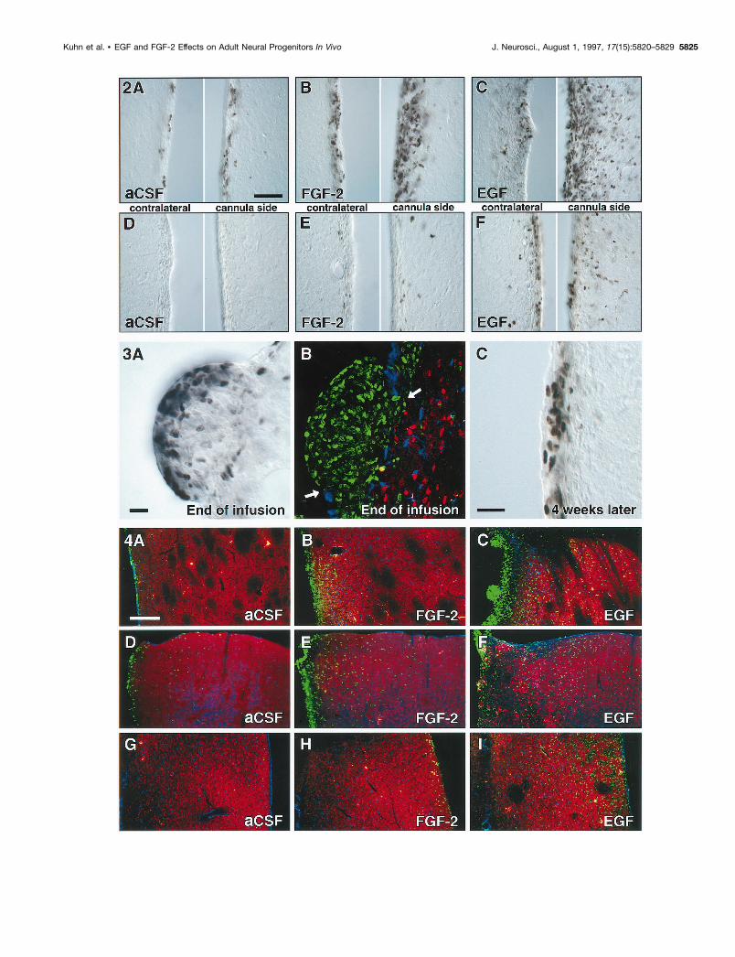
Figure 2. BrdU-positive cells in the SVZ at the end of and 4 weeks after intracerebroventricular infusion of aCSF (A, D), FGF-2 (B, E), and EGF (C,F ). Note the large expansion of the SVZ and the density of newborn cells in the striatum after FGF-2 administration ( B), which are even more dramaticafter EGF administration (C). Proliferation was more pronounced on the side of the cannula, as compared with the contralateral side. Four weeks aftergrowth factor withdrawal, a high density of BrdU-positive cells was still present in the SVZ of EGF-treated animals (F). Scale bar in A, 50 mm.
Figure 3. “Polyp-like” hyperplasia in the SVZ of EGF-treated animals at the end of treatment (2 weeks). A, High density of BrdU-positive cells at theconvex pole of a hyperplasia, which protrudes into the CSF-filled ventricle. B, BrdU-positive cells are immunonegative for neuronal (NeuN, red) andastrocytic markers (S100b, blue). The ependymal layer (S100b, blue) is discontinuous (arrows) in areas of growth. C, Density of BrdU-labeled cells isstill increased; however, the hyperplastic changes completely regress 4 weeks after EGF withdrawal. Scale bars in A, C, 25 mm.
Figure 4. Increased number of BrdU-positive cells in the striatum ( A–C), cortex ( D–F), and medial septum ( G–I) of EGF- and FGF-2-treated animalsat the end of infusion. D–F, Note the increase of BrdU-positive cells along the cannula tract in the cerebral cortex (on the lef t side of the images). Shownare confocal microscopic images with immunofluorescent triple labeling for BrdU ( green), NeuN (red), and S100b (blue). Scale bar in A, 200 mm.
5824 J. Neurosci., August 1, 1997, 17(15):5820–5829 Kuhn et al. • EGF and FGF-2 Effects on Adult Neural Progenitors In Vivo
Kuhn et al. • EGF and FGF-2 Effects on Adult Neural Progenitors In Vivo J. Neurosci., August 1, 1997, 17(15):5820–5829 5825
5826 J. Neurosci., August 1, 1997, 17(15):5820–5829 Kuhn et al. • EGF and FGF-2 Effects on Adult Neural Progenitors In Vivo
the majority of precursor cells from the SVZ and hippocampusdifferentiates into neurons in their appropriate target regions(Kaplan and Hinds, 1977; Bayer, 1983; Cameron et al., 1993).However, in animals treated with EGF, the ratio of newbornneurons to astrocytes was altered, favoring glial differentiation.Among others, three alternative underlying cellular mechanismsare possible. (1) EGF could have opposite effects on separate glialand neuronal precursor populations, thus inducing proliferationof glial and reducing proliferation of neuronal progenitors. (2)Another explanation involves the effect of cell death on changesin progenitor populations. Developmental studies have showndirect evidence (Gould et al., 1991; Naruse and Keino, 1995;Blaschke et al., 1996) and studies of adult neurogenesis haveshown indirect evidence (Morshead and van der Kooy, 1992) thatnaturally occurring cell death might play an important role incontrolling the number of neuronal progenitors from SVZ andhippocampus. Therefore, general stimulation of proliferation byEGF in combination with increased cell death of neuronal pro-genitors could produce an increase in newborn glial cells withouthaving a specific stimulatory effect of glial progenitors. The datafrom SVZ/OB could be interpreted in this way, because theincrease in SVZ proliferation is equivalent to the increase innewborn OB glia. However, in the hippocampus the .100-foldincrease in gliogenesis is not matched by a significantly higherproliferation. (3) The recent finding that multipotent neural stemcells exist in the adult rodent brain (Kilpatrick and Bartlett, 1993;Lois and Alvarez-Buylla, 1993; Morshead et al., 1994; Gage et al.,1995b; Palmer et al., 1995; Gritti et al., 1996; Reynolds and Weiss,1996; Svendsen et al., 1996) suggests that EGF infusion couldstimulate proliferation of stem cells in the brain but also couldinfluence the fate of these multipotent cells toward a glial lineage.
The limited effect of FGF-2 on hippocampal progenitors in vivocontrasts with previous reports of the ability of FGF to maintainproliferative hippocampal progenitors in vitro (Ray et al., 1993;Gage et al., 1995a). It may be possible that hippocampal progen-itors in their natural environment are not responsive to exoge-nous FGF-2 in the dose provided in this study. Alternatively, lowpenetration efficiency might reduce the availability of FGF-2 inthe brain parenchyma (Gonzalez et al., 1994). Improved penetra-tion could be achieved by addition of soluble FGF-binding mol-ecules, such as heparan sulfate proteoglycans, to the infusion
solution to prevent rapid absorption of FGF-2 by extracellularmatrix molecules during infusion (Rapraeger et al., 1994).
Our findings are in part consistent with and in part in contrastto a recent study in adult mice (Craig et al., 1996). As in our study,EGF induced an expansion of the SVZ and an increased densityof newborn cells into the adjacent striatum, cortex, and septum(Fig. 4). Whether the newborn cells migrated into these areas orwere stimulated locally cannot be decided from our data, becausemultiple BrdU injections prevent the exact determination of birthplace and time for these cells. However, in contrast to ourfindings, immunofluorescent double labeling of striatal and cor-tical cells with NeuN and BrdU in the previous study in mice(Craig et al., 1996) had indicated that newborn cells showed aneuronal phenotype. In our study three-dimensional confocalanalysis revealed that NeuN and BrdU invariably were detectedin separate cells (Fig. 5). A portion of the BrdU-positive cells thatwere juxtaposed to the NeuN-immunoreactive neurons expressedS100b, indicating that they were of astrocytic origin. Perineuronalsatellite cells were described as early as 1913 by Ramon y Cajal asbeing positioned closely to neuronal perikarya and being of as-trocytic and oligodendrocytic origin (Penfield, 1932; Ludwin,1979, 1984). However, not all of the closely juxtaposed cells wereS100b-positive, so we cannot exclude the possibility that some ofthe “unclassified” cells are uncommitted progenitor cells, whichmay differentiate into neurons at a later time point. In summary,although some experimental conditions, such as continuous EGFinfusion, daily EGF doses, and immunohistochemical markers(BrdU, NeuN, and S100b), were comparable between the twostudies, species differences (rat vs mouse) and, in particular,different histological analyses may account for the discrepancies.
A surprising finding of chronic EGF stimulation was the induc-tion of pronounced hyperplasias in the ventricular wall, whichprotruded into the CSF-filled space (Fig. 3). Although receptorsfor both EGF and FGF-2 are expressed by subependymal cells(Gonzalez et al., 1995; Weickert and Blum, 1995; Craig et al.,1996), only EGF induced this hyperplasia. Numerous studies haveshown the involvement of the EGF receptor family in tumorogen-esis of the CNS (for review, see Berger et al., 1992; Collins, 1995;von Deimling et al., 1995). Four weeks after treatment the EGF-induced hyperplasia regressed completely, indicating that the con-tinuous presence of EGF was required for the abnormal growth.
4
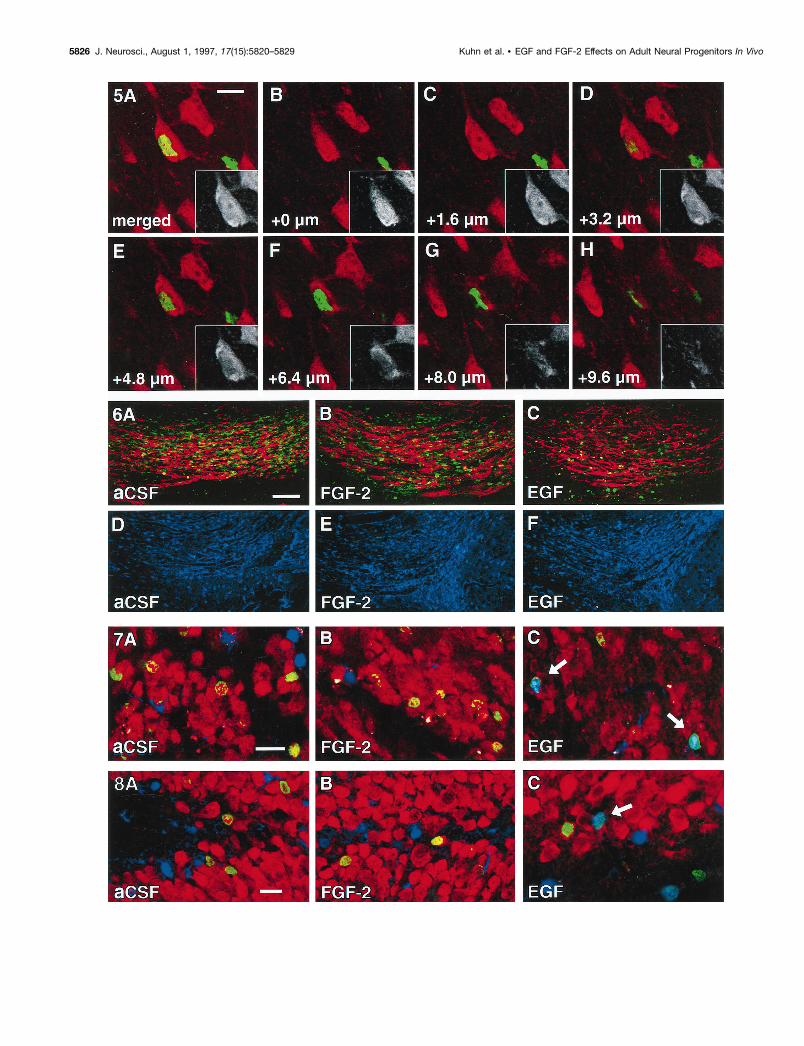
Figure 5. Close association of neurons with newborn cells (satellite cells). A, A NeuN-positive neuron (red and black/white inset) appeared to becolabeled with BrdU in a merged image resembling a regular fluorescent microscope image. B–H, Z-series analysis revealed that the NeuN-positiveneuronal cell body is situated in a different focal plane from the BrdU-positive nucleus. Note that the NeuN-positive nucleus with nucleolus is visiblein C and the BrdU-positive nucleus in E and F. Scale bar in A, 10 mm.
Figure 6. Reduced rostral migration of BrdU-positive cells at the end of EGF and FGF-2 infusions. Compared with aCSF controls (A), the numberof BrdU-positive cells ( green) in the RMS is decreased in FGF-2 (B) and decreased further in EGF-treated animals (C). However, PSA-NCAM (red),which is required for migration of progenitors within the RMS, is present in all groups. No BrdU-positive cells can be found at 4 weeks after infusion(D–F ). Images D–F are immunofluorescent double labelings for BrdU ( green) and S100b (blue). Note that scanning parameters for the confocalmicroscope are identical for A–F. Scale bar in A, 50 mm.
Figure 7. Cellular phenotype of newborn cells in the olfactory bulb. Cells in the olfactory granule cell layer were characterized at 4 weeks after infusionof aCSF (A), FGF-2 (B), or EGF (C) for BrdU ( green), NeuN (red), and S100b (blue). After aCSF or FGF-2 treatment the vast majority of newborncells double labels for NeuN ( green/red). Note the reduced number of BrdU-positive cells that reach the olfactory bulb in EGF-treated animals (C; seealso Table 1). The differentiation of EGF-induced progenitors was shifted toward a glial lineage. Arrows indicate newborn astrocytes ( green/blue). Scalebar in A, 20 mm.
Figure 8. Cellular phenotype of newborn cells in the dentate gyrus. Cells in the hippocampal granule cell layer were characterized at 4 weeks afterinfusion of aCSF (A), FGF-2 (B), or EGF (C) for BrdU ( green), NeuN (red), and S100b (blue). After aCSF or FGF-2 treatment the vast majority ofnewborn cells double labels for NeuN ( green/red). The differentiation of EGF-induced progenitors has shifted toward a glial lineage (C). Arrow indicatesa newborn astrocyte ( green/blue). Scale bar in A, 20 mm.
Kuhn et al. • EGF and FGF-2 Effects on Adult Neural Progenitors In Vivo J. Neurosci., August 1, 1997, 17(15):5820–5829 5827
In vitro models have been excellent tools for analyzing signalsthat influence the proliferation and fate of neural progenitor cells,but it has been difficult to determine how well these in vitroobservations relate to signaling in vivo. By testing mitogensknown to be effective in vitro, we have been able to show thatprogenitor populations in the adult rodent brain respond, in part,differently from in vitro. The site-specific responsiveness of pro-genitors to exogenous factors indicates that local cues play animportant role in regulating neurogenesis in vivo. In the absenceof in vivo signals, progenitors cultured in the presence of EGFproliferate and differentiate into neurons and glia, yet, in vivo,EGF has a stimulatory influence on proliferation and the genesisof glia but an unexpected limiting effect on the generation ofneurons. This dichotomy emphasizes the importance of obtainingin vivo and in vitro results to identify more completely the factorsthat direct site-specific neuronal differentiation.
REFERENCESAltman J, Das GD (1965) Autoradiographic and histological evidence of
postnatal hippocampal neurogenesis in rats. J Comp Neurol124:319–335.
Bayer SA (1983) 3H-thymidine-radiographic studies of neurogenesis inthe rat olfactory bulb. Exp Brain Res 50:329–340.
Berger F, Laine M, Hoffmann D, Verna JM, Charffanet M, Chauvin C,Rost N, Nissou MF, Benabid AL (1992) The EGF receptor pathwayin human cerebral tumors. Neurochirurgie 38:257–266.
Blaschke AJ, Staley K, Chun J (1996) Widespread programmed celldeath in proliferative and postmitotic regions of the fetal cerebralcortex. Development 122:1165–1174.
Bonfanti L, Theodosis DT (1994) Expression of polysialylated neuralcell adhesion molecule by proliferating cells in the subependymal layerof the adult rat, in its rostral extension, and in the olfactory bulb.Neuroscience 62:291–305.
Bouvier MM, Mytilineou C (1995) Basic fibroblast growth factor in-creases division and delays differentiation of dopamine precursors invitro. J Neurosci 15:7141–7149.
Boyes BE, Kim SU, Lee V, Sung SC (1986) Immunohistochemical co-localization of S-100b and the glial fibrillary acidic protein in rat brain.Neuroscience 17:857–865.
Calof AL (1995) Intrinsic and extrinsic factors regulating vertebrateneurogenesis. Curr Opin Neurobiol 5:19–27.
Cameron HA, Woolley CS, McEwen BS, Gould E (1993) Differentia-tion of newly born neurons and glia in the dentate gyrus of the adult rat.Neuroscience 56:337–344.
Cattaneo E, McKay R (1991) Identifying and manipulating neuronalstem cells. Trends Neurosci 14:338–340.
Collins VP (1995) Gene amplification in human gliomas. Glia15:289–296.
Corotto FS, Henegar JA, Maruniak JA (1993) Neurogenesis persists inthe subependymal layer of the adult mouse brain. Neurosci Lett149:111–114.
Craig CG, Tropepe V, Morshead CM, Reynolds BA, Weiss S, van derKooy D (1996) In vivo growth factor expansion of endogenous sub-ependymal neural precursor cell populations in the adult mouse brain.J Neurosci 16:2649–2658.
Gage FH (1994) Neuronal stem cells: their characterization and utiliza-tion. Neurobiol Aging 15(Suppl 2):S191.
Gage FH, Coates PW, Palmer TD, Kuhn HG, Fisher LJ, Suhonen JO,Peterson DA, Suhr ST, Ray J (1995a) Survival and differentiation ofadult neuronal progenitor cells transplanted to the adult brain. ProcNatl Acad Sci USA 92:11879–11883.
Gage FH, Ray J, Fisher LJ (1995b) Isolation, characterization, and useof stem cells from the CNS. Annu Rev Neurosci 18:159–192.
Goldman JE (1995) Lineage, migration, and fate determination of post-natal subventricular zone cells in the mammalian CNS. J Neurooncol24:61–64.
Gonzalez AM, Carman LS, Ong M, Ray J, Gage FH, Shults CW, BairdA (1994) Storage, metabolism, and processing of 125I-fibroblastgrowth factor-2 after intracerebral injection. Brain Res 665:285–292.
Gonzalez AM, Berry M, Maher PA, Logan A, Baird A (1995) A com-prehensive analysis of the distribution of FGF-2 and FGFR1 in the ratbrain. Brain Res 701:201–226.
Gould E, Woolley CS, McEwen BS (1991) Naturally occurring celldeath in the developing dentate gyrus of the rat. J Comp Neurol304:408–418.
Gritti A, Cova L, Parati EA, Galli R, Vescovi AL (1995) Basic fibroblastgrowth factor supports the proliferation of epidermal growth factor-generated neuronal precursor cells of the adult mouse CNS. NeurosciLett 185:151–154.
Gritti A, Parati EA, Cova L, Frolichsthal P, Galli R, Wanke E, FaravelliL, Morassutti DJ, Roisen F, Nickel DD, Vescovi AL (1996) Multipo-tential stem cells from the adult mouse brain proliferate and self-renewin response to basic fibroblast growth factor. J Neurosci 16:1091–1100.
Hauke C, Ackermann I, Korr H (1995) Cell proliferation in the sub-ependymal layer of the adult mouse in vivo and in vitro. Cell Prolif28:595–607.
Hu H, Tomasiewicz H, Magnuson T, Rutishauser U (1996) The role ofpolysialic acid in migration of olfactory bulb interneuron precursors inthe subventricular zone. Neuron 16:735–743.
Kaplan MS, Bell DH (1983) Neuronal proliferation in the 9-month-oldrodent—radioautographic study of granule cells in the hippocampus.Exp Brain Res 52:1–5.
Kaplan MS, Hinds JW (1977) Neurogenesis in the adult rat: electronmicroscopic analysis of light radioautographs. Science 197:1092–1094.
Kilpatrick TJ, Bartlett PF (1993) Cloning and growth of multipotentialneural precursors: requirements for proliferation and differentiation.Neuron 10:255–265.
Kilpatrick TJ, Bartlett PF (1995) Cloned multipotential precursors fromthe mouse cerebrum require FGF-2, whereas glial restricted precursorsare stimulated with either FGF-2 or EGF. J Neurosci 15:3653–3661.
Kuhn HG, Dickinson-Anson H, Gage FH (1996) Neurogenesis in thedentate gyrus of the adult rat: age-related decrease of neuronal pro-genitor proliferation. J Neurosci 16:2027–2033.
Levison SW, Goldman JE (1993) Both oligodendrocytes and astrocytesdevelop from progenitors in the subventricular zone of postnatal ratforebrain. Neuron 10:201–212.
Levison SW, Chuang C, Abramson BJ, Goldman JE (1993) The migra-tional patterns and developmental fates of glial precursors in the ratsubventricular zone are temporally regulated. Development119:611–622.
Lewis PD (1968) A quantitative study of cell proliferation in the sub-ependymal layer of the adult rat brain. Exp Neurol 20:203–207.
Lois C, Alvarez-Buylla A (1993) Proliferating subventricular zone cellsin the adult mammalian forebrain can differentiate into neurons andglia. Proc Natl Acad Sci USA 90:2074–2077.
Lois C, Alvarez-Buylla A (1994) Long-distance neuronal migration inthe adult mammalian brain. Science 264:1145–1148.
Lois C, Garcia-Verdugo JM, Alvarez-Buylla A (1996) Chain migrationof neuronal precursors. Science 271:978–981.
Ludwin SK (1979) The perineuronal satellite oligodendrocyte. A role inremyelination. Acta Neuropathol (Berl) 47:49–53.
Ludwin SK (1984) The function of perineuronal satellite oligodendro-cytes: an immunohistochemical study. Neuropathol Appl Neurobiol10:143–149.
Luskin MB (1993) Restricted proliferation and migration of postnatallygenerated neurons derived from the forebrain subventricular zone.Neuron 11:173–189.
Luskin MB, McDermott K (1994) Divergent lineages for oligodendro-cytes and astrocytes originating in the neonatal forebrain subventricu-lar zone. Glia 11:211–226.
Menezes JRL, Smith CM, Nelson KC, Luskin MB (1995) The divisionof neuronal progenitor cells during migration in the neonatal mamma-lian forebrain. Mol Cell Neurosci 6:496–508.
Michel RP, Cruz-Orive LM (1988) Application of the Cavalieri princi-ple and vertical sections method to lung: estimation of volume andpleural surface area. J Microsc 150:117–136.
Morshead CM, van der Kooy D (1992) Postmitotic death is the fate ofconstitutively proliferating cells in the subependymal layer of the adultmouse brain. J Neurosci 12:249–256.
Morshead CM, Reynolds BA, Craig CG, McBurney MW, Staines WA,Morassutti D, Weiss S, van der Kooy D (1994) Neural stem cells in theadult mammalian forebrain: a relatively quiescent subpopulation ofsubependymal cells. Neuron 13:1071–1082.
Mullen RJ, Buck CR, Smith AM (1992) NeuN, a neuronal specific nu-clear protein in vertebrates. Development 116:201–211.
Naruse I, Keino H (1995) Apoptosis in the developing CNS. Prog Neu-robiol 47:135–155.
5828 J. Neurosci., August 1, 1997, 17(15):5820–5829 Kuhn et al. • EGF and FGF-2 Effects on Adult Neural Progenitors In Vivo
Ono K, Tomasiewicz H, Magnuson T, Rutishauser U (1994) N-CAMmutation inhibits tangential neuronal migration and is phenocopied byenzymatic removal of polysialic acid. Neuron 13:595–609.
Palmer TD, Ray J, Gage FH (1995) FGF-2-responsive neuronal progen-itors reside in proliferative and quiescent regions of the adult rodentbrain. Mol Cell Neurosci 6:474–486.
Paxinos G, Watson C (1986) The rat brain in stereotaxic coordinates.San Diego: Academic.
Penfield W (1932) Neuroglia and microglia, the interstitial tissue of thecentral nervous system. In: Special cytology (Cowdry EV, ed), pp1147–1182. New York: Hoeber.
Privat A, Leblond CP (1972) The subependymal layer and neighboringregion in the brain of the young rat. J Comp Neurol 146:277–302.
Rapraeger AC, Guimond S, Krufka A, Olwin BB (1994) Regulation byheparan sulfate in fibroblast growth factor signaling. Methods Enzymol245:219–240.
Ray J, Peterson DA, Schinstine M, Gage FH (1993) Proliferation, dif-ferentiation, and long-term culture of primary hippocampal neurons.Proc Natl Acad Sci USA 90:3602–3606.
Reynolds BA, Weiss S (1992) Generation of neurons and astrocytesfrom isolated cells of the adult mammalian central nervous system.Science 255:1707–1710.
Reynolds BA, Weiss S (1996) Clonal and population analyses demon-strate that an EGF-responsive mammalian embryonic CNS precursor isa stem cell. Dev Biol 175:1–13.
Richards LJ, Kilpatrick TJ, Bartlett PF (1992) De novo generation ofneuronal cells from the adult mouse brain. Proc Natl Acad Sci USA89:8591–8595.
Santa-Olalla J, Covarrubias L (1995) Epidermal growth factor (EGF),transforming growth factor-alpha (TGF-alpha), and basic fibroblastgrowth factor (bFGF) differentially influence neural precursor cells ofmouse embryonic mesencephalon. J Neurosci Res 42:172–183.
Sensenbrenner M, Deloulme JC, Gensburger C (1994) Proliferation ofneuronal precursor cells from the central nervous system in culture.Rev Neurosci 5:43–53.
Sterio DC (1984) The unbiased estimation of number and sizes of arbi-trary particles using the disector. J Microsc 134:127–136.
Suhonen JO, Peterson DA, Ray J, Gage FH (1996) Differentiation ofadult hippocampus-derived progenitors into olfactory neurons in vivo.Nature 383:624–627.
Svendsen CN, Clarke DJ, Rosser AE, Dunnett SB (1996) Survival anddifferentiation of rat and human epidermal growth factor-responsiveprecursor cells following grafting into the lesioned adult nervous sys-tem. Exp Neurol 137:376–388.
Thomas LB, Gates MA, Steindler DA (1996) Young neurons from theadult subependymal zone proliferate and migrate along an astrocyte,extracellular matrix-rich pathway. Glia 17:1–14.
Vescovi AL, Reynolds BA, Fraser DD, Weiss S (1993) bFGF regulatesthe proliferative fate of unipotent (neuronal) and bipotent (neuronal /astroglial) EGF-generated CNS progenitor cells. Neuron 11:951–966.
von Deimling A, Louis DN, Wiestler OD (1995) Molecular pathways inthe formation of gliomas. Glia 15:328–338.
Weickert CS, Blum M (1995) Striatal TGF-alpha: postnatal develop-mental expression and evidence for a role in the proliferation ofsubependymal cells. Brain Res Dev Brain Res 86:203–216.
Kuhn et al. • EGF and FGF-2 Effects on Adult Neural Progenitors In Vivo J. Neurosci., August 1, 1997, 17(15):5820–5829 5829