endotracheal intubation by paramedics using neodymium...
TRANSCRIPT
Research ArticleEndotracheal Intubation by Paramedics Using NeodymiumMagnet and Modified Stylet in Simulated Difficult Airway: AProspective, Randomized, Crossover Manikin Study
Sedat Bilge ,1 Onur Tezel,1 Yahya Ayhan Acar ,1 Guclu Aydin,2 Attila Aydin,3
and Gokhan Ozkan4
1Department of Emergency Medicine, Gulhane Medicine Faculty, Health Sciences University, Ankara 06010, Turkey2Department of Emergency Medicine, Gulhane Training and Research Hospital, Health Sciences University,Ankara 06010, Turkey3Miaclinics, Atasehir, Istanbul 34758, Turkey4Department of Anesthesiology and Reanimation, Gulhane Training and Research Hospital, Health Sciences University,Ankara 06010, Turkey
Correspondence should be addressed to Yahya Ayhan Acar; [email protected]
Received 31 May 2019; Revised 22 August 2019; Accepted 25 September 2019; Published 15 October 2019
Academic Editor: Jeffrey R. Avner
Copyright © 2019 Sedat Bilge et al. )is is an open access article distributed under the Creative Commons Attribution License,which permits unrestricted use, distribution, and reproduction in any medium, provided the original work is properly cited.
Background. )e present study evaluates the success and efficacy of endotracheal intubation (ETI) using a modified intubationstylet and a magnet system to direct the stylet into the trachea. )e system was developed by the researchers in an attempt toincrease the success and efficacy of ETI. Methods. ETI procedures were performed on an airway management manikin byemergency medical technicians with at least four years of experience in ETI. )e technicians used a stylet modified with an ironball affixed to the tip and a neodymium magnet, designed specifically for the study. )e intention was to guide the endotrachealtube into the trachea at the level of the thyroid and cricoid cartilages on the manikin with the aid of the modified stylet and themagnetic force of the neodymium magnet. )e success rate, completion time, and degree of difficulty of two procedures werecompared: magnetic endotracheal intubation (METI) and classic ETI (CETI). Results. )e success rate was 100% in both groups.)e mean completion times for the METI and CETI procedures were 18.31± 2.46 s and 20.01± 1.95 s, respectively. )ere weresignificant differences in completion time and degree of difficulty between the METI and CETI procedures (both p � 0.001).Conclusions. We found the use of a neodymium magnet and modified stylet to be an effective method to guide the endotrachealtube into the trachea. )e present study may provide a basis for future studies.
1. Introduction
Endotracheal intubation (ETI) is the optimum method forprotecting the patency of the airway and maintaining ox-ygenation and ventilation in patients requiring advanced lifesupport [1–3]. Various factors can complicate securing theairway, such as anatomical variations in the airway andrelated structures and cervical immobilization [4–8].
To overcome the factors complicating ETI procedures,noninvasive intubation methods and devices are available,such as intubating laryngeal mask airways, video-assistedintubation, fiberoptic intubation, and laryngoscope-based
lighted stylets [2, 9–11].)e use of invasive methods, such asretrograde ETI and surgical airway management, can lead toincreased costs, loss of time, and various catastrophiccomplications that require management [12, 13]. Althoughmagnetic ETI (METI) techniques have been described in theliterature, some limitations have also been reported [14–16].)ey are not widely applicable in clinical practice due tothese limitations. We aimed to optimize METI use in dif-ficult airway management. An intubation stylet combinedwith a metal ball was developed by the researchers. Aneodymium magnet was used with this modified intubationstylet (MIS). )is study aimed to compare the procedural
HindawiEmergency Medicine InternationalVolume 2019, Article ID 5804260, 7 pageshttps://doi.org/10.1155/2019/5804260
success rate, degree of difficulty, and completion time of thistechnique with those of classic ETI (CETI).
2. Materials and Methods
2.1. Study Design. )is study was approved by the GulhaneEthics Board of the University of Health Sciences on May22nd, 2018 (meeting no: 2018/7; decision number: 18/129).Written voluntary informed consent has been obtained fromall participants before performing the procedures. )e studywas designed as a randomized, crossover manikin study.
2.2. Participants. All interventions were performed by 20ambulance and emergency care technicians who weregraduates of the Gulhane Military Medical AcademyNoncommissioned Officer Health College. All participantshad more than four years of emergency medical serviceexperience and were trained in advanced cardiac life supportand advanced trauma life support. )e operators were givenrefresher training lasting for at least 30 minutes on the ETIprocedure by specialists in emergency medicine prior to thestudy. )ese training programs (theoretical and practical)included anatomical landmarks and standard proceduraltools on ETI.
2.3. Equipment. An airway management manikin (LaerdalAirway Management Trainer; Laerdal Medical, Stavanger,Norway, 2013) was used in the study, fitted with a rigidcervical collar (Ambu® Perfit ACE extrication collar; AmbuInc., Columbia, MD, USA). )e distance between the skinand trachea at the vocal cord level of the airwaymanagementmanikin was measured as 18.5mm using the image obtainedvia fluoroscopy (Infinix-i Core INFX-8000F; Canon MedicalSystems, Japan) and the device’s computer. )e Macintosh(MAC) blade (no. 3) laryngoscope (Teutotechnik Inc.,Germany) with a laryngoscope handle and a size 8 cuffedendotracheal tube (ETT) (Kaishou Inc., China) was used inall ETI procedures. Lubricant gel, a bag valve mask (BVM),and a 20ml syringe were also used to inflate the cuff.
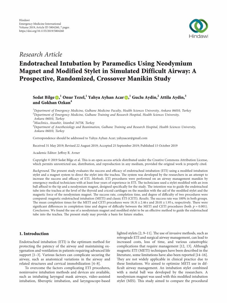
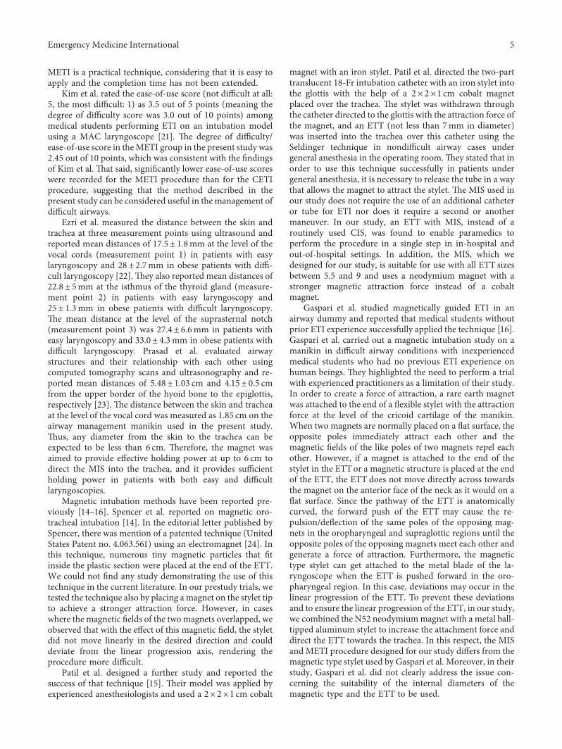
)e classic intubation stylet (CIS) was a 12 Ch-sizedstylet with a length of 40 cm and made of malleable alu-minum covered with a synthetic plastic polymer (polyvinylchloride) (Plasti-med Inc., Turkey). )e MIS comprised aniron-rich metal ball measuring 5mm in diameter soldered tothe tip of the classic stylet (Figure 1(a)). )e neodymiummagnet used was manufactured specifically for this study(Manyet Manyetik Tutucular Inc., Ankara, Turkey). )eproperties of the magnet reported by the manufacturer wereas follows. )e magnetic plate was composed of four40× 40× 40mm grade N52 magnets. )e magnets wereattached to an ST52 iron plate and covered with 2mm ofstainless protective tin. )e grade N52 magnetic plate had a14,400 Gauss magnetic field, producing a 4,500 Gaussmagnetic field on the stainless plate (Figures 1(a) and 1(b)).)e holding power applied by the magnet on the MIS wasmeasured by simple dynamometry in the physics teachinglaboratory of Middle East Technical University. All deviceswere used according to the manufacturers’ instructions.
2.4. Study Protocol. )e operators received theoretical andpractical training before the study for 60 minutes on three-dimensional laryngeal anatomy, standard laryngoscopy, andthe use of the MIS and the N52 neodymium magnet. At theend of the training, the participants were given the op-portunity to practice on a model for a total of 10 minutes,with five minutes set aside for each technique. A difficultairway was simulated by fitting a cervical collar to the ETImodel. Only one intervention attempt was allowed for eachtechnique.
(a) CETI: intubation performed with an ETT fitted witha CISSteps of the CETI procedure: (i) A CIS-fitted ETTwasready on the table with the laryngoscope blade attachedbefore the procedure. (ii) )e practitioner started theCETI procedure with the help of the laryngoscopewhen a “start” command was issued by the researcher.(iii) )e CIS was withdrawn by the practitioner, theBVM was attached to the ETT, and the procedure wasterminated when lung inflation was detected.
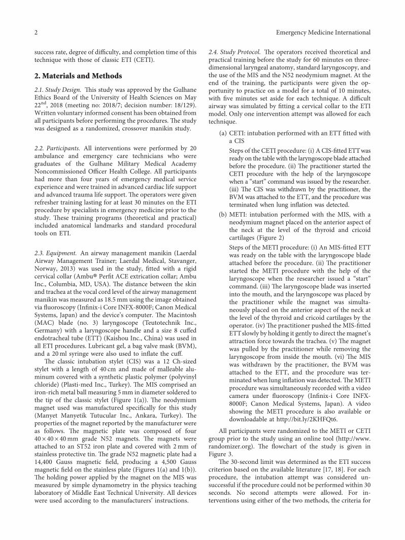
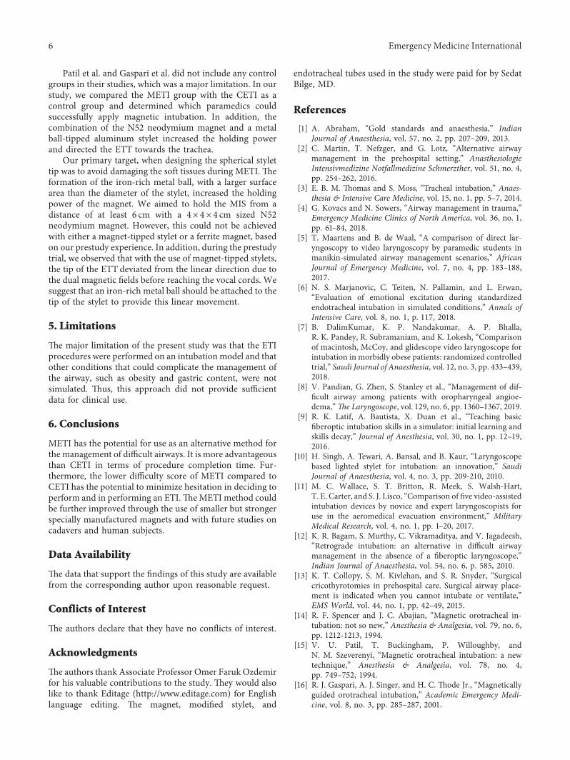
(b) METI: intubation performed with the MIS, with aneodymium magnet placed on the anterior aspect ofthe neck at the level of the thyroid and cricoidcartilages (Figure 2)Steps of the METI procedure: (i) An MIS-fitted ETTwas ready on the table with the laryngoscope bladeattached before the procedure. (ii) )e practitionerstarted the METI procedure with the help of thelaryngoscope when the researcher issued a “start”command. (iii) )e laryngoscope blade was insertedinto the mouth, and the laryngoscope was placed bythe practitioner while the magnet was simulta-neously placed on the anterior aspect of the neck atthe level of the thyroid and cricoid cartilages by theoperator. (iv) )e practitioner pushed the MIS-fittedETTslowly by holding it gently to direct the magnet’sattraction force towards the trachea. (v) )e magnetwas pulled by the practitioner while removing thelaryngoscope from inside the mouth. (vi) )e MISwas withdrawn by the practitioner, the BVM wasattached to the ETT, and the procedure was ter-minated when lung inflation was detected.)eMETIprocedure was simultaneously recorded with a videocamera under fluoroscopy (Infinix-i Core INFX-8000F; Canon Medical Systems, Japan). A videoshowing the METI procedure is also available ordownloadable at http://bit.ly/2KHFQt6.
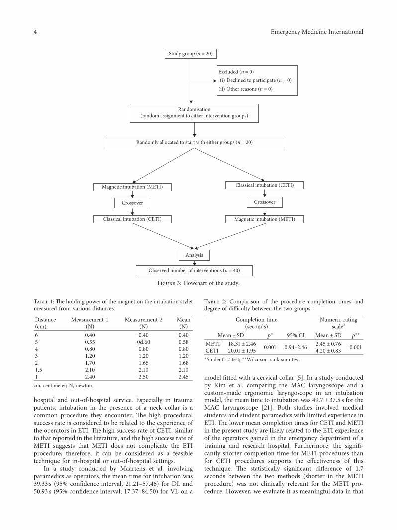
All participants were randomized to the METI or CETIgroup prior to the study using an online tool (http://www.randomizer.org). )e flowchart of the study is given inFigure 3.
)e 30-second limit was determined as the ETI successcriterion based on the available literature [17, 18]. For eachprocedure, the intubation attempt was considered un-successful if the procedure could not be performed within 30seconds. No second attempts were allowed. For in-terventions using either of the two methods, the criteria for
2 Emergency Medicine International
successful intubation were the completion of the procedureby the operator within 30 seconds and the observation oflung inflation on the manikin by the investigators. )ecompletion time was defined as the interval between whenthe “start” instruction was issued by the investigators towhen the BVM was attached to the ETT. )e primaryendpoint of the study was successful intubation, and thesecondary endpoints were completion time and the degree ofdifficulty reported by the operators. Feedback was receivedfrom the operators regarding the degree of difficulty of theprocedure, rated using a numeric rating scale (not difficult atall: 0; the most difficult: 10).
2.5. Power Analysis. In the power analysis, based on pre-vious study findings (completion time 14.46± 2.31 s) [19],assuming a completion time of 17.0 s for ETI with an 80%power and a two-sided error margin of 0.05, 18 participantswere required. )us, we planned to include a total of 20participants who would be randomized to the study groups.
2.6. Statistical Analyses. Statistical analyses were performedusing SPSS for Windows version 22.0 (SPSS Inc., Chicago, IL,USA). Descriptive data are expressed as the mean± standarddeviation. A Kolmogorov–Smirnov test was used to assess ifthe data were normally distributed. )e Student’s t-test wasused to compare normally distributed paired groups, and theWilcoxon rank sum test was used to compare the degree ofdifficulty between the procedures. A p value <0.05 wasconsidered statistically significant.
3. Results
)e holding power applied by the neodymiummagnet to theiron ball fixed to the tip of the MIS was measured from 1 cmto 6 cm using a calibrated dynamometer and recorded asapproximately 0.4N at 6 cm and 2.45N at 1 cm (Table 1). Atotal of 20 paramedics (100%) successfully performed 40interventions, 20 each in the METI and CETI groups; therewere no cases of failed intubation. )ere was a statisticallysignificant difference in completion time between the METIand CETI groups, with the mean completion time beinglower in the METI group (p � 0.001) (Table 2). A com-parison of the degree of difficulty rated using the numericrating scale showed lower scores in the METI group, and thedifference between the groups was statistically significant(p � 0.001) (Table 2).
4. Discussion
Alter et al. assessed ETI attempts by paramedics in theprehospital care of 2,299 patients using direct laryngoscopy(DL) with either MAC or miller (MIL) blades and reportedthat ETI was successfully performed with DL using a MACblade (n� 1865), MIL blade (n� 367), and both blades(n� 67). )e first-pass success rates of ETI performed byparamedics with MAC and MIL blades were reported to be86% and 73%, respectively [20]. )e success rate of CETI inthe present study (100%) was higher than that reported byAlter et al. )e operators (n� 20) in the present study wereselected from among paramedics who are responsible forETIs as part of trauma resuscitation teams in the emergencydepartment of a tertiary training and research hospital,admitting 1,200 emergency patients daily, and who had atleast four years of professional experience. )e paramedicsparticipate in difficult airway management both in in-
(a) (b)
Figure 1: (a) Modified intubation stylet produced by soldering an iron-rich metal ball onto the tip of a standard stylet; (b) the neodymiummagnet designed for the study.
Endotracheal tube
Modifiedintubation stylet
Laryngoscope(curved blade)
Cuff inflating tube Magnet
Vocal cords
Trachea
Esophagus
Figure 2: Illustration of endotracheal intubation performed withan endotracheal tube fitted over a modified intubation stylet and aneodymiummagnet placed on the anterior aspect of the neck at thelevel of the thyroid and cricoid cartilages.
Emergency Medicine International 3
hospital and out-of-hospital service. Especially in traumapatients, intubation in the presence of a neck collar is acommon procedure they encounter. )e high proceduralsuccess rate is considered to be related to the experience ofthe operators in ETI. )e high success rate of CETI, similarto that reported in the literature, and the high success rate ofMETI suggests that METI does not complicate the ETIprocedure; therefore, it can be considered as a feasibletechnique for in-hospital or out-of-hospital settings.
In a study conducted by Maartens et al. involvingparamedics as operators, the mean time for intubation was39.33 s (95% confidence interval, 21.21–57.46) for DL and50.93 s (95% confidence interval, 17.37–84.50) for VL on a
model fitted with a cervical collar [5]. In a study conductedby Kim et al. comparing the MAC laryngoscope and acustom-made ergonomic laryngoscope in an intubationmodel, the mean time to intubation was 49.7± 37.5 s for theMAC laryngoscope [21]. Both studies involved medicalstudents and student paramedics with limited experience inETI. )e lower mean completion times for CETI and METIin the present study are likely related to the ETI experienceof the operators gained in the emergency department of atraining and research hospital. Furthermore, the signifi-cantly shorter completion time for METI procedures thanfor CETI procedures supports the effectiveness of thistechnique. )e statistically significant difference of 1.7seconds between the two methods (shorter in the METIprocedure) was not clinically relevant for the METI pro-cedure. However, we evaluate it as meaningful data in that
Randomly allocated to start with either groups (n = 20)
Magnetic intubation (METI)
Crossover
Classical intubation (CETI)
Classical intubation (CETI)
Crossover
Magnetic intubation (METI)
Analysis
Observed number of interventions (n = 40)
Study group (n = 20)
Excluded (n = 0)Declined to participate (n = 0)Other reasons (n = 0)
Randomization(random assignment to either intervention groups)
(i)(ii)
Figure 3: Flowchart of the study.
Table 1: )e holding power of the magnet on the intubation styletmeasured from various distances.
Distance(cm)
Measurement 1(N)
Measurement 2(N)
Mean(N)
6 0.40 0.40 0.405 0.55 0d.60 0.584 0.80 0.80 0.803 1.20 1.20 1.202 1.70 1.65 1.681.5 2.10 2.10 2.101 2.40 2.50 2.45cm, centimeter; N, newton.
Table 2: Comparison of the procedure completion times anddegree of difficulty between the two groups.
Completion time(seconds)
Numeric ratingscale#
Mean± SD p∗ 95% CI Mean± SD p∗∗
METI 18.31± 2.46 0.001 0.94–2.46 2.45± 0.76 0.001CETI 20.01± 1.95 4.20± 0.83∗Student’s t-test; ∗∗Wilcoxon rank sum test.
4 Emergency Medicine International
METI is a practical technique, considering that it is easy toapply and the completion time has not been extended.
Kim et al. rated the ease-of-use score (not difficult at all:5, the most difficult: 1) as 3.5 out of 5 points (meaning thedegree of difficulty score was 3.0 out of 10 points) amongmedical students performing ETI on an intubation modelusing a MAC laryngoscope [21]. )e degree of difficulty/ease-of-use score in theMETI group in the present study was2.45 out of 10 points, which was consistent with the findingsof Kim et al. )at said, significantly lower ease-of-use scoreswere recorded for the METI procedure than for the CETIprocedure, suggesting that the method described in thepresent study can be considered useful in themanagement ofdifficult airways.
Ezri et al. measured the distance between the skin andtrachea at three measurement points using ultrasound andreported mean distances of 17.5± 1.8mm at the level of thevocal cords (measurement point 1) in patients with easylaryngoscopy and 28± 2.7mm in obese patients with diffi-cult laryngoscopy [22]. )ey also reported mean distances of22.8± 5mm at the isthmus of the thyroid gland (measure-ment point 2) in patients with easy laryngoscopy and25± 1.3mm in obese patients with difficult laryngoscopy.)e mean distance at the level of the suprasternal notch(measurement point 3) was 27.4± 6.6mm in patients witheasy laryngoscopy and 33.0± 4.3mm in obese patients withdifficult laryngoscopy. Prasad et al. evaluated airwaystructures and their relationship with each other usingcomputed tomography scans and ultrasonography and re-ported mean distances of 5.48± 1.03 cm and 4.15± 0.5 cmfrom the upper border of the hyoid bone to the epiglottis,respectively [23]. )e distance between the skin and tracheaat the level of the vocal cord was measured as 1.85 cm on theairway management manikin used in the present study.)us, any diameter from the skin to the trachea can beexpected to be less than 6 cm. )erefore, the magnet wasaimed to provide effective holding power at up to 6 cm todirect the MIS into the trachea, and it provides sufficientholding power in patients with both easy and difficultlaryngoscopies.
Magnetic intubation methods have been reported pre-viously [14–16]. Spencer et al. reported on magnetic oro-tracheal intubation [14]. In the editorial letter published bySpencer, there was mention of a patented technique (UnitedStates Patent no. 4.063.561) using an electromagnet [24]. Inthis technique, numerous tiny magnetic particles that fitinside the plastic section were placed at the end of the ETT.We could not find any study demonstrating the use of thistechnique in the current literature. In our prestudy trials, wetested the technique also by placing amagnet on the stylet tipto achieve a stronger attraction force. However, in caseswhere the magnetic fields of the two magnets overlapped, weobserved that with the effect of this magnetic field, the styletdid not move linearly in the desired direction and coulddeviate from the linear progression axis, rendering theprocedure more difficult.
Patil et al. designed a further study and reported thesuccess of that technique [15]. )eir model was applied byexperienced anesthesiologists and used a 2× 2×1 cm cobalt
magnet with an iron stylet. Patil et al. directed the two-parttranslucent 18-Fr intubation catheter with an iron stylet intothe glottis with the help of a 2× 2×1 cm cobalt magnetplaced over the trachea. )e stylet was withdrawn throughthe catheter directed to the glottis with the attraction force ofthe magnet, and an ETT (not less than 7mm in diameter)was inserted into the trachea over this catheter using theSeldinger technique in nondifficult airway cases undergeneral anesthesia in the operating room.)ey stated that inorder to use this technique successfully in patients undergeneral anesthesia, it is necessary to release the tube in a waythat allows the magnet to attract the stylet. )e MIS used inour study does not require the use of an additional catheteror tube for ETI nor does it require a second or anothermaneuver. In our study, an ETT with MIS, instead of aroutinely used CIS, was found to enable paramedics toperform the procedure in a single step in in-hospital andout-of-hospital settings. In addition, the MIS, which wedesigned for our study, is suitable for use with all ETT sizesbetween 5.5 and 9 and uses a neodymium magnet with astronger magnetic attraction force instead of a cobaltmagnet.
Gaspari et al. studied magnetically guided ETI in anairway dummy and reported that medical students withoutprior ETI experience successfully applied the technique [16].Gaspari et al. carried out a magnetic intubation study on amanikin in difficult airway conditions with inexperiencedmedical students who had no previous ETI experience onhuman beings. )ey highlighted the need to perform a trialwith experienced practitioners as a limitation of their study.In order to create a force of attraction, a rare earth magnetwas attached to the end of a flexible stylet with the attractionforce at the level of the cricoid cartilage of the manikin.When two magnets are normally placed on a flat surface, theopposite poles immediately attract each other and themagnetic fields of the like poles of two magnets repel eachother. However, if a magnet is attached to the end of thestylet in the ETTor a magnetic structure is placed at the endof the ETT, the ETT does not move directly across towardsthe magnet on the anterior face of the neck as it would on aflat surface. Since the pathway of the ETT is anatomicallycurved, the forward push of the ETT may cause the re-pulsion/deflection of the same poles of the opposing mag-nets in the oropharyngeal and supraglottic regions until theopposite poles of the opposing magnets meet each other andgenerate a force of attraction. Furthermore, the magnetictype stylet can get attached to the metal blade of the la-ryngoscope when the ETT is pushed forward in the oro-pharyngeal region. In this case, deviations may occur in thelinear progression of the ETT. To prevent these deviationsand to ensure the linear progression of the ETT, in our study,we combined the N52 neodymiummagnet with a metal ball-tipped aluminum stylet to increase the attachment force anddirect the ETT towards the trachea. In this respect, the MISand METI procedure designed for our study differs from themagnetic type stylet used by Gaspari et al. Moreover, in theirstudy, Gaspari et al. did not clearly address the issue con-cerning the suitability of the internal diameters of themagnetic type and the ETT to be used.
Emergency Medicine International 5
Patil et al. and Gaspari et al. did not include any controlgroups in their studies, which was a major limitation. In ourstudy, we compared the METI group with the CETI as acontrol group and determined which paramedics couldsuccessfully apply magnetic intubation. In addition, thecombination of the N52 neodymium magnet and a metalball-tipped aluminum stylet increased the holding powerand directed the ETT towards the trachea.
Our primary target, when designing the spherical stylettip was to avoid damaging the soft tissues during METI. )eformation of the iron-rich metal ball, with a larger surfacearea than the diameter of the stylet, increased the holdingpower of the magnet. We aimed to hold the MIS from adistance of at least 6 cm with a 4× 4× 4 cm sized N52neodymium magnet. However, this could not be achievedwith either a magnet-tipped stylet or a ferrite magnet, basedon our prestudy experience. In addition, during the prestudytrial, we observed that with the use of magnet-tipped stylets,the tip of the ETT deviated from the linear direction due tothe dual magnetic fields before reaching the vocal cords. Wesuggest that an iron-rich metal ball should be attached to thetip of the stylet to provide this linear movement.
5. Limitations
)e major limitation of the present study was that the ETIprocedures were performed on an intubationmodel and thatother conditions that could complicate the management ofthe airway, such as obesity and gastric content, were notsimulated. )us, this approach did not provide sufficientdata for clinical use.
6. Conclusions
METI has the potential for use as an alternative method forthe management of difficult airways. It is more advantageousthan CETI in terms of procedure completion time. Fur-thermore, the lower difficulty score of METI compared toCETI has the potential to minimize hesitation in deciding toperform and in performing an ETI.)eMETI method couldbe further improved through the use of smaller but strongerspecially manufactured magnets and with future studies oncadavers and human subjects.
Data Availability
)e data that support the findings of this study are availablefrom the corresponding author upon reasonable request.
Conflicts of Interest
)e authors declare that they have no conflicts of interest.
Acknowledgments
)e authors thank Associate Professor Omer Faruk Ozdemirfor his valuable contributions to the study. )ey would alsolike to thank Editage (http://www.editage.com) for Englishlanguage editing. )e magnet, modified stylet, and
endotracheal tubes used in the study were paid for by SedatBilge, MD.
References
[1] A. Abraham, “Gold standards and anaesthesia,” IndianJournal of Anaesthesia, vol. 57, no. 2, pp. 207–209, 2013.
[2] C. Martin, T. Nefzger, and G. Lotz, “Alternative airwaymanagement in the prehospital setting,” AnasthesiologieIntensivmedizine Notfallmedizine Schmerzther, vol. 51, no. 4,pp. 254–262, 2016.
[3] E. B. M. )omas and S. Moss, “Tracheal intubation,” Anaes-thesia & Intensive Care Medicine, vol. 15, no. 1, pp. 5–7, 2014.
[4] G. Kovacs and N. Sowers, “Airway management in trauma,”Emergency Medicine Clinics of North America, vol. 36, no. 1,pp. 61–84, 2018.
[5] T. Maartens and B. de Waal, “A comparison of direct lar-yngoscopy to video laryngoscopy by paramedic students inmanikin-simulated airway management scenarios,” AfricanJournal of Emergency Medicine, vol. 7, no. 4, pp. 183–188,2017.
[6] N. S. Marjanovic, C. Teiten, N. Pallamin, and L. Erwan,“Evaluation of emotional excitation during standardizedendotracheal intubation in simulated conditions,” Annals ofIntensive Care, vol. 8, no. 1, p. 117, 2018.
[7] B. DalimKumar, K. P. Nandakumar, A. P. Bhalla,R. K. Pandey, R. Subramaniam, and K. Lokesh, “Comparisonof macintosh, McCoy, and glidescope video laryngoscope forintubation in morbidly obese patients: randomized controlledtrial,” Saudi Journal of Anaesthesia, vol. 12, no. 3, pp. 433–439,2018.
[8] V. Pandian, G. Zhen, S. Stanley et al., “Management of dif-ficult airway among patients with oropharyngeal angioe-dema,”7e Laryngoscope, vol. 129, no. 6, pp. 1360–1367, 2019.
[9] R. K. Latif, A. Bautista, X. Duan et al., “Teaching basicfiberoptic intubation skills in a simulator: initial learning andskills decay,” Journal of Anesthesia, vol. 30, no. 1, pp. 12–19,2016.
[10] H. Singh, A. Tewari, A. Bansal, and B. Kaur, “Laryngoscopebased lighted stylet for intubation: an innovation,” SaudiJournal of Anaesthesia, vol. 4, no. 3, pp. 209-210, 2010.
[11] M. C. Wallace, S. T. Britton, R. Meek, S. Walsh-Hart,T. E. Carter, and S. J. Lisco, “Comparison of five video-assistedintubation devices by novice and expert laryngoscopists foruse in the aeromedical evacuation environment,” MilitaryMedical Research, vol. 4, no. 1, pp. 1–20, 2017.
[12] K. R. Bagam, S. Murthy, C. Vikramaditya, and V. Jagadeesh,“Retrograde intubation: an alternative in difficult airwaymanagement in the absence of a fiberoptic laryngoscope,”Indian Journal of Anaesthesia, vol. 54, no. 6, p. 585, 2010.
[13] K. T. Collopy, S. M. Kivlehan, and S. R. Snyder, “Surgicalcricothyrotomies in prehospital care. Surgical airway place-ment is indicated when you cannot intubate or ventilate,”EMS World, vol. 44, no. 1, pp. 42–49, 2015.
[14] R. F. Spencer and J. C. Abajian, “Magnetic orotracheal in-tubation: not so new,” Anesthesia & Analgesia, vol. 79, no. 6,pp. 1212-1213, 1994.
[15] V. U. Patil, T. Buckingham, P. Willoughby, andN. M. Szeverenyi, “Magnetic orotracheal intubation: a newtechnique,” Anesthesia & Analgesia, vol. 78, no. 4,pp. 749–752, 1994.
[16] R. J. Gaspari, A. J. Singer, and H. C. )ode Jr., “Magneticallyguided orotracheal intubation,” Academic Emergency Medi-cine, vol. 8, no. 3, pp. 285–287, 2001.
6 Emergency Medicine International
[17] K. Y. Tseng, S. W. Chau, M. P. Su, C. K. Shih, I. C. Lu, andK. I. Cheng, “A comparison of trachway intubating stylet andairway scope for tracheal intubation by novice operators: amanikin study,” 7e Kaohsiung Journal of Medical Sciences,vol. 28, no. 8, pp. 448–451, 2012.
[18] E. J. Wipfler, L. E. Heiskel, J. Smith, and J. E. Campbell,“Tactical medicine essentials,” in Advanced Airway Man-agement, pp. 176–195, Jones and Bartlett Publishers, Bur-lington, MA, USA, 2012.
[19] A. Aydın, S. Bilge, C. Aydın, M. Bilge, E. Çevik, andM. Eryılmaz, “)e success of endotracheal intubation with amodified laryngoscope using night vision goggles,” TurkishJournal of Trauma & Emergency Surgery, vol. 24, no. 2,pp. 97–103, 2018.
[20] S. M. Alter, E. D. Haim, A. H. Sullivan, and L. M. Clayton,“Intubation of prehospital patients with curved laryngoscopeblade is more successful than with straight blade,” 7eAmerican Journal of Emergency Medicine, vol. 36, no. 10,pp. 1807–1809, 2018.
[21] S. H. Kim, J. Kwon, Y. J. Kim et al., “Impact of a custom-made3D printed ergonomic grip for direct laryngoscopy on noviceintubation performance in a simulated easy and difficultairway scenario – a manikin study,” PLoS One, vol. 13, no. 11,Article ID e0207445, 2018.
[22] T. Ezri, G. Gewurtz, D. I. Sessler et al., “Prediction of difficultlaryngoscopy in obese patients by ultrasound quantification ofanterior neck soft tissue,” Anaesthesia, vol. 58, no. 11,pp. 1111–1114, 2003.
[23] A. Prasad, E. Yu, D. T. Wong, R. Karkhanis, P. Gullane, andV. W. S. Chan, “Comparison of sonography and computedtomography as imaging tools for assessment of airwaystructures,” Journal of Ultrasound in Medicine, vol. 30, no. 7,pp. 965–972, 2011.
[24] R. G. McKenna, “Direction control device for endotrachealtube,” United States Patent, )e Signal Companies, Inc.,Beverly, CA, USA, 1977.
Emergency Medicine International 7

Stem Cells International
Hindawiwww.hindawi.com Volume 2018
Hindawiwww.hindawi.com Volume 2018
MEDIATORSINFLAMMATION
of
EndocrinologyInternational Journal of
Hindawiwww.hindawi.com Volume 2018
Hindawiwww.hindawi.com Volume 2018
Disease Markers
Hindawiwww.hindawi.com Volume 2018
BioMed Research International
OncologyJournal of
Hindawiwww.hindawi.com Volume 2013
Hindawiwww.hindawi.com Volume 2018
Oxidative Medicine and Cellular Longevity
Hindawiwww.hindawi.com Volume 2018
PPAR Research
Hindawi Publishing Corporation http://www.hindawi.com Volume 2013Hindawiwww.hindawi.com
The Scientific World Journal
Volume 2018
Immunology ResearchHindawiwww.hindawi.com Volume 2018
Journal of
ObesityJournal of
Hindawiwww.hindawi.com Volume 2018
Hindawiwww.hindawi.com Volume 2018
Computational and Mathematical Methods in Medicine
Hindawiwww.hindawi.com Volume 2018
Behavioural Neurology
OphthalmologyJournal of
Hindawiwww.hindawi.com Volume 2018
Diabetes ResearchJournal of
Hindawiwww.hindawi.com Volume 2018
Hindawiwww.hindawi.com Volume 2018
Research and TreatmentAIDS
Hindawiwww.hindawi.com Volume 2018
Gastroenterology Research and Practice
Hindawiwww.hindawi.com Volume 2018
Parkinson’s Disease
Evidence-Based Complementary andAlternative Medicine
Volume 2018Hindawiwww.hindawi.com
Submit your manuscripts atwww.hindawi.com