encephalopathy following gastro-enteritis · pdf fileinfantile gastro-enteritis is not, on the...
TRANSCRIPT
ENCEPHALOPATHY FOLLOWING INFANTILEGASTRO-ENTERITIS
BY
L. CROMEFrom the Department of Neuropathology, the Fountain Hospital, London
(RECEIVED FOR PUBLICATION MARCH 3, 1952)
The following case illustrates an unusual neuro-logical hazard of infantile gastro-enteritis.
Case ReportThe patient was the third child in a family free from
nervous disease. The first sibling was stillborn. Thesecond and fourth are alive and well.The patient's early development was normal, and
special care was taken to confirm this. It was ascer-tained that he could hold up his head, smile, reach forobjects, grasp them and knock them against the cot.At about the age of 6 months he was beginning to sit up.At that time he was vaccinated and the vaccination isreported to have taken with a moderate local and generalreaction. A week later he developed gastro-enteritisand was admitted to hospital in a state of extremedehydration. Some improvement followed early treat-ment, but he relapsed again, and remained gravely illduring the following month. Three weeks after admis-sion he developed strabismus and a high-pitched cry.Some twitching of the limbs was noticed and he lapsedinto unconsciousness. After that, the gastro-enteritisgradually improved. The cerebrospinal fluid wasexamined and was found to be normal. On his dis-charge, four months after admission, he was quadriplegic,blind and characterized as a decerebrate animal. Thecondition was at that time believed to have been causedby bilateral cortical venous thrombosis.Soon after his discharge he developed petit mal attacks
which were partly controlled by tridione. A report on anelectro-encephalogram, taken when he was 1 years old,read as follows: 'Severe dysrhythmia in all areas, with noasymmetry. High voltage delta activity is dominant,and occasional spike complexes are seen with no localiz-ing signs.' An air encephalogram showed markedinternal hydrocephalus. At about the same time somepallor of the temporal sides was observed in both opticdiscs and the patient was also noticed to have spasmof the right hand. He did not appear to see although thepupils reacted to light. Involuntary movements of thelimbs became more frequent and assumed an athetoidcharacter.The patient was admitted to the Fountain Hospital
at the age of 34. After that he was often seen ina position of opisthotonos. The limbs were hypotonic.
468
The tendon jerks were present and equal. Plantarresponses were flexor. He had petit mal attacks andathetoid movements. His mental level was that of ahelpless idiot. Two months after admission he had ascreaming attack which was followed by marked pallorand death.
Necropsy ReportThe only relevant findings besides those in the central
nervous system were in the lungs, which showed onmicroscopical examination a patchy and varied pattern oflobular collapse, bronchiolitis and catarrhal pneumonia.The brain weighed 840 g. and presented no obvious
naked eye abnormality other than a relatively slightopacity and thickening of the meninges at the base andover the Sylvian fissure. Coronal sections of the brainat the level of the basal ganglia showed a narrowing ofthe putamen and moderate dilatation of the lateralventricles.
HISTOLOGY. Coronal sections of the frontal, parietal,temporal and occipital lobes, and of the basal ganglia, thecerebellum, mid-brain, pons and medulla as well asrepresentative levels of the cord were embedded incelloidin and sections stained with the usual neurologicalmethods. Frozen sections were used for scarlet R stain-ing and for silver impregnation methods.The meninges and the meningeal blood vessels were
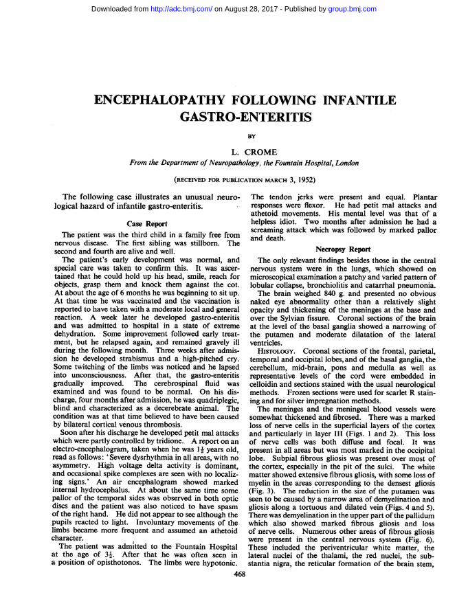
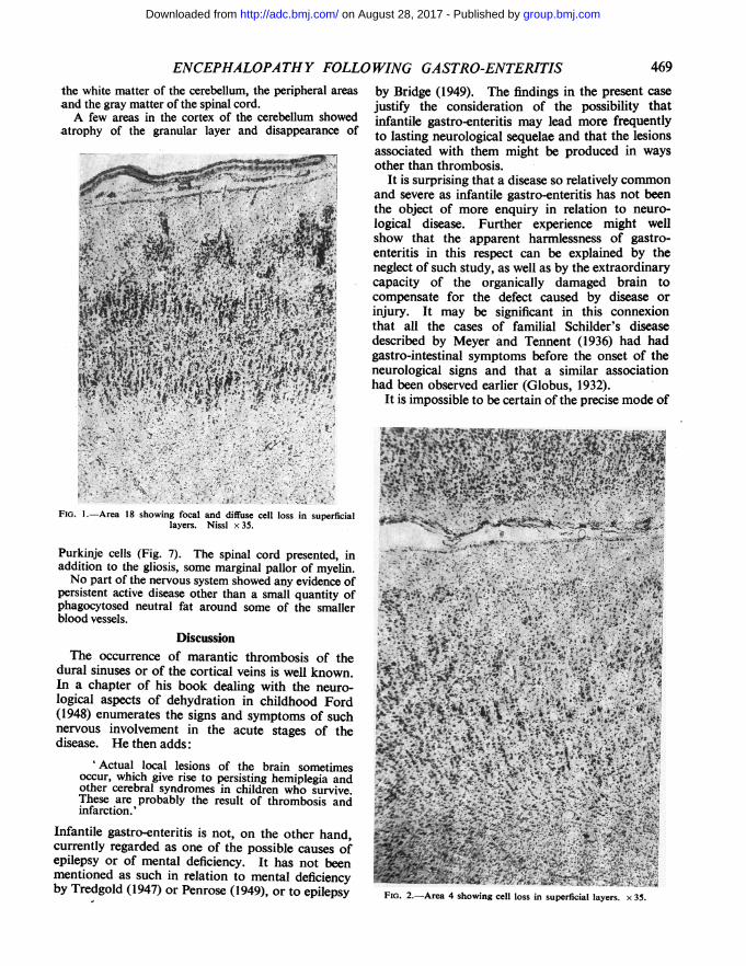
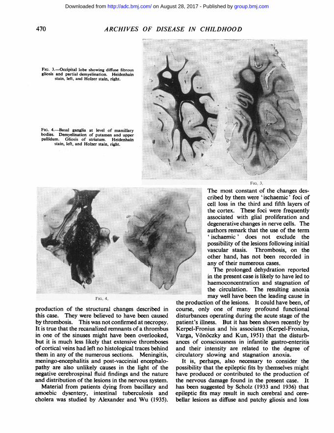
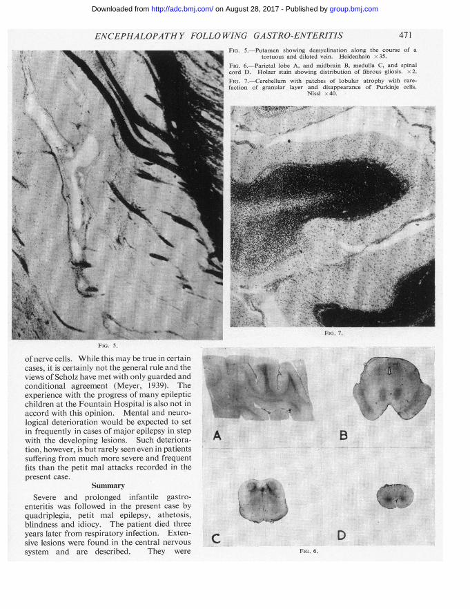
somewhat thickened and fibrosed. There was a markedloss of nerve cells in the superficial layers of the cortexand particularly in layer III (Figs. 1 and 2). This lossof nerve cells was both diffuse and focal. It waspresent in all areas but was most marked in the occipitallobe. Subpial fibrous gliosis was present over most ofthe cortex, especially in the pit of the sulci. The whitematter showed extensive fibrous gliosis, with some loss ofmyelin in the areas corresponding to the densest gliosis(Fig. 3). The reduction in the size of the putamen wasseen to be caused by a narrow area of demyelination andgliosis along a tortuous and dilated vein (Figs. 4 and 5).There was demyelination in the upper part of the pallidumwhich also showed marked fibrous gliosis and lossof nerve cells. Numerous other areas of fibrous gliosiswere present in the central nervous system (Fig. 6).These included the periventricular white matter, thelateral nuclei of the thalami, the red nuclei, the sub-stantia nigra, the reticular formation of the brain stem,
group.bmj.com on August 28, 2017 - Published by http://adc.bmj.com/Downloaded from
ENCEPHALOPATHY FOLLOWING GASTRO-ENTERITISthe white matter of the cerebellum, the peripheral areasand the gray matter of the spinal cord.A few areas in the cortex of the cerebellum showed
atrophy of the granular layer and disappearance of
by Bridge (1949). The findings in the present casejustify the consideration of the possibility thatinfantile gastro-enteritis may lead more frequentlyto lasting neurological sequelae and that the lesionsassociated with them might be produced in waysother than thrombosis.
It is surprising that a disease so relatively commonand severe as infantile gastro-enteritis has not beenthe object of more enquiry in relation to neuro-logical disease. Further experience might wellshow that the apparent harmlessness of gastro-enteritis in this respect can be explained by theneglect of such study, as well as by the extraordinarycapacity of the organically damaged brain tocompensate for the defect caused by disease orinjury. It may be significant in this connexionthat all the cases of familial Schilder's diseasedescribed by Meyer and Tennent (1936) had hadgastro-intestinal symptoms before the onset of theneurological signs and that a similar associationhad been observed earlier (Globus, 1932).
It is impossible to be certain of the precise mode of
FIG. 1.-Area 18 showing focal and diffuse cell loss in superficiallayers. Nissl x 35.
Purkinje cells (Fig. 7). The spinal cord presented, inaddition to the gliosis, some marginal pallor of myelin.No part of the nervous system showed any evidence of
persistent active disease other than a small quantity ofphagocytosed neutral fat around some of the smallerblood vessels.
DiscussionThe occurrence of marantic thrombosis of the
dural sinuses or of the cortical veins is well known.In a chapter of his book dealing with the neuro-logical aspects of dehydration in childhood Ford(1948) enumerates the signs and symptoms of suchnervous involvement in the acute stages of thedisease. He then adds:
'Actual local lesions of the brain sometimesoccur, which give rise to persisting hemiplegia andother cerebral syndromes in children who survive.These are probably the result of thrombosis andinfarction.'
Infantile gastro-enteritis is not, on the other hand,currently regarded as one of the possible causes ofepilepsy or of mental deficiency. It has not beenmentioned as such in relation to mental deficiencyby Tredgold (1947) or Penrose (1949), or to epilepsy FIG. 2.-Area 4 showing cell loss in superficial layers. x 35.
469
group.bmj.com on August 28, 2017 - Published by http://adc.bmj.com/Downloaded from
.70 ARCHIVES OF DISEASE IN CHILDHOOD
FIG. 3.-Occipital lobe showing diffuse fibrousgliosis and partial demyelination. Heidenhain
stain, left, and Holzer stain, right.
FIG. 4.-Basal ganglia at level of mamillarybodies. Demyelination of putamen and upperpallidum. Gliosis of striatum. Heidenhain
stain, left, and Holzer stain, right.
FIG. 3.
FIG. 4.
production of the structural changes described inthis case. They were believed to have been causedby thrombosis. This was not confirmed at necropsy.It is true that the recanalized remnants of a thrombusin one of the sinuses might have been overlooked,but it is much less likely that extensive thrombosesof cortical veins had left no histological traces behindthem in any of the numerous sections. Meningitis,meningo-encephalitis and post-vaccinial encephalo-pathy are also unlikely causes in the light of thenegative cerebrospinal fluid findings and the natureand distribution of the lesions in the nervous system.
Material from patients dying from bacillary andamoebic dysentery, intestinal tuberculosis andcholera was studied by Alexander and Wu (1935).
The most constant of the changes des-cribed by them were 'ischaemic' foci ofcell loss in the third and fifth layers ofthe cortex. These foci were frequentlyassociated with glial proliferation anddegenerative changes in nerve cells. Theauthors remark that the use of the term'ischaemic' does not exclude thepossibility of the lesions following initialvascular stasis. Thrombosis, on theother hand, has not been recorded inany of their numerous cases.The prolonged dehydration reported
in the present case is likely to have led tohaemoconcentration and stagnation ofthe circulation. The resulting anoxiamay well have been the leading cause in
the production of the lesions. It could have been, ofcourse, only one of many profound functionaldisturbances operating during the acute stage of thepatient's illness. But it has been shown recently byKerpel-Fronius and his associates (Kerpel-Fronius,Varga, Vonoczky and Kun, 1951) that the disturb-ances of consciousness in infantile gastro-enteritisand their intensity are related to the degree ofcirculatory slowing and stagnation anoxia.
It is, perhaps, also necessary to consider thepossibility that the epileptic fits by themselves mighthave produced or contributed to the production ofthe nervous damage found in the present case. Ithas been suggested by Scholz (1933 and 1936) thatepileptic fits may result in such cerebral and cere-bellar lesions as diffuse and patchy gliosis and loss
4
...:
T
group.bmj.com on August 28, 2017 - Published by http://adc.bmj.com/Downloaded from
ENCEPHALOPATHY FOLLO WING GASTRO-ENTERITISFIG. 5.-Putamen showing demyelination along the course of a
tortuous and dilated vein. Heidenhain x 35.
FIG. 6.-Parietal lobe A, and midbrain B, medulla C, and spinalcord D. Holzer stain showing distribution of fibrous gliosis. x2.FIG. 7.-Cerebellum with patches of lobular atrophy with rare-faction of granular layer and disappearance of Purkinje cells.
Nissl x 40.
riu. i.
FIo. 5.
ofnerve cells. While this may be true in certaincases, it is certainly not the general rule and theviews of Scholz have met with only guarded andconditional agreement (Meyer, 1939). Theexperience with the progress of many epilepticchildren at the Fountain Hospital is also not inaccord with this opinion. Mental and neuro-lcgical deterioration would be expected to setin frequently in cases of major epilepsy in stepwith the developing lesions. Such deteriora-tion, however, is but rarely seen even in patientssuffering from much more severe and frequentfits than the petit mal attacks recorded in thepresent case.
SummarySevere and prolonged infantile gastro-
enteritis was followed in the present case byquadriplegia, petit mal epilepsy, athetosis,blindness and idiocy. The patient died threeyears later from respiratory infection. Exten-sive lesions were found in the central nervoussystem and are described. They were
B.
......
.C DFiG. 6.
471
group.bmj.com on August 28, 2017 - Published by http://adc.bmj.com/Downloaded from
472 ARCHIVES OF DISEASE IN CHILDHOOD
characterized chiefly by a focal and diffuse cellloss in the superficial layers of the cortex.Numerous areas of gliosis and demyelination werealso present throughout the nervous system. It issuggested that haemoconcentration with stagnationof the cerebral circulation and the resulting anoxiamight have been the main links in the chain ofevents leading to the production of the lesions.
I am greatly indebted to my lay and medical colleaguesat the Fountain Hospital for their cooperation and theuse of their case records. Professor A. Meyer andDr. B. H. Kirman have given much useful advice andcriticism. Miss M. F. Craib provided the social andearly history of the patient's development. Mr. J. F.
Watt and Mr. J. E. Stevens have assisted in the histologi-cal and photographic work.
REFERENCES
Alexander, L. and Wu, T. T. (1935). Arch. Neurol. Psychiat., Chicago,33, 72.
Bridge, E. M. (1949). Epilepsy and Convulsive Disorders in Children,New York.
Ford, F. R. (1948). Diseases of the Nervous System in Infancy, Child-hood and Adolescence, 2nd ed., p. 758, Springfield.
Globus, J. H. (1932). In Cytology and Cellular Pathology of theNervous System, ed. W. Penfield: vol. 3, p. 1145, New York.
Kerpel-Fronius, E., Varga, F., Vonoczky, J. and Kun, K. (1951).Helv. paediat. Acta, 6, 377.
Meyer, A. (1939). J. ment. Sci., 85, 927.-- and Tennent, T. (1936). Brain, 59, 100.Penrose, L. S. (1949). The Biology of Mental Defect. London.Scholz, W. (1933). Z. ges. Neurol. Psychiat., 145, 471.- (1936). Allg. Z. Psychiat., 104, 89.Tredgold, A. F. (1947). A Textbook of Mental Deficiency, 7th ed.
London.
Scholarships in Aid of Scientific ResearchThe Council of the British Medical Association is
prepared to receive applications for Research Scholar-ships, as follows:
An Emest Hart Memorial Scholarship of the valueof £250
A Walter Dixon Scholarship of the value of £250One or More Research Scholarships each of the
value of £200These scholarships are given to candidates whom the
Science Committee of the Association recommends asqualified to undertake research in any subject (includingstate medicine) relating to the causation, prevention, ortreatment of disease.Each scholarship is tenable for one year, beginning
on October 1, 1953. A scholar may be re-appointed for
not more than two additional terms. A scholar is notnecessarily required to devote the whole of his or hertime to the work of research, but may be a member ofH.M. Forces or may hold a junior appointment at auniversity, medical school or hospital, provided theduties of such appointment will not, in the opinion ofthe Science Committee, interfere with his or her workas a scholar.
Applications for Scholarships must be made notlater than March 31, 1953, on the prescribed form, acopy of which will be supplied by the Secretary onapplication.
Applicants are required to furnish the names of threereferees who are competent to speak as to their capacityfor the research contemplated.
group.bmj.com on August 28, 2017 - Published by http://adc.bmj.com/Downloaded from
Infantile Gastro-EnteritisEncephalopathy following
L. Crome
doi: 10.1136/adc.27.135.4681952 27: 468-472 Arch Dis Child
http://adc.bmj.com/content/27/135/468.citationUpdated information and services can be found at:
These include:
serviceEmail alerting
online article. article. Sign up in the box at the top right corner of the Receive free email alerts when new articles cite this
Notes
http://group.bmj.com/group/rights-licensing/permissionsTo request permissions go to:
http://journals.bmj.com/cgi/reprintformTo order reprints go to:
http://group.bmj.com/subscribe/To subscribe to BMJ go to:
group.bmj.com on August 28, 2017 - Published by http://adc.bmj.com/Downloaded from