elevated liver enzymes - boca raton regional …...introduction •evaluation of abnormal liver...
TRANSCRIPT
ELEVATED LIVER ENZYMES
Eric F. Martin, MD Transplant Hepatology
Assistant Professor of Clinical Medicine Medical Director of Living Donor Liver Transplant University of Miami ~ Miami Transplant Institute
Financial Disclosures
• None
Objectives
1. Identify the components of the liver biochemistry profile and understand their meaning if abnormal
2. Identify and understand the significance of the true liver function test “LFTs”
3. Develop a differential diagnosis for abnormal liver biochemistries, including AST and/or ALT >1000
4. Follow an organized approach to evaluate abnormal liver biochemistries
Introduction
• Evaluation of abnormal liver enzymes in an otherwise healthy patient may pose challenge to most experienced clinician
• May not be necessary to pursue extensive evaluation for all abnormal tests, due to unnecessary expenses and procedural risks
• On the other hand, failure to investigate mild or moderate liver enzyme abnormalities could mean missing the early diagnosis of potentially life-threatening, but otherwise treatable conditions
• Liver enzymes are readily available and included in many routine labs
• Estimated that 1%-9% of asymptomatic patients have elevated liver enzyme levels when screened with standard “liver function panels”
• All persistent elevations of liver enzymes require methodical evaluation and appropriate working diagnosis
Am J Gastroenterol 2017;12:18-35
Introduction
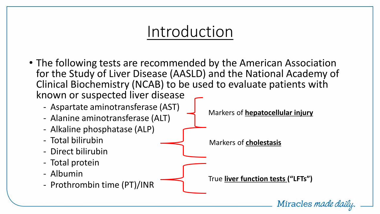
• The following tests are recommended by the American Association for the Study of Liver Disease (AASLD) and the National Academy of Clinical Biochemistry (NCAB) to be used to evaluate patients with known or suspected liver disease
- Aspartate aminotransferase (AST) - Alanine aminotransferase (ALT) - Alkaline phosphatase (ALP) - Total bilirubin - Direct bilirubin - Total protein - Albumin - Prothrombin time (PT)/INR
Markers of hepatocellular injury
Markers of cholestasis
True liver function tests (“LFTs”)
Lab Orders
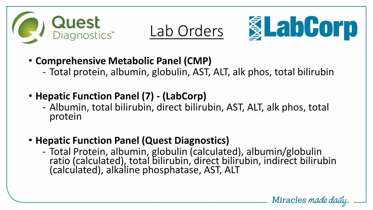
• Comprehensive Metabolic Panel (CMP) - Total protein, albumin, globulin, AST, ALT, alk phos, total bilirubin
• Hepatic Function Panel (7) - (LabCorp)
- Albumin, total bilirubin, direct bilirubin, AST, ALT, alk phos, total protein
• Hepatic Function Panel (Quest Diagnostics) - Total Protein, albumin, globulin (calculated), albumin/globulin
ratio (calculated), total bilirubin, direct bilirubin, indirect bilirubin (calculated), alkaline phosphatase, AST, ALT
Lab Orders
• Comprehensive Metabolic Panel (CMP) - Total protein, albumin, globulin, AST, ALT, alk phos, total bilirubin
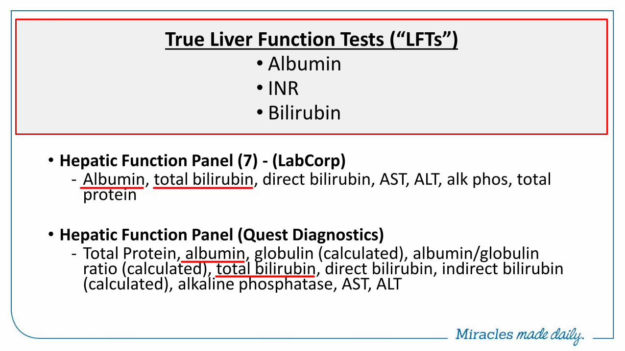
• Hepatic Function Panel (7) - (LabCorp)
- Albumin, total bilirubin, direct bilirubin, AST, ALT, alk phos, total protein
• Hepatic Function Panel (Quest Diagnostics) - Total Protein, albumin, globulin (calculated), albumin/globulin
ratio (calculated), total bilirubin, direct bilirubin, indirect bilirubin (calculated), alkaline phosphatase, AST, ALT
True Liver Function Tests (“LFTs”) • Albumin • INR • Bilirubin
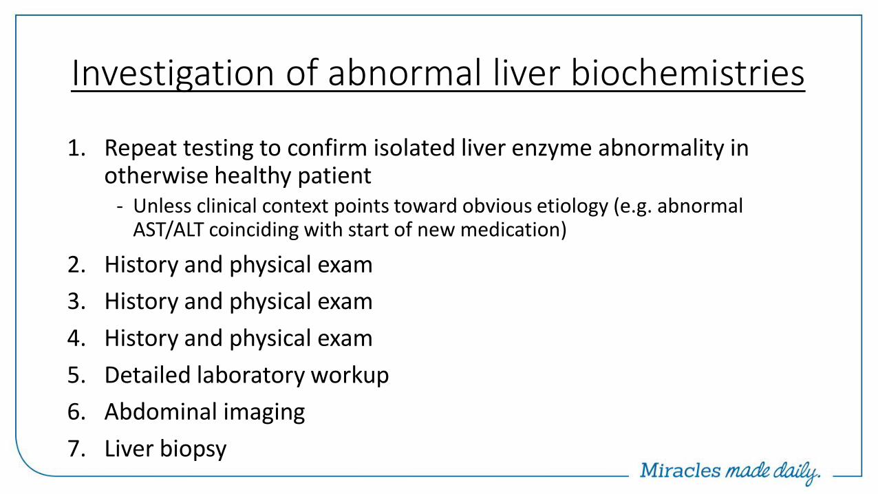
Investigation of abnormal liver biochemistries
1. Repeat testing to confirm isolated liver enzyme abnormality in otherwise healthy patient
- Unless clinical context points toward obvious etiology (e.g. abnormal AST/ALT coinciding with start of new medication)
2. History and physical exam
3. History and physical exam
4. History and physical exam
5. Detailed laboratory workup
6. Abdominal imaging
7. Liver biopsy
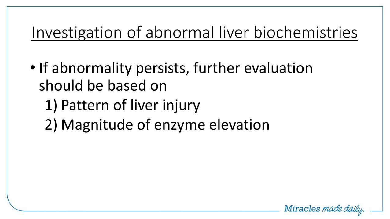
Investigation of abnormal liver biochemistries
• If abnormality persists, further evaluation should be based on 1) Pattern of liver injury 2) Magnitude of enzyme elevation
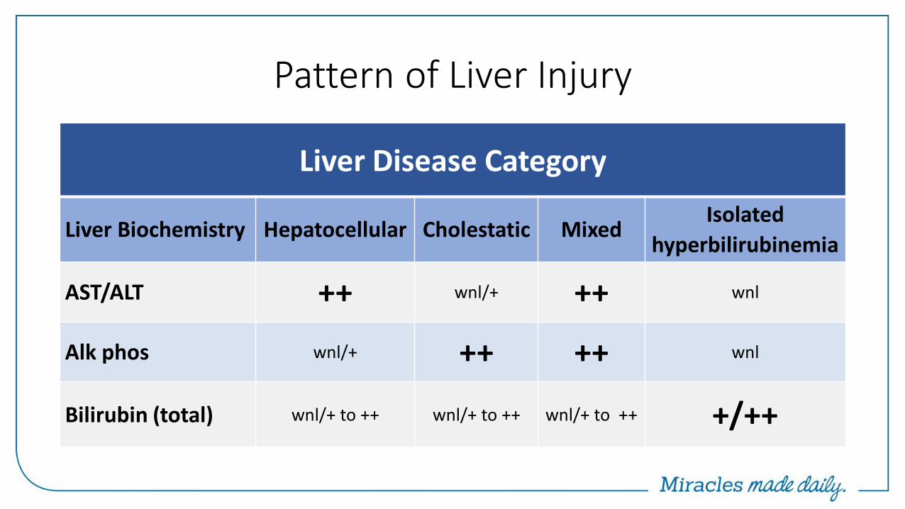
Pattern of Liver Injury
Liver Disease Category
Liver Biochemistry Hepatocellular Cholestatic Mixed Isolated
hyperbilirubinemia
AST/ALT ++ wnl/+ ++ wnl
Alk phos wnl/+ ++ ++ wnl
Bilirubin (total) wnl/+ to ++ wnl/+ to ++ wnl/+ to ++ +/++
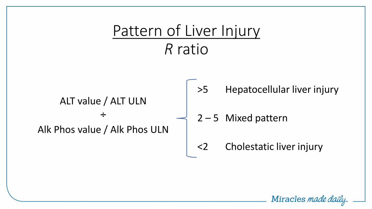
Pattern of Liver Injury R ratio
ALT value / ALT ULN
÷
Alk Phos value / Alk Phos ULN
>5 Hepatocellular liver injury
2 – 5 Mixed pattern
<2 Cholestatic liver injury
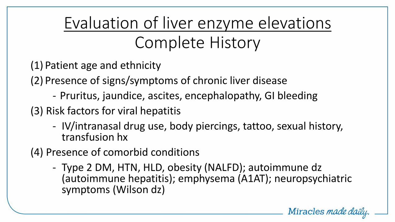
Evaluation of liver enzyme elevations Complete History
(1) Patient age and ethnicity
(2) Presence of signs/symptoms of chronic liver disease
- Pruritus, jaundice, ascites, encephalopathy, GI bleeding
(3) Risk factors for viral hepatitis
- IV/intranasal drug use, body piercings, tattoo, sexual history, transfusion hx
(4) Presence of comorbid conditions
- Type 2 DM, HTN, HLD, obesity (NALFD); autoimmune dz (autoimmune hepatitis); emphysema (A1AT); neuropsychiatric symptoms (Wilson dz)
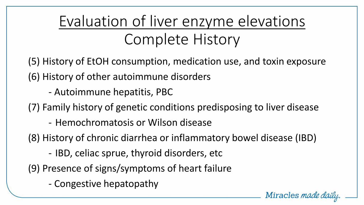
Evaluation of liver enzyme elevations Complete History
(5) History of EtOH consumption, medication use, and toxin exposure
(6) History of other autoimmune disorders
- Autoimmune hepatitis, PBC
(7) Family history of genetic conditions predisposing to liver disease
- Hemochromatosis or Wilson disease
(8) History of chronic diarrhea or inflammatory bowel disease (IBD)
- IBD, celiac sprue, thyroid disorders, etc
(9) Presence of signs/symptoms of heart failure
- Congestive hepatopathy
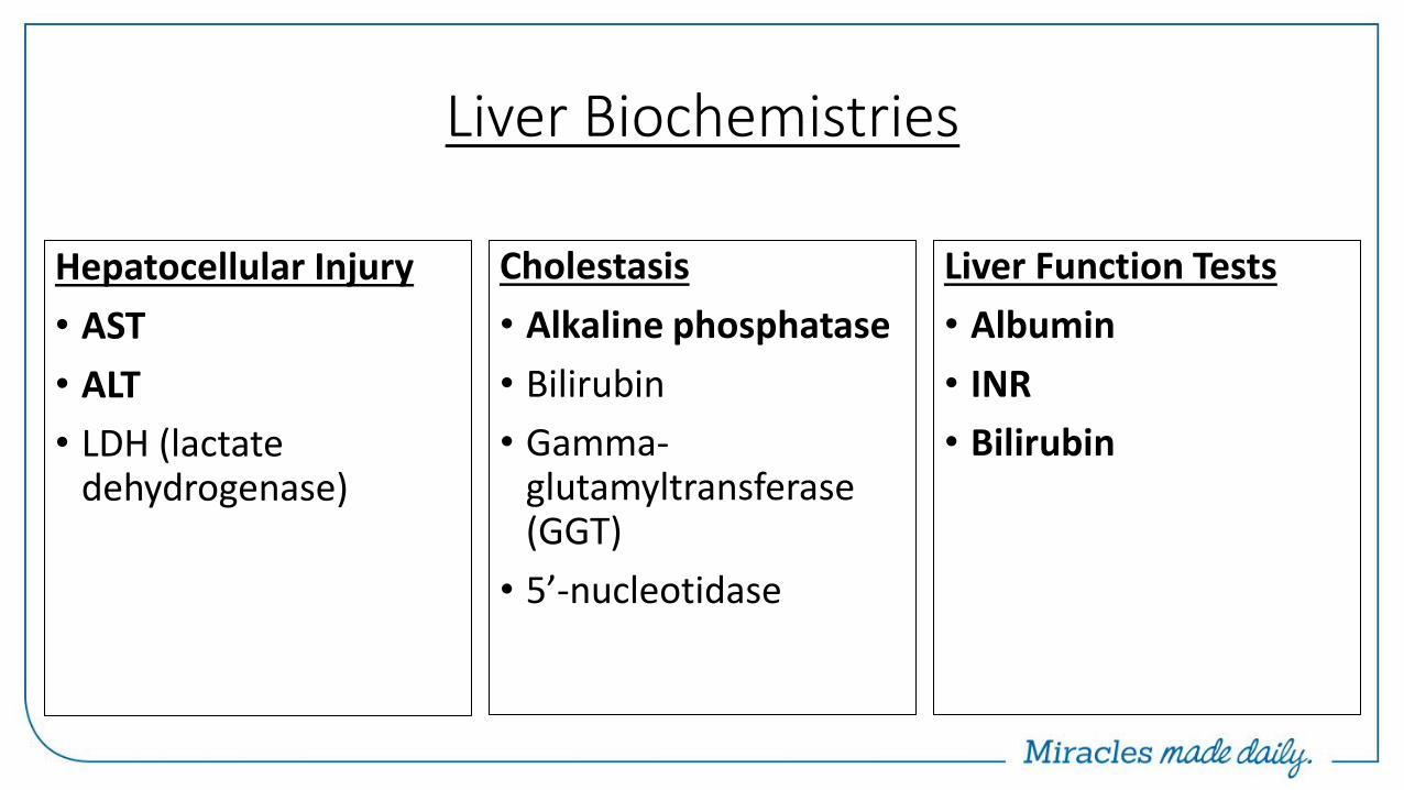
Liver Biochemistries
Cholestasis
• Alkaline phosphatase
• Bilirubin
• Gamma-glutamyltransferase (GGT)
• 5’-nucleotidase
Liver Function Tests
• Albumin
• INR
• Bilirubin
Hepatocellular Injury
• AST
• ALT
• LDH (lactate dehydrogenase)
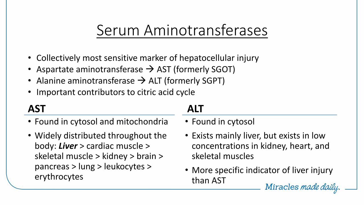
Serum Aminotransferases
AST • Found in cytosol and mitochondria
• Widely distributed throughout the body: Liver > cardiac muscle > skeletal muscle > kidney > brain > pancreas > lung > leukocytes > erythrocytes
ALT • Found in cytosol
• Exists mainly liver, but exists in low concentrations in kidney, heart, and skeletal muscles
• More specific indicator of liver injury than AST
• Collectively most sensitive marker of hepatocellular injury • Aspartate aminotransferase AST (formerly SGOT) • Alanine aminotransferase ALT (formerly SGPT) • Important contributors to citric acid cycle
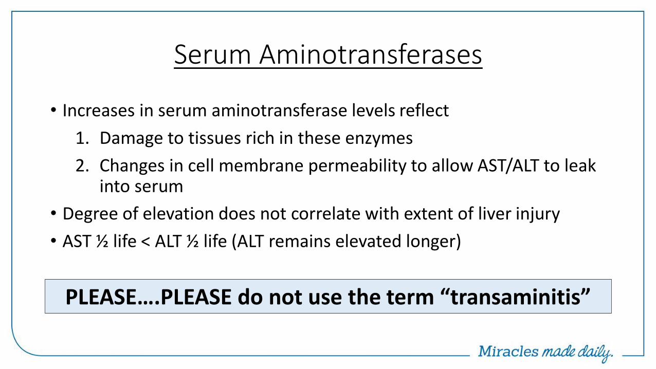
Serum Aminotransferases
• Increases in serum aminotransferase levels reflect
1. Damage to tissues rich in these enzymes
2. Changes in cell membrane permeability to allow AST/ALT to leak into serum
• Degree of elevation does not correlate with extent of liver injury
• AST ½ life < ALT ½ life (ALT remains elevated longer)
PLEASE….PLEASE do not use the term “transaminitis”
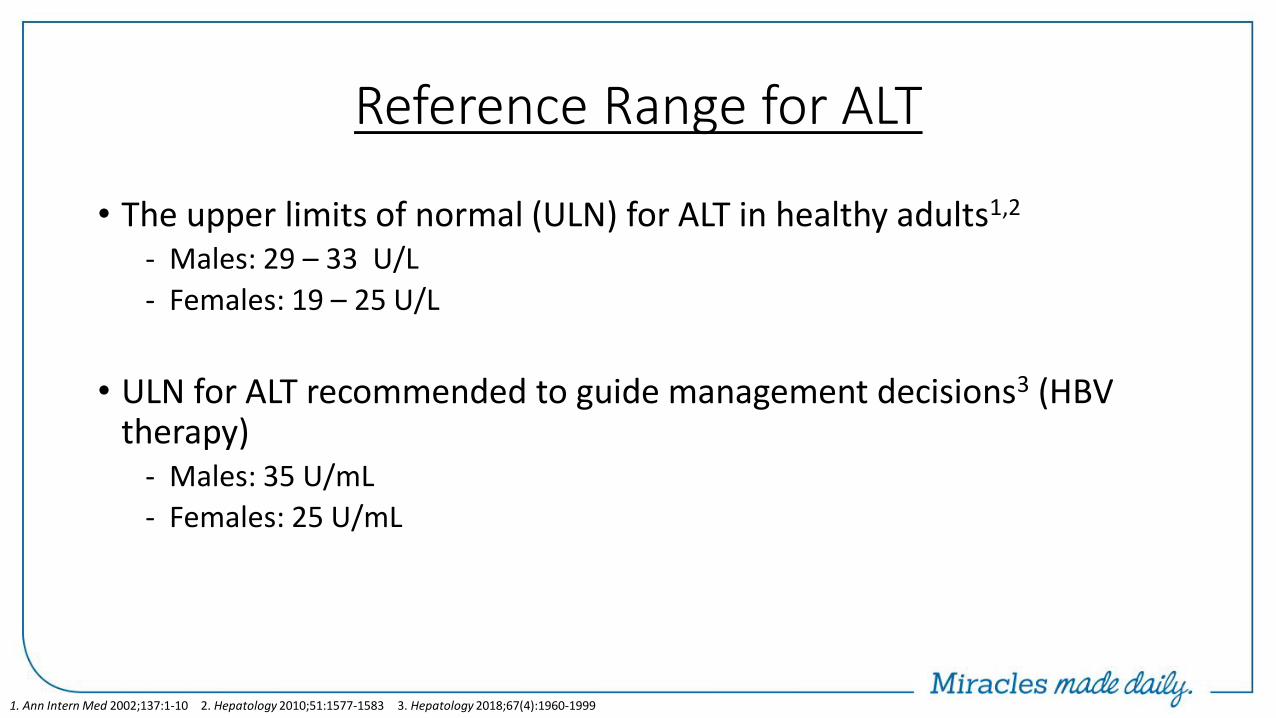
Reference Range for ALT
• The upper limits of normal (ULN) for ALT in healthy adults1,2
- Males: 29 – 33 U/L
- Females: 19 – 25 U/L
• ULN for ALT recommended to guide management decisions3 (HBV therapy)
- Males: 35 U/mL
- Females: 25 U/mL
1. Ann Intern Med 2002;137:1-10 2. Hepatology 2010;51:1577-1583 3. Hepatology 2018;67(4):1960-1999
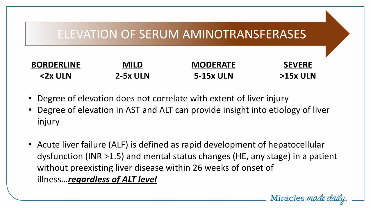
MILD 2-5x ULN
MODERATE 5-15x ULN
SEVERE >15x ULN
ELEVATION OF SERUM AMINOTRANSFERASES
BORDERLINE <2x ULN
• Degree of elevation does not correlate with extent of liver injury • Degree of elevation in AST and ALT can provide insight into etiology of liver
injury
• Acute liver failure (ALF) is defined as rapid development of hepatocellular dysfunction (INR >1.5) and mental status changes (HE, any stage) in a patient without preexisting liver disease within 26 weeks of onset of illness…regardless of ALT level
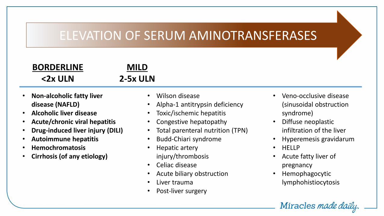
• Non-alcoholic fatty liver disease (NAFLD)
• Alcoholic liver disease • Acute/chronic viral hepatitis • Drug-induced liver injury (DILI) • Autoimmune hepatitis • Hemochromatosis • Cirrhosis (of any etiology)
• Wilson disease • Alpha-1 antitrypsin deficiency • Toxic/ischemic hepatitis • Congestive hepatopathy • Total parenteral nutrition (TPN) • Budd-Chiari syndrome • Hepatic artery
injury/thrombosis • Celiac disease • Acute biliary obstruction • Liver trauma • Post-liver surgery
• Veno-occlusive disease (sinusoidal obstruction syndrome)
• Diffuse neoplastic infiltration of the liver
• Hyperemesis gravidarum • HELLP • Acute fatty liver of
pregnancy • Hemophagocytic
lymphohistiocytosis
ELEVATION OF SERUM AMINOTRANSFERASES
MILD 2-5x ULN
MODERATE
5-15x ULN
SEVERE
>15x ULN BORDERLINE
<2x ULN
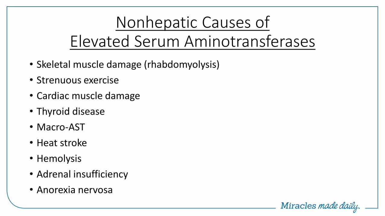
Nonhepatic Causes of Elevated Serum Aminotransferases
• Skeletal muscle damage (rhabdomyolysis)
• Strenuous exercise
• Cardiac muscle damage
• Thyroid disease
• Macro-AST
• Heat stroke
• Hemolysis
• Adrenal insufficiency
• Anorexia nervosa
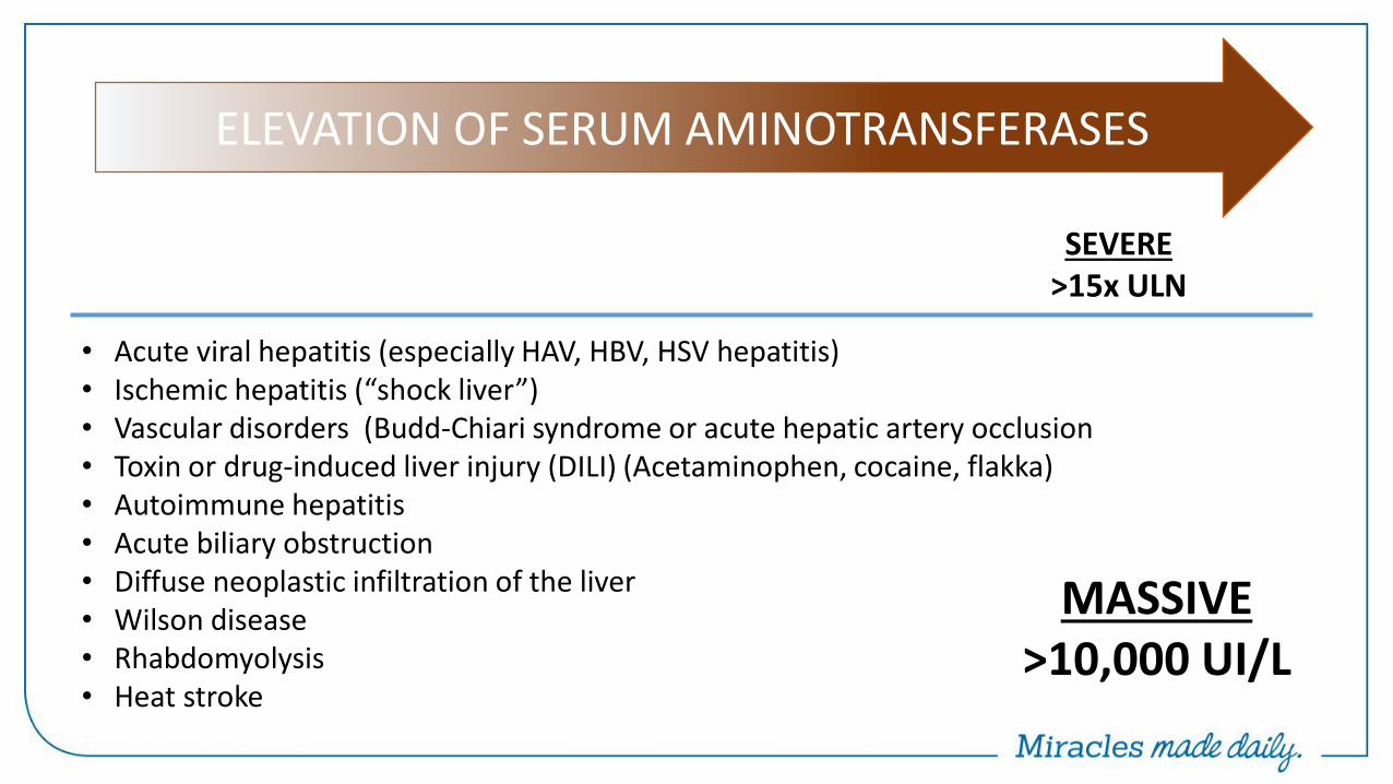
• Acute viral hepatitis (especially HAV, HBV, HSV hepatitis) • Ischemic hepatitis (“shock liver”) • Vascular disorders (Budd-Chiari syndrome or acute hepatic artery occlusion • Toxin or drug-induced liver injury (DILI) (Acetaminophen, cocaine, flakka) • Autoimmune hepatitis • Acute biliary obstruction • Diffuse neoplastic infiltration of the liver • Wilson disease • Rhabdomyolysis • Heat stroke
ELEVATION OF SERUM AMINOTRANSFERASES
MILD
2-5x ULN
MODERATE
5-15x ULN
SEVERE >15x ULN
BORDERLINE
<2x ULN
MASSIVE >10,000 UI/L
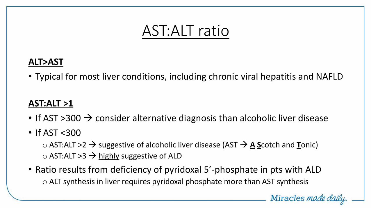
AST:ALT ratio
ALT>AST
• Typical for most liver conditions, including chronic viral hepatitis and NAFLD
AST:ALT >1
• If AST >300 consider alternative diagnosis than alcoholic liver disease
• If AST <300 o AST:ALT >2 suggestive of alcoholic liver disease (AST A Scotch and Tonic)
o AST:ALT >3 highly suggestive of ALD
• Ratio results from deficiency of pyridoxal 5’-phosphate in pts with ALD o ALT synthesis in liver requires pyridoxal phosphate more than AST synthesis
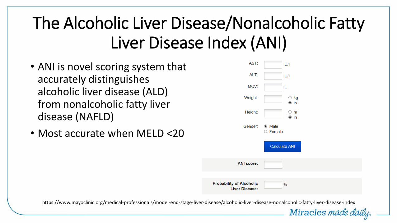
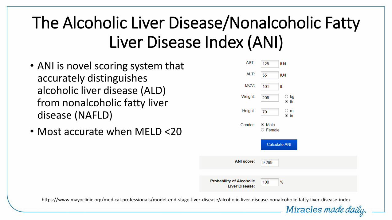
The Alcoholic Liver Disease/Nonalcoholic Fatty Liver Disease Index (ANI)
• ANI is novel scoring system that accurately distinguishes alcoholic liver disease (ALD) from nonalcoholic fatty liver disease (NAFLD)
• Most accurate when MELD <20
https://www.mayoclinic.org/medical-professionals/model-end-stage-liver-disease/alcoholic-liver-disease-nonalcoholic-fatty-liver-disease-index
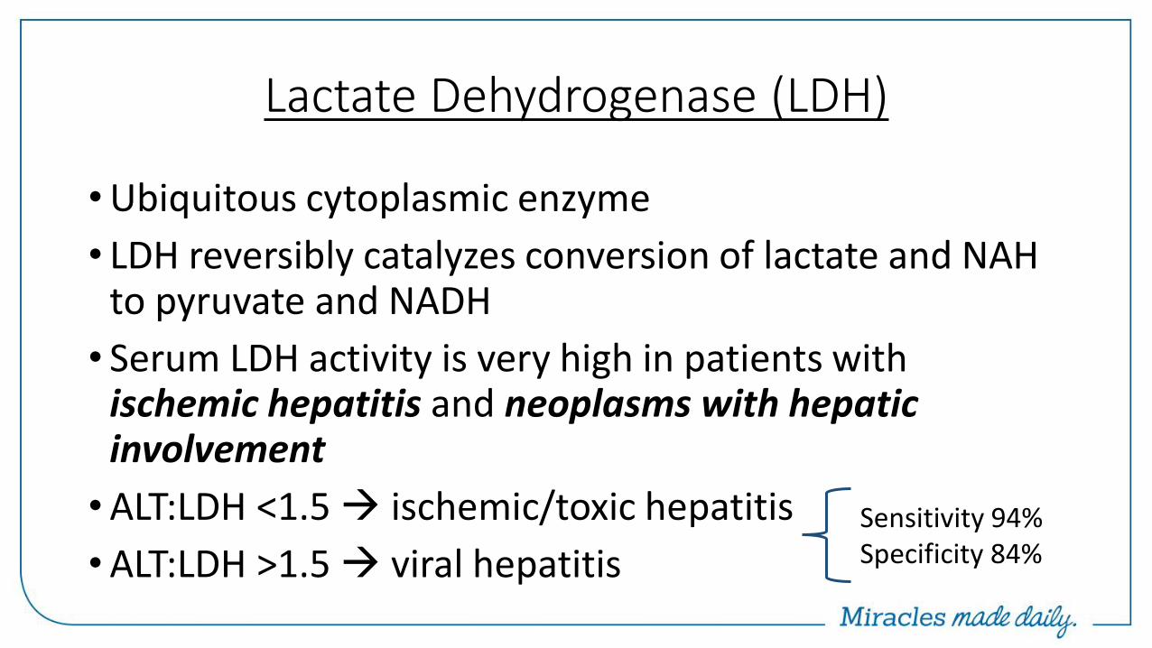
Lactate Dehydrogenase (LDH)
• Ubiquitous cytoplasmic enzyme
• LDH reversibly catalyzes conversion of lactate and NAH to pyruvate and NADH
• Serum LDH activity is very high in patients with ischemic hepatitis and neoplasms with hepatic involvement
• ALT:LDH <1.5 ischemic/toxic hepatitis
• ALT:LDH >1.5 viral hepatitis Sensitivity 94% Specificity 84%
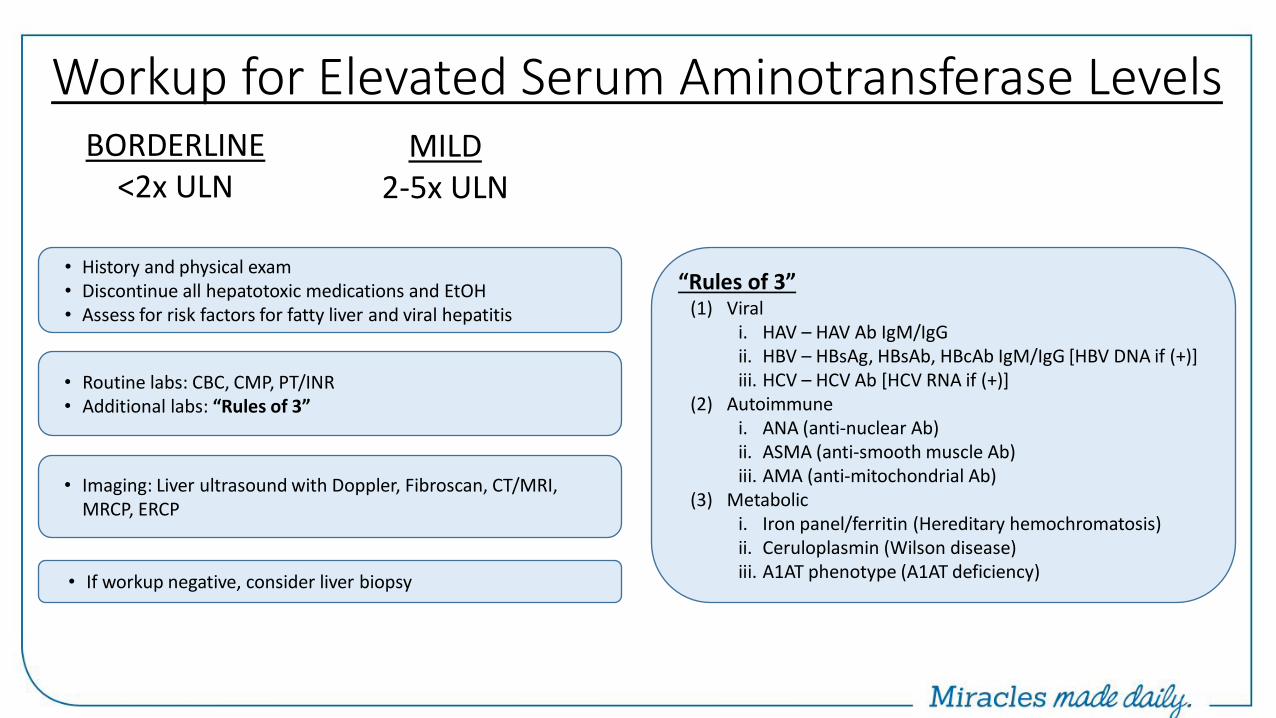
Workup for Elevated Serum Aminotransferase Levels
• History and physical exam • Discontinue all hepatotoxic medications and EtOH • Assess for risk factors for fatty liver and viral hepatitis
• Routine labs: CBC, CMP, PT/INR • Additional labs: “Rules of 3”
• Imaging: Liver ultrasound with Doppler, Fibroscan, CT/MRI, MRCP, ERCP
• If workup negative, consider liver biopsy
MILD 2-5x ULN
BORDERLINE <2x ULN
“Rules of 3” (1) Viral
i. HAV – HAV Ab IgM/IgG ii. HBV – HBsAg, HBsAb, HBcAb IgM/IgG [HBV DNA if (+)] iii. HCV – HCV Ab [HCV RNA if (+)]
(2) Autoimmune i. ANA (anti-nuclear Ab) ii. ASMA (anti-smooth muscle Ab) iii. AMA (anti-mitochondrial Ab)
(3) Metabolic i. Iron panel/ferritin (Hereditary hemochromatosis) ii. Ceruloplasmin (Wilson disease) iii. A1AT phenotype (A1AT deficiency)
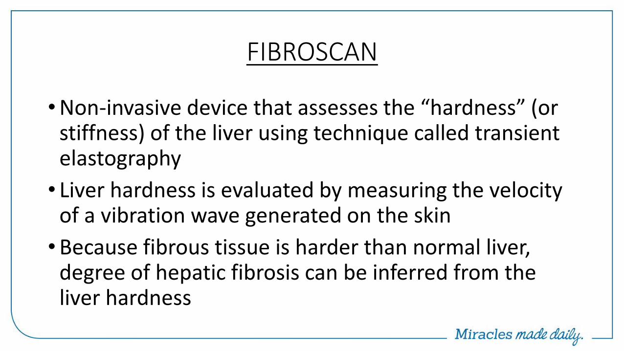
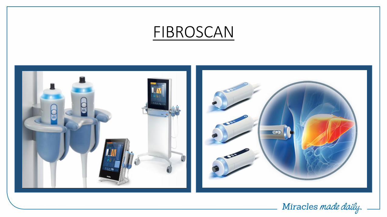
FIBROSCAN
• Non-invasive device that assesses the “hardness” (or stiffness) of the liver using technique called transient elastography
• Liver hardness is evaluated by measuring the velocity of a vibration wave generated on the skin
• Because fibrous tissue is harder than normal liver, degree of hepatic fibrosis can be inferred from the liver hardness
FIBROSCAN
FIBROSCAN
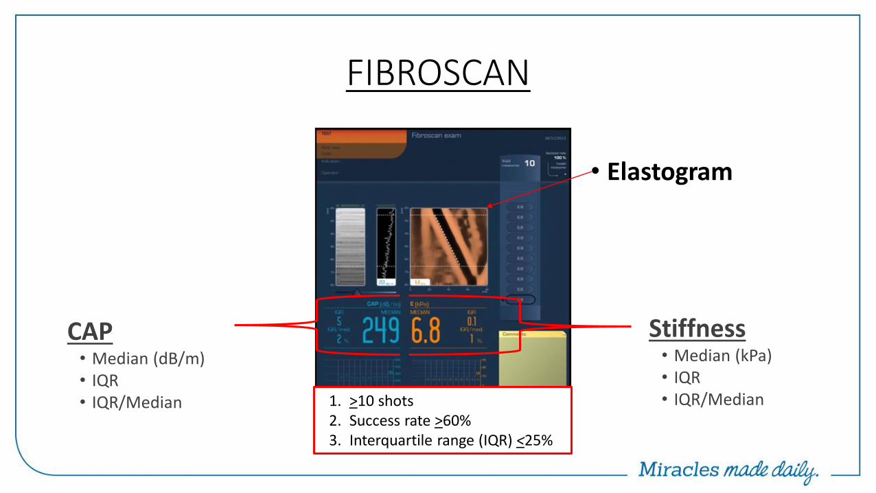
• Elastogram
1. >10 shots 2. Success rate >60% 3. Interquartile range (IQR) <25%
Stiffness • Median (kPa) • IQR • IQR/Median
CAP • Median (dB/m) • IQR • IQR/Median
FIBROSCAN
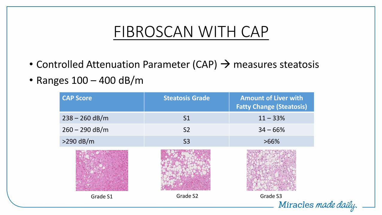
FIBROSCAN WITH CAP
CAP Score Steatosis Grade Amount of Liver with Fatty Change (Steatosis)
238 – 260 dB/m S1 11 – 33%
260 – 290 dB/m S2 34 – 66%
>290 dB/m S3 >66%
Grade S1 Grade S2 Grade S3
• Controlled Attenuation Parameter (CAP) measures steatosis
• Ranges 100 – 400 dB/m
https://livertox.nih.gov
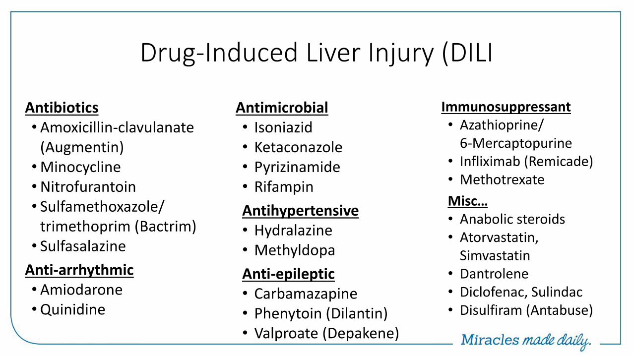
Drug-Induced Liver Injury (DILI
Antibiotics • Amoxicillin-clavulanate
(Augmentin) • Minocycline • Nitrofurantoin • Sulfamethoxazole/
trimethoprim (Bactrim) • Sulfasalazine
Anti-arrhythmic • Amiodarone • Quinidine
Immunosuppressant • Azathioprine/
6-Mercaptopurine • Infliximab (Remicade) • Methotrexate
Misc… • Anabolic steroids • Atorvastatin,
Simvastatin • Dantrolene • Diclofenac, Sulindac • Disulfiram (Antabuse)
Antimicrobial • Isoniazid • Ketaconazole • Pyrizinamide • Rifampin
Antihypertensive • Hydralazine • Methyldopa
Anti-epileptic • Carbamazapine • Phenytoin (Dilantin) • Valproate (Depakene)
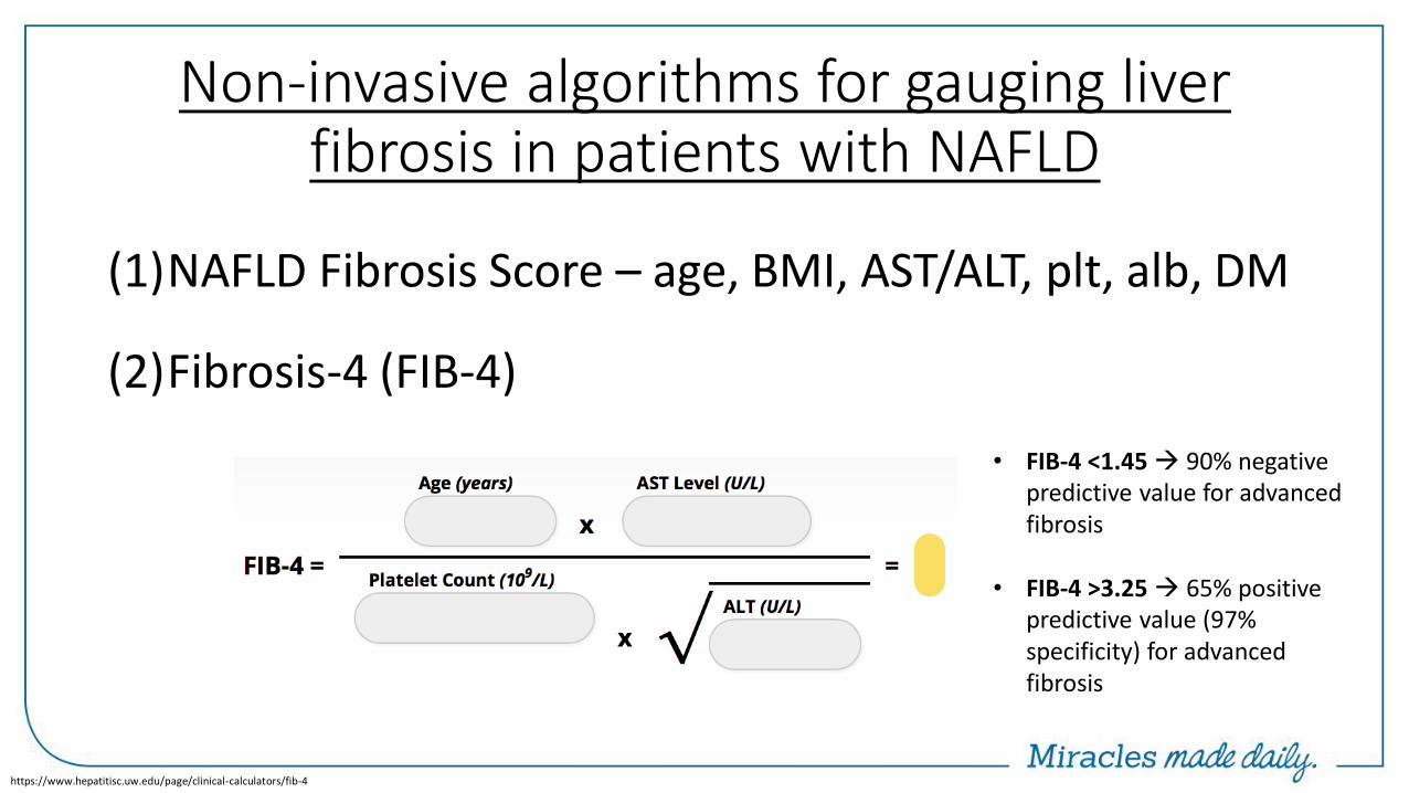
Non-invasive algorithms for gauging liver fibrosis in patients with NAFLD
(1)NAFLD Fibrosis Score – age, BMI, AST/ALT, plt, alb, DM
(2)Fibrosis-4 (FIB-4)
https://www.hepatitisc.uw.edu/page/clinical-calculators/fib-4
• FIB-4 <1.45 90% negative predictive value for advanced fibrosis
• FIB-4 >3.25 65% positive predictive value (97% specificity) for advanced fibrosis
ENZYMES FOR DETECTION OF CHOLESTASIS
• Cholestasis develops from (1) Defect in bile synthesis (2) Defect in bile secretion (3) Obstruction to bile flow
• Serum alkaline phosphatase (ALP) is traditional indirect marker of cholestasis
• Other enzymes that reflect that reflect cholestasis - 5’-nucleotidase - γ-glutamyltransferase (GGT)
ALKALINE PHOSPHATASE
• Group of enzymes that catalyze hydrolysis of organic phosphate esters at an alkaline pH
• Precise function is not yet known, but likely related to secretory activities of intrahepatic biliary epithelium
• Produced mainly in the liver (from biliary epithelium) and the bone
• Also found in smaller quantities in the - Intestine
- Kidney
- Placenta
- White blood cells
ALKALINE PHOSPHATASE
• Normal distribution of the enzyme in the serum varies with age; normal serum range 20 – 140 IU/mL
• ALP activity 3x higher in young adults due to bone growth
• ALP activity 3-4x ULN in late pregnancy
• Intestinal contribution of ALP enhanced in patients with blood groups O and B after ingestion of fatty meals
ALKALINE PHOSPHATASE
• Elevation in ALP is not completely specific for cholestasis
• Mild elevation of ALP 2-3x ULN can be seen with almost any type of liver disease
• Elevations of ALP >4x ULN - Cholestatic liver disease
- Infiltrative liver disease
- Rapid bone turnover
• When alk phos is elevated in isolation, important to determine whether alk phos is of hepatic or non-hepatic origin
- Obtaining additional enzyme testing with GGT and/or 5’-nucleotidase
- Measurement of ALP isoenzymes using electrophoresis
5’-nucleotidase
• Enzymes found in the bile canalicular and sinusoidal membrane, but also found in the intestines, brain, heart, blood vessels, and pancreas
• Despite widespread distribution, ↑ serum levels of 5’-nucleotidase seen in pts with cholestasis similar to alk phos activity
• Negligible increase in pts with bone disease; therefore, used to confirm liver origin of ↑ alk phos in pts with suspected liver disease
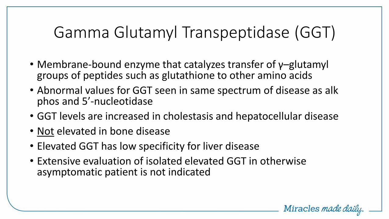
Gamma Glutamyl Transpeptidase (GGT)
• Membrane-bound enzyme that catalyzes transfer of γ–glutamyl groups of peptides such as glutathione to other amino acids
• Abnormal values for GGT seen in same spectrum of disease as alk phos and 5’-nucleotidase
• GGT levels are increased in cholestasis and hepatocellular disease
• Not elevated in bone disease
• Elevated GGT has low specificity for liver disease
• Extensive evaluation of isolated elevated GGT in otherwise asymptomatic patient is not indicated
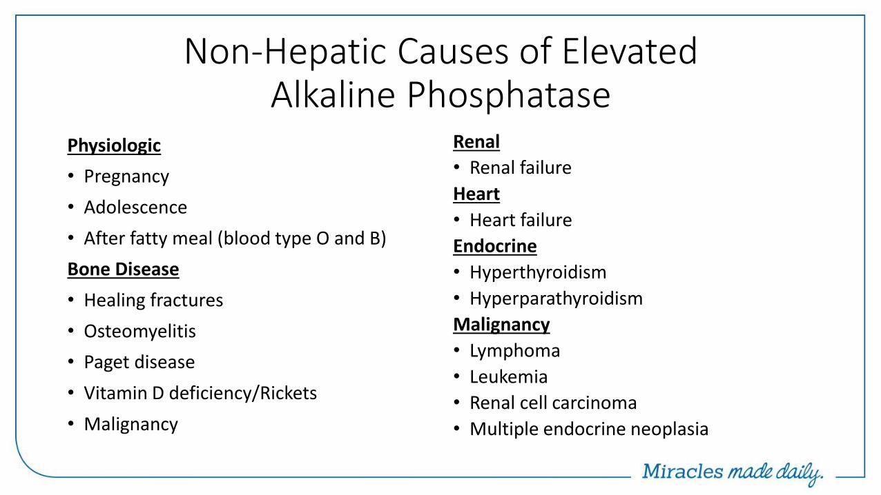
Non-Hepatic Causes of Elevated Alkaline Phosphatase
Physiologic
• Pregnancy
• Adolescence
• After fatty meal (blood type O and B)
Bone Disease
• Healing fractures
• Osteomyelitis
• Paget disease
• Vitamin D deficiency/Rickets
• Malignancy
Renal
• Renal failure
Heart
• Heart failure
Endocrine
• Hyperthyroidism
• Hyperparathyroidism
Malignancy
• Lymphoma
• Leukemia
• Renal cell carcinoma
• Multiple endocrine neoplasia
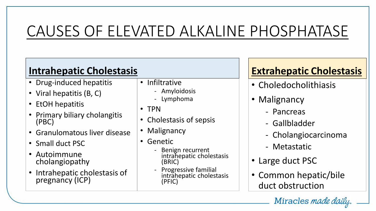
Intrahepatic Cholestasis • Drug-induced hepatitis
• Viral hepatitis (B, C)
• EtOH hepatitis
• Primary biliary cholangitis (PBC)
• Granulomatous liver disease
• Small duct PSC
• Autoimmune cholangiopathy
• Intrahepatic cholestasis of pregnancy (ICP)
• Infiltrative - Amyloidosis - Lymphoma
• TPN
• Cholestasis of sepsis
• Malignancy
• Genetic - Benign recurrent
intrahepatic cholestasis (BRIC)
- Progressive familial intrahepatic cholestasis (PFIC)
• Choledocholithiasis
• Malignancy - Pancreas
- Gallbladder
- Cholangiocarcinoma
- Metastatic
• Large duct PSC
• Common hepatic/bile duct obstruction
Extrahepatic Cholestasis
CAUSES OF ELEVATED ALKALINE PHOSPHATASE
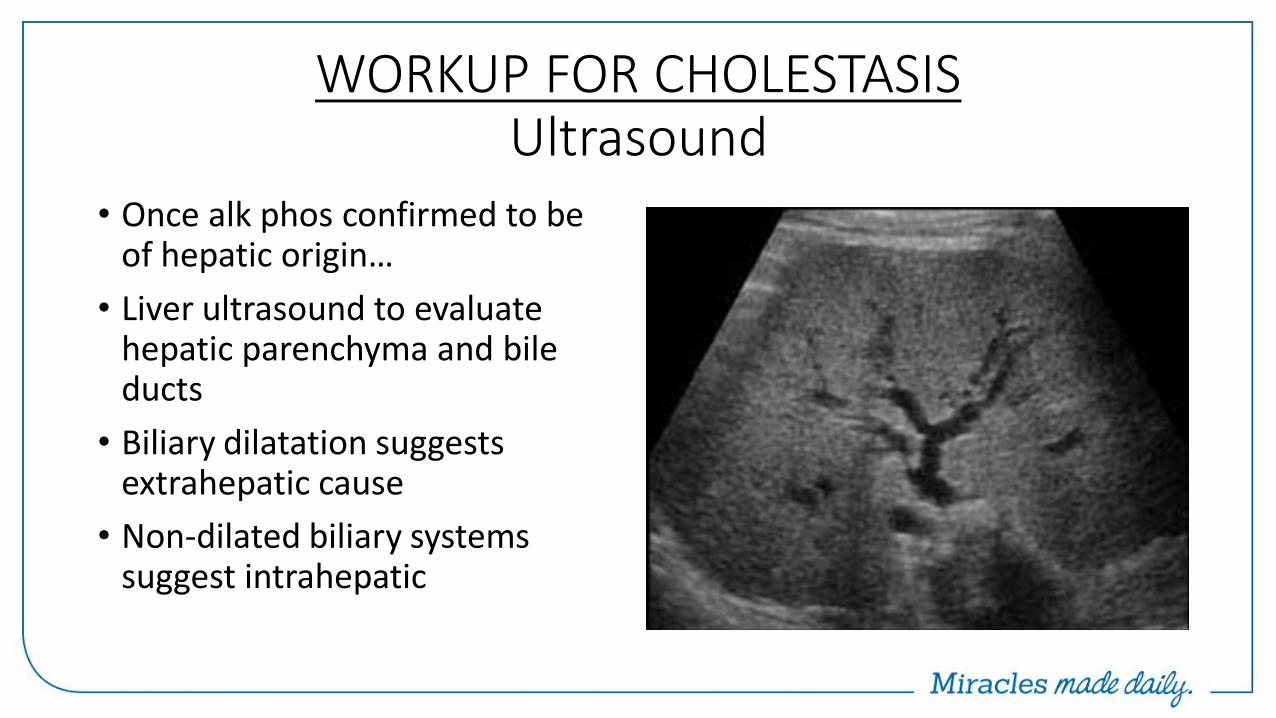
WORKUP FOR CHOLESTASIS Ultrasound
• Once alk phos confirmed to be of hepatic origin…
• Liver ultrasound to evaluate hepatic parenchyma and bile ducts
• Biliary dilatation suggests extrahepatic cause
• Non-dilated biliary systems suggest intrahepatic
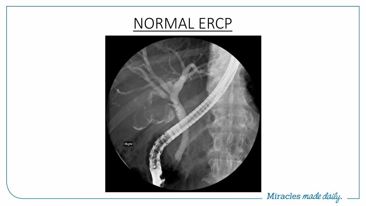
NORMAL ERCP
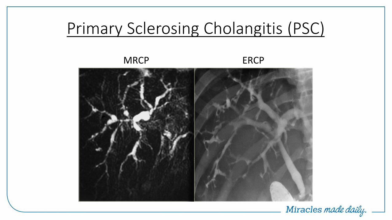
Primary Sclerosing Cholangitis (PSC)
MRCP ERCP
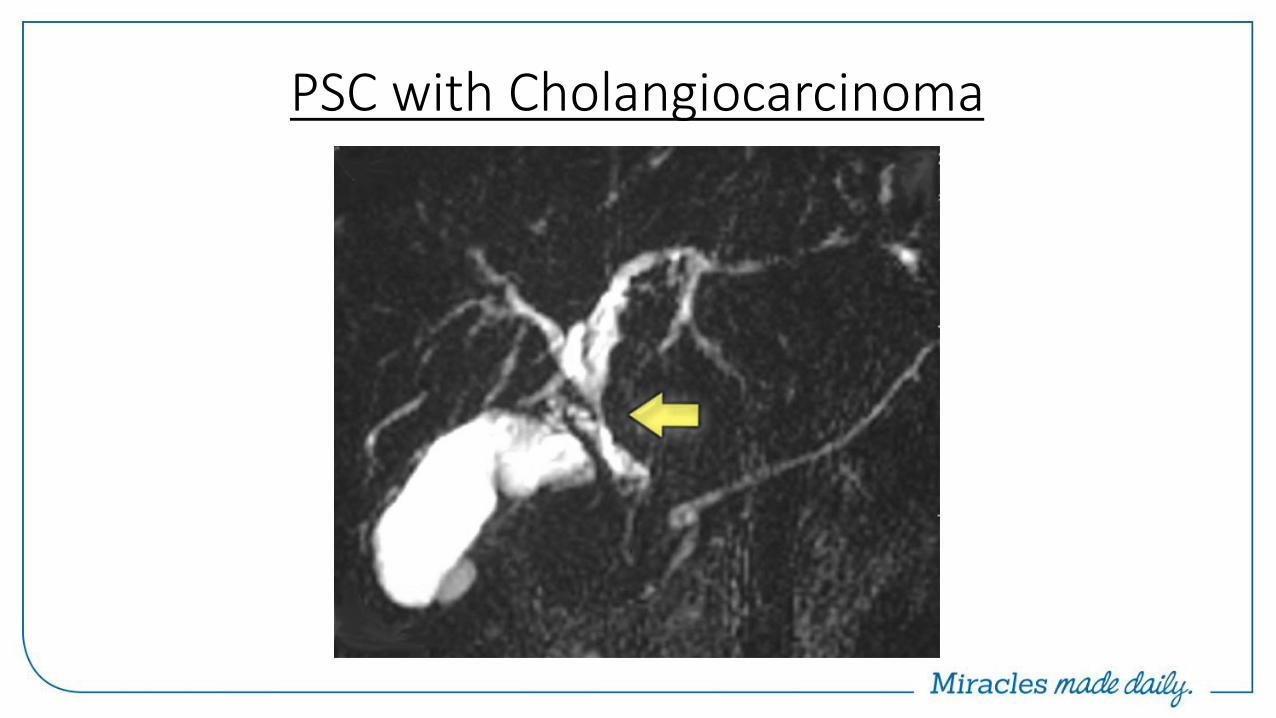
PSC with Cholangiocarcinoma
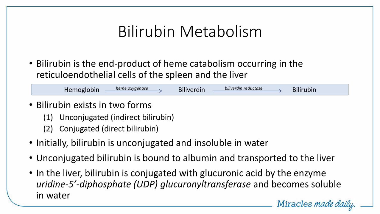
Bilirubin Metabolism
• Bilirubin is the end-product of heme catabolism occurring in the reticuloendothelial cells of the spleen and the liver
• Bilirubin exists in two forms (1) Unconjugated (indirect bilirubin)
(2) Conjugated (direct bilirubin)
• Initially, bilirubin is unconjugated and insoluble in water
• Unconjugated bilirubin is bound to albumin and transported to the liver
• In the liver, bilirubin is conjugated with glucuronic acid by the enzyme uridine-5’-diphosphate (UDP) glucuronyltransferase and becomes soluble in water
Hemoglobin heme oxygenase Biliverdin biliverdin reductase Bilirubin
Bilirubin Metabolism
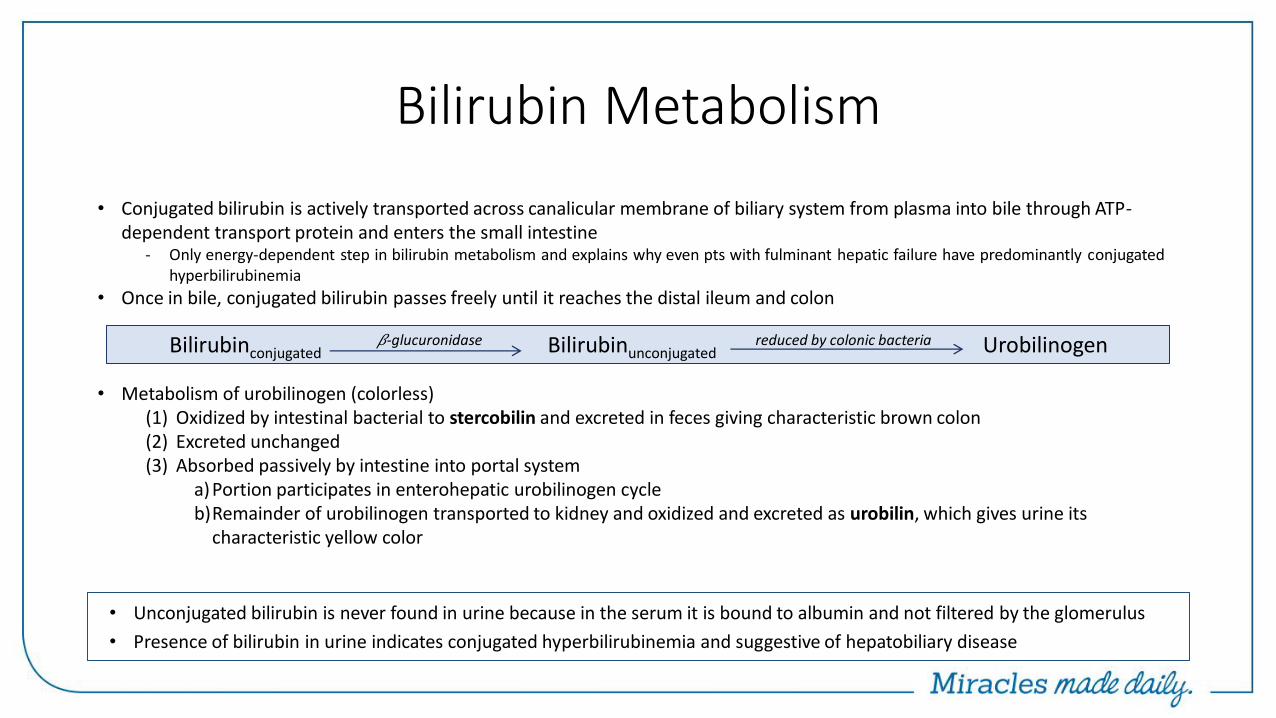
• Conjugated bilirubin is actively transported across canalicular membrane of biliary system from plasma into bile through ATP-dependent transport protein and enters the small intestine
- Only energy-dependent step in bilirubin metabolism and explains why even pts with fulminant hepatic failure have predominantly conjugated hyperbilirubinemia
• Once in bile, conjugated bilirubin passes freely until it reaches the distal ileum and colon
• Metabolism of urobilinogen (colorless) (1) Oxidized by intestinal bacterial to stercobilin and excreted in feces giving characteristic brown colon (2) Excreted unchanged (3) Absorbed passively by intestine into portal system
a)Portion participates in enterohepatic urobilinogen cycle b)Remainder of urobilinogen transported to kidney and oxidized and excreted as urobilin, which gives urine its
characteristic yellow color
Bilirubinconjugated 𝛽-glucuronidase Bilirubinunconjugated reduced by colonic bacteria Urobilinogen
• Unconjugated bilirubin is never found in urine because in the serum it is bound to albumin and not filtered by the glomerulus
• Presence of bilirubin in urine indicates conjugated hyperbilirubinemia and suggestive of hepatobiliary disease
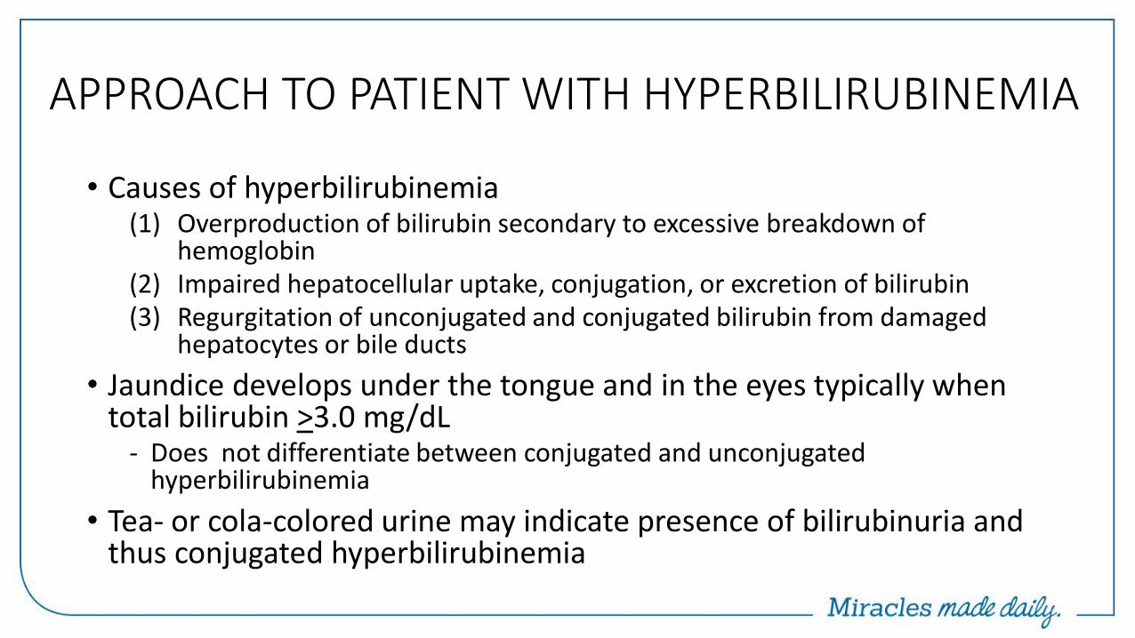
APPROACH TO PATIENT WITH HYPERBILIRUBINEMIA
• Causes of hyperbilirubinemia (1) Overproduction of bilirubin secondary to excessive breakdown of
hemoglobin (2) Impaired hepatocellular uptake, conjugation, or excretion of bilirubin (3) Regurgitation of unconjugated and conjugated bilirubin from damaged
hepatocytes or bile ducts
• Jaundice develops under the tongue and in the eyes typically when total bilirubin >3.0 mg/dL
- Does not differentiate between conjugated and unconjugated hyperbilirubinemia
• Tea- or cola-colored urine may indicate presence of bilirubinuria and thus conjugated hyperbilirubinemia
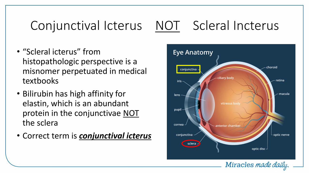
Conjunctival Icterus NOT Scleral Incterus
• “Scleral icterus” from histopathologic perspective is a misnomer perpetuated in medical textbooks
• Bilirubin has high affinity for elastin, which is an abundant protein in the conjunctivae NOT the sclera
• Correct term is conjunctival icterus
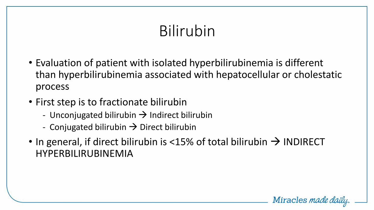
Bilirubin
• Evaluation of patient with isolated hyperbilirubinemia is different than hyperbilirubinemia associated with hepatocellular or cholestatic process
• First step is to fractionate bilirubin - Unconjugated bilirubin Indirect bilirubin
- Conjugated bilirubin Direct bilirubin
• In general, if direct bilirubin is <15% of total bilirubin INDIRECT HYPERBILIRUBINEMIA
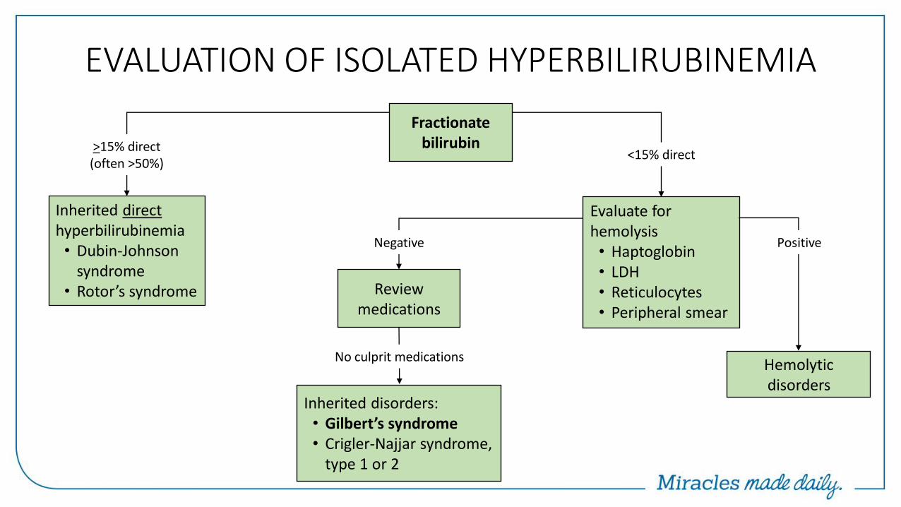
EVALUATION OF ISOLATED HYPERBILIRUBINEMIA
Fractionate bilirubin
Inherited direct hyperbilirubinemia • Dubin-Johnson
syndrome • Rotor’s syndrome
>15% direct (often >50%)
<15% direct
Evaluate for hemolysis • Haptoglobin • LDH • Reticulocytes • Peripheral smear
Review medications
Negative Positive
Inherited disorders: • Gilbert’s syndrome • Crigler-Najjar syndrome,
type 1 or 2
No culprit medications Hemolytic disorders
Albumin
• Protein that is produced exclusively in the liver
• Multiple biological actions - Maintenance of oncotic pressure
- Binding of other substances (unconjugated bilirubin, fatty acids, thyroid hormones, and drugs)
- Metabolism of compounds
- Antioxidant properties
• Normal serum values for albumin 3.5 – 4.5 g/dL
Albumin
• Long half-life of 15-20 days; thus, changes are not good indicator of acute liver dysfunction
• Hypoalbuminemia is more common in chronic liver disorders, e.g. cirrhosis, due to reduced albumin synthesis
• Serum albumin level <3 g/dL associated with liver disease should raise suspicious for chronic liver disease +/- cirrhosis
• Although serum albumin concentration is considered marker of synthetic function of the liver, albumin concentrations may be reduced in many clinical situations
- Nephrotic syndrome - Malabsorption - Protein-losing enteropathies - SIRS associated with chronic infections
Prothrombin time (PT) and INR
• Liver is major site of synthesis of all major blood coagulation proteins – Factors I, II, V, VII, IX, X, XII, XIII o Exception Factor VIII is produced by vascular endothelial cells
• Blood clotting parameters used to measure liver function • In significant liver injury, results in reduction in clotting factor production
and subsequent coagulopathy ↑ PT/INR • Prolonged PT not specific to liver disease; may result from congenital or
acquired conditions - Congenital deficiency of clotting factors - Consumption of clotting factors [disseminated intravascular coagulation (DIC) or
severe GI bleeding] - Certain medications (coumadin) - Vitamin K deficiency
Prothrombin time (PT) and INR
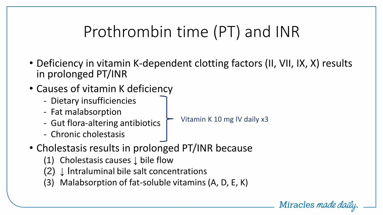
• Deficiency in vitamin K-dependent clotting factors (II, VII, IX, X) results in prolonged PT/INR
• Causes of vitamin K deficiency - Dietary insufficiencies - Fat malabsorption - Gut flora-altering antibiotics - Chronic cholestasis
• Cholestasis results in prolonged PT/INR because (1) Cholestasis causes ↓ bile flow (2) ↓ Intraluminal bile salt concentrations (3) Malabsorption of fat-soluble vitamins (A, D, E, K)
Vitamin K 10 mg IV daily x3
Prothrombin time (PT) and INR
• International normalized ratio (INR) devised and validated to standardize PT across laboratories in pts receiving anticoagulation therapy with vitamin K antagonists (e.g. Coumadin)
• INR is not accurate measure of bleeding risk in patients with cirrhosis; prolonged PT/INR does not accurately predict risk of GI bleeding
• Likewise, prolonged INR does not provide a protective role in development of DVT or PVT
Hematologic Disorders in Cirrhosis
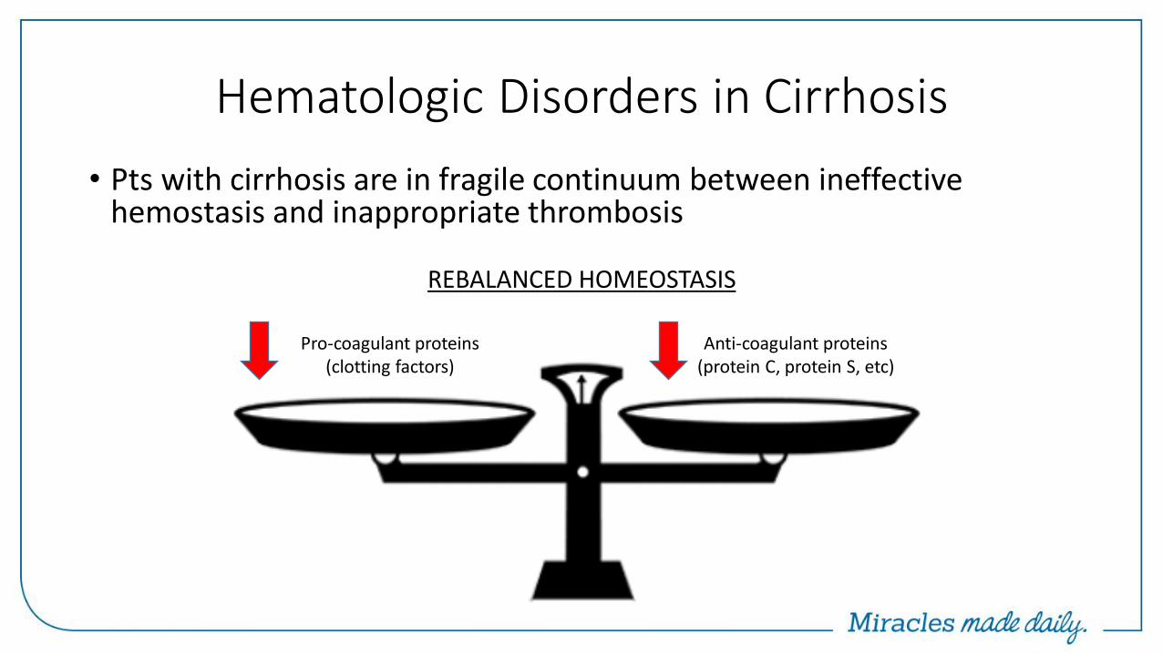
• Pts with cirrhosis are in fragile continuum between ineffective hemostasis and inappropriate thrombosis
Pro-coagulant proteins (clotting factors)
Anti-coagulant proteins (protein C, protein S, etc)
REBALANCED HOMEOSTASIS
Prognostic Markers of PT/INR
• Unlike albumin, PT is helpful is assessing acute liver dysfunction
• PT/INR has a high prognostic value and is included in multiple prognostication models
(1) King’s College criteria (acute liver failure) (2) Maddrey’s discriminant function (alcoholic hepatitis) (3) Child-Turcotte-Pugh score (4) Model for End-stage Liver Disease (MELD) score
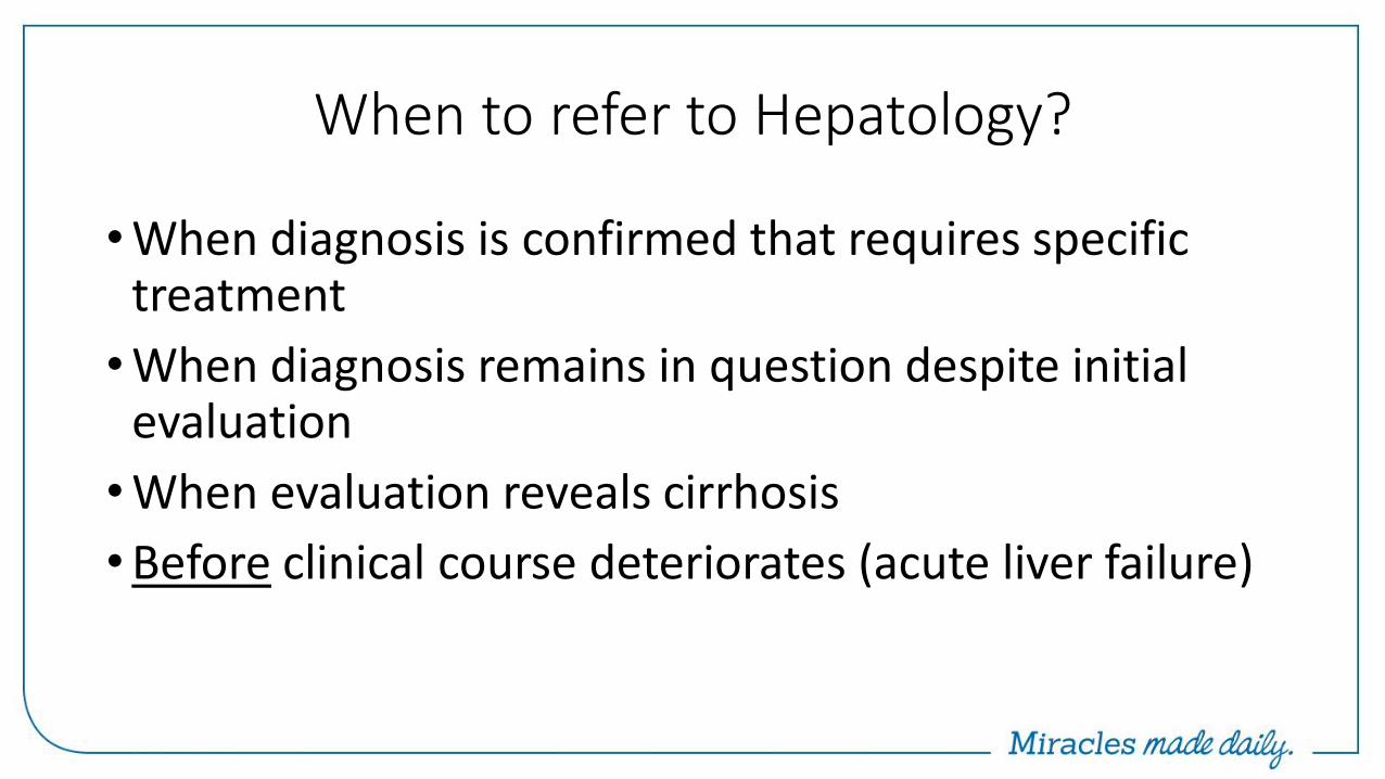
When to refer to Hepatology?
• When diagnosis is confirmed that requires specific treatment
• When diagnosis remains in question despite initial evaluation
• When evaluation reveals cirrhosis
• Before clinical course deteriorates (acute liver failure)
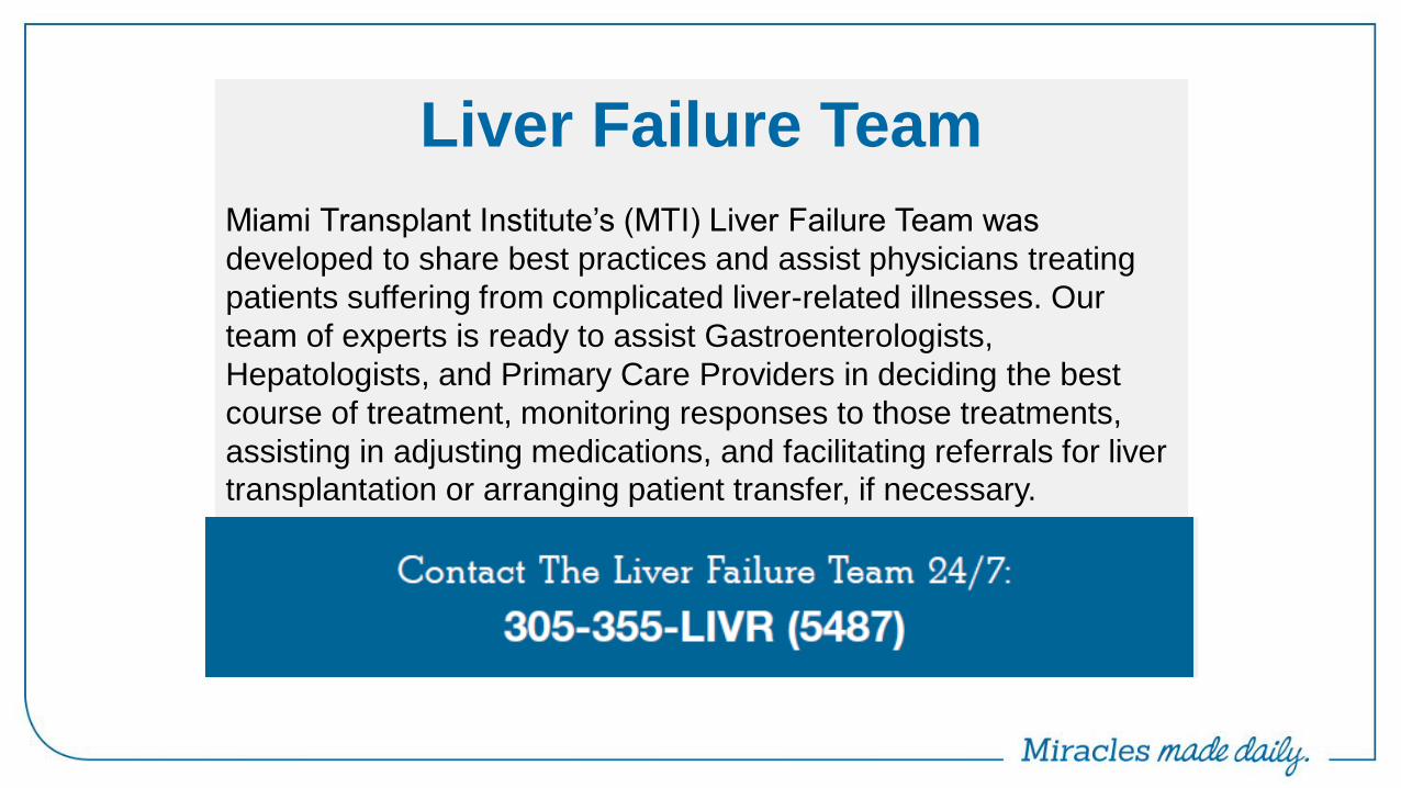
Liver Failure Team
Miami Transplant Institute’s (MTI) Liver Failure Team was
developed to share best practices and assist physicians treating
patients suffering from complicated liver-related illnesses. Our
team of experts is ready to assist Gastroenterologists,
Hepatologists, and Primary Care Providers in deciding the best
course of treatment, monitoring responses to those treatments,
assisting in adjusting medications, and facilitating referrals for liver
transplantation or arranging patient transfer, if necessary.
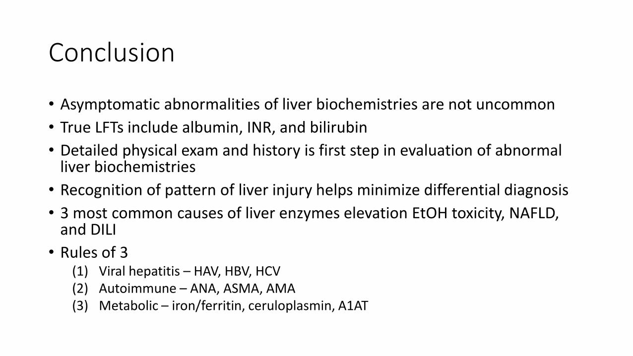
Conclusion
• Asymptomatic abnormalities of liver biochemistries are not uncommon
• True LFTs include albumin, INR, and bilirubin
• Detailed physical exam and history is first step in evaluation of abnormal liver biochemistries
• Recognition of pattern of liver injury helps minimize differential diagnosis
• 3 most common causes of liver enzymes elevation EtOH toxicity, NAFLD, and DILI
• Rules of 3 (1) Viral hepatitis – HAV, HBV, HCV (2) Autoimmune – ANA, ASMA, AMA (3) Metabolic – iron/ferritin, ceruloplasmin, A1AT