electrochemistry and electrogenerated chemiluminescence of...
TRANSCRIPT

REVIEW
Electrochemistry and electrogenerated chemiluminescenceof organic nanoparticles
Jungdon Suk & Allen J. Bard
Received: 31 March 2011 /Revised: 26 May 2011 /Accepted: 27 May 2011 /Published online: 12 July 2011# Springer-Verlag 2011
Abstract This review discusses briefly the preparation,electrochemistry, and electrogenerated chemiluminescence(ECL) as well as spectroscopic properties of organicnanoparticles. Organic nanoparticles, ranging from severaltens of nanometers to hundreds of nanometers in diameter,were successfully prepared by various methods. Using asimple reprecipitation method, organic nanoparticles of avery small size can be prepared and show uniqueelectrochemical and ECL characteristics. As with inorganicnanoparticles, organic nanoparticles suggest possible appli-cations, like labels for the analysis of biological materialswith ECL.
Keywords Electrochemistry . Electrogeneratedchemiluminescence . Organic nanoparticles
Introduction
Nanoparticles are particles in the nanometer-size range, andthose of a size where the properties differ from those of thebulk material are of special interest. Metal and semicon-ductor nanoparticles have been extensively studied andoften show new physical phenomena, distinct from the bulkmaterial, such as quantum confinement or finite size effects[1–4]. This can affect the thermal, electrical, and electronicproperties. For example, the band gap (or color) ofsemiconductor nanoparticles in the few nanometer diameterregion is a function of size and is different from that of the
bulk material [5–7]. When the size of a semiconductorparticle is reduced to around a 1- to 10-nm radius, theexcitons (bound electron–hole pairs) are confined withinthe particle. This phenomenon, called the size confinementeffect, has been studied by optical spectroscopy [8–12] andscanning tunneling spectroscopy [13].
As discussed below, nanoparticles of organic materialscan also be synthesized and are of interest. In contrast to theextensive research that has been done on metal andinorganic semiconductor nanoparticles, the study of organicnanoparticles is still at a very early stage. The fundamentalprinciples of the properties of organic nanoparticles are notyet well established. For many organic species, a size effectis not expected because electrons are not delocalized overnanometer domains. However, organic nanoparticles haverecently received attention because of the huge range ofdifferent molecular structures, the flexibility in materialsynthesis and preparation, and the ability to tailor theirbinding affinity toward various materials [14–16]. Inapplications, organic nanoparticles are used as advancedmaterials for display elements, inks, toners, drugs, andcosmetics.
As a complement to spectroscopic methods, electro-chemical ones can provide valuable insight into the size-dependent quantization effect with metal and semiconduc-tor nanoparticles. For example, the electrochemistry of goldnanoparticles as a function of particle size has showndiscrete single electron charging behavior [17–19], andthere have been many studies of the electrocatalyticproperties of metal nanoparticles, including those at thesingle particle level [20]. For semiconductor particles, whenthe electrochemically reduced or oxidized nanoparticles arestable, they can react with sufficient energy to form anemitting excited state and electrogenerated chemilumines-cence (ECL) occurs. ECL has many advantages as an
J. Suk :A. J. Bard (*)Center for Electrochemistry, Department of Chemistry andBiochemistry, The University of Texas at Austin,Austin, TX 78712, USAe-mail: [email protected]
J Solid State Electrochem (2011) 15:2279–2291DOI 10.1007/s10008-011-1449-x

analytical technique compared to fluorescence, such as highsensitivity and low background. ECL has been extensivelystudied for organic molecules [21–23] and several semi-conductor nanoparticles [24–32]. Electrochemical and ECLstudies of organic nanoparticles have only recently started.A challenge in research with organic nanoparticles iscontrolling the particle size and shape in the same way asone can with metals and semiconductors and understandinghow electrochemical and optical properties depend on sizeand shape. Some studies show particle-size dependence ofoptical constants of molecular crystals [33–35]. As dis-cussed below, a challenge in electrochemical studies is thelarge size of nanoparticles currently available, resulting insmall diffusion coefficients and the low concentration inaqueous solutions.
The article is arranged as follows: In “Preparation of organicnanoparticles” section, we discuss various techniques tosynthesize organic nanoparticles. In “Electrochemistry andelectrogenerated chemiluminescence of organic nanoparticles”section, we consider electrochemical and ECL characteristicsof organic nanoparticles. These electrochemical properties oforganic nanoparticles suggest possible applications. Finally,spectroscopic properties of organic nanoparticles areaddressed.
Preparation of organic nanoparticles
While a large number of different approaches for thefabrication of semiconductor and metal nanoparticles havebeen developed, only a few techniques have been reportedfor the preparation of organic nanoparticles. The top-downtechnique of preparing nanoparticles is difficult to apply toorganic materials because of their lower melting temper-atures, thermal instability, and poor mechanical properties.
One approach to the preparation of sub-micrometerparticles is by the mechanical milling method. The millingmethod is a simple and well-understood process. The sizeof particles obtained depends on impact and shear forces.This process has several limitations; for example, it is notappropriate for explosive, low-melting, or temperature-sensitive compounds [36]. Moreover, the processing timenecessary to approach the target size can be long, and themilling agent can introduce impurities, discoloration, andfractionation. Because of aggregation during the millingprocess, it is difficult with this method to produce uniformorganic nanoparticles having diameters smaller than300 nm [37].
To obtain smaller nanoparticles, several methods havebeen developed, such as solvent deposition [38–42], laserablation [43–45], sol–gel phase transition [46], and repre-cipitation methods [47–51]. In the deposition method,organic molecules are evaporated in a vacuum chamber,
and nanoscale organic aggregates are deposited on asubstrate. This technique allows production of largevolumes of concentrated (several milligrams per milliliter)organic nanoparticles in a single step. However, it also hasdrawbacks such as broad size distribution and deformationof crystal structure due to a mechanothermal effect [39, 40].Tamaki et al. first applied the laser ablation technique toprepare organic nanoparticles from phthalocyanines andaromatic hydrocarbons [52]. These organic crystals absorblaser light, leading to a local increase in temperature andevaporation of a small fraction of material from the crystalsurface [53]. Moreover, the high photon density of the laserinduces surface fragmentation or cracking of the materials.A surrounding liquid helps to cool the vaporized materialsrapidly and to form nanoparticles. Recently, differentphthalocyanine [54], tetracene, and pentacene [55, 56]nanoparticles formed by the laser ablation technique werereported. The laser ablation technique makes more concen-trated nanoparticle dispersions than other techniques andprovides easy control of size and molecular packing bychanging laser wavelength, pulse duration, fluence, repeti-tion rate, irradiation period, solvent, and temperature [44,45]. A limitation of this method is the requirement of highphoton density and a narrow laser beam. Moreover, theintense laser light may cause severe photochemical damagein many organic materials.
In 1992, Kasai et al. proposed the reprecipitation methodand demonstrated that nanoparticles smaller than 100 nmdispersed in water could be prepared [48]. Among the manypreparation methods, this bottom-up type of technique hasbeen widely used in nanoparticle preparation for variouskinds of molecules, for example, π-conjugated organicnanocrystals [57–59], polydiacetylene derivatives [34, 60],low molecular weight aromatic compounds [61, 62], andorganic functional chromophores [63–65]. The usualreprecipitation method is shown in Fig. 1. First, a targetcompound is dissolved at a concentration on the order ofmillimolars in a “good solvent” like tetrahydrofuran (THF)or alcohol (in which the compound is highly soluble). Next,a small volume (a few microliters) of this solution isinjected rapidly at constant temperature into a much largervolume (~10 mL) of a vigorously stirred “poor solvent”such as water (in which the target compound is less solubleor insoluble). Smaller nanoparticles can be produced bylowering the concentration of the target compound and byreducing the volume of the injected solution. Ideally, thewater should be warm (60°C), but also just below theboiling point of the good solvent, in order to make smallernanoparticles. The key to this method is a large differencein solubilities of the target compound in the good solventand the poor solvent and good miscibility of the twosolvents. The rapid mixing of the two solvents produces amicro-environment for the target compound molecules,
2280 J Solid State Electrochem (2011) 15:2279–2291

leading to supersaturation, precipitation, and crystallizationof the dispersed nanoparticles.
Unlike other preparation methods, with this process onecan easily control particle size from several tens ofnanometers to several micrometers. There are severalvariables useful in controlling the crystal size and shapein the reprecipitation process, e.g., stirring rate, injectionrate, concentration of parent solution, injection volume, andtemperature of the poor solvent [49]. It is, however, usuallynot possible with injections into water to produce particlessmaller than about 20 nm. Additives, such as polymers orsurfactants, can promote the formation of smaller particles.The nanoparticles formed this way can be stable from daysto months. However, the presence of surfactants can have anegative effect on electrochemical reactions and thegeneration of ECL because the encapsulation of a particlewithin a layer of surfactant decreases the electron transferrate.
Alternatively, water-soluble organic compounds such as4′-dimethylamino-N-methylstilbazolum p-toluenesulfonate(DAST) nanoparticles are fabricated by the inverse repre-cipitation method. Nanoparticles are prepared by injectingan ethanol solution of DAST into vigorously stirred decalinat room temperature. The crystal size in this case variedfrom about 200 to 600 nm [66, 67].
We fabricated nanoparticles dispersed in acetonitrile(MeCN) by a reprecipitation method (Suk et al.,submitted for publication). Unlike many aromatic com-pounds, the 9-naphthylanthracene-based dimer synthe-sized by Wang et al. [68] is not soluble in MeCN, but issoluble in dichloromethane and THF. MeCN is one of thepreferred solvents for electrochemical studies and ECLbecause of its wide electrochemical window. Nanoparticleswere fabricated by injecting a THF solution of the 9-naphthylanthracene-based dimer into vigorously stirredMeCN (10 mL) at room temperature to produce particlesas small as 16 nm. The electrochemical and ECL resultsare discussed below.
Electrochemistry and electrogeneratedchemiluminescence of organic nanoparticles
ECL is a type of luminescence that involves the generation ofoxidized and reduced species, often radical ions, at electrodesurfaces that undergo a fast electron transfer reaction toproduce an excited state [22, 69–71]. It is desirable in ECL tohave stable and long-lived radical ions with apparent highphotoluminescence (PL) quantum yield to obtain strong andbright ECL. Developing such highly efficient and stable ECLemitters over a broad spectrum of wavelength has been ofinterest for many years, particularly for application as anECL label for analytical purposes, for example, for DNAdetermination and immunoassay [72, 73]. ECL has beenextensively studied for organic molecules [22–24, 74] and afew semiconductor nanoparticles [25–28]. In commercialapplications, the ECL label has largely been Ru(bpy)3
2+
because of its good solubility in water and high ECLefficiency. One driving force in the study of organicnanoparticles, which can also be used in aqueous solution,is the possibility that, if optimized, they may find applicationin practical assays. The electrochemical and emissionproperties of organic nanoparticles have been the subject ofrelatively few studies compared to inorganic nanoparticles.Organic molecules have the advantage of being easilyfunctionalized by versatile synthetic strategies, possiblytailoring the electronic and optical properties of organicnanoparticles [75]. Organic nanoparticles have the potentialfor many practical applications by expanding the broadspectrum of wavelengths available in ECL. ECL can begenerated by annihilation (electron transfer between) theparent radical ions or by reaction of the radical with acoreactant. A coreactant is a compound that can produce astrong oxidizing or reducing agent using a reaction thatfollows the electrochemical electron transfer reaction. Thecoreactant must be energetic enough to oxidize or reduceradical ions of luminophores to produce their excited states.Typical coreactants for oxidation are tri-n-propylamine
Fig. 1 Scheme of thereprecipitation method
J Solid State Electrochem (2011) 15:2279–2291 2281

(TPrA) and oxalate ion (C2O42−) and those for reduction are
benzoyl peroxide and peroxydisulfate (S2O82−). Each cor-
eactant has different potentials to produce oxidant orreductant. Depending on the electrochemical properties ofthe studied compound in organic solvent, a suitablecoreactant can be chosen to oxidize or reduce the morestable radical ion.
Conjugated polymer nanoparticles
Organic conjugated molecules are highly fluorescentmaterials and are used for photo- or electroluminescentdevices [76], e.g., light-emitting displays [77–79] andphotovoltaics [80–83]. They are easily synthesized andcan be polyfunctionalized, which allow their optical,electronic, and chemical properties to be tailored [84–86].The first reports of colloidal dispersions of nanoparticles ofconjugated polymers appeared in the 1980s, motivated bythe difficult processing of conjugated polymers due tointractability and insolubility in organic solvents [87].Conjugated polymer nanoparticles were studied in thepreparation of nanoscale multiphase films for photovoltaicsor for biomolecule labeling and sensing due to the highintensity and photostability of fluorescent nanoparticles ascompared to conventional dyes [88, 89]. Our group hasstudied the electrochemical oxidation [90] and ECL [91] ofnanoparticles of a conjugated copolymer, poly(9,9-dioctyl-fluorene-co-benzothiadiazole) (F8BT; Fig. 2). By the
reprecipitation method, produced F8BT nanoparticles witha size of 25±15 nm. F8BT nanoparticles were immobilizedon an ITO working electrode in an electrochemical cell(Fig. 3a) with a gold counter electrode and a silverquasireference electrode. F8BT showed electrochemicallyirreversible oxidation (Fig. 3b). As shown in Fig. 3c, ECLemission was produced when the potential was scannedfrom 0 to + 1.8 V to oxidize both F8BT nanoparticles andthe coreactant, TPrA, which generates a strong reducingagent. The nanoparticle luminescence intensity increasedsharply as the potential reached +1.8 V. This was the firstECL observed result from sub-25 nm single nanoparticles[92]. Studies of the electrochemistry of single immobilizedparticles were tracked by observing the loss of fluorescenceon oxidation [91].
Aromatic hydrocarbon nanoparticles
Aromatic hydrocarbons have been widely used in ECL andsolid-state electroluminescence studies. Several new anthra-cene derivatives have been developed and studied to tunetheir photophysical properties, so that new derivatives showgood color purity, high efficiency, and good stability. ECLfrom nanoparticles of aromatic hydrocarbon compoundssuch as rubrene, 9,10-diphenylanthracene (DPA), and 9-naphthylanthracene-based dimer (NA) have been reportedin an aqueous solution. Rubrene, DPA [92], and NA (Suk etal., submitted for publication) compounds (Fig. 2) dissolved
Fig. 2 Formulas of organicnanoparticles used in ECLstudies
2282 J Solid State Electrochem (2011) 15:2279–2291

in organic solvents have been widely studied electrochem-ically, spectroscopically, and by ECL. They show goodreversibility in electrochemistry and ECL by an annihilationreaction in organic solvent produces strong light.
Figure 4 shows the TEM image of rubrene nanoparticlesand the SEM image of DPA nanoparticles after they weredispersed in water. Both nanoparticles in aqueous solution
were prepared by a simple reprecipitation method, in whicha small amount of an organic compound in a good solventsuch as THF and MeCN was injected rapidly and stirred
Fig. 4 a TEM image of freshly prepared rubrene NPs dispersed inwater. b SEM image of DPA NPs (fresh sample). c TEM image ofnanowires of DPA (after 1-week aging in solution) [99]
Fig. 3 a Schematic diagram of single molecule spectroelectrochem-istry (SMS-EC) cell. CE counter electrode, QRE quasireferenceelectrode, WE working electrode. b Cyclic voltammogram of F8BTin Nafion thin film on ITO in 0.2 M LiClO4/acetonitrile solution atpotential scan rate of 0.1 V/s. Right inset cyclic voltammogram of aNafion film showing very small background currents over the scannedrange. c Ensemble average of single nanoparticle ECL intensity (blackcurve) with 0.1 M TPrA and its corresponding voltammogram (redcurve) [90, 91]
J Solid State Electrochem (2011) 15:2279–2291 2283

vigorously in a poor solvent, water. The sizes of rubrenenanoparticles determined from TEM images and dynamiclight scattering measurement were ~50 nm. In the dark,rubrene nanoparticles were stable for a month without anysurfactants. However, DPA nanoparticles were initiallyformed as nanorods (50 nm in diameter and 500 nm inlength, Fig. 4b) that converted into large nanowires, about1 μm in diameter and 10 μm in length (Fig. 4c). Althoughsurfactants such as Triton X-100 and cetyltrimethyl ammo-nium bromide helped to decrease the size and avoid theformation of aggregates because of the encapsulation by thesurfactants, electron transfer to the particles was hinderedand this limited the generation of ECL.
Because of the limited potential window in aqueoussolution and low concentration of nanoparticles, thedistinctive features that could be clearly assigned to eitherthe oxidation or reduction of the nanoparticles themselves(rather than the coreactant, TPrA) could not be distin-guished. Moreover, the small diffusion coefficients of theserather large nanoparticles also result in small electrochem-ical signals. However, emission that can be attributed to theECL can be observed by reaction with TPrA (Fig. 5a).Nanoparticles in the presence of a coreactant such as TPrA[93–97] or oxalate ion showed redox behavior and strongECL light. As shown in Fig. 5a, the ECL emission was
produced when the potential was scanned from 0 to +1.2 Vto oxidize both rubrene nanoparticles and TPrA, whichgenerates a strong reducing agent. Transient ECL generatedfrom the reaction of oxidized nanoparticles and theenergetic intermediates of TPrA was obtained by steppingthe electrode potential with different pulse widths from 0.0to +1.1 V versus Ag/AgCl (Fig. 5b). ECL emission wasproduced when the electrode potential was stepped to+1.1 V to oxidize both nanoparticles and TPrA, and noECL was seen when the potential was stepped back to0.0 V. The ECL signal intensity was not stable with timebecause of the instability of the oxidized nanoparticles inwater. In the case of rubrene nanoparticles prepared fromdimethylformamide (DMF), the ECL intensity was weaker;this can be attributed to the bigger particles producedbecause of some miscibility of the solvents with waterduring the reprecipitation process [98]. The ECL intensitywas not strong enough to record the ECL spectrum with aCCD camera. The low concentration and diffusion coeffi-cient of the nanoparticles and possible reactions betweenreducing agent and water or oxygen could explain the verylow intensity of the ECL signal [99].
In the case of DPA nanoparticles, transient ECLgenerated from the reaction of oxidized nanoparticles andthe energetic intermediate, CO2
•−, from oxalate ion oxida-tion was obtained by stepping the electrode potential withdifferent pulse widths from 0.0 to +1.5 V versus Ag/AgCl(Fig. 6). TPrA is not energetic enough to generate theexcited state of DPA, so a stronger reducing agent (CO2
•−)was used to oxidize the DPA nanoparticles [100]. Due tothe very small diffusion coefficients of the nanoparticles,the ECL intensity was weak.
The 9-naphthylanthracene-based dimer (NA, Fig. 2)synthesized by Wang’s group has poor solubility in organicsolvents such as MeCN, benzene, DMF and dimethylsulfoxide. Among them, MeCN is a preferred solvent inECL studies because of its wide electrochemical window toobserve electrochemically generated radical cations andanions. NA nanoparticles can be prepared by a reprecipi-tation method, in which a small amount of NA in a goodsolvent (THF) is injected rapidly and stirred vigorously into
Fig. 6 Transient ECL experiment, electrochemical current (black line)and ECL intensity (red line) for DPA NPs (prepared from THF) inaqueous 0.1 M NaClO4 with 0.1 M Na2C2O4, sampling time 1 ms,pulsing pattern 0 to 1.5 V, pulse width is 0.1 s. Reprinted from [99]
a bFig. 5 a Cyclic voltammogramof rubrene NPs (preparedfrom THF) in aqueous 0.1 MNaClO4 with 0.1 M TPrA at ascan rate of 500 mV/s. bChronoamperometry (black line)and ECL transient (red line) ofrubrene NPs, pulse width 0.1 s.WE Pt disk, CE Pt coil, REAg/AgCl. Reprinted from [99]
2284 J Solid State Electrochem (2011) 15:2279–2291

a poor solvent (either water or MeCN). Figure 7 shows theTEM image of NA nanoparticles dispersed in both water andMeCN. Because of the different miscibility of THF withwater and with MeCN, the size of NA nanoparticles obtainedwas different. NA nanoparticles prepared in water aresignificantly larger, ~40 nm, and had a less spherical shapethan those in MeCN. They were stable for only a few days.On the other hand, NA nanoparticles dispersed in MeCNproduced spherical, small, and well-dispersed nanoparticles.
CVs of the NA nanoparticles dispersed in aqueous 0.1 MNaClO4 displayed no distinctive peaks, and no ECLemission was observed because of the limited potentialwindow in water, low concentration of nanoparticles andsmall diffusion coefficients (due to their large radii).However, CVs of the NA nanoparticles dispersed in MeCNare different. There is a distinctive peak at around −1.8 V,which is similar to the potential region where reduction ofthe NA molecule in THF occurs. Weak but noticeable ECL
a bFig. 8 a Cyclic voltammogram(black line) and ECLintensity (red line) of NAnanoparticles in water with0.1 M NaClO4/S2O8
2− at scanrate of 50 mV/s and b transientECL experiment, electrochemi-cal current (black line) andECL intensity (red line) forNA nanoparticles in water.Sampling time 1 ms, pulsingpattern 0 to −1.8 V. Pulse widthis 2 s (Suk et al., in preparation)
a b
c d
Fig. 7 a TEM Image oforganic nanoparticles of NAdispersed in water and bhistogram of size distributionof NA nanoparticles.The average size of the NAnanoparticles was 40±15 nm.c TEM image of organicnanoparticles of NA dispersedin MeCN and d histogram ofsize distribution of NAnanoparticles. The average sizeof NA nanoparticles was 15±6 nm (Suk et al., in preparation)
J Solid State Electrochem (2011) 15:2279–2291 2285

emission from the annihilation reaction was produced whenthe potential was scanned first toward negative and thenpositive potentials.
We can get information about the redox behavior and theECL emission with a coreactant in both water and MeCN.In the case of NA nanoparticles dispersed in water, a muchstronger ECL signal was observed (Fig. 8a) when thepotential was scanned from 0 to −1.8 V in 0.1 M NaClO4
containing 0.1 M K2S2O8, (a coreactant which forms astrong oxidizing agent, SO4
•−) (Ered≥3.15 V vs. SCE)[101]. Moreover, strong ECL emission was produced bypulsing between 0 and −1.8 V. The intensity was stable withtime but not strong enough to obtain an ECL spectrum(Fig. 8b). A strong ECL signal was produced with NAnanoparticles dispersed in MeCN when the potential wasscanned from 0 to −1.8 V in 0.1 M MeCN containing 0.1 MS2O8
− as a coreactant (Fig. 9a). As shown in Fig. 9b, strongECL emission was produced when the electrode potentialwas stepped to −1.7 V to reduce both nanoparticles andS2O8
−. The increasing ECL signal on cycling perhapsindicates adsorption with formation of an emitting film,although there is no evidence of electrode fouling. The ECLsignal was strong enough to obtain an ECL spectrum(Fig. 10, red line). The ECL spectrum of nanoparticles
dispersed in MeCN showed a blue emission peak atwavelength of ~430 nm, which is close to the ECL peakof NA molecules dissolved in THF (Fig. 8, black line).
Nanoparticles of C2-symmetric donor–acceptor compounds
ECL from nanoparticles of the recently synthesizedfluorescent molecules spiro-BTA [102] and azide-BTA(Suk et al., manuscript submitted) (Fig. 2) has also beenobserved. Both compounds are donor–acceptor (DA)compounds that show a single reversible reduction wave(Eo
red ¼ �1:48 V vs. SCE) and two reversible oxidationwaves (Eo
1; ox ¼ 0:9 V, Eo2; ox ¼ 1:34V vs. SCE) and pro-
duce red emission with a high PL quantum yield. Therehave been a number of studies of the electrochemical,spectroscopic, and ECL behavior of DA molecules insolution [103–108].
Spiro-BTA is a C2-symmetric compound, consisting ofspirobifluorene as a π-bridge core connected to two 2,1,3-benzothiadiazole (BTA) units, the acceptors (A), and end-capped with triphenylamines as the donors (D), with anoverall structure D–A–π–A–D. A reprecipitation methodwas used to prepare well-dispersed organic nanoparticlesof the spiro-BTA in aqueous solution. Spiro-BTA nano-particles were synthesized by injecting 100 μL of spiro-BTA in THF into 10 mL of deionized water undersonication at room temperature. Figure 11 shows the
Fig. 11 SEM image of organic nanoparticles of Spiro-BTA preparedin water. Scale marker is 300 nm [102]
Fig. 10 ECL spectra of NA molecules in THF with 0.1 M TBAPF6/S2O8
2- (black line) and NA nanoparticles in MeCN with 0.1 MTBAPF6/S2O8
2- (red line) [Suk et al., submitted for publication]
a bFig. 9 a Cyclicvoltammogram (black line) andECL intensity (red line) of NAnanoparticles in MeCN with0.1 M TBAPF6/S2O8
2− at scanrate of 100 mV/s and b transientECL experiment, electrochemi-cal current (black line) and ECLintensity (red line) for NAnanoparticles in MeCN with0.1 M TBAPF6/S2O8
2−.Sampling time 1 ms, pulsingpattern 0 to −1.7 V. Pulse widthis 1 s (Suk et al., in preparation)
2286 J Solid State Electrochem (2011) 15:2279–2291

SEM image of fresh spiro-BTA nanoparticles dispersed inwater with an average diameter of ~130 nm. The particlesize was stable for 1 week under ambient conditionswithout any surfactants.
Like other aromatic compound nanoparticles dispersedin water, there were no distinctive features that could beclearly assigned to either oxidation or reduction of nano-particles because of the limited potential window inaqueous solutions. No ECL emission from an annihilationreaction was produced. However, with a coreactant, TPrA,ECL was obtained from well-dispersed spiro-BTA nano-particles in an aqueous medium, as shown in Fig. 12. TheECL emission was produced when the electrode potentialwas stepped to +1.2 V to oxidize both nanoparticles andTPrA, and no ECL was produced when the electrodepotential was stepped back to 0.0 V. The ECL signal wasmoderate and increased, like the result of NA nanoparticlesdispersed in MeCN with S2O8
−. The increasing ECL signalon cycling might be from the formation of an emitting film,but again in this case there was no evidence of electrodefouling.
Azide-BTA is also a C2-symmetric DA compound,consisting of two 2,1,3-BTA groups as the acceptors andtriphenylamine as the donors bridged by a fluorene moiety.
A reprecipitation method was used to synthesize thesenanoparticles. Figure 13 shows the TEM image of well-dispersed and spherical nanoparticles with an averagediameter of 16 nm produced by reprecipitation in water;these are the smallest nanoparticles produced so far by thereprecipitation technique. The size distribution indicatesthat the measured diameters range from a few to 40 nm.The very small size of the nanoparticles was controlled bythe preparation conditions, e.g., concentration of azide-BTAin THF, water temperature, stirring rate, and droppingmethod into water. The production of small nanoparticleswith a narrow size distribution is important to increase thediffusion coefficients. However, CVs of dispersed azide-BTA nanoparticles in water with 0.1 M NaClO4 displayedno distinctive peaks in the oxidation and reduction region.ECL emission from the annihilation reaction was producedwhen the potential was first scanned negative and thenpositive (Fig. 14a). The transient ECL generated fromannihilation is weakly observed when the electrode poten-tial is stepped from −2.2 to + 2.3 V versus Ag/AgCl(Fig. 14b). ECL emission was stable with time, but the lightintensity was not strong enough to obtain a spectrum. Eventhough the size of nanoparticles was small, low concentra-tion and small diffusion coefficient of nanoparticles allowedonly a weak light intensity.
Unlike Spiro-BTA, TPrA was not a good coreactant forazide-BTA. However, the oxidation of azide-BTA nano-particles in the presence of oxalate ions produced ECL by apotential sweep or step (Fig. 14c, d). A much stronger ECLsignal was observed when the potential was scanned from 0to −1.8 V in 0.1 M NaClO4 containing 0.1 M K2S2O8 (acoreactant that forms a strong oxidizing agent) (Fig. 12e).Moreover, pulsing between 0 and −1.8 V produced strongECL emission. The intensity was stable with time andstrong enough to obtain an ECL spectrum (Fig. 14f). TheECL spectrum (Fig. 15, red dotted line) of nanoparticlesshowed a red emission peak at a wavelength of ~670 nm,
Fig. 12 i–t ECL of Spiro-BTA nanoparticles in water with0.1 M TPrA as a coreactant, pulsing 0.0 to +1.2 V vs. Ag/AgCl [102]
Fig. 13 Left TEM image oforganic nanoparticles of azide-BTA dispersed in water. Righthistogram of size distribution.The average size of azide-BTAnanoparticles is 16±5 nm (Suket al., in preparation)
J Solid State Electrochem (2011) 15:2279–2291 2287

which is close to the ECL peak of azide-BTA molecules(Fig. 15, red solid line). The strong and broad ECLspectrum (with ~200-nm half width) from nanoparticlesseems to consist of multiple peaks due to the various sizesof the nanoparticles. This is the first ECL spectrum fromorganic nanoparticles dispersed in water.
Spectroscopic properties of organic nanoparticles
Unlike metal and inorganic semiconductor nanoparticles,which show typical nanometer-size dependence, the spec-troscopy of organic nanoparticles has been the subject ofrelatively few studies. Only several cases of size-dependentoptical properties in molecular crystals have been reported[33, 36, 109–111]. Only some organic nanoparticles such asperylene [35, 112], phthalocyanine [112, 113], and pyrazo-
line nanoparticles [114] have shown size effects in theabsorption and fluorescence spectra, the latter attributed tofluorescence emission enhancement because of conforma-tional changes of the molecules [114, 115]. In the perylenesystem, a blue shift in the absorption band maximum ofabout 30 nm was observed on a decreasing particle sizefrom 200 to 50 nm [112]. A blue shift of the absorptionmaximum by about 15 nm was also found with decreasingparticle size from 150 to 70 nm [35]. The fluorescencespectra of various sizes of organic nanoparticles can exhibita similar result. However, a study of these phenomena toexplain and define a confinement effect suffers from thecurrent limitation in preparing particles smaller than~20 nm. Intra- and intermolecular effects by fluorophoreaggregation might induce these phenomena [116–119]. Theintramolecular effect depends on conformational changes ofthe fluorophores. For example, nanoparticles made by
a b
c d
e f
Fig. 14 a Cyclic voltammo-gram (black line) and ECLintensity (red line) of azide-BTAnanoparticles in water with0.1 M NaClO4 at a scan rate of100 mV/s. b Transient ECLexperiment, electrochemicalcurrent (black line) and ECLintensity (red line) for azide-BTA nanoparticles in water with0.1 M NaClO4. Sampling time1 ms, pulsing pattern: from −2to 2.7 V, pulse width is 1 s. c CVof azide-BTA nanoparticles inaqueous 0.1 M NaClO4 with0.1 M oxalate. d Transient ECLexperiment for azide-BTA nano-particles with 0.1 M oxalate,sampling time 1 ms, pulsingpattern 0 to 2.5 V, pulse width is2 s. e CV of azide-BTA nano-particles in aqueous 0.1 MNaClO4 with 0.1 M peroxydisulfate. f Transient ECL ex-periment for azide-BTA nano-particles with 0.1 M peroxydisulfate, sampling time 1 ms,pulsing pattern 0 to −1.8 V, pulsewidth is 2 s. WE Pt disk, CE Ptcoil, RE Ag/AgCl (Suk et al., inpreparation)
2288 J Solid State Electrochem (2011) 15:2279–2291
oligophenylenevinylenes with a series of substituentsshowed fluorescence enhancement by intramolecular twist-ing, which suppresses the radiation process [120].Intermolecular effects are correlated with aggregates suchas H- and J-aggregates. In the absorption case, thetransition of an H-aggregate (card-stack structure) [121,122] from the ground state to a lower coupled excited stateis forbidden [123]. As a result, the absorption of the H-aggregate appears blue-shifted [124]. In this case, emis-sion of the H-aggregate is quenched because the internalconversion from a higher electronic state to a lowerelectronic state is faster than emission. For the J-aggregate structure, the molecules are arranged in head-to-tail direction. This structure induces a red-shiftedabsorption and stronger fluorescence than that of themonomer because the transition from the ground state to alower coupled excited state is allowed [125, 126].Figure 16 shows a comparison of absorption spectra ofdifferent β-carotene formulations that have different
aggregate structures and particle sizes [127]. The absorp-tion spectrum of the amorphous nanoparticles showed anincreasing blue shift with decreasing particle size, whereasa red shift with change in band structure characterizes thespectra of the crystalline dispersion colloids.
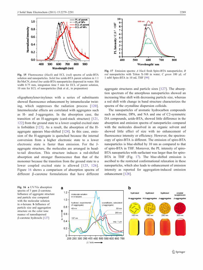
The nanoparticles of aromatic hydrocarbon compoundssuch as rubrene, DPA, and NA and one of C2-symmetricDA compounds, azide-BTA, showed little difference in theabsorption and emission spectra of nanoparticles comparedwith the molecules dissolved in an organic solvent andshowed little effect of size with no enhancement offluorescence intensity or efficiency. However, the spectros-copy of spiro-BTA is different. The emission of spiro-BTAnanoparticles is blue-shifted by 10 nm as compared to thatof spiro-BTA in THF. Moreover, the PL intensity of spiro-BTA nanoparticles with surfactant was larger than for spiro-BTA in THF (Fig. 17). The blue-shifted emission isascribed to the restricted conformational relaxation in thesenanoparticles, which also leads to enhancement of emissionintensity as reported for aggregation-induced emissionenhancement [128].
Fig. 17 Emission spectra: A black fresh Spiro-BTA nanoparticles; Bred nanoparticles with Triton X-100 in water; C green 100 μL of1 mM Spiro-BTA in 10 mL THF [99]
Fig. 15 Fluorescence (black) and ECL (red) spectra of azide-BTAsolution and nanoparticles. Solid line azide-BTA parent solution in 1:1Bz/MeCN, dotted line azide-BTA nanoparticles dispersed in water. Slitwidth 0.75 mm, integration time 5 min for ECL of parent solution,10 min for ECL of nanoparticles (Suk et al., in preparation)
Fig. 16 a UV/Vis absorptionspectra of 5 ppm β-carotene.Influence of aggregate structureand particle size comparedwith the molecular solutionin n-hexane. b Influence ofparticle size and aggregationstructure on the color tonenuance of nanodispersedβ-carotene hydrosols [127]
J Solid State Electrochem (2011) 15:2279–2291 2289

Summary and outlook
In this article, we have tried to provide a perspective aboutthe possibility of studying organic nanoparticles electro-chemically and by ECL. Currently, the research based onorganic nanoparticles and nanomaterials is still in itsinfancy and much remains to be done in this area. Size-dependent optical and electronic properties have not beenelucidated completely, due to limitations of synthesizingvery small nanoparticles in significant quantities. Newapproaches are needed for the preparation of monodispersenanoparticles of controlled size. Studies of the effect ofsurface modification are also needed. If suitable stable andefficient nanoparticles can be found, applications like labelsfor biological material analysis by ECL would be possible.
Acknowledgments We acknowledge the support of this researchfrom Roche Diagnostics, Inc. and the Robert A. Welch Foundation(F-0021).
References
1. Kubo R, Kawabata A, Kobayashi S (1984) Annu Rev Mater Sci14:49
2. Rossetti R, Ellison JL, Gibson JM, Brus LE (1984) J Chem Phys80:4464
3. Hanamura E (1988) Phys Rev B: Condens Matter 38:12284. Medintz IL, Uyeda HT, Goldman ER, Mattoussi H (2005) Nat
Mater 4:4355. Alivisatos AP (1996) Science 271:9336. Guzelian AA, Katari JEB, Kadavanich AV, Banin U, Hamad K,
Juban E, Alivisatos AP, Wolters RH, Arnold CC, Heath JR(1996) J Phys Chem 100:7212
7. Trindade T, O’Brien P, Pickett NL (2001) Chem Mater 13:38438. Mittleman DM, Schoenlein RW, Shiang JJ, Colvin VL, Alivisatos
AP, Shank CV (1994) Phys Rev B: Condens Matter 49:144359. de Paula AM, Barbosa LC, Cruz CHB, Alves OL, Sanjurjo JA,
Cesar CL (1996) Appl Phys Lett 69:35710. Freire PTC, Silva MAA, Reynoso VCS, Vaz AR, Lemos V
(1997) Phys Rev B: Condens Matter 55:674311. Carbone L, Nobile C, De Giorgi M, Sala FD, Morello G, Pompa
P, Hytch M, Snoeck E, Fiore A, Franchini IR, Nadasan M,Silvestre AF, Chiodo L, Kudera S, Cingolani R, Krahne R,Manna L (2007) Nano Lett 7:2942
12. Dzhagan V, Valakh MY, Kolny-Olesiak J, Lokteva I, Zahn DRT(2009) Appl Phys Lett 94:243101
13. Jdira L, Liljeroth P, Stoffels E, Vanmaekelbergh D, Speller S(2006) Phys Rev B: Condens Matter 73:115305
14. Peng AD, Xiao DB, Ma Y, Yang WS, Yao JN (2005) Adv Mater17:2070
15. Patra A, Hebalkar N, Sreedhar B, Sarkar M, Samanta A,Radhakrishnan TR (2006) Small 2:650
16. Kaeser A, Schenning APHJ (2010) Adv Mater 22:298517. Chen SW, Murray RW, Feldberg SW (1998) J Phys Chem B
102:989818. Hicks JF, Zamborini FP, Osisek A, Murray RW (2001) J Am
Chem Soc 123:704819. Hicks JF, Miles DT, Murray RW (2002) J Am Chem Soc
124:1332220. Bard AJ, Zhou HJ, Kwon SJ (2010) Is J Chem 50:267
21. Bard AJ (ed) (2004) Electrogenerated chemiluminescence.Marcel Dekker, New York
22. Richter MM (2004) Chem Rev 104:300323. Miao WJ (2008) Chem Rev 108:250624. Ding ZF, Quinn BM, Haram SK, Pell LE, Korgel BA, Bard AJ
(2002) Science 296:129325. Myung N, Ding ZF, Bard AJ (2002) Nano Lett 2:131526. Myung N, Bae Y, Bard AJ (2003) Nano Lett 3:105327. Bae Y, Myung N, Bard AJ (2004) Nano Lett 4:115328. Myung N, Lu XM, Johnston KP, Bard AJ (2004) Nano Lett
4:18329. Bard AJ, Ding ZF, Myung N (2005) Struct Bond 118:130. Ren T, Xu JZ, Tu YF, Xu S, Zhu JJ (2005) Electrochem Commun
7:531. Chen M, Pan LJ, Huang ZQ, Cao JM, Zheng YD, Zhan HQ
(2007) Mater Chem Phys 101:31732. Shen LH, Cui XX, Qi HL, Zhang CX (2007) J Phys Chem C
111:817233. Horn D, Honigmann B (1974) XII FATIPEC Kongress. Verlag
Chemie, Weinheim, p 18134. Katagi H, Kasai H, Okada S, Oikawa H, Komatsu K, Matsuda H,
Liu ZF, Nakanishi H (1996) Jpn J Appl Phys Part 2 35:L136435. Katagi H, Kasai H, Okada S, Oikawa H, Matsuda H, Nakanishi
H (1997) J Macromol Sci Pure Appl Chem A34:201336. Texter J (2001) J Dispers Sci Technol 22:49937. Rabinow BE (2004) Nat Rev Drug Discov 3:78538. Seko T, Ogura K, Kawakami Y, Sugino H, Toyotama H, Tanaka J
(1998) Chem Phys Lett 291:43839. Balzer F, Bordo VG, Simonsen AC, Rubahn HG (2003) Appl
Phys Lett 82:1040. Liu HB, Li YL, Xiao SQ, Gan HY, Jiu TG, Li HM, Jiang L, Zhu
DB, Yu DP, Xiang B, Chen YF (2003) J Am Chem Soc125:10794
41. Chiu JJ, Kei CC, Perng TP, Wang WS (2003) Adv Mater 15:136142. Ong BS, Wu YL, Liu P, Gardner S (2004) J Am Chem Soc
126:337843. Tamaki Y, Asahi T, Masuhara H (2002) J Phys Chem A 106:213544. Tamaki Y, Asahi T, Masuhara H (2003) Jpn J Appl Phys Part 1
42:272545. Asahi T, Sugiyama T, Masuhara H (2008) Acc Chem Res
41:179046. Ibanez A, Maximov S, Guin A, Chaillout C, Baldeck PL (1998)
Adv Mater 10:154047. Kasai H, Nalwa HS, Oikawa H, Okada S, Matsuda H, Minami
N, Kakuta A, Ono K, Mukoh A, Nakanishi H (1992) Jpn J ApplPhys Part 2 31:L1132
48. Kasai H, Oikawa H, Okada S, Nakanishi H (1998) Bull ChemSoc Jpn 71:2597
49. Wu CF, Peng HS, Jiang YF, McNeill J (2006) J Phys Chem B110:14148
50. Kaneko Y, Shimadai S, Onodera T, Kimura T, Matsuda H, OkadaS, Kasai H, Oikawa H, Kakudate Y, Nakanishi H (2007) Jpn JAppl Phys Part 1 46:6893
51. Mori J, Miyashita Y, Oliveira D, Kasai H, Oikawa H, NakanishiH (2009) J Cryst Growth 311:553
52. Tamaki Y, Ashshi T, Masuhara H (2000) Appl Surf Sci 168:8553. Kostler S, Rudorfer A, Haase A, Satzinger V, Jakopic G, Ribitsch
V (2009) Adv Mater 21:250554. Masuhara H, Ashahi T (2003) Single organic nanoparticles.
Springer, Berlin, p 3255. Kita S, Masuo S, Machida S, Itaya A (2006) Jpn J Appl Phys
Part 1 45:650156. Kostler S, Rudorfer A, Berghauser R, Jakopic G, Ribitsch V
(2006) Adv Materi Forum Iii, Pts 1 and 2 514–516:123557. Nalwa HS, Kasai H, Okada S, Oikawa H, Matsuda H, Kakuta A,
Mukoh A, Nakanishi H (1993) Adv Mater 5:758
2290 J Solid State Electrochem (2011) 15:2279–2291

58. Grey JK, Kim DY, Norris BC, Miller WL, Barbara PF (2006) JPhys Chem B 110:25568
59. Cui S, Liu HB, Gan LB, Li YL, Zhu DB (2008) Adv Mater20:2918
60. Matsuda H, Yamada S, VanKeuren E, Katagi H, Kasai H, OkadaS, Oikawa H, Nakanishi H, Smith EC, Kar AK, Wherrett BS(1997) Photosensit Opt Mater Devices 2998:241
61. Kim HY, Bjorklund TG, Lim SH, Bardeen CJ (2003) Langmuir19:3941
62. Oliveira D, Baba K, Mori J, Miyashita Y, Kasai H, Oikawa H,Nakanishi H (2010) J Cryst Growth 312:431
63. Kasai H, Kamatani H, Yoshikawa Y, Okada S, Oikawa H,Watanabe A, Itoh O, Nakanishi H (1997) Chem Lett 1181
64. Gesquiere AJ, Uwada T, Asahi T, Masuhara H, Barbara PF(2005) Nano Lett 5:1321
65. Olive AGL, Del Guerzo A, Schafer C, Belin C, Raffy G,Giansante C (2010) J Phys Chem C 114:10410
66. Oikawa H, Fujita S, Kasai H, Okada S, Tripathy SK, NakanishiH (2000) Colloids Surf A 169:251
67. Okazoe S, Fujita S, Kasai H, Okada S, Oikawa H, Nakanishi H(2001) Mol Cryst Liq Cryst 367:11
68. Wang L, Wong WY, Lin MF, Wong WK, Cheah KW, Tam HL,Chen CH (2008) J Mater Chem 18:4529
69. Faulkner LR, Bard AJ (1977) In: Bard AJ (ed) Electroanalyticalchemistry, vol 10. Dekker, New York, pp 1–95
70. Faulkner LR, Glass RS (1982) In: Waldemar A, Giuseppe C(eds) Chemical and biological generation of excited states. NewYork, Academic, Chapter 6
71. Bard AJ, Debad JD, Leland JK, Sigal GB, Wilbur JL,Wohlstadter JN (2002) In: Meyers RA (ed) Encyclopedia ofanalytical chemistry: applications, theory and instrumentation,vol 11. Wiley, New York, p 9842
72. Namba Y, Sawada T, Suzuki O (2000) Anal Sci 16:75773. Cao WD, Ferrance JP, Demas J, Landers JP (2006) J Am Chem
Soc 128:757274. Knight AW, Greenway GM (1994) Analyst 119:87975. Balazs AC, Emrick T, Russell TP (2006) Science 314:110776. Burroughes JH, Bradley DDC, Brown AR, Marks RN, Mackay
K, Friend RH, Burns PL, Holmes AB (1990) Nature 347:53977. Grimsdale AC, Chan KL, Martin RE, Jokisz PG, Holmes AB
(2009) Chem Rev 109:89778. Mullen K, Scherf U (2005) Organic light-emitting devices.
Wiley-VCH, Weinheim79. Nalwa HS, Rohwer LS (2003) Handbook of luminescence
display materials and devices. American Scientific, StevensonRanch
80. Scharber MC, Wuhlbacher D, Koppe M, Denk P, Waldauf C,Heeger AJ, Brabec CL (2006) Adv Mater 18:789
81. Chen JW, Cao Y (2009) Acc Chem Res 42:170982. Peet J, Heeger AJ, Bazan GC (2009) Acc Chem Res 42:170083. Cheng YJ, Yang SH, Hsu CS (2009) Chem Rev 109:586884. Hoeben FJM, Jonkheijm P, Meijer EW, Schenning APHJ (2005)
Chem Rev 105:149185. Yamamoto Y, Fukushima T, Suna Y, Ishii N, Saeki A, Seki S,
Tagawa S, Taniguchi M, Kawai T, Aida T (2006) Science314:1761
86. Grimsdale AC, Mullen K (2005) Angew Chem Int Ed 44:559287. Gunes S, Neugebauer H, Sariciftci NS (2007) Chem Rev
107:132488. Chan WCW, Nie SM (1998) Science 281:201689. Bruchez M, Moronne M, Gin P, Weiss S, Alivisatos AP (1998)
Science 281:201390. Palacios RE, Fan FRF, Grey JK, Suk J, Bard AJ, Barbara PF
(2007) Nat Mater 6:68091. Chang YL, Palacios RE, Fan FRF, Bard AJ, Barbara PF (2008) J
Am Chem Soc 130:8906
92. Maloy JT, Bard AJ (1971) J Am Chem Soc 93:596893. Smith JH, Mann CK (1969) J Org Chem 34:182194. Noffsinger JB, Danielson ND (1987) Anal Chem 59:86595. Zu YB, Bard AJ (2000) Anal Chem 72:322396. Kanoufi F, Cannes C, Zu YB, Bard AJ (2001) J Phys Chem B
105:895197. Miao WJ, Choi JP, Bard AJ (2002) J Am Chem Soc 124:1447898. Chung HR, Kwon E, Kawa H, Kasai H, Nakanishi H (2006) J
Cryst Growth 294:45999. Omer KM, Bard AJ (2009) J Phys Chem C 113:11575
100. Lai RY, Bard AJ (2003) J Phys Chem A 107:3335101. Memming R (1969) J Electrochem Soc 116:785102. Omer KM, Ku SY, Cheng JZ, Chou SH, Wong KT, Bard AJ
(2011) J Am Chem Soc 133:5492–5499103. Fungo F, Wong KT, Ku SY, Hung YY, Bard AJ (2005) J Phys
Chem B 109:3984104. Rashidnadimi S, Hung TH, Wong KT, Bard AJ (2008) J Am
Chem Soc 130:634105. Omer KM, Ku SY, Wong KT, Bard AJ (2009) Angew Chem Int
Ed 48:9300106. Omer KM, Ku SY, Wong KT, Bard AJ (2009) J Am Chem Soc
131:10733107. Shen M, Rodriguez-Lopez J, Lee YT, Chen CT, Fan FRF, Bard
AJ (2010) J Phys Chem C 114:9772108. Omer KM, Ku SY, Chen YC, Wong KT, Bard AJ (2010) J Am
Chem Soc 132:10944109. Kasai H, Kamatani H, Okada S, Oikawa H, Matsuda H,
Nakanishi H (1996) Jpn J Appl Phys Part 2 35:L221110. Fu HB, Ji XH, Zhang XH, Wu SK, Yao JN (1999) J Colloid
Interface Sci 220:177111. Fu HB, Yao JN (2001) J Am Chem Soc 123:1434112. Kasai H, Yoshikawa Y, Seko T, Okada S, Oikawa H, Mastuda H,
Watanabe A, Ito O, Toyotama H, Nakanishi H (1997) Mol CrystLiq Cryst Sci Technol Sect A 294:173
113. Komai Y, Kasai H, Hirakoso H, Hakuta Y, Okada S, Oikawa H,Adschiri T, Inomata H, Arai K, Nakanishi H (1998) Mol CrystLiq Cryst Sci Technol Sect A 322:167
114. An BK, Kwon SK, Jung SD, Park SY (2002) J Am Chem Soc124:14410
115. Feng X, Tong B, Shen J, Shi J, Han T, Chen L, Zhi J, Lu P, Ma Y,Dong Y (2010) J Phys Chem B 114:16731
116. Miteva T, Palmer L, Kloppenburg L, Neher D, Bunz UHF (2000)Macromolecules 33:652
117. Deans R, Kim J, Machacek MR, Swager TM (2000) J Am ChemSoc 122:8565
118. Luo JD, Xie ZL, Lam JWY, Cheng L, Chen HY, Qiu CF, KwokHS, Zhan XW, Liu YQ, Zhu DB, Tang BZ (2001) ChemCommun 1740
119. Walters KA, Ley KD, Schanze KS (1999) Langmuir 15:5676120. Oelkrug D, Tompert A, Gierschner J, Egelhaaf HJ, Hanack M,
Hohloch M, Steinhuber E (1998) J Phys Chem B 102:1902121. Kasha M, Rawls HR, El-Bayouni A (1965) Pure Appl Chem
11:371122. Dahne L, Biller E (1998) Adv Mater 10:241123. Li S, He L, Ziong F, Li Y, Yang G (2004) J Phys Chem B
108:10887124. Gruszecki WI (1991) J Biol Phys 18:99125. Akins DL, Macklin JW (1989) J Phys Chem 93:5999126. Chowdhury A, Wachsmann-Hogiu S, Bangal PR, Raheem I,
Peteanu LA (2001) J Phys Chem B 105:12196127. Horn D, Lűddecke E (1996) In: Pelizzetti E (ed) Fine particles
science and technology—from micro to nanoparticles. Kluwer,Dordrecht, p 761
128. Zeng Q, Li Z, Dong Y, Di C, Qin A, Hong Y, Ji L, Zhu Z, JimKW, Yu G, Li Q, Li Z, Liu Y, Qin J, Tang BZ (2007) ChemCommun 1:70
J Solid State Electrochem (2011) 15:2279–2291 2291