elective intubation - respiratory carerc.rcjournal.com/content/respcare/59/6/825.full.pdf ·...
TRANSCRIPT

Elective Intubation
Charles G Durbin Jr MD FAARC, Christopher T Bell MD, and Ashley M Shilling MD
IntroductionIntubation: Perioperative Versus Outside the Operating Room
Patient Examination and Airway EvaluationPredictors of a Difficult AirwayMouth Opening and DentitionOral Cavity and Neck Mobility EvaluationPhysiologic Risks of IntubationEnvironmental Assessment and PreparationManaging Airway Risks Outside the Operating RoomMonitoringIntubation: Plans A, B, and C
Preparing for IntubationPhysical Considerations and Time-OutDenitrogenation or Preoxygenation
Intubation Using Direct LaryngoscopyInitial PlanConfirmation of Tracheal IntubationPhysiologic Changes and Management Strategies
Alternative Airway PlansPostintubation PlansTransfer of Care
After the Difficult Intubation: A Case ReviewConclusions
Endotracheal intubation is a commonly performed operating room (OR) procedure that providessafe delivery of anesthetic gases and airway protection during surgery. The most common intuba-tion technique in the perioperative environment is direct laryngoscopy with orotracheal tube in-sertion. Infrequently, difficulties that require an alternative intubation technique are encountereddue to patient anatomy, equipment limitations, or patient pathophysiology. Careful patient evalu-ation, advanced planning, equipment preparation, system redundancy, use of checklists, familiaritywith airway algorithms, and availability of additional help when needed during OR intubationshave resulted in exceptional success and safety. Airway difficulties during intubation outside thecontrolled environment of the OR are more frequent and more serious. Translating the intubationprocesses practiced in the OR to intubations outside the perioperative setting should improvepatient safety. This paper considers each step in the OR intubation process in detail and proposesways of incorporating perioperative procedures into intubations outside the OR. Management ofthe physiologic impact of intubation, lack of readily available specialized equipment and experi-enced help, and planning for transfer of care following intubation are all challenges during theseintubations. Key words: tracheal intubation; manual ventilation; artificial airway; endotracheal tube;difficult airway; difficult intubation; airway emergency; direct laryngoscopy; fiberoptic intubation. [RespirCare 2014;59(6):825–849. © 2014 Daedalus Enterprises]
RESPIRATORY CARE • JUNE 2014 VOL 59 NO 6 825

Introduction
Endotracheal intubation is a commonly performed pro-cedure. It was calculated by the Centers for Disease Con-trol and Prevention that over 51 million surgeries wereperformed in the United States in 2010.1 An estimated 15million (30%) of these included insertion of an endotra-cheal tube (ETT). During this same time period, at least650,000 intubations were performed in hospitals outsidethe operating room (OR) and procedure areas. In addition,almost 300,000 additional intubations were performed inthe emergency room in 2010.2 This does not include thoseETT placements performed during resuscitation attemptsoutside the hospital setting. The number of ETT intuba-tions performed to facilitate surgery or procedural inter-ventions continues to rise in the United States and through-out the world.
Airway management is often complex and can be asource of patient morbidity and mortality. The AmericanSociety of Anesthesiologists Closed Claims Project data-base allows structured analyses of adverse anesthetic out-comes obtained from reviewing closed claims files of 35professional liability insurance companies representing an-esthesiologists in the United States. While this database isbiased toward the worst outcomes, it can be used to iden-tify areas and practices in which improvements can bemade. Remarkably, the most common serious outcomesoccurred during airway management and intubation. Ofthe first 1,540 cases reported in 1990, 522 (34%) wererelated to airway problems.3 Death or severe brain injuryoccurred in 83% of these cases. As many as 72% of thesewere judged preventable with improved clinical care. Air-way issues were not confined to the time of anesthesiainduction. Over 6% of the complications occurred outsidethe OR at the time of extubation, and these carried a higherrisk of death or severe injury than those occurring duringanesthesia induction and intubation. With the developmentof algorithms and new devices to deal with expected and
unexpected difficulties, airway safety has improved mark-edly in the OR environment.
Preparing for and executing a safe and successful elec-tive intubation require completion of a complex series ofactivities that should occur in a prescribed order. While thecorrect placement and securing of the artificial airway in apatient’s trachea are the ultimate goals and hallmarks ofsuccess, each individual step leading to this goal must becarefully considered and accomplished to be effective. Un-like an emergency situation requiring an expeditious air-way insertion to preserve life, performing an elective in-tubation allows time for a systematic evaluation andoptimization of environmental and patient factors that willinfluence ultimate success in intubation with a minimumof physiologic trespass. In this discussion, a systematicapproach used for intubation as part of the provision ofanesthetic care during surgery will be developed and pro-posed as a model to approach intubations being performedoutside the OR. While scientific evidence is available insupport of many components of contemporary anesthesiaairway practice, some of the routine approaches and safetymeasures have not been (and likely will never be) evalu-ated in randomized prospective trials. Published data, whenavailable, will be cited to support recommendations. Byanalyzing the intubation process in the OR, the criticalcomponents, preparations, and actions that are associatedwith safety and success can be identified and adapted forintubations performed outside the OR.
Intubation: Perioperative Versus Outside theOperating Room
As listed in detail in Table 1, airway management andintubation in the operative setting include the followingsequential activities: examination and identification of spe-cific patient issues, development of an airway plan, envi-ronmental preparation, managing the actual airway andperforming intubation, confirming and maintaining correcttube placement, and managing the patient’s physiologicresponse to intubation. When intubation is performed out-side the OR, an additional step is transitioning care of thepatient with the artificial airway to another clinician. Trans-fer of care also occurs following a surgical procedure whenextubation is not immediately appropriate. This is true fortransfers to ICUs and when an intubated patient is placedin the post-anesthesia care unit. It is important to note thatextubation is also a time of increased airway risk, andknowledge of any issues encountered during intubation isa priority for the team responsible for removing the arti-ficial airway. Timely and accurate communications be-tween teams are essential for patient safety at extubation.Consistent methods for documenting and conveying de-tails of airway management to the next and future clini-cians have received considerable attention.
The authors are affiliated with the Department of Anesthesiology, Uni-versity of Virginia Health Science System, Charlottesville, Virginia.
Dr Durbin is on the board of medical advisors of the Masimo Foundationand has consulted on airway products for Kimberly-Clark. The otherauthors have disclosed no conflicts of interest.
Dr Durbin presented a version of this paper at the 52nd RESPIRATORY
CARE Journal Conference, “Adult Artificial Airways and Airway Ad-juncts,” held June 14 and 15, 2013, in St Petersburg, Florida.
Correspondence: Charles G Durbin Jr MD FAARC, Department of An-esthesiology, University of Virginia Health Science System, Box 800710,Charlottesville, VA 22908. E-mail: [email protected].
DOI: 10.4187/respcare.02802
ELECTIVE INTUBATION
826 RESPIRATORY CARE • JUNE 2014 VOL 59 NO 6

Difficulties during airway management outside the ORare encountered more frequently than during intubationsperformed in the controlled OR setting. In one study, ur-
gent intubation in ICU patients by airway experts (indi-viduals with � 6 months of anesthesiology training) re-sulted in a difficult intubation rate (defined as requiring
Table 1. Steps in Preparing for and Managing an Intubation Under Anesthesia for Surgery in the Operating Room
Activity Details Objectives
Patient and airway evaluation Pay particular attention to airway history and examination. Predict airway difficulty.Assess existing patient medical issues. Identify patient physiologic tolerance.Identify potentially modifiable patient medical risks.
Intubation plan development Choose a primary intubation plan based on the patientevaluation.
Define an initial plan and alternative plansfor intubation.
Include additional options to deal with anticipated orunanticipated difficulties that may be encountered.
Consider airway algorithms and protocols.Confirm all equipment that may be needed.
Environment analysis andpreparation
Use a checklist.Locate, prepare, and test all essential and emergency equipment.
• Oxygen source and backup• Suction• Manual ventilation devices• CPR equipment• Defibrillator
Establish patient monitoring.Identify and confirm function of communication system to get
help.Plan for obtaining additional intubation equipment if needed.Establish or confirm function of intravenous access.Prepare drugs for sedation and intubation.Locate and prepare drugs and fluids to modify patient
physiologic responses to intubation.Organize assistants and assign tasks.Communicate plans for intubation and emergencies.
Prepare equipment and assistants forintubation.
Guarantee that all essential equipment ispresent and working.
Provide intravenous access.Record vital signs.Review airway plans with the team.Confirm availability of help in an emergency.
Actual intubation Call a time-out, check-in, or pause. Focus the team.• Identify the patient, procedure, and correct surgical site. Safely perform the intubation.• Confirm presence of critical equipment and personnel.• Review intubation and surgical plans.• Identify potential and actual risks.• Discuss what will be needed and by whom if difficulties
are encountered.Begin and complete intubation.Anticipate and respond to undesired responses in
hemodynamics.Postintubation tasks Confirm correct ETT placement. Confirm tracheal placement.
• Physical examination Prevent accidental extubation.• Auscultation Normalize vital signs after intubation.• CO2 measurement• Pulse oximetry monitoring
Secure the ETT and provide appropriate ventilation.Monitor vital signs and gas exchange.Continue management of expected and unanticipated changes in
hemodynamics and respiratory function.Airway issue communication Record details of the intubation.
Convey methods used to resolve airway problems.Inform the patient and family members of all issues.Communicate with the team and future caregivers of airway
risks and their successful management.
Document airway difficulties in medicalrecord.
Provide the patient with information.Guarantee essential information is available
when needed in future.
CPR � cardiopulmonary resuscitationETT � endotracheal tube
ELECTIVE INTUBATION
RESPIRATORY CARE • JUNE 2014 VOL 59 NO 6 827

� 2 attempts at ETT placement) of 8%.4 In addition, deathwithin 30 min of intubation occurred in 3% of patients.Although the authors speculated that insufficient environ-mental preparations, inadequate equipment, fewer experi-enced staff members assisting, and intubator deficienciesmay contribute to the increased incidence of complica-tions, no definitive conclusions implicating any of thesefactors were presented in this paper. Published data sup-port the assertion that patients undergoing intubations out-side the OR are 10–30 times more likely to experienceairway complications. A poor laryngeal view on directlaryngoscopy is encountered in the OR in 0.13–0.3% ofcases; however, failure to intubate is much lower.5 Whilepoor glottic views are seen more frequently in intubationsoccurring outside the OR, intubation can typically still besuccessful with simple interventions.6 Having a facultyanesthesiologist present during ICU intubations was asso-ciated with fewer complications: 21.7 versus 6.1%. Over-all, there were no intubation-related deaths during any ofthe 322 intubations.7 Applying the systematic operationalmodel used in the OR to intubations performed outside theOR may improve the outcome from unanticipated airwayevents during these higher risk situations.8 Each of thesetasks involved in a successful intubation in the OR will beconsidered as it could apply to intubations performed out-side the OR.
Patient Examination and Airway Evaluation
Perioperative airway evaluation begins by obtaining ahistory of previous airway problems and performing a pa-tient examination specifically directed at identifying pa-tient characteristics known to correlate with airway diffi-culties. The steps in this airway evaluation process arereviewed and summarized in Table 2.
During an elective surgical procedure, patients are typ-ically seen and the airway is evaluated prior to entering theOR suite.9 An important part of this evaluation includesprevious surgeries and intubations in order to predict thelikelihood of airway difficulties during the intubation pro-cess. One of the most important predictors of difficult-airway management is a history of difficult intubation. Theclinician may be alerted to this by a patient or a familymember. A very sore throat after surgery and dental dam-age after an intubation are clues that the patient was dif-ficult to intubate. It is critical to obtain as many details ofthe event as possible and, most importantly, to identify theintubation method that was finally successful. With cur-rent anesthesia practice and the expanding use of elec-tronic records, a detailed contemporaneous record of theevents may be available. However, many patients will betold it was difficult to place their ETT, but none of thedetails will be available. Because of this, there is growingsentiment in the anesthesia community to standardize cod-
ing for airway events and to create a consistent method forconveying difficult intubation details to patients and futurecaregivers. One such proposal, shown in Figure 1, wascreated by the Anesthesia Patient Safety Foundation.10 Tothis point, no consensus of how to standardize this docu-mentation has been established. Within hospital systems,patients experiencing a difficult intubation event may belabeled as such, much in the way an allergy will be iden-tified. Arm bands can be placed on the patient, a stickermay be placed in the record, a tag may be attached to theETT, or a sign may be placed on the patient’s door iden-tifying the event. At a minimum, a note in the medicalrecord should detail the events and their resolution.
Predictors of a Difficult Airway. There are many con-ditions that can lead to problems managing a patient’sairway. Difficulty with airway management is typicallydivided into difficult mask ventilation and difficult intu-bation. These are different situations that demand differentapproaches and pose markedly different patient risks. Anairway emergency is defined as the situation in which theclinician is unable to ventilate AND unable to intubate apatient. This may result in a fall in PaO2
, leading to cardiacarrest and brain injury or death. Prevention of lethal hy-poxemia by applying extraordinary airway techniques, in-cluding surgical airway access, is mandatory and must bea part of all elective airway management plans.11
Physical examination of patients and their airways shouldhelp identify predictors of difficult ventilation or intuba-tion. In a large study of over 53,000 general anesthetics, 77(0.15%) patients could not be ventilated by mask or witha simple airway adjuvant.12 Of those who failed ventila-tion, 74% were able to be intubated with 3 or less attemptsemploying conventional and specialized intubation tech-niques; 25% were found to have a difficult intubation aswell. Two of these required an urgent surgical airway,whereas 2 were awakened and electively intubated awake.None suffered adverse neurologic events during care. Evenwith careful preparation, the situation of cannot ventilate/cannot intubate will occur. For the group of patients dis-cussed above, predictors for impossible ventilation areshown in Table 3. With the exception of head and neckradiation and addition of obesity, these are generally rec-ognized as predictors for difficult ventilation.
In many patients, no concrete information about previ-ous airway problems is available from the record or fromthe patient at the time it is sought. Self-reported airwaydifficulties should serve as a red flag to perform a carefulairway exam in an attempt to understand what the prob-lems might have been, develop plans, have appropriateequipment, and anticipate a difficult-airway situation. Typ-ical questions and physical findings that are helpful inidentifying a high risk of airway problems during electiveintubation are listed in Table 2.
ELECTIVE INTUBATION
828 RESPIRATORY CARE • JUNE 2014 VOL 59 NO 6

Table 2. Important Details of the History and Physical Exam That May Help Predict a Difficult Airway or Intubation
Factor Important Questions Implications
History of a difficultintubation
What were the details of the difficulty?When did it occur?During what kind of surgery or procedure did it occur?How was it managed?Were there any sequelae?
Increased risk of intubation difficultiesKnown successful management plan
History of an airwayproblem
What were the details of the difficulty?When did it occur?During what kind of surgery or procedure did it occur?How was it managed?Were there any sequelae?
Increased risk of ventilation difficultiesAnticipation of other problems
History of a previoustracheostomy
Why was it performed?When was it performed?When was it removed?Were there any complications?Are there any symptoms of airway narrowing now?
Possible need for smaller ETTPossible need for repeat surgical airway
Mouth opening Is the mouth opening small? Laryngoscope may not fit.Is the mouth distance (upper to lower incisor) � 3 cm? Supraglottic airway (LMA) may not fit through oral opening.Is pain a limiting factor to opening? Possible dental damage
Anesthesia may improve oral access.Nasal or retrograde intubation may be necessary.
Teeth Is the patient edentulous?Are the teeth in poor condition?Does the patient have prominent upper incisors
(overbite)?
More difficulty with manual ventilationEasy DL and intubationIncreased loss risk, aspiration riskIncreased tooth damage riskIncreased DL difficulty
Oral view What is the Mallampati score? Increasing score predicts poorer laryngeal view and possibledifficult intubation.
Neck mobility Does the patient have limited extension?Does the patient have no extension?Does the patient have symptoms of neurologic
compromise?Is the neck in orthopedic fixation or a rigid collar?
Ventilation and DL intubation may be difficult.Difficult ventilation: early use of airway adjuncts may be
needed.Low probability of DL intubationNeed to use other means for placement of ETT (ie, FOI or
indirect laryngoscope)Surgical airway may not be possible.Dangerous ventilation: early use of airway adjuncts may be
needed.Neck immobility during ventilation and intubation requiredDangerous ventilation: early use of airway adjuncts may be
needed.Neck immobility during ventilation and intubation requiredAwake intubation (FOI) preferred to evaluate neurologic
function after intubationJaw Is the jaw short?
Is the jaw thick: � 3 finger tips between inside of jawand hyoid bone?
Creates anterior larynx phenomenonDifficult intubation likelyDifficult to displace tongue and intubate
Submental tissues Are they stiff?Are they solid?
Radiation therapy or healed burns can make tonguedisplacement impossible and intubation difficult.
It may be impossible to intubate with DL; FOI may be onlysuccessful approach.
Body size Is the patient morbidly obese? Difficult ventilation but usually normal DL intubationMay require device to elevate head and shoulders (ramp) to
improve ventilation and intubationMay have OSA syndromeWill desaturate quickly during intubation attempts
LMA � laryngeal mask airwayDL � direct laryngoscopyFOI � flexible fiberoptic intubationOSA � obstructive sleep apnea
ELECTIVE INTUBATION
RESPIRATORY CARE • JUNE 2014 VOL 59 NO 6 829

Fig. 1. Suggested letter to be given to a patient with a difficulty airway experience to be shared with future caregivers, who then can usethis information to prepare for and prevent future tragedies. DOB � date of birth. LMA � laryngeal mask airway. Courtesy the AnesthesiaPatient Safety Foundation.
ELECTIVE INTUBATION
830 RESPIRATORY CARE • JUNE 2014 VOL 59 NO 6

Elective intubation is usually performed under directvision using a laryngoscope. This technique is referred toas direct laryngoscopy (DL). The laryngoscope will haveeither a straight or a curved blade and a light source nearits tip to illuminate the airway structures. After insertingthe blade through the mouth, the laryngoscope is used tomove and control the tongue and upper airway structures.By lifting the tongue and jaw, the larynx is seen. A directline of vision from the mouth to the glottis is used when
inserting an ETT. The directed airway examination is usedto identify obstacles to establishing a direct line of visionto the larynx. If the physical examination suggests that DLwill be difficult, an alternative intubation technique shouldbe considered. Some of the many techniques that can beused for intubation are listed in Table 4.
Mouth Opening and Dentition. Inserting a standard la-ryngoscope blade into the mouth and passing a tube intothe trachea of an adult requires a reasonably sized mouthopening. A normal size mouth opening is demonstrated inFigure 2. Patients with a � 1.5-cm mouth opening be-tween the upper and lower front teeth (or gums if edentu-lous) may be impossible to intubate with conventional DLsince the blade flange is often larger than this size. If asmall mouth opening is identified during airway examina-tion, the cause of the restriction should be determined. Ifpain from temporomandibular joint disease or an acutemandibular fracture is the cause, use of analgesics or an-esthesia with or without a muscle relaxant may increasethe mouth opening and allow a conventional intubationtechnique. If the mouth opening is fixed and severely lim-ited, then an alternative intubation technique such as oral
Table 4. Selected Intubation Techniques That May Be Useful Under a Variety of Circumstances
Intubation Techniques Comments
Direct laryngoscopy and intubation Best known skill with most practiceCurved blade Tongue control and working space bestStraight blade Best laryngeal view, less easy tube placementSedation/anesthesia with muscle relaxant Best view, no patient cooperationSedation with topical local anesthesia Continued spontaneous ventilation, less pain with curved bladeAwake with topical local anesthesia Cooperative patient, good success without burning bridges
Indirect laryngoscopic intubation Visual view excellent and seen by allSedation/anesthesia with muscle relaxant Best successSedation with topical local anesthesia Not well tolerated by patients unless deeply sedated
Fiberoptic lighted stylet Dark room needed, blinded techniqueSedation/anesthesia with muscle relaxant Best success, but a problem if it failsSedation/anesthesia with spontaneous ventilation Safest approach as spontaneous ventilation is maintained
Flexible fiberoptic intubation Accepted standard for difficult-airway managementSedation/anesthesia with muscle relaxant Secretions and blood can greatly reduce successOral Difficulty greater than nasal, but adjuncts can helpNasal Direct intubation route, bleeding riskSedation with spontaneous ventilation Improved safety, as no bridge is burntAwake with topical local anesthesia Standard for known or anticipated difficult airway
Retrograde intubation Minimally invasive blind technique, technical details difficult to executeCricothyrotomy Emergency airway when all else fails, and death is imminentPercutaneous tracheostomy Preferred technique for long-term artificial airway
Almost as fast as cricothyrotomy for emergency surgical airway with less potential forairway damage
Requires special equipment and skillTranstracheal jet ventilation Needle or catheter for entry
Least invasive surgical emergency airwayMust have high pressure gas source and jet deviceComplications frequent
Table 3. Patient Factors Found to Be Associated With ImpossibleVentilation Identified in � 50,000 Subjects Cared for inthe Operating Room
Predictor POdds Ratio(95% CI)
Radiation changes to neck .002 7.1 (2.1–24.4)Male sex � .001 3.3 (1.8–6.3)OSA syndrome .005 2.4 (1.3–4.3)Mallampati III or IV .01 2.0 (1.1–3.4)Beard .02 1.9 (1.1–3.3)
Data from Reference 12.OSA � obstructive sleep apnea
ELECTIVE INTUBATION
RESPIRATORY CARE • JUNE 2014 VOL 59 NO 6 831

or nasal flexible fiberoptic intubation should be chosen forthe first intubation attempt. Prominent or misaligned upperincisors may also present a challenge to intubation by DL.This can be subjectively evaluated by viewing the subjectin profile. A normal mandibular profile is seen in Figure 3.The tip of the chin is in line with or in front of the upperlip. An abnormal profile suggesting intubation difficultieswould show a jaw tip that is substantially behind the upperlip.
Poor dental hygiene and periodontal disease may resultin loose or broken teeth and may increase the risk for toothdamage or loss during intubation. Care to prevent pulmo-nary aspiration of a dislodged tooth or crown is essential.If native dentition or prostheses are at risk of damage dueto disease or mouth anatomy, the patient should be madeaware of this risk. Cost of dental repairs necessitated bytooth injury during elective intubation are usually borne bythe patient and rarely covered by health insurance.
Oral Cavity and Neck Mobility Evaluation. After ex-amining the mouth opening and dentition, the oral cavityshould be examined. One of the most frequently used toolsto quantitate the risk of a difficult intubation is the Mal-lampati (MP) score.13 Prospectively tested and reported in1985, the clinical sign of concealment of the faucial pillars(palatoglossal and palatopharyngeal arches) and uvula by
the base of the tongue predicts difficulty with visualizationof the laryngeal opening during DL. With maximal tongueprotrusion in the upright sitting position and without pho-nation, the degree of visual obstruction of the posteriormouth and palate is graded from MP I (the entire faucialpillars and uvula are seen) to MP III (only the soft palateand the base of the uvula are visualized). As the MP scoreincreases, the risk of visualization difficulty during DLalso increases. In the original report, there were no laryn-geal view or intubation difficulties with any the 155 pa-tients with an MP I oral view. Of the 40 patients with anMP II oral view, 26 patients had a good laryngeal gradeview, and all 40 were successfully intubated. Of the 15patients with an MP III airway, only one had a good glotticview. In 9 of these patients, only the arytenoids were seen,and in 5 patients, no identified laryngeal structure could beseen. Also noted in this initial report, in the 4 patients withlimited neck extension, 2 patients had poor glottic viewsdespite an MP I or II oral view. Since this original report,an MP IV airway score has been added, in which only thehard palate can be visualized. The MP system is widelyused and easy to apply. An MP score of I or II is reassuringbut, by itself, does not guarantee good visualization oreasy intubation by DL. A higher MP score is associatedwith less optimal visualization of the glottis but does notnecessarily predict a difficult intubation.
Fig. 2. This subject demonstrates an wide oral opening and stabledentition.
Fig. 3. The normal profile of this subject suggests that his man-dibular length is adequate and that manual ventilation and intu-bation should not be difficult.
ELECTIVE INTUBATION
832 RESPIRATORY CARE • JUNE 2014 VOL 59 NO 6
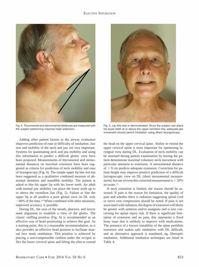
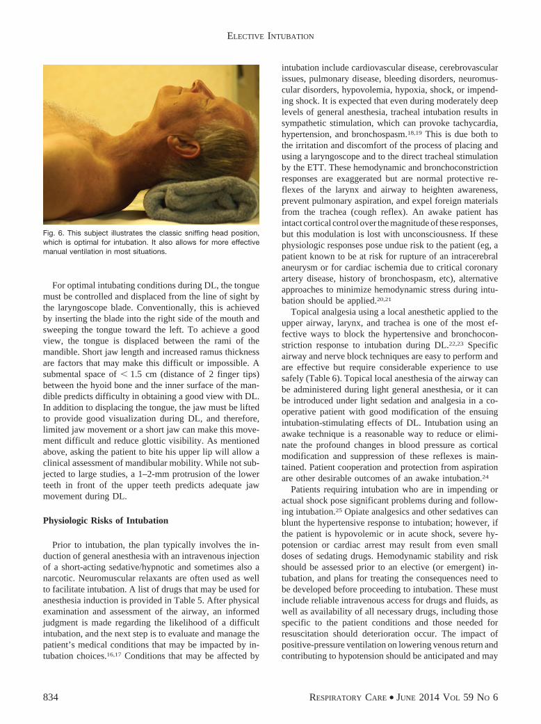
Adding other patient factors to the airway evaluationimproves prediction of ease or difficulty of intubation. Jawsize and mobility of the neck and jaw are very important.Systems for quantitating neck and jaw mobility and usingthis information to predict a difficult glottic view havebeen proposed. Measurements of thyromental and sterno-mental distances on maximal extension have been sug-gested as criteria for prediction of neck mobility and easeof laryngoscopy (Fig. 4). The simple upper lip bite test hasbeen suggested as a qualitative combined measure of ab-normal dentition and mandible mobility. The patient isasked to bite his upper lip with his lower teeth. An adultwith normal jaw mobility can place the lower teeth up toor above the vermilion line (Fig. 5). Failure to bite theupper lip at all predicts a poor glottal view on DL only�40% of the time.14 When combined with other measures,improved accuracy is possible.
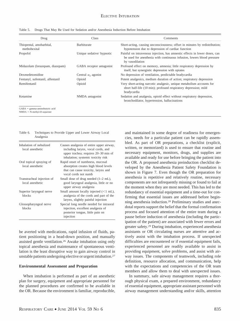
During DL, the axes of the mouth, pharynx, and larynxneed alignment to establish a view of the glottis. Theclassic sniffing position (Fig. 6) is recommended as aneffective way of head positioning to achieve this goal. Asa starting point, this is a reasonable recommendation, as italso provides an effective head position to facilitate man-ual face mask ventilation. This position is achieved byplacing a non-compressible cushion under the occiput toflex the lower cervical spine and lifting the chin to extend
the head on the upper cervical spine. Ability to extend theupper cervical spine is most important for optimizing la-ryngeal view during DL. Evaluation of neck mobility canbe assessed during patient examination by having the pa-tient demonstrate maximal voluntary neck movement withparticular attention to extension. A sternomental distanceof � 9 cm predicts adequate extension. Correction for pa-tient height may improve positive prediction of a difficultlaryngoscopic view on DL (short sternomental measure-ment), but use of even this corrected measurement is � 50%accurate.15
If neck extension is limited, the reason should be as-sessed. If pain is the reason for limitation, the quality ofpain and whether there is radiation suggesting spinal cordor nerve root compression should be noted. If pain is notassociated with radiation, the degree of extension will likelybe greater with sedation and/or analgesia and is less con-cerning for spinal injury risk. If there is significant limi-tation of extension and no pain, this represents a fixedbony issue that is unlikely to improve with medications.The presence of a known instability of the spine prohibitsextension and makes safe intubation with DL difficult,and an alternative approach is mandated, eg, fiberopticintubation. Additional intubation techniques are listed inTable 4.
Fig. 5. Lip bite test is demonstrated. Since the subject can placehis lower teeth at or above the upper vermilion line, adequate jawmovement should permit intubation using direct laryngoscopy.
Fig. 4. Thyromental and sternomental distances are measured withthe subject performing maximal head extension.
ELECTIVE INTUBATION
RESPIRATORY CARE • JUNE 2014 VOL 59 NO 6 833
For optimal intubating conditions during DL, the tonguemust be controlled and displaced from the line of sight bythe laryngoscope blade. Conventionally, this is achievedby inserting the blade into the right side of the mouth andsweeping the tongue toward the left. To achieve a goodview, the tongue is displaced between the rami of themandible. Short jaw length and increased ramus thicknessare factors that may make this difficult or impossible. Asubmental space of � 1.5 cm (distance of 2 finger tips)between the hyoid bone and the inner surface of the man-dible predicts difficulty in obtaining a good view with DL.In addition to displacing the tongue, the jaw must be liftedto provide good visualization during DL, and therefore,limited jaw movement or a short jaw can make this move-ment difficult and reduce glottic visibility. As mentionedabove, asking the patient to bite his upper lip will allow aclinical assessment of mandibular mobility. While not sub-jected to large studies, a 1–2-mm protrusion of the lowerteeth in front of the upper teeth predicts adequate jawmovement during DL.
Physiologic Risks of Intubation
Prior to intubation, the plan typically involves the in-duction of general anesthesia with an intravenous injectionof a short-acting sedative/hypnotic and sometimes also anarcotic. Neuromuscular relaxants are often used as wellto facilitate intubation. A list of drugs that may be used foranesthesia induction is provided in Table 5. After physicalexamination and assessment of the airway, an informedjudgment is made regarding the likelihood of a difficultintubation, and the next step is to evaluate and manage thepatient’s medical conditions that may be impacted by in-tubation choices.16,17 Conditions that may be affected by
intubation include cardiovascular disease, cerebrovascularissues, pulmonary disease, bleeding disorders, neuromus-cular disorders, hypovolemia, hypoxia, shock, or impend-ing shock. It is expected that even during moderately deeplevels of general anesthesia, tracheal intubation results insympathetic stimulation, which can provoke tachycardia,hypertension, and bronchospasm.18,19 This is due both tothe irritation and discomfort of the process of placing andusing a laryngoscope and to the direct tracheal stimulationby the ETT. These hemodynamic and bronchoconstrictionresponses are exaggerated but are normal protective re-flexes of the larynx and airway to heighten awareness,prevent pulmonary aspiration, and expel foreign materialsfrom the trachea (cough reflex). An awake patient hasintact cortical control over themagnitudeof these responses,but this modulation is lost with unconsciousness. If thesephysiologic responses pose undue risk to the patient (eg, apatient known to be at risk for rupture of an intracerebralaneurysm or for cardiac ischemia due to critical coronaryartery disease, history of bronchospasm, etc), alternativeapproaches to minimize hemodynamic stress during intu-bation should be applied.20,21
Topical analgesia using a local anesthetic applied to theupper airway, larynx, and trachea is one of the most ef-fective ways to block the hypertensive and bronchocon-striction response to intubation during DL.22,23 Specificairway and nerve block techniques are easy to perform andare effective but require considerable experience to usesafely (Table 6). Topical local anesthesia of the airway canbe administered during light general anesthesia, or it canbe introduced under light sedation and analgesia in a co-operative patient with good modification of the ensuingintubation-stimulating effects of DL. Intubation using anawake technique is a reasonable way to reduce or elimi-nate the profound changes in blood pressure as corticalmodification and suppression of these reflexes is main-tained. Patient cooperation and protection from aspirationare other desirable outcomes of an awake intubation.24
Patients requiring intubation who are in impending oractual shock pose significant problems during and follow-ing intubation.25 Opiate analgesics and other sedatives canblunt the hypertensive response to intubation; however, ifthe patient is hypovolemic or in acute shock, severe hy-potension or cardiac arrest may result from even smalldoses of sedating drugs. Hemodynamic stability and riskshould be assessed prior to an elective (or emergent) in-tubation, and plans for treating the consequences need tobe developed before proceeding to intubation. These mustinclude reliable intravenous access for drugs and fluids, aswell as availability of all necessary drugs, including thosespecific to the patient conditions and those needed forresuscitation should deterioration occur. The impact ofpositive-pressure ventilation on lowering venous return andcontributing to hypotension should be anticipated and may
Fig. 6. This subject illustrates the classic sniffing head position,which is optimal for intubation. It also allows for more effectivemanual ventilation in most situations.
ELECTIVE INTUBATION
834 RESPIRATORY CARE • JUNE 2014 VOL 59 NO 6
be averted with medications, rapid infusion of fluids, pa-tient positioning in a head-down position, and manuallyassisted gentle ventilation.26 Awake intubation using onlytopical anesthesia and maintenance of spontaneous venti-lation is the least disruptive way to gain airway control inunstable patients undergoing elective or urgent intubation.27
Environmental Assessment and Preparation
When intubation is performed as part of an anestheticplan for surgery, equipment and appropriate personnel forthe planned procedures are confirmed to be available inthe OR. Because the environment is familiar, reproducible,
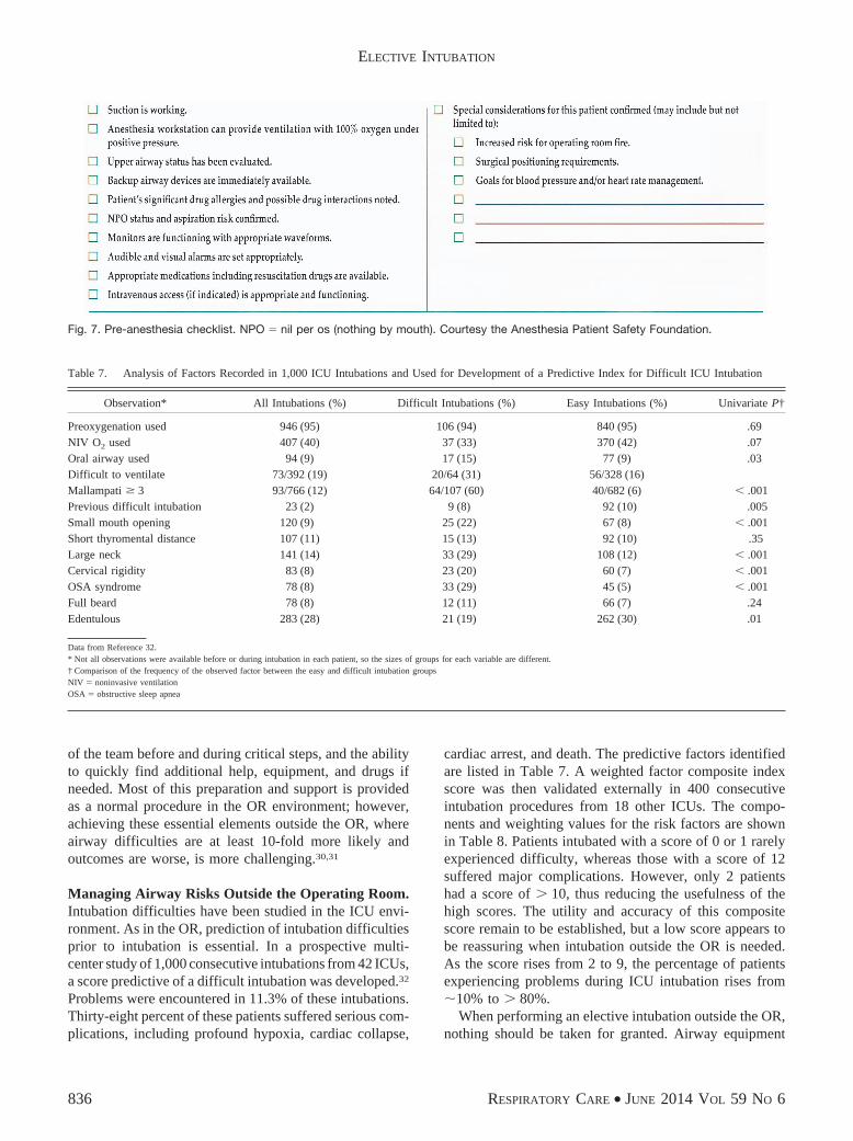
and maintained in some degree of readiness for emergen-cies, needs for a particular patient can be rapidly assem-bled. As part of OR preparations, a checklist (explicit,written, or memorized) is used to ensure that routine andnecessary equipment, monitors, drugs, and supplies areavailable and ready for use before bringing the patient intothe OR. A proposed anesthesia preinduction checklist de-veloped by the Anesthesia Patient Safety Foundation isshown in Figure 7. Even though the OR preparation foranesthesia is repetitive and relatively routine, necessarycomponents are not infrequently missing or found to fail atthe moment when they are most needed. This has led to theredundancy of essential equipment and a time-out for con-firming that essential issues are addressed before begin-ning anesthesia induction.28 Preliminary studies and anec-dotal reports support the belief that the formal confirmationprocess and focused attention of the entire team during apause before induction of anesthesia (including the partic-ipation of the patient) are associated with fewer errors andgreater safety.29 During intubation, experienced anesthesiaassistants or OR circulating nurses are attentive and ac-tively assist with the intubation process. If unexpecteddifficulties are encountered or if essential equipment fails,experienced personnel are readily available to assist inproviding equipment, solve problems, and assist with air-way issues. The components of teamwork, including roledefinition, resource allocation, and communication, helpwith the expectations and competencies of the OR teammembers and allow them to deal with unexpected issues.
In summary, safe airway management requires a thor-ough physical exam, a prepared environment, redundancyof essential equipment, appropriate assistant personnel withairway management understanding and/or skills, attention
Table 5. Drugs That May Be Used for Sedation and/or Anesthesia Induction Before Intubation
Drug Class Comments
Thiopental, amobarbital,methohexital
Barbiturate Short-acting, causing unconsciousness; offset in minutes by redistribution;hypotension due to depression of cardiac function
Propofol Unique sedative/ hypnotic Painful on intravenous injection, has amnestic effects in lower doses, canbe used for anesthesia with continuous infusion, lowers blood pressureby vasodilation
Midazolam (lorazepam, diazepam) GABA receptor antagonist Profound effect on memory, amnesia; little respiratory depression byitself, but synergistic depression with opiates
Dexmedetomidine Central �2 agonist No depression of ventilation, predictable bradycardiaFentanyl, sufentanil, alfentanil Opioid Potent analgesics, medium duration of action; respiratory depressionRemifentanil Opioid Very short-acting narcotic analgesic, unique metabolism accounts for
short half-life (10 min); profound respiratory depression; mildbradycardia
Ketamine NMDA antagonist Sedation and analgesia, opioid effect without respiratory depression,bronchodilator, hypertension, hallucinations
GABA � gamma-aminobutyric acidNMDA � N-methyl-D-aspartate
Table 6. Techniques to Provide Upper and Lower Airway LocalAnalgesia
Inhalation of nebulizedlocal anesthetic
Causes analgesia of entire upper airway,including larynx, vocal cords, andupper trachea; requires 20–30 min ofinhalation; systemic toxicity risk
Oral topical spraying oflocal anesthetic
Rapid onset of numbness, mucosalabsorption creates high blood levelsthat can cause toxicity, larynx andvocal cords not numb
Transtracheal injection oflocal anesthetic
Small dose of drug needed (1–2 mL),good laryngeal analgesia, little or noupper airway analgesia
Superior laryngeal nerveblocks
Small amount locally injected (�1 mL),analgesia of the cords and part of thelarynx, slightly painful injection
Glossopharyngeal nerveblocks
Special long needle needed for intraoralinjection, excellent analgesia ofposterior tongue, little pain oninjection
ELECTIVE INTUBATION
RESPIRATORY CARE • JUNE 2014 VOL 59 NO 6 835
of the team before and during critical steps, and the abilityto quickly find additional help, equipment, and drugs ifneeded. Most of this preparation and support is providedas a normal procedure in the OR environment; however,achieving these essential elements outside the OR, whereairway difficulties are at least 10-fold more likely andoutcomes are worse, is more challenging.30,31
Managing Airway Risks Outside the Operating Room.Intubation difficulties have been studied in the ICU envi-ronment. As in the OR, prediction of intubation difficultiesprior to intubation is essential. In a prospective multi-center study of 1,000 consecutive intubations from 42 ICUs,a score predictive of a difficult intubation was developed.32
Problems were encountered in 11.3% of these intubations.Thirty-eight percent of these patients suffered serious com-plications, including profound hypoxia, cardiac collapse,
cardiac arrest, and death. The predictive factors identifiedare listed in Table 7. A weighted factor composite indexscore was then validated externally in 400 consecutiveintubation procedures from 18 other ICUs. The compo-nents and weighting values for the risk factors are shownin Table 8. Patients intubated with a score of 0 or 1 rarelyexperienced difficulty, whereas those with a score of 12suffered major complications. However, only 2 patientshad a score of � 10, thus reducing the usefulness of thehigh scores. The utility and accuracy of this compositescore remain to be established, but a low score appears tobe reassuring when intubation outside the OR is needed.As the score rises from 2 to 9, the percentage of patientsexperiencing problems during ICU intubation rises from�10% to � 80%.
When performing an elective intubation outside the OR,nothing should be taken for granted. Airway equipment
Fig. 7. Pre-anesthesia checklist. NPO � nil per os (nothing by mouth). Courtesy the Anesthesia Patient Safety Foundation.
Table 7. Analysis of Factors Recorded in 1,000 ICU Intubations and Used for Development of a Predictive Index for Difficult ICU Intubation
Observation* All Intubations (%) Difficult Intubations (%) Easy Intubations (%) Univariate P†
Preoxygenation used 946 (95) 106 (94) 840 (95) .69NIV O2 used 407 (40) 37 (33) 370 (42) .07Oral airway used 94 (9) 17 (15) 77 (9) .03Difficult to ventilate 73/392 (19) 20/64 (31) 56/328 (16)Mallampati � 3 93/766 (12) 64/107 (60) 40/682 (6) � .001Previous difficult intubation 23 (2) 9 (8) 92 (10) .005Small mouth opening 120 (9) 25 (22) 67 (8) � .001Short thyromental distance 107 (11) 15 (13) 92 (10) .35Large neck 141 (14) 33 (29) 108 (12) � .001Cervical rigidity 83 (8) 23 (20) 60 (7) � .001OSA syndrome 78 (8) 33 (29) 45 (5) � .001Full beard 78 (8) 12 (11) 66 (7) .24Edentulous 283 (28) 21 (19) 262 (30) .01
Data from Reference 32.* Not all observations were available before or during intubation in each patient, so the sizes of groups for each variable are different.† Comparison of the frequency of the observed factor between the easy and difficult intubation groupsNIV � noninvasive ventilationOSA � obstructive sleep apnea
ELECTIVE INTUBATION
836 RESPIRATORY CARE • JUNE 2014 VOL 59 NO 6

and monitors may be different and unfamiliar to the cli-nician. There are often no trained assistants who can beexpected to anticipate critical needs. Therefore, a compre-hensive explicit environmental checklist should be carriedout, and staff should be made familiar with how to retrieveemergency assistance. Each of the aspects listed in Table9 should be specifically identified and confirmed. Missingelements need to be addressed, and alternative plans shouldbe developed. A physical checklist can be used and re-viewed, item by item. This is not too basic an approach,especially in an area where intubations are rarely per-formed. While oversights and missing items in the OR cangenerally be quickly remedied, outside the OR, this israrely possible; these environments are much more hostile.Local staff may know where things are kept in their areabut will not be able to anticipate what is needed withoutspecific direction from the intubation team.
Intubating devices are best brought to the intubationlocation. At a minimum, the institution should provide astandardized fully stocked airway cart that contains allusual and special devices and equipment, including thosefor emergency surgical airway access. The organization ofthe cart and location of critical components should befamiliar to the intubation team, allowing necessary butmissing components to be quickly identified and acquiredbefore attempting intubation. At least 2 experienced air-way experts should be present at elective intubations. Thisdegree of preparation and support is less than that pro-vided routinely in the OR. During an emergent intubation,there is likely to be less time for preparation and lesssupport. However, in the case of an elective intubationoutside the OR, even if urgent, appropriate preparationsand equipment must be available even if a delay in theprocedure is necessary.
Monitoring
Minimal acceptable monitoring standards have been ad-opted for all patients undergoing anesthesia. These includea dedicated monitor (a person not performing surgery)able to observe the patient and electronic monitoring de-vices. The intubating team should be able to respond tochanges in vital signs, administer fluids and drugs, andmanage the patient’s airway. Minimal patient monitoringincludes continuous visual electrocardiogram display of atleast 2 leads, continuous pulse oximetry, and intermittent(at least every 3 min) arterial blood pressure measure-ments. In the OR, capnography, FIO2
, and anesthetic gasmonitoring are also routine. Outside the OR, either CO2
measurement devices or capnography for intubation con-firmation should be available.33 In addition, the patient’sclinical status may mandate additional needs such as mon-itoring of intra-arterial pressure, central venous pressure,cardiac output, and intracranial pressure.
Intubation: Plans A, B, and C
After the environment is optimized, equipment con-firmed present and working, and appropriate help avail-
Table 8. Factors and Weighting Used to Derive a Composite ScorePredictive of Problems or Difficulties During IntubationPerformed in an ICU
Factors Points
Factors related to patientMallampati III or IV 5Obstructive sleep apnea syndrome 2Reduced mobility of cervical spine 1Limited mouth opening � 3 cm 1
Factors related to pathologyComa 1Severe hypoxemia (SpO2
� 80%) 1Factor related to intubator
Non-anesthesiologist 1
From Reference 32, with permission. The score can range from 0 to 12.
Table 9. Essential Requirements for Safety and Success WhenPerforming an Elective Intubation Outside of the OperatingRoom
Oxygen source and backup sourceAirway equipment
Assorted clear plastic ventilation masksSelf-inflating manual ventilation deviceNon-self-inflating airway ventilation deviceOral, nasal, upper airway devicesAssorted sizes and types of supraglottic ventilating airwaysEsophageal blocking and lung ventilation deviceSuction system with tracheal and oral aspirating devices
Intubating equipmentLaryngoscope handles (at least 2 working)Laryngoscope blades (assortment of different sizes and shapes)Appropriate assortment of cuffed ETTsMalleable styletsGum elastic bougie or tracheal identifying styletHead-and-shoulder positioning devicesSurgical emergency airway device
Difficult airway or difficult intubation expectedIndirect laryngoscope and styletSupraglottic intubating airway devices and tubesFlexible fiberoptic intubating scope and light source
Designated staff to helpMethod to get additional and appropriate help quicklyFunctioning intravenous accessDrugs for intravenous injection and qualified person to administer
them
ETT � endotracheal tube
ELECTIVE INTUBATION
RESPIRATORY CARE • JUNE 2014 VOL 59 NO 6 837

able, a primary intubation sequence is chosen and dis-cussed with the team. If the airway examination isreassuring, the patient’s physiologic risks are low, and thepatient is not an aspiration risk, intubation using DL afterinduction of deep sedation or general anesthesia with orwithout a muscle relaxant is a reasonable first choice. Ifthe intubation is predicted to be difficult, a different ap-proach, such as use of an indirect laryngoscope, may bechosen for the initial intubation attempt. If the patient is athigh risk of vomiting (eg, bowel obstruction) and aspirat-ing (eg, altered mental status), this must be taken intoaccount in determining the first choice of an intubationtechnique. The so-called rapid-sequence intubation, whichincludes induction with immediate paralysis, may be pref-erable in some of these cases.34 The risk of an adversephysiologic response or inability to intubate with DL mustbe weighed when choosing to perform a rapid-sequenceintubation. Direct posterior cricoid ring pressure duringrapid-sequence intubation (Sellick’s maneuver) is oftenused to prevent passive regurgitation; however, its effec-tiveness in this regard has been questioned.35 Additionally,intubation with cricoid pressure is more difficult and maydelay insertion of the tube and inflation of the tube cuff,which protects the airway.36 Other approaches, includingmethods for awake intubation (discussed above), can besuccessful in preventing aspiration in patients at high risk.
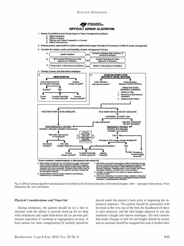
Airway management failures, including intubation is-sues, are a cause of patient morbidity in and out of the OR.Delay in recognizing a problem and failing to change courseresult in worsening patient outcomes. This has led to thedevelopment and promotion of difficult-airway manage-ment algorithms by various professional societies. Onesuch guideline is the difficult-airway algorithm37 createdand revised by the American Society of Anesthesiologists(Fig. 8).38 This guide can serve as a road map for choosingthe next option to establish a patent airway when the firstor subsequent attempted airway technique has failed. Thestarting point of this and other guidelines is to establisheffective manual ventilation with optimal head positionusing simple supraglottic airway devices (eg, oral and na-sal pharyngeal airways). An important addition to ensuringgas exchange was provided with the invention of the la-ryngeal mask airway (LMA). Designed by Dr Brain in thelate 1980s, this device bypasses the tongue and upper air-way structures, seats in the lower pharynx, and conformsto the opening of the larynx. A seal is formed by inflationof the mask cuff pressing against the pharyngeal walls, andmanual ventilation can be provided through a standardETT connector. Once in place, the LMA is remarkablywell tolerated even without deep anesthesia. LMA inser-tion and confirmation techniques are easily taught andquickly mastered. Use of this device has been added to theairway algorithm as a rescue airway and when other ap-proaches have failed. Of note, an LMA does not protect a
patient from aspiration and, although an excellent tempo-rizing technique, is not a safe or definitive airway whenaspiration is a concern.
No matter which primary intubation technique is cho-sen, explicit plans must be made in advance for the nextsteps if intubation fails or unexpected difficulties occuronce the primary intubation sequence is begun. This plan-ning should be done before beginning the intubation, andthese alternative plans should be shared with the clinicianspresent and assisting. If the patient is able to understandand cooperate, the intubation plan (as well as plans forcomfort measures after intubation) should be discussedwith the patient, and verbal or written consent should beobtained.
Preparing for Intubation
After the primary and additional contingency plans forintubation are developed, all indicated and backup airwaytools are available and working, and necessary drugs areavailable and prepared, patient preparation can then begin.Often overlooked, a functioning and adequate source ofsuction is imperative in case the patient vomits or hassignificant secretions or blood in the airway. This shouldinclude suction tips capable of removing massive quanti-ties of gastric material from the upper airway and mouth ifthe patient vomits before or during intubation, as well aslower airway catheters if aspiration occurs. During an elec-tive intubation, routine and indicated additional monitorsshould be placed and functioning. Frequent blood pressuremeasurements are needed to detect and correct the ex-pected physiologic changes that occur with sedation/anes-thesia, intubation, and initiation of mechanical ventilation.Monitoring should begin prior to starting the intubationsequence so that baseline values and variability can benoted. As mentioned before, a means of measuring ex-haled carbon dioxide is needed for confirmation of intu-bation. Continuous CO2 monitoring during the intubationprocess and with mechanical ventilation is recommendedto detect airway problems before tragedy occurs.39 De-pending on individual patient risk factors and concern forthe response to intubation, continuous intra-arterial pres-sure measurements may be needed to provide safe careand to avoid cardiovascular collapse. In situations in whichchanges in intracranial pressure are of concern, direct pres-sure monitoring with an intracranial device is useful. Drugsthat can modify intracranial pressure changes should beavailable, as well as the expertise to order and administerthem. A peripheral nerve stimulator can be used to detectmaximal paralysis following administration of a neuro-muscular blocker.
ELECTIVE INTUBATION
838 RESPIRATORY CARE • JUNE 2014 VOL 59 NO 6
Physical Considerations and Time-Out
During intubation, the patient should be on a bed orstretcher with the ability to provide head-up tilt (to helpwith intubation) and rapid head-down tilt (to prevent pul-monary aspiration if vomiting or regurgitation occurs). Ahard surface for chest compressions (if needed) should be
placed under the patient’s back prior to beginning the in-tubation sequence. The patient should be positioned withhis head at the very top of the bed, the headboard (if thereis one) removed, and the bed height adjusted to suit theintubator’s height and chosen technique. The bed controlsthat make changes in bed tilt and height should be tested,and an assistant should be assigned this task if needed after
Fig. 8. Difficult airway algorithm developed and modified by the American Society of Anesthesiologists. LMA � laryngeal mask airway. FromReference 38, with permission.
ELECTIVE INTUBATION
RESPIRATORY CARE • JUNE 2014 VOL 59 NO 6 839

the intubation process begins. Because intubations occur-ring outside the OR are unscheduled and the staff involvedis usually unfamiliar with the intubation process and se-quence, a formal time-out is useful to bring the team to-gether. During this pause, immediately before beginningthe intubation sequence, the patient is identified, the statusand plan are reviewed, critical points are identified, equip-ment is confirmed to be present, and tasks are assigned.Alternative plans should be discussed at this time. Thepatient should be explicitly identified, the initial intubationplan should be verbalized, and the team members shouldbe identified by name and assignment. Medications, aller-gies, intravenous access, oxygen source, suction, monitors,manual ventilatory devices, simple airway devices, intu-bating equipment, the selected and tested ETT, and ad-juncts to be used if intubation fails should be verballyconfirmed as present and functional. As these are con-firmed, the individual responding should identify them-selves by name, professional role, and tasks they will beassuming. If someone or something that is needed is miss-ing, this situation must be remedied before starting theintubation process. During a pause after reviewing thechecklist (see Table 9), the group should be asked, “Doesanyone have a question or a safety concern?” If there areno concerns from the group, the beginning of the intuba-tion sequence should be announced, and patient prepara-tion should begin with oxygen administration.
Denitrogenation or Preoxygenation
Prevention of hypoxemia and its deleterious effects onpatients is the paramount consideration in airway manage-ment situations. The term preoxygenation is commonlybut wrongly applied to a patient inhaling a high concen-tration (usually 100%) of oxygen immediately before be-ginning medication administration for intubation. The ac-tual process is effected by removal of nitrogen from thelungs and body storage spaces and should more accuratelybe called denitrogenation. Clinically effective denitroge-nation can be achieved during a short period of spontane-ous or assisted ventilation with a very high oxygen con-centration. This provides a margin of safety for periods ofapnea of at least 5–10 min without critical oxygen desatu-ration (Fig. 9). With prolonged oxygen breathing (� 45 minof 100%), duration of apnea without hypoxemia for aslong as 30–45 min can be achieved.41 Of course, PaCO2
will rise during apnea, and levels exceeding 150 mm Hghave been reported.42 This degree of hypercarbic acidosisis remarkably well tolerated since hypoxemia is prevented.Several methods have been suggested to achieve a marginof safety in a brief period, and these include 3 min ofspontaneous breathing with 100% O2, 4–5 maximal inha-lations and exhalations with a high-flow non-rebreathingcircuit,43 breathing with face mask CPAP or noninvasive
mechanical ventilation, and 100% O2 for a longer periodof time.44 While any of these are generally effective ifintubation is rapidly successful, in compromised patientsor during prolonged unsuccessful attempts at intubation,saturation may fall to unacceptable levels. Successive in-tubation attempts should be interrupted with periods ofmask ventilation to restore baseline oxygenation. Beforebeginning intubation, the ability to provide positive-pres-sure ventilation with a mask should be confirmed. If man-ual mask ventilation is difficult or unsuccessful, use of anLMA should be considered, and avoidance of neuromus-cular blockers should also be considered. If failed venti-lation continues despite maximal efforts, desaturation oc-curs to dangerous levels (as indicated by a falling heartrate), and one or two attempts at intubation have failed,then a surgical airway should be rapidly placed, and ade-quate oxygenation should be restored. Surgical airways arelisted in Table 4. Transtracheal jet ventilation through alarge-bore intravenous catheter is a reasonable alternativeto other surgical airways and offers a low risk of compli-cations and reasonable success if equipment is close athand and if undue delay is avoided.45
Intubation Using Direct Laryngoscopy
Initial Plan
If a difficult intubation is not anticipated and if thepatient’s risk for deterioration during or immediately afterintubation is low, then an acceptable approach is asedated/anesthetized intubation under direct vision with astandard straight or curved laryngoscope blade with laryn-geal exposure facilitated by use of a neuromuscular blocker.This is a standard technique in most OR cases. After deni-trogenation, unconsciousness is induced; manual ventila-tion may be attempted; and if manual mask ventilation iseasy, the relaxant may be administered. The head shouldbe in ideal intubating position before starting: flexed onthe lower cervical spine and occiput slightly elevated on a
Fig. 9. Comparison of 2 methods of preoxygenation. Data fromReference 40.
ELECTIVE INTUBATION
840 RESPIRATORY CARE • JUNE 2014 VOL 59 NO 6

non-compressible head support, with extension of the up-per cervical spine. To further improve the view of thelarynx, some suggest placing the bed in a slight reverseTrendelenburg position. This also has the advantage ofreducing airway pressures and lowering gastric reflux po-tential. Intubation using DL is the most frequently taughtand mastered intubation technique and is therefore the firstchosen and most often successful technique. Mastery ofthis intubation technique is achieved by anesthesia traineesafter performing between 100 and 200 intubations.46 Oth-ers with less experience or who intubate only rarely shouldhave immediately available assistance from a more expe-rienced person during an elective out-of-OR intubation,when conditions are less controlled, help is farther away,and complications are more frequent than in the OR.
Intubation is a risky and complex team process withspecific actions requiring full attention of all members ofthe team. The ongoing activities of patient care create adegree of chaos as each individual performs his own taskat the patient’s bedside. Safe intubation requires a quietand calm environment and the attention of all who arepresent to focus on this critical process. The person per-forming the intubation should assertively announce, “I amstarting the intubation.” This will get everyone’s attentionand allow the group to perform better as a team.
A functional and tested peripheral or central intravenousline should be used to deliver sedating and anesthetizingmedications. The chosen medication is injected into therunning intravenous line, usually by another team memberat the direction of the person performing the intubation.The appropriate sterile technique is consistently applied tothe injection process. Once the patient has lost conscious-ness and eyelash reflex is extinguished, airway patencyand ability to ventilate are tested by performing one ormore manual breaths. Success of manual ventilation isconfirmed by observing the chest rise and fall, noting ex-haled humidity in the mask, listening with a stethoscope tothe trachea or lung fields, and/or observing the capnographtracing. If a muscle relaxant is planned, it is then admin-istered, and ventilation is provided until maximal relax-ation is obtained.
DL begins by opening the patient’s mouth and insertingthe blade from the right side of the mouth, sweeping thetongue to the left. A crossed-finger technique to force themouth open and to dislocate the jaw is recommended.Alternatively, use of the right hand to rotate the headbackwards (neck extension) allows the mouth to fall openand accept the blade. The blade is moved from right to left,moving the tongue past the midline. As the blade is ad-vanced, the oral anatomy comes into view, and any ab-normalities are noted. With further advancement, the tip ofthe epiglottis should be seen. The larynx is visualized bycatching the tip of the epiglottis if using a straight blade orby advancing into the vallecula (the space above the epi-glottis) if using a curved blade. Lifting the blade (hangingthe patient’s jaw from the blade) brings the laryngeal struc-tures into view.47 The Cormack-Lehane system of gradingthe laryngeal view is often used to describe the best viewobtained during laryngoscopy. Grade 1 is assigned if theentire larynx and both commissures are seen, grade 2 ifhalf of the larynx and cords and only the posterior com-missure are seen, grade 3 if none of the laryngeal openingis seen and only the posterior structures (the arytenoidcartilages) can be identified, and grade 4 if no identifiablelaryngeal anatomy can be seen (Fig. 10).6 Even when theglottic view is poor, blind intubation with a styleted ETTor over a bougie may not be difficult.
The selected ETT, which has been prepared and tested,is inserted from the right side of the mouth, out of thedirect visual field, with the tube tip pointing upward. TheETT is then advanced upward through the laryngeal open-ing and into the trachea. This is best done with the intu-bator’s head at least 2 feet from the patient’s face, allow-ing binocular vision and depth perception to be maintainedand facilitating controlled manipulation of the tip of thetube into the larynx. The tube should be placed so that thecuff is about 1 cm below the cords, but not farther to avoidendobronchial placement. If the best glottic view obtainedis a grade 3 or 4 view, changing the head position orchoosing a different length or shaped blade can be triednext. When an unexpected poor view of the larynx isencountered during DL, a malleable stylet is often helpful
Fig. 10. Cormack-Lehane grades.
ELECTIVE INTUBATION
RESPIRATORY CARE • JUNE 2014 VOL 59 NO 6 841

in directing the ETT up into the so-called anterior larynx.Experienced clinicians will use a stylet to stiffen and shapethe flexible ETT, giving it a hockey stick curve to be usedduring any high-risk intubation. This advanced preparationallows a blind insertion when only a small or no part of thelarynx is visible. After the tip is blindly inserted a shortdistance (enough to pass through the vocal cords), thestylet is pulled back in the tube to allow the ETT tip tobend downward and follow as the trachea descends intothe thorax. If a malleable stylet is routinely placed beforeall intubations and if the tube is formed in an ideal shape,the ETT will always be ready when an unexpected poor-grade laryngeal view is encountered. After inflation of theETT cuff, confirmation that the ETT is in the trachea andnot the esophagus is necessary following all intubationsand especially after a blind insertion.
Confirmation of Tracheal Intubation
Even if the ETT is placed by directly observing the tubeenter the larynx and carefully advancing it an appropriatedistance in the trachea, additional measures are used toconfirm correct placement. The first sign of tracheal place-ment is humidity forming in the tube during gentle venti-lation. Three or 4 breaths are delivered with a manualventilatory device, and humidity is noted inside the tube,appearing and disappearing coincident with ventilation. ACO2 monitor or detector is then attached to the tube, andappropriate levels of CO2 should be detected in the nextfew breaths with tracheal placement. With esophagealplacement, low levels of exhaled CO2 can be seen withinitial ventilation, but the CO2 level will not be as high asexpected from the trachea and will fall farther with eachadditional breath. Hyperventilation of the lungs can alsoreduce the measured CO2 even if the ETT is correctlyplaced in the trachea. For this and other reasons, hyper-ventilation should be avoided. Next, both lung fields andthe stomach area should be carefully auscultated. This canreliably identify an esophageal placement (loud ventilationsounds over the stomach and muted or no sounds over thelungs) and help avoid bronchial placement (no sounds overthe stomach and asymmetric breath sounds over the lungfields). If bronchial intubation is suspected, the ETT canbe withdrawn until lung breath sounds are equalized, andthe tube can then be fixed in place. The distance marker onthe tube at the teeth (or gums) should be noted when thebreath sounds are equal and used as a reference pointshould the tube be moved in the future. There are manyways an ETT may be secured to the patient. Each has itadvantages and disadvantages. The method chosen shouldbe familiar to the caregivers who will manage the patientafter the intubation team turns over care.
Physiologic Changes and Management Strategies
As mentioned earlier, blood pressure changes duringand following intubation can be dramatic and life-threat-ening. If the patient is at risk of not tolerating these ex-pected changes, plans to modify these responses are needed.These include choice of method for intubation, use oftopical analgesia, and administration of cardiovascularmedications. If drugs are needed, those that have rapidonset and are of short duration should be chosen. Hyper-tension and tachycardia during tracheal tube insertion areoften followed by hypotension as reflex responses occur.Patients receiving active treatments for underlying hyper-tension or cardiac disease may have a paradoxical responseto intubation, that is, hypotension. The application of pos-itive-pressure ventilation after intubation will reduce ve-nous return to the heart, resulting in reduced cardiac outputand hypotension. This must be anticipated, and fluids and/ormedications should be ready and administered in antici-pation of this effect. Ventilation should be initiated withthe lowest acceptable rate and tidal volume to minimizehypotension. In hypovolemia or patients with low cardiacreserve, intubation and ventilation can cause cardiac ar-rest. Resuscitation measures should be available and ap-plied as needed without delay. While medication choicesare not the domain of this paper, some useful drugs formanaging the hemodynamic responses during airway man-agement are listed in Table 10.
Table 10. Techniques and Drugs Useful for Managing PhysiologicalTrespass Incurred by Tracheal Intubation and Positive-Pressure Ventilation
Topical airway anesthesiaSystemic treatment options
Deep sedation, opioids, general anesthesiaCardiac drugs
� Blockers (esmolol)Vascular drugs
Direct dilators (hydralazine, nitroprusside, nicardipine)� Blockers (trimethaphan camsylate)Combined effects (labetalol)
Central �2 agents (dexmedetomidine)Bronchodilators
� AgonistsVolatile anesthetics
Hypotension treatmentsFluidsVasopressors (ephedrine, phenylephrine, vasopressin)Inotropic agents (epinephrine, norepinephrine)Inodilators (amrinone, milrinone)
CPRDefibrillation
CPR � cardiopulmonary resuscitation
ELECTIVE INTUBATION
842 RESPIRATORY CARE • JUNE 2014 VOL 59 NO 6
Alternative Airway Plans
With any intubation, changes in plans may be necessarydepending on what happens during the initially chosensequence. Major decision points are indicated in the air-way algorithm in Figure 8. For instance, if manual venti-lation (without a relaxant) is difficult, a short-acting re-laxant rather than a longer acting one may be chosen.Also, the patient may be allowed to awaken, and an alter-native intubation technique could be used. When the in-tubation is being performed outside the OR, explicit plansfor dealing with the difficulties that may occur must beestablished prior to starting the intubation so that appro-priate equipment and personnel are available. If a difficultintubation is predicted or known from previous intuba-tions, all reasonable alternatives for achieving a successfulintubation must be available. These must include, at aminimum, the airway equipment and the plan that wassuccessful in the past but must also include a plan for asurgical airway if these fail. The patient’s consent shouldinclude a discussion of this potential outcome. It may beprudent, if the situation allows, to move the patient to theOR when an extremely difficult airway or intubation isanticipated and to have a surgeon or other airway expertavailable.
The components of the difficult-airway algorithm aregenerally available in the OR suite. For intubations outsidethe OR, however, a lifesaving piece of equipment may notbe available when problems occur. Having a dedicateddifficult-airway cart that is mobile and stocked with allnecessary devices and delivered on demand is one solutionto this dangerous situation. A system solution should bedeveloped in any health system where out-of-OR intuba-tions are necessary on more than the occasional basis.
Postintubation Plans
Once intubation is successful, plans for continued main-tenance of the ETT and patient comfort need to be initi-ated. As mentioned previously, manual ventilation or ven-tilation with a mechanical ventilator may induce the needfor hemodynamic support due to the effects of positive-pressure ventilation affecting cardiac function. Vasopres-sors and fluids should be available and administered asneeded. Use of sedation, analgesia, and neuromuscularblockade should be considered and may be part of theairway maintenance plan. Inadvertent extubation is a risk,and an effective method to secure an ETT should be ap-plied. No one method is appropriate for all patients andsituations. The consequences of tube displacement are moregrave for more difficult intubations. Under these condi-tions, use of the most effective method of securing the tubeis required. While placement of the tube may be the focusof the team initially, planning for its ultimate removal at a
later time is essential. Next to intubation, extubation (ei-ther planned or accidental) is one of the most dangeroustimes in airway management. The risks at extubation of apatient after a difficult intubation are actually greater thanthose at intubation and are often underestimated by care-givers. Planning for extubation should be started at thetime of intubation. Where and when to extubate should beinfluenced by the issues surrounding intubation.48 Algo-rithms for stratification and management of airway risks atextubation have been proposed.49
Transfer of Care
Often during intubations outside the OR environment,the person placing the ETT is not the one who will becaring for the patient and the airway during the subsequentperiod. Depending on the reason for intubation (eg, toallow a brief diagnostic or therapeutic procedure or a morepersistent need such as progressive respiratory failure fromCOPD or congestive heart failure), complex medical careplans will be carried out that may impact airway stability.If the person performing the intubation is leaving the bed-side, a change of care will occur. It is now well recognizedthat turnover of care carries significant risk. Transmissionof critical patient information is frequently faulty. Idealmethods to summarize and pass on relevant and completeinformation at transfer points have not been established;however, checklists are often used to avoid omission ofessential information and to delineate required activities.50
In general, the transfer communications regarding airwaymanagement should include the medications administered;the patient’s response to them; and the expected durationof those used to facilitate airway placement, to reduceconsciousness, and to modify the physiologic changes dur-ing and following intubation.
Since the subsequent care is often not provided by anairway expert, explicit lists of drugs and doses used andwhether the patient’s response was normal, resistant, orsensitive should be conveyed. In addition, a description(verbal and written) of the intubation sequence, adjunctsused, and the ultimately successful technique should be
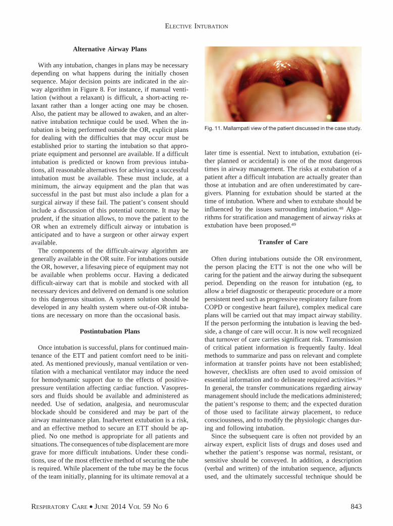
Fig. 11. Mallampati view of the patient discussed in the case study.
ELECTIVE INTUBATION
RESPIRATORY CARE • JUNE 2014 VOL 59 NO 6 843

communicated to the next team. Any deviations from theinitial plan and why they were invoked should be de-scribed. Additional information about the intubation andsuggestions that could improve the process if re-intubationbecomes necessary should be included. The method usedto confirm correct tracheal placement (ie, expired CO2,direct vision, breath sounds, and fiberoptic observation ofthe trachea through the ETT) should be conveyed anddocumented. The method used to secure the tube and thedepth of the tube at the point it is secured to the patient arepart of the transfer process and notes.
After the Difficult Intubation: A Case Review
Following an unexpected difficult-airway managementissue, additional communication measures are required. Anote in the medical record detailing the event should beentered as soon as possible so that the details are clearlyrecalled and accurately described.
The following is an actual case report of an unexpecteddifficult intubation in which one of the authors (CGD Jr)recently took part. The patient’s preoperative airway eval-uation is shown in Figures 2–6, and the MP view is shownin Figure 11. The patient was essentially normal by allusual measures, demonstrating no markers predictive of adifficult airway. Intubation was planned using DL afterintravenous induction of general anesthesia. Figure 12 isthe note entered into the medical record describing theairway events that ensued. After an uneventful anesthesiainduction, mask ventilation was easily maintained. Severalattempts at intubation were tried using DL and severaldifferent blades, but no laryngeal view was able to beobtained (Cormack-Lehane grade 4 view). An LMA wasthen inserted, and ventilation was easily maintained. TheLMA was changed to a larger size and used as a conduitto facilitate a fiberoptic scope to enter the trachea. The
scope was used to place a guide, and the LMA was re-moved. An ETT was advanced over the guide into thetrachea. Successful tracheal placement was confirmed withcapnography. The surgery proceeded uneventfully. Fol-lowing the procedure, a discussion of the airway eventswas held with the patient and his wife.
The cause of the difficult intubation encountered in thepatient above was subsequently identified as the presenceof massive mandibular tori (Fig. 13). These benign bonyoutgrowths were not noted during the preoperative airwayexamination. Such tori are commonly seen by dental sur-geons, as their presence is a challenge to providing dental
Fig. 13. The cause of the intubation difficulty in the case study wasmassive mandibular tori.
Fig. 12. Note dictated and entered into the medical record of the patient in the case study. LMA � laryngeal mask airway.
ELECTIVE INTUBATION
844 RESPIRATORY CARE • JUNE 2014 VOL 59 NO 6

prostheses and tooth repair. By functionally narrowing thesubmental space, these mandibular processes (when large)prevent displacement of the tongue during DL and lead tothe difficult intubation that was encountered in this pa-tient.51
To alert subsequent caregivers in the hospital that asignificant airway event has occurred, a warning label canbe placed on the ETT, the patient’s chart or arm, the bedheadboard, or the room door. An entry using the medicalrecord critical alert system can also be used. In the casedescribed above, an armband was placed and remaineduntil hospital discharge (Fig. 14). Communication withfuture caregivers of the unexpected difficulties confrontedduring airway management begins with a discussion of theevents with the patient and his family. This discussionshould occur at an appropriate time before discharge. Thenote shown (Fig. 12) became a part of the medical record,and a copy of it was given to the patient. Some expertsadvise a patient experiencing a difficult intubation to ac-quire a MedicAlert bracelet or medallion to notify futureclinicians of the airway problem at a time when the patientor family may not be able to supply necessary information.Many professional societies have advocated providing thepatient with a standard difficult-intubation letter as de-scribed previously (Fig. 1), which can be included in thepatient’s medical record and given to the patient to beshared with future caregivers.
Conclusions
Management of elective intubations in the OR are fre-quently and safely performed daily throughout the world.The OR intubation forms a body of experience and infor-mation that can be used to improve the intubation processwhen performed elsewhere. Areas of highest risk that needattention are preintubation patient evaluation, intubatingenvironment optimization, postintubation physiology man-agement, and communication between caregivers. Docu-mentation of airway difficulties needs consistent attentionin the OR domain as well as when the intubation is per-formed elsewhere.
REFERENCES
1. Centers for Disease Control and Prevention. Inpatient surgery. http://www.cdc.gov/nchs/fastats/insurg.htm. Accessed June 23, 2013.
2. Centers for Disease Control and Prevention. NAMCS and NHAMCSWeb Tables. http://www.cdc.gov/nchs/ahcd/web_tables.htm#2010.Accessed June 23, 2013.
3. Caplan RA, Posner KL, Ward RJ, Cheney FW. Adverse respiratoryevents in anesthesia: a closed claims analysis. Anesthesiology 1990;72(5):828-833.
4. Schwartz DE, Matthay MA, Cohen NH. Death and other complica-tions of emergency airway management in critically ill adults. Aprospective investigation of 297 tracheal intubations. Anesthesiol-ogy 1995;82(2):367-376.
5. Williams KN, Carli F, Cormack RS. Unexpected difficult laryngos-copy: a prospective survey in routine general surgery. Br J Anaesth1991;66(1):38-44.
6. Cormack RS, Lehane J. Difficult tracheal intubation in obstetrics.Anaesthesia 1984;39(11):1105-1111.
7. Schmidt UH, Kumwilaisak K, Bittner E, George E, Hess D. Effectsof supervision by attending anesthesiologists on complications ofemergency tracheal intubation. Anesthesiology 2008;109(6):973-977.
8. Jaber S, Amraoui J, Lefrant JY, Arich C, Cohendy R, Landreau L, etal. Clinical practice and risk factors for immediate complications ofendotracheal intubation in the intensive care unit: A prospective,multiple-center study. Crit Care Med 2006;34(9):2355-2361.
9. American Society of Anesthesiologists Task Force on PreanesthesiaEvaluation. Practice advisory for preanesthesia evaluation: a reportby the American Society of Anesthesiologists Task Force on Prean-esthesia Evaluation. Anesthesiology 2002;96(2):485-496.
10. Koenig HM. No more difficult airway, again! Time for consistentstandardized written patient notification of a difficult airway.Anesthesia Patient Safety Foundation Newsletter: Summer 2010.http://www.apsf.org/newsletters/html/2010/summer/06_diffairway.htm. Accessed May 13, 2014.
11. Hamaekers AE, Henderson JJ. Equipment and strategies for emer-gency tracheal access in the adult patient. Anaesthesia 2011;66 (Suppl2):65-80.
12. Kheterpal S, Martin L, Shanks AM, Tremper KK. Prediction andoutcomes of impossible mask ventilation: a review of 50,000 anes-thetics. Anesthesiology 2009;110(4):891-897.
13. Mallampati SR, Gatt SP, Gugino LD, Desai SP, Waraksa B, FreibergerD, Liu PL. A clinical sign to predict difficult laryngoscopy: a pro-spective study. Can Anaesth Soc J 1985;32(4):429-434.
14. Khan ZH, Mohammadi M, Rasouli MR, Farrokhnia F, Khan RH.The diagnostic value of the upper lip bite test combined with ster-nomental distance, thyromental distance, and interincisor distance
Fig. 14. A difficult-airway warning band was placed on the patient,alerting hospital caregivers of the potential airway problem.
ELECTIVE INTUBATION
RESPIRATORY CARE • JUNE 2014 VOL 59 NO 6 845
for prediction of easy laryngoscopy and intubation: a prospectivestudy. Anesth Analg 2009;109(3):822-824.
15. Safavi M, Honarmand A, Zare N. A comparison of the ratio ofpatient’s height to thyromental distance with the modified Mallam-pati and the upper lip bite test in predicting difficult laryngoscopy.Saudi J Anaesth 2011;5(3):258-263.
16. Pedersen T, Eliasen K, Henriksen E. A prospective study of riskfactors in cardiopulmonary complications associated with anaesthe-sia and surgery: risk indicators of cardiopulmonary morbidity. ActaAnaesthesiol Scand 1990;34(2):144-155.
17. Pedersen T, Viby-Mogensen J, Ringsted C. Anaesthetic practice andpostoperative pulmonary complications. Acta Anaesthesiol Scand1992;36(8):812-818.
18. Perkins ZB, Gunning M, Crilly J, Lockey D, O’Brien B. The hae-modynamic response to pre-hospital RSI in injured patients. Injury2013;44(5):618-623.
19. Bedford RF, Feinstein B. Hospital admission blood pressure: a pre-dictor for hypertension following endotracheal intubation. AnesthAnalg 1980;59(5):367-370.
20. Pedersen T. Complications and death following anaesthesia. A pro-spective study with special reference to the influence of patient-,anaesthesia-, and surgery-related risk factors. Dan Med Bull 1994;41(3):319-331.
21. Pedersen T, Johansen SH. Serious morbidity attributable to anaes-thesia. Considerations for prevention. Anaesthesia 1989;44(6):504-508.
22. Stoelting RK. Circulatory changes during direct laryngoscopy andtracheal intubation: influence of duration of laryngoscopy with orwithout prior lidocaine. Anesthesiology 1977;47(4):381-384.
23. Hamill JF, Bedford RF, Weaver DC, Colohan AR. Lidocaine beforeendotracheal intubation: intravenous or laryngotracheal? Anesthesi-ology 1981;55(5):578-581.
24. Morris IR. Continuing medical education: fibreoptic intubation. CanJ Anesth 1994;41(10):996-1007; discussion 1007-1008.
25. Heffner AC, Swords DS, Nussbaum ML, Kline JA, Jones AE. Pre-dictors of the complication of postintubation hypotension duringemergency airway management. J Crit Care 2012;27(6):587-593.
26. Murphy BA, Durbin CG Jr. Using ventilator and cardiovasculargraphics in the patient who is hemodynamically unstable. RespirCare 2005;50(2):262-274; discussion 274.
27. Kopman AF, Wollman SB, Ross K, Surks SN. Awake endotrachealintubation: a review of 267 cases. Anesth Analg 1975;54(3):323-377.
28. DeMaria S Jr, Neustein SM. Production pressure, medical errors, andthe pre-anesthesia checkout. Middle East J Anesthesiol 2010;20(5):631-638.
29. Hart EM, Owen H. Errors and omissions in anesthesia: a pilot studyusing a pilot’s checklist. Anesth Analg 2005;101(1):246-250.
30. Heuer JF, Barwing TA, Barwing J, Russo SG, Bleckmann E, QuintelM, Moerer O. Incidence of difficult intubation in intensive carepatients: analysis of contributing factors. Anaesth Intensive Care2012;40(1):120-127.
31. Robbertze R, Posner KL, Domino KB. Closed claims review ofanesthesia for procedures outside the operating room. Curr OpinAnaesthesiol 2006;19(4):436-442.
32. De Jong A, Molinari N, Terzi N, Mongardon N, Arnal JM, GuittonC, et al. Early identification of patients at risk for difficult intubation
in the intensive care unit: development and validation of theMACOCHA score in a multicenter cohort study. Am J Respir CritCare Med 2013;187(8):832-839.
33. Goldhill DR, Cook TM, Waldmann CS. Airway incidents in criticalcare, the NPSA, medical training and capnography. Anaesthesia 2009;64(4):354-357.
34. Ehrenfeld JM, Cassedy EA, Forbes VE, Mercaldo ND, SandbergWS. Modified rapid sequence induction and intubation: a survey ofUnited States current practice. Anesth Analg 2012;115(1):95-101.
35. Brimacombe JR, Berry AM. Cricoid pressure. Can J Anaesth 1997;44(4):414-425.
36. Snider DD, Clarke D, Finucane BT. The ‘BURP’ maneuver worsensthe glottic view when applied in combination with cricoid pressure.Can J Anaesth 2005;52(1):100-104.
37. Practice guidelines for management of the difficult airway: a report bythe American Society of Anesthesiologists Task Force on Managementof the Difficult Airway. Anesthesiology 1993;78(3):597-602.
38. American Society of Anesthesiologists Task Force on Managementof the Difficult Airway. Practice guidelines for management of dif-ficult airway: an updated report by the American Society of Anes-thesiologists Task Force on Management of the Difficult Airway.Anesthesiology 2003;98(5):1269-1277.
39. Mort TC. The incidence and risk factors for cardiac arrest during emer-gency tracheal intubation: a justification for incorporating theASAguide-lines in the remote location. J Clin Anesth 2004;16(7):508-516.
40. Valentine SJ, Marjot R, Monk CR. Preoxygenation in the elderly: acomparison of the four-maximal-breath and three-minute techniques.Anesth Analg; 1990;71(5):516-519.
41. Teller LE, Alexander CM, Frumin MJ, Gross JB. Pharyngeal insuf-flation of oxygen prevents arterial desaturation during apnea. Anes-thesiology 1988;69(6):980-982.
42. Frumin MJ, Epstein RM, Cohen G. Apneic oxygenation in man.Anesthesiology 1959;20(6):789-798.
43. Tanoubi I, Drolet P, Donati F. Optimizing preoxygenation in adults.Can J Anaesth 2009;56(6):449-466.
44. Weingart SD, Levitan RM. Preoxygenation and prevention of de-saturation during emergency airway management. Ann Emerg Med2012;59(3):165.e1-175.e1.
45. Ross-Anderson DJ, Ferguson C, Patel A. Transtracheal jet ventila-tion in 50 patients with severe airway compromise and stridor. Br JAnaesth 2011;106(1):140-144.
46. Clarke RC, Gardner AI. Anaesthesia trainees’ exposure to airwaymanagement in an Australian tertiary adult teaching hospital. An-aesth Intensive Care 2008;36(4):513-515.
47. McIntyre JW. Laryngoscope design and the difficult adult trachealintubation. Can J Anaesth 1989;36(1):94-98.
48. Cavallone LF, Vannucci A. Review article: extubation of the diffi-cult airway and extubation failure. Anesth Analg 2013;116(2):368-383.
49. Difficult Airway Society Extubation Guidelines Group, Popat M,Mitchell V, Dravid R, Patel A, Swampillai C, Higgs A. DifficultAirway Society Guidelines for the management of tracheal extuba-tion. Anaesthesia 2012;67(3):318-340.
50. Hales BM, Pronovost PJ. The checklist–a tool for error managementand performance improvement. J Crit Care 2006;21(3):231-235.
51. Durrani MA, Barwise JA. Difficult endotracheal intubation associ-ated with torus mandibularis. Anesth Analg 2000;90(3):757-759.
ELECTIVE INTUBATION
846 RESPIRATORY CARE • JUNE 2014 VOL 59 NO 6
Discussion
Hurford: One of the problems withchecklists is that you need to actuallycomplete the checklist. In our electronicmedical record, the process for complet-ing the checklist can only be done afteryou’ve managed the airway. Bizarre. Iwas taught 30 years ago a mental check-list that’s really simple called SOAP: Sfor suction, O for oxygen, A for airway,and P for pharmacy. Suction, as youmentioned is what you always forget,comes first. The oxygen (do I have away of administering O2 to the patient?)is next. All my airway equipment, asyou mentioned, is included. Pharmacyis a functioning I.V. [intravenous line]and drugs to put into it. I’ve never seenit studied, but it works because it’s avery simple mnemonic device that, evenwith me, 30 years later, I can remem-ber, and I still use it every time I put anairway in.
Durbin: Bill, how would you mod-ify something like that to take it out-side the OR [operating room]?
Hurford: I use that everywhere I go,whether it’s in the ICU or an emer-gent intubation anywhere. I’m alwayslooking. Do we have suction? Do wehave the bag and mask? Do we haveoxygen hooked up? Is the Ambu bag[manual resuscitation bag] hooked upto the air rotameter? Do I have anairway to put in? Where’s the cart?Are we going to use medications, anddo I have an I.V.? Outside the OR, allfour of those are often missing. Yougo to the patient, somebody’s tryingto manually ventilate, and nothing isprepared, but this enables me to askfor what I need very quickly and ef-fectively.
Davies: Our therapists carry a packor code bag, and it’s arranged exactlythe way you’re describing. The firstthings accessed are the stabilizationtools: oxygen equipment, resuscitationbag, and suction equipment. Once the
stabilization equipment has been em-ployed, another clinician can then ac-cess the laryngoscope, stylet, syringe,ETCO2
[end-tidal carbon dioxide] de-tector, etc. The standardized code bagarrangement helps clinicians accessthe equipment faster than if they hadto root around looking for somethingdue to unfamiliarity.
Durbin: One of the things I includedin my presentation but didn’t promotestrongly is a time-out to start the ac-tual intubation sequence. This can bea time when everybody in the envi-ronment pays attention to the patient,the process, and the patient’s upcom-ing needs. Do any of you use some-thing like this as a way to focus at-tention during out-of-OR intubations?Do you say, “We’re ready to intubate,the patient is X, we have the equip-ment and supplies, and now we’re go-ing to intubate?”
Davies: In an acute situation whereintubation is considered imminent, it’snot a time-out per se, but we have tomake the announcement, “Do youwant us to intubate?” Once we get thisapproval, we go ahead with the intu-bation. In the case of an elective in-tubation (where time is not as much ofa priority), there will be a time-outperformed to bring the focus to thepatient.
Ramachandran: Let me go back toemergent intubations. With a 10% dif-ficult intubation rate outside the ORversus about a 1% difficult intubationrate with DL [direct laryngoscopy], Ithink one of the challenges we face iswho’s going out to do these intuba-tions with a 10 times increased risk. AMassachusetts General Hospital studyshowed that the presence of an attend-ing anesthesiologist reduced the riskof failed intubation.1 We didn’t findthat at our institution.2 I wonderedwhat your thoughts are about that andalso about the use of advanced videolaryngoscopy as first choice, which is
increasingly en vogue, but not withvery good evidence.
Durbin: The initial reports with ev-ery new device suggest that it is muchbetter, easier to use, and always suc-cessful. Further reports identify fail-ures, unexpected problems, and limi-tations. The reality is that every deviceor technique has advantages and dis-advantages. This is not different withVL [video laryngoscopy]. As more ex-perience is gained, the appropriate in-dications and limitations of this tech-nique are becoming more clear.Individuals also have to have a signif-icant amount of hands-on experienceto appropriately use them. The initiallearning curve with video laryngo-scopic intubation is steep, and begin-ners quicklydevelopbasic skills.How-ever, knowing when to deviate fromthe normal plan takes more experi-ence. In the case of emergent intuba-tions, the first choice should the de-vice with which you have the mostexperience; the second choice is thealternative technique with which youhave the most experience. Tradition-ally, during anesthesia today, the firstchoice is DL with a straight or curvedblade, and the second choice is a sty-let or a tracheal bougie to facilitate ablind intubation. I believe that supra-glottic airways are the solution forcan’t-ventilate patients, and an intu-bating LMA [laryngeal mask airway]has made a difference in patient out-comes. Since the incidence of venti-lation failure is so low in the OR, datasupporting this contention are anec-dotal. The LMA takes a difficult-to-ventilate or failure-to-ventilate emer-gency situation and turns it into “I cannow ventilate and think about what todo next” situation. The questions “DoI need more help?” and “Do I need adifferent device?” can now be enter-tained. Video laryngoscopes are notperfect; they have their own problems.The reassuring airway exam is differ-ent than with DL: more room betweenthe teeth is needed, an anterior larynxis not as much of a problem, and neck
ELECTIVE INTUBATION
RESPIRATORY CARE • JUNE 2014 VOL 59 NO 6 847

mobility is less of a issue. A smallmouth opening or large teeth can de-rail intubation with a video laryngo-scope since both the scope and thestiffly styleted tube must be manipu-lated through the mouth. If individu-als are now being trained with VL astheir primary or backup technique,then using it during an emergent in-tubation makes good sense. You usethe technique that you have the mostexperience with first; you recognizeits limitations.WhenVLfails, you thenuseyour secondary technique.Thebig-gest problem with VL is that the de-vice may not be available in emer-gency locations. These devicescertainly should be available in elec-tive intubations, which is where I thinktheir primary role will be. They canbe used either as a secondary device(DL being primary) because the air-way suggests it would be easier to man-age that way or as a primary devicebecause you happen to have VL avail-able at every intubating situation.
Collins: I would like to highlight myconcern that new technologies likevideo laryngoscopy can be dangerousif new trainees are not learning andgaining firm skills in traditional meth-odologies such as direct laryngoscopywith straight or curved blades. Thismight be a problem if trainees consis-tently use such technology as a go-todevice without a skill set in other moretraditional techniques. This sentimenthas been mentioned in at least onerecent editorial3 that I recall.
Napolitano: First, in the ICU for theunanticipated difficult airway, we’veseen great benefit from the use of stan-dardized airway carts that have ourequipment in close proximity and al-low us to train our health care provid-ers about where the equipment is andthen when to use it. There are not a lotof data to show that that actuallyworks,but it seems reasonable. So, commentson whether that’s useful or not use-ful?
Durbin: Yes, I agree there are littleor no data on this, but it’s certainlylogical. If you can move what you’reused to using to where you need it,that makes sense. The ASA [Ameri-can Society of Anesthesiologists] dif-ficult-airway algorithm includes vari-ous airway devices that should beavailable should an unexpected diffi-cult airway occur.4 Having these de-vices available and placing them to-gether on a cart is one way toaccomplish this task outside the OR.Having a restocking system and per-sonnel to bring the cart to where it isneeded is also necessary. Such a sys-tem works well in the hospital andmaybe in rescue squads, but there areplaces where it doesn’t work terriblywell. I think you have to modify yourprocess to suit the needs of the situa-tions in which you’re going to findyourself. I think that’s what most peo-ple do. I saw a paper from the UnitedKingdom5 where the availability ofemergency airway carts, or trolleys asthey call them, and their contents werelimited. The authors commented thatthey did not meet the recommendedstandards in most of their institutions.They didn’t have data on their patientoutcomes, so we don’t know whetherthe cart makes a difference, but it prob-ably does.
Davies: One of the problems we runinto with the airway carts in the ICUsand other areas is that neither the RTs[respiratory therapists] nor anesthesiol-ogists stock them. When certain equip-ment is needed, there can be a delay dueto unfamiliarity. In the majority of air-way procedures, the RT code bag is used(if available) to limit this potential delayin establishing the airway.
Napolitano: We’ve had that issue ofwho is responsible for the airway cartsin the ICU recently, and we had torevise who was going to be responsi-ble, what the equipment was going tobe, what needed to be there and par-ticularly which pharmacologic optionsfor induction and neuromuscular
blockade, and how those drugs wouldbe stocked as well. My second com-ment/question is that we struggle withthe difficult-airway algorithm andteaching it to critical care fellows. Outof Melbourne, there’s a new very sim-ple approach called the Vortex ap-proach6 for management of the unan-ticipated difficult airway, where theybasically have a circle for nonsurgicalairway techniques, including facemask, ETT [endotracheal tube], andLMA, and in the center of the vortexis the surgical airway. So it’s a verysimplistic approach. We’ve been con-templating whether we should teachour fellows this approach, but we’venot had any experience, and I won-dered if anybody has.
Durbin: I do not have any experi-ence with that approach.
Ramachandran: I think the pointLena [Napolitano] is trying to make isthe importance of communicationaround an emergent intubation. Themost important piece of communica-tion should be when to stop intubationand look at other options. If you lookat medicolegal cases where adverseneurological outcomes occurred, theyoccurred because people hadn’t paidattention to the oxygenation and con-tinued with their attempts at intuba-tion. Now, difficult and failed intuba-tions are very difficult situationsbecause someone’s got the laryngo-scope, trying to secure the airwaywhile everyone else in the room isessentially doing little else. This situ-ation could be helped by communica-tion related to what are the limits, whenare we going to stop, and what’s ourbackup? And have one person be re-sponsible for ensuring timeliness andcalling for backup. Like for CPR [car-diopulmonary resuscitation], it’s im-perative to have somebody there witha timer and telling people what to doand when, but unfortunately, we don’talways have a similar approach forairway management.
ELECTIVE INTUBATION
848 RESPIRATORY CARE • JUNE 2014 VOL 59 NO 6
Durbin: I agree, it’s challenging. Ithink most of us feel like our skill set,whatever it may be, should work. Andwe’ll go to the end of the earth toprove that we can do it alone. It’s es-sential to get the ego out of the wayand ask for help. The first recom-mended action when encountering anunexpected difficult airway is to callfor help. This is essential to do imme-diately when the help is far away.There must be a system in place to gethelp and knowledge of how to acti-vate the system. Most importantly, youmust be able to say to yourself thatit’s OK to ask for help. My most mem-orable bad airway experience was witha pregnant lady being anesthetized off-site for a urologic procedure. She hadbeen easily intubated just the weekbefore and had a reassuring airwayexam. I did not think there would beproblems. I was working with a juniorresident, who I allowed to manage theairway. The resident had some diffi-culty achieving satisfactory ventilationand was unsuccessful at visualizingthe larynx after a muscle relaxant wasgiven. When I took over, I also strug-gled to maintain gas exchange andcould not see any of the larynx. Myfirst call was for help, which was faraway. Fortunately, somebody in thatenvironment asked, “Would you likean intubating LMA?” I was surprisedand responded, “Oh, you guys haveone here?!” I inserted the device and
was able to ventilate, and gas exchangeimproved. The arterial saturations,which were in the 30s, rose to 100%.The ETT was then placed blindlythrough the device and confirmed to bein the trachea. Having the device whenneeded probably saved the patient’s life.By the time thehelparrivedabout10minlater, they were not needed, but callingfor help was appropriate, and calling forhelp early is key because it can take along time to arrive.
Davies: When you mentioned theego part of it, we run into that sce-nario from time to time. The clinicianmay want to take additional attemptsto establish the airway before seekinghelp so that they won’t be viewed as afailure. The other thing is a decisionabout who’s going to do the intuba-tion. For instance, in our ICUs, thereare times when it is not in the bestinterests of the patient for a clinicianwith limited experience to make theinitial attempt at intubation (eg, a pa-tient with anatomical indicators for air-way difficulty). In cases of potentialairway difficulty, the attending physi-cian should decide who would be theclinician most capable of having suc-cess without complication.
Haas: Charlie, you talked abouttime-outs and checklists for intuba-tions. Are there such devices, or doyou recommend these for extubations
as well? To make sure the right peo-ple are there, and the right equipmentis available?
Durbin: I’ll defer on commenting onthat because we will have a paper onthe subject of difficult extubation atanother time.
REFERENCES
1. Schmidt UH, Kumwilaisak K, Bittner E,George E, Hess D. Effects of supervisionby attending anesthesiologists on compli-cations of emergency tracheal intubation.Anesthesiology 2008;109(6):973-977.
2. Mhyre JM, Martin LD, Ramachandran SK,Kheterpal S. Is faculty presence duringemergent tracheal intubations justified? An-esthesiology 2009;111(1):217-218; authorreply 218-219.
3. Sharma D. Is Glidescope the best way tointubate? Anesthesiology 2010;113(1):258-259; author reply 259.
4. Apfelbaum JL, Hagberg CA, Caplan RA,Blitt CD, Connis RT, Nickinovich DG, etal. Practice guidelines for the managementof the difficult airway: an updated reportby the American Society of Anesthesiolo-gists Task Force on Management of theDifficult Airway. Anesthesiology 2013;118(2):251-270.
5. Walsh K, Cummins F. Difficult airwayequipment in departments of emergencymedicine in Ireland: results of a nationalsurvey. Eur J Anaesthesiol 2004;21(2):128-131.
6. Chrimes N, Fritz P. The Vortex approach.http://vortexapproach.com/vortex_approach/vortex.html. Accessed March 4, 2014.
This article is approved for Continuing Respiratory Care Educationcredit. For information and to obtain your CRCE
(free to AARC members) visitwww.rcjournal.com
ELECTIVE INTUBATION
RESPIRATORY CARE • JUNE 2014 VOL 59 NO 6 849