el el ele electrolyte & water balance el 2 electrolyte and acid-base balance 3 chapter 3 water...
TRANSCRIPT

H+
O-H+
H+
O-
H+
H+O-
H+
Na+
H+
O-
H+
H+
O-
H+
H+
H+
Cl-
O-
H+
O-
H+Electrolyte & Water Balance
In Calves
Developed ByRob Costello, Technical Specialist
H+
O-H+H+
O-
H+
H+O-
H+
Na+
H+O-
H+
Merrick Animal NutritionThe Performance Leader In Baby Animal Nutrition
Electrolyte And Water Balance In Calves
Contents
Introduction i
Chapter1 Digestion,AbsorptionandWaterMovement 1
Chapter2 ElectrolyteAndAcid-BaseBalance 3
Chapter3 WaterLossMechanismsandDiarrhea 9
Chapter4 OralRehydrationTherapy 13
Chapter5 ComponentsofElectrolyteSolutions 16
Chapter6 ElectrolyteFormulationandFunction 18
LiteratureReview 27
IntroductionThe importance of administering electrolytes to scouring calves is well recognized. Most calf facilities include electrolytes as a standard item in their arsenal of diarrhea medications and treatments. It is interesting that even though electrolytes are widely used, the method of administration, the amount given, timing, frequency, expected outcomes and actual results are quite variable. That’s not surprising considering the misunderstanding, confusion and disagreement surrounding the basic principles of electrolyte and water balance in the body.
The discussions that follow begin with the routine processes of water movement into and out of the digestive tract. This basic treatment of digestion and absorption sets the stage for discussions of water loss, rehydration therapy and electrolyte formulation and function that follow in later sections. The second chapter, Electrolyte and Acid-Base Balance, provides a more detailed technical look at how the body regulates the chemical composition of blood, maintaining control over electrolytes, water and acid-base balance. These principles provide a solid foundation for assessing actual situations and developing successful, cost effective treatment and prevention protocols. Chapter 6 continues this in-depth exploration by applying many of the principles introduced in Chapter 2 to electrolyte formulation and function. The information presented in this text is not unique to baby calves, and is applicable to electrolyte and water movement in other animal species.
©2011 Rob Costello, Merrick’s Inc., Electrolyte and Water Balance In Calves i
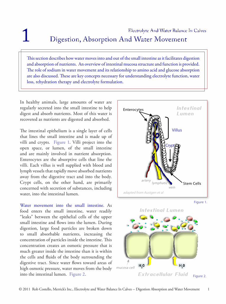
In healthy animals, large amounts of water are regularly secreted into the small intestine to help digest and absorb nutrients. Most of this water is recovered as nutrients are digested and absorbed.
The intestinal epithelium is a single layer of cells that lines the small intestine and is made up of villi and crypts. Figure 1. Villi project into the open space, or lumen, of the small intestine and are mainly involved in nutrient absorption. Enterocytes are the absorptive cells that line the villi. Each villus is well supplied with blood and lymph vessels that rapidly move absorbed nutrients away from the digestive tract and into the body. Crypt cells, on the other hand, are primarily concerned with secretion of substances, including water, into the intestinal lumen.
Water movement into the small intestine. As food enters the small intestine, water readily “leaks” between the epithelial cells of the upper small intestine and flows into the lumen. During digestion, large food particles are broken down to small absorbable nutrients, increasing the concentration of particles inside the intestine. This concentration creates an osmotic pressure that is much greater inside the intestine than it is within the cells and fluids of the body surrounding the digestive tract. Since water flows toward areas of high osmotic pressure, water moves from the body into the intestinal lumen. Figure 2.
Enterocytes
Figure1.
Villus
Crypt
StemCells
Intestinal Lumen
veinlymphatic
artery
adaptedfromAustgenetal
Electrolyte And Water Balance In Calves
Digestion, Absorption And Water Movement
This section describes how water moves into and out of the small intestine as it facilitates digestion and absorption of nutrients. An overview of intestinal mucosa structure and function is provided. The role of sodium in water movement and its relationship to amino acid and glucose absorption are also discussed. These are key concepts necessary for understanding electrolyte function, water loss, rehydration therapy and electrolyte formulation.
1
Figure2.
Intestinal Lumen
mucosacell
Extracellular Fluid
© 2011 Rob Costello, Merrick’s Inc., Electrolyte and Water Balance In Calves -- Digestion Absorption and Water Movement 1
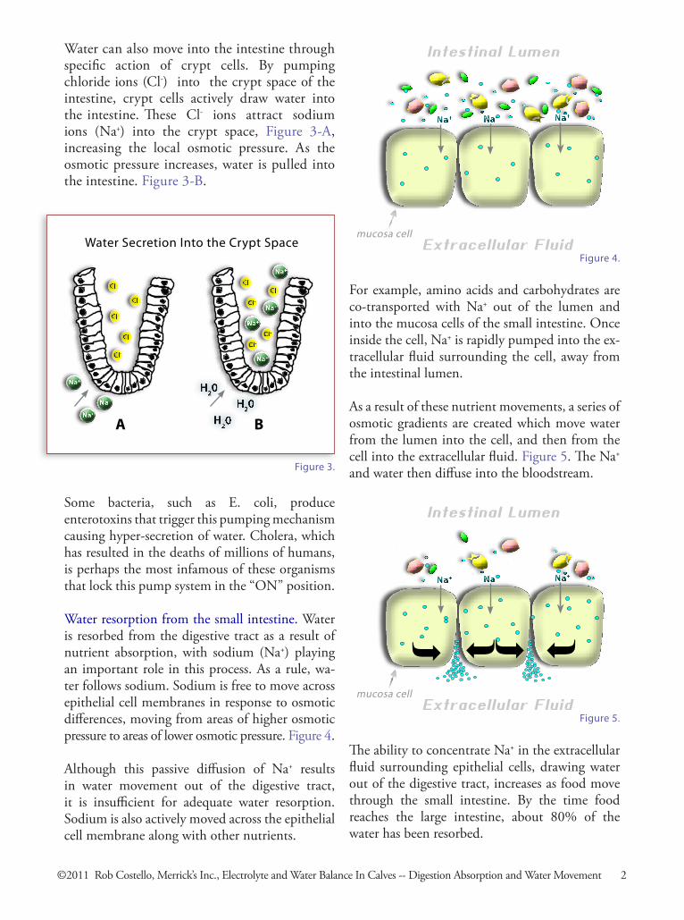
For example, amino acids and carbohydrates are co-transported with Na+ out of the lumen and into the mucosa cells of the small intestine. Once inside the cell, Na+ is rapidly pumped into the ex-tracellular fluid surrounding the cell, away from the intestinal lumen.
As a result of these nutrient movements, a series of osmotic gradients are created which move water from the lumen into the cell, and then from the cell into the extracellular fluid. Figure 5. The Na+ and water then diffuse into the bloodstream.
Water can also move into the intestine through specific action of crypt cells. By pumping chloride ions (Cl-) into the crypt space of the intestine, crypt cells actively draw water into the intestine. These Cl- ions attract sodium ions (Na+) into the crypt space, Figure 3-A, increasing the local osmotic pressure. As the osmotic pressure increases, water is pulled into the intestine. Figure 3-B.
Some bacteria, such as E. coli, produce enterotoxins that trigger this pumping mechanism causing hyper-secretion of water. Cholera, which has resulted in the deaths of millions of humans, is perhaps the most infamous of these organisms that lock this pump system in the “ON” position.
Water resorption from the small intestine. Water is resorbed from the digestive tract as a result of nutrient absorption, with sodium (Na+) playing an important role in this process. As a rule, wa-ter follows sodium. Sodium is free to move across epithelial cell membranes in response to osmotic differences, moving from areas of higher osmotic pressure to areas of lower osmotic pressure. Figure 4.
Although this passive diffusion of Na+ results in water movement out of the digestive tract, it is insufficient for adequate water resorption. Sodium is also actively moved across the epithelial cell membrane along with other nutrients.
Figure4.
Intestinal Lumen
mucosacell Extracellular Fluid
Na+ Na+Na+
Figure3.
WaterSecretionIntotheCryptSpace
A B
Na+
Na+
Na+
Na+
Na+
Na+
Na+
The ability to concentrate Na+ in the extracellular fluid surrounding epithelial cells, drawing water out of the digestive tract, increases as food move through the small intestine. By the time food reaches the large intestine, about 80% of the water has been resorbed.
Figure5.
Intestinal Lumen
mucosacell Extracellular Fluid
Na+ Na+Na+
©2011 Rob Costello, Merrick’s Inc., Electrolyte and Water Balance In Calves -- Digestion Absorption and Water Movement 2
Individual cells work in conjunction with the kid-neys and lungs to regulate and maintain normal water and electrolyte balance within the body. The kidneys have the primary responsibility for main-taining blood chemistry. The combined actions of the kidneys and lungs provide the necessary regula-tory control over acid-base balance and the home-ostasis of body fluids. These processes involve both electrolytes and water.
Electrolyte And Water Balance In Calves
Electrolyte And Acid-Base Balance
This section describes how individual cells, the kidneys and the lungs regulate the body’s electrolytes and water. The kidneys and lungs work in concert to regulate the chemical composition of blood, providing primary control over electrolytes, water and acid-base balance. This elementary exploration of the physical chemistry of biological solutions is one of the more technical discussions in this publication. Although an understanding of this material is not required before reading each of the remaining sections, these discussions provide a good foundation for understanding electrolyte and water balance, dehydration, and electrolyte formulation.
2
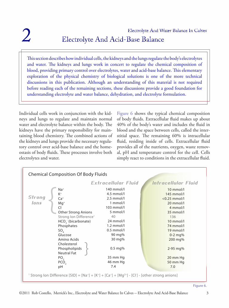
Figure 6 shows the typical chemical composition of body fluids. Extracellular fluid makes up about 40% of the body’s water and includes the fluid in blood and the space between cells, called the inter-stitial space. The remaining 60% is intracellular fluid, residing inside of cells. Extracellular fluid provides all of the nutrients, oxygen, waste remov-al, pH and temperature control for the cell. Cells simply react to conditions in the extracellular fluid.
Intracellular Fluid10mmol/l
145mmol/l<0.25mmol/l
20mmol/l4mmol/l
35mmol/l136
10mmol/l74mmol/l19mmol/l
0-2mg%200mg%
2-95mg%
20mmHg50mmHg
7.0
140mmol/l4.5mmol/l2.5mmol/l
1mmol/l103mmol/l
5mmol/l40
24mmol/l1.2mmol/l0.5mmol/l
90mg%30mg%
0.5mg%
35mmHg46mmHg
7.4
Extracellular FluidNa+
K+
Ca+
Mg+
Cl-
OtherStrongAnionsStrongIonDifference1
HCO3-(bicarbonate)
PhosphatesSO4GlucoseAminoAcidsCholesterolPhospholipidsNeutralFatPO2PCO2pH
{
{
StrongIons
ChemicalCompositionOfBodyFluids
1StrongIonDifference[SID]=[Na+]+[K+]+[Ca+]+[Mg2+]-[Cl-]-[otherstronganions]
Figure6.
©2011 Rob Costello, Merrick’s Inc., Electrolyte and Water Balance In Calves -- Electrolyte And Acid-Base Balance 3
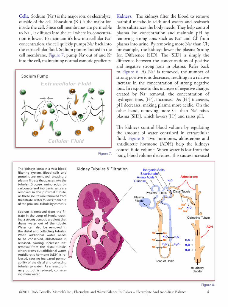
Cells. Sodium (Na+) is the major ion, or electrolyte, outside of the cell. Potassium (K+) is the major ion inside the cell. Since cell membranes are permeable to Na+, it diffuses into the cell where its concentra-tion is lower. To maintain it’s low intracellular Na+ concentration, the cell quickly pumps Na+ back into the extracellular fluid. Sodium pumps located in the cell membrane, Figure 7, pump Na+ out of and K+ into the cell, maintaining normal osmotic gradients.
Kidneys. The kidneys filter the blood to remove harmful metabolic acids and wastes and reabsorb those substances the body needs. They help control plasma ion concentration and maintain pH by removing strong ions such as Na+ and Cl- from plasma into urine. By removing more Na+ than Cl-, for example, the kidneys lower the plasma Strong Ion Difference [SID]. The [SID] is simply the difference between the concentrations of positive and negative strong ions in plasma. Refer back to Figure 6. As Na+ is removed, the number of strong positive ions decreases, resulting in a relative increase in the concentration of strong negative ions. In response to this increase of negative charges created by Na+ removal, the concentration of hydrogen ions, [H+], increases. As [H+] increases, pH decreases, making plasma more acidic. On the other hand, removing more Cl- than Na+ raises plasma [SID], which lowers [H+] and raises pH.
The kidneys control blood volume by regulating the amount of water contained in extracellular fluid. Figure 8. Two hormones, aldosterone and antidiuretic hormone (ADH) help the kidneys control fluid volume. When water is lost from the body, blood volume decreases. This causes increased
Na+
Na+
K+
Na+Na+
Na+
Na+
K+
K+K+
K+
Extracellular Fluid
Cellular Fluid
Sodium Pump
Figure7.
GlucoseAmino Acids
Inorganic Salts
Proximal Tubule Distal Tubule
to urinarybladder
Collecting Tubule
Loop of Henle
BicarbonateAldosterone
ADH
Na+ H2O
H2O
H2O
H2O
PlasmaFiltrate
The kidneys contain a vast blood filteringsystem. Blood cells and proteins areremoved, creating a plasma filtrate thatpasses into the tubules. Glucose, aminoacids, bicarbonate and inorganic salts areremoved in the proximal tubule. As thesesolutes are removed from the filtrate,water follows them out of the proximaltubule by osmosis.
Sodium is removed from the filtrate in theLoop of Henle, creating a strong osmoticgradient that draws water out of the tubule.Water can also be removed in the distaland collecting tubules. When additionalwater needs to be conserved, aldosteroneis released, causing increased Na+
removal from the distal tubule, which drawsout additional water. Antidiuretic hormone(ADH) is released, causing increasedpermeability of the distal and collectingtubules to water. As a result, urinaryoutput is reduced, conserving more water.
Na+
Na+
H2O H2O
H2O H2ONa+ Na+
Na+ Na+
Na+ Na+
H2O
The kidneys contain a vast bloodfiltering system. Blood cells andproteins are removed, creating aplasmafiltratethatpassesintothetubules. Glucose, amino acids, bi-carbonate and inorganic salts areremoved in the proximal tubule.Asthesesolutesareremovedfromthefiltrate,waterfollowsthemoutoftheproximaltubulebyosmosis.
Sodium is removed from the fil-trate in the Loop of Henle, creat-ingastrongosmoticgradientthatdraws water out of the tubule.Water can also be removed inthe distal and collecting tubules.When additional water needsto be conserved, aldosterone isreleased, causing increased Na+removal from the distal tubule,whichdrawsoutadditionalwater.Antidiuretichormone(ADH) isre-leased, causing increased perme-abilityofthedistalandcollectingtubules to water. As a result, uri-nary output is reduced, conserv-ingmorewater.
Figure8.
KidneyTubules&Filtration
©2011 Rob Costello Merrick’s Inc., Electrolyte and Water Balance In Calves -- Electrolyte And Acid-Base Balance 4
K+
Na+
aldosterone and ADH production. Aldosterone increases Na+ pump activity in the kidney tubules, removing more Na+ from the distal tubules. This concentrates Na+ within the kidney rather than excreted it in urine. By concentrating Na+ in kidney tissue an osmotic gradient is created that pulls water out of the tubules. ADH works along with aldosterone by increasing the permeability of the tubules to water, allowing water to follow Na+ out of the tubules, thereby reducing urinary water loss.
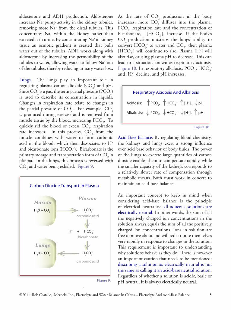
Lungs. The lungs play an important role in regulating plasma carbon dioxide (CO2) and pH. Since CO2 is a gas, the term partial pressure (PCO2)is used to describe its concentration in liquids. Changes in respiration rate relate to changes in the partial pressure of CO2. For example, CO2 is produced during exercise and is removed from muscle tissue by the blood, increasing PCO2. To quickly rid the blood of excess CO2, respiration rate increases. In this process, CO2 from the muscle combines with water to form carbonic acid in the blood, which then dissociates to H+ and bicarbonate ions (HCO3
-). Bicarbonate is the primary storage and transportation form of CO2 in plasma. In the lungs, this process is reversed with CO2 and water being exhaled. Figure 9.
MuscleH20+CO2
Plasma
H2CO3-
H++HCO3-
carbonicacid
bicarbonate
H2CO3-
carbonicacid
LungsH20+CO2
CarbonDioxideTransportInPlasma
Figure9.
As the rate of CO2 production in the body increases, more CO2 diffuses into the plasma. PCO2, respiration rate and the concentration of bicarbonate, [HCO3
-], increase. If the body’s CO2 production outstrips the lungs’ ability to convert HCO3
- to water and CO2, then plasma [HCO3
-] will continue to rise. Plasma [H+] will also rise, causing plasma pH to decrease. This can lead to a situation known as respiratory acidosis. Figure 10. In respiratory alkalosis, PCO2, HCO3
- and [H+] decline, and pH increases.
Acid-Base Balance. By regulating blood chemistry, the kidneys and lungs exert a strong influence over acid base behavior of body fluids. The power of the lungs to excrete large quantities of carbon dioxide enables them to compensate rapidly, while the smaller capacity of the kidneys corresponds to a relatively slower rate of compensation through metabolic means. Both must work in concert to maintain an acid-base balance.
An important concept to keep in mind when considering acid-base balance is the principle of electrical neutrality: all aqueous solutions are electrically neutral. In other words, the sum of all the negatively charged ion concentrations in the solution always equals the sum of all the positively charged ion concentrations. Ions in solution are free to move about and will redistribute themselves very rapidly in response to changes in the solution. This requirement is important to understanding why solutions behave as they do. There is however an important caution that needs to be mentioned: describing a solution as electrically neutral is not the same as calling it an acid-base neutral solution. Regardless of whether a solution is acidic, basic or pH neutral, it is always electrically neutral.
Acidosis:PCO2,HCO3-,[H+],pH
Alkalosis:PCO2,HCO3-,[H+],pH
RespiratoryAcidosisAndAlkalosis
Figure10.
©2011 Rob Costello, Merrick’s Inc., Electrolyte and Water Balance In Calves -- Electrolyte And Acid-Base Balance 5
H+
O-H+H+
O-
H+
H+O-
H+
Na+
H+O-
H+
H+
O-H+
H+
H+
Cl-O-
H+
O-
H+
Figure11.
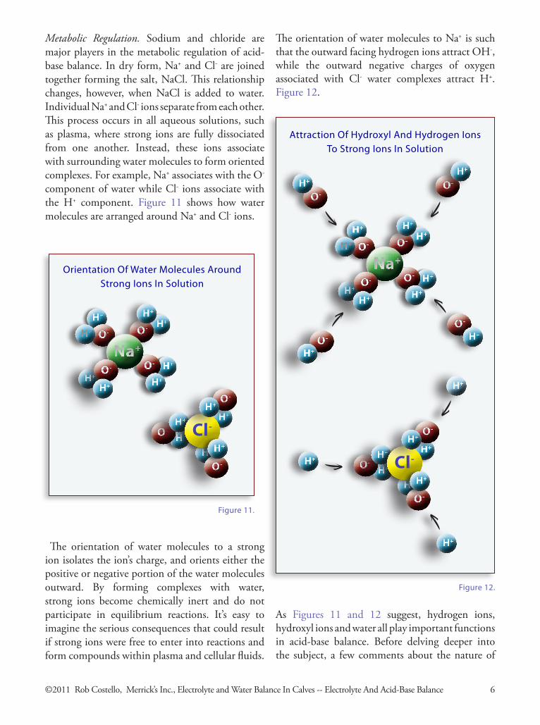
Metabolic Regulation. Sodium and chloride are major players in the metabolic regulation of acid-base balance. In dry form, Na+ and Cl- are joined together forming the salt, NaCl. This relationship changes, however, when NaCl is added to water. Individual Na+ and Cl- ions separate from each other. This process occurs in all aqueous solutions, such as plasma, where strong ions are fully dissociated from one another. Instead, these ions associate with surrounding water molecules to form oriented complexes. For example, Na+ associates with the O- component of water while Cl- ions associate with the H+ component. Figure 11 shows how water molecules are arranged around Na+ and Cl- ions.
The orientation of water molecules to a strong ion isolates the ion’s charge, and orients either the positive or negative portion of the water molecules outward. By forming complexes with water, strong ions become chemically inert and do not participate in equilibrium reactions. It’s easy to imagine the serious consequences that could result if strong ions were free to enter into reactions and form compounds within plasma and cellular fluids.
Figure12.
H+
O-H+H+
O-
H+
H+O-
H+
Na+
H+O-
H+
O-
H+
O-
H+
O-
H+
O-
H+
H+
H+
H+
H+O-H+
H+
Cl-O-
H+
O-
H+
H+
The orientation of water molecules to Na+ is such that the outward facing hydrogen ions attract OH-, while the outward negative charges of oxygen associated with Cl- water complexes attract H+. Figure 12.
As Figures 11 and 12 suggest, hydrogen ions, hydroxyl ions and water all play important functions in acid-base balance. Before delving deeper into the subject, a few comments about the nature of
OrientationOfWaterMoleculesAroundStrongIonsInSolution
AttractionOfHydroxylAndHydrogenIonsToStrongIonsInSolution
©2011 Rob Costello, Merrick’s Inc., Electrolyte and Water Balance In Calves -- Electrolyte And Acid-Base Balance 6
H+
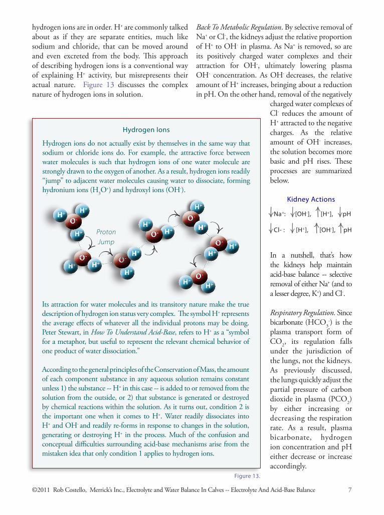
hydrogen ions are in order. H+ are commonly talked about as if they are separate entities, much like sodium and chloride, that can be moved around and even excreted from the body. This approach of describing hydrogen ions is a conventional way of explaining H+ activity, but misrepresents their actual nature. Figure 13 discusses the complex nature of hydrogen ions in solution.
Hydrogen ions do not actually exist by themselves in the same way that sodium or chloride ions do. For example, the attractive force between water molecules is such that hydrogen ions of one water molecule are strongly drawn to the oxygen of another. As a result, hydrogen ions readily “jump” to adjacent water molecules causing water to dissociate, forming hydronium ions (H3O
+) and hydroxyl ions (OH-).
H+
O-H+
H+O-
H+H+ O-
H+
H+O-
H+
H+O-
H+
H+
O-
H+
H+
O-
H+
HydrogenIons
ProtonJump
Its attraction for water molecules and its transitory nature make the true description of hydrogen ion status very complex. The symbol H+ represents the average effects of whatever all the individual protons may be doing. Peter Stewart, in How To Understand Acid-Base, refers to H+ as a “symbol for a metaphor, but useful to represent the relevant chemical behavior of one product of water dissociation.”
According to the general principles of the Conservation of Mass, the amount of each component substance in any aqueous solution remains constant unless 1) the substance -- H+ in this case -- is added to or removed from the solution from the outside, or 2) that substance is generated or destroyed by chemical reactions within the solution. As it turns out, condition 2 is the important one when it comes to H+. Water readily dissociates into H+ and OH- and readily re-forms in response to changes in the solution, generating or destroying H+ in the process. Much of the confusion and conceptual difficulties surrounding acid-base mechanisms arise from the mistaken idea that only condition 1 applies to hydrogen ions.
Back To Metabolic Regulation. By selective removal of Na+ or Cl-, the kidneys adjust the relative proportion of H+ to OH- in plasma. As Na+ is removed, so are its positively charged water complexes and their attraction for OH-, ultimately lowering plasma OH- concentration. As OH- decreases, the relative amount of H+ increases, bringing about a reduction in pH. On the other hand, removal of the negatively
Figure13.
charged water complexes of Cl- reduces the amount of H+ attracted to the negative charges. As the relative amount of OH- increases, the solution becomes more basic and pH rises. These processes are summarized below.
Na+:[OH-],[H+],pH
Cl-:[H+],[OH-],pH
In a nutshell, that’s how the kidneys help maintain acid-base balance -- selective removal of either Na+ (and to a lesser degree, K+) and Cl-.
Respiratory Regulation. Since bicarbonate (HCO3
-) is the plasma transport form of CO2, its regulation falls under the jurisdiction of the lungs, not the kidneys. As previously discussed, the lungs quickly adjust the partial pressure of carbon dioxide in plasma (PCO2) by either increasing or decreasing the respiration rate. As a result, plasma bicarbonate, hydrogen ion concentration and pH either decrease or increase accordingly.
KidneyActions
©2011 Rob Costello, Merrick’s Inc., Electrolyte and Water Balance In Calves -- Electrolyte And Acid-Base Balance 7
Combined Effect of Respiratory & Metabolic Regulation. Although the lungs and the kidneys have their own regulatory processes, they act together to bring about changes in acid-base balance. As an example, consider a baby calf that is undergoing the common summertime problem of heat stress. As the outside temperature goes up, the calf ’s body temperature can begin to rise. In an attempt to get rid of this additional heat load, the calf ’s respiration rate increases.
Although rapid breathing rids the body of some excess heat, it also causes a loss of CO2, which lowers the partial pressure of carbon dioxide (PCO2) in the calf ’s plasma. As PCO2 decreases, the amount of CO2 in the calf ’s plasma decreases. Since most of this CO2 is in the form of bicarbonate, HCO3
-, the concentration of HCO3- decreases.
Since less HCO3- is being produced, less H+
is being generated through this process (H+ is generated when H2O and CO2 are combined into HCO3
-). As a result, plasma [H+] decreases. This reduction in [H+] brings about a relative increase in [OH-] causing plasma to become more alkaline, increasing pH. This situation is generally referred to as respiratory alkalosis. Figure 14 summarizes the changes that occur in the calf as a result of increased respiration rate due to heat stress.
CO2,PCO2,HCO3-,[H+],[OH-],pH
EffectOfIncreasedRespirationRateInResponseToHeatStress
(RespiratoryAlkalosis)
Figure14.
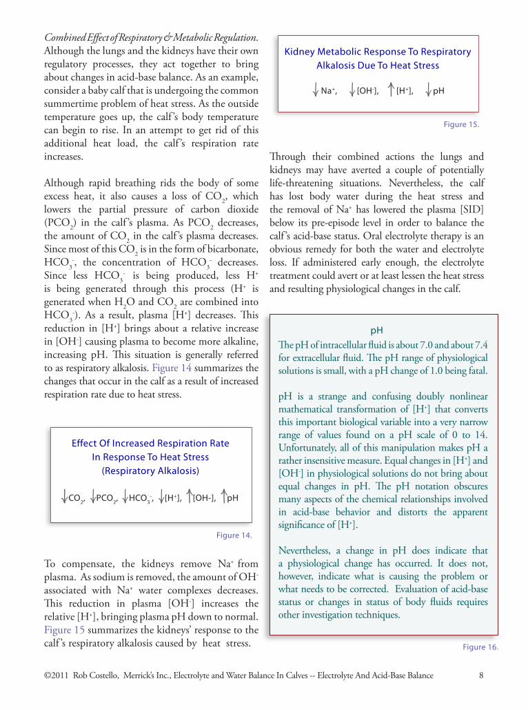
To compensate, the kidneys remove Na+ from plasma. As sodium is removed, the amount of OH- associated with Na+ water complexes decreases. This reduction in plasma [OH-] increases the relative [H+], bringing plasma pH down to normal. Figure 15 summarizes the kidneys’ response to the calf ’s respiratory alkalosis caused by heat stress.
Na+,[OH-],[H+],pH
KidneyMetabolicResponseToRespiratoryAlkalosisDueToHeatStress
Figure15.
Through their combined actions the lungs and kidneys may have averted a couple of potentially life-threatening situations. Nevertheless, the calf has lost body water during the heat stress and the removal of Na+ has lowered the plasma [SID] below its pre-episode level in order to balance the calf ’s acid-base status. Oral electrolyte therapy is an obvious remedy for both the water and electrolyte loss. If administered early enough, the electrolyte treatment could avert or at least lessen the heat stress and resulting physiological changes in the calf.
The pH of intracellular fluid is about 7.0 and about 7.4 for extracellular fluid. The pH range of physiological solutions is small, with a pH change of 1.0 being fatal.
pH is a strange and confusing doubly nonlinear mathematical transformation of [H+] that converts this important biological variable into a very narrow range of values found on a pH scale of 0 to 14. Unfortunately, all of this manipulation makes pH a rather insensitive measure. Equal changes in [H+] and [OH-] in physiological solutions do not bring about equal changes in pH. The pH notation obscures many aspects of the chemical relationships involved in acid-base behavior and distorts the apparent significance of [H+].
Nevertheless, a change in pH does indicate that a physiological change has occurred. It does not, however, indicate what is causing the problem or what needs to be corrected. Evaluation of acid-base status or changes in status of body fluids requires other investigation techniques.
pH
Figure16.
©2011 Rob Costello, Merrick’s Inc., Electrolyte and Water Balance In Calves -- Electrolyte And Acid-Base Balance 8
Electrolyte And Water Balance In Calves
Water Loss Mechanisms And Diarrhea3Pathogens, feed characteristics and management influence digestive function and can result in water loss through the digestive tract. There are four basic types of digestive water loss. Each is described in this section. A diarrheic animal may actually suffer from more than one type of water loss at the same time. The process of dehydration and the clinical signs associated with progressive water loss in calves are also discussed
One of the primary functions of the intestinal epithelium is to provide a barrier between the animal and the environment inside the intestine. Body fluids are retained, large food particles, microorganisms and toxins are excluded from the body and water and solute movements are regulated. If epithelial function is disrupted, the volume of liquid inside the intestinal lumen increases. A wide variety of nutritional, management and microbial factors can contribute to water loss through the digestive tract. Calf diarrhea, or scours, can be the result of several different water loss mechanisms.
Increased Permeability. Microbes cause inflammation and damage to the intestinal epithelium resulting in increased water movement into the intestine. This type of water loss is commonly caused by viruses (rotavirus, coronavirus) and protozoa (coccidia, cryptosporidia).
Rotavirus and coronaviruses damage the enterocytes, the cells that line intestinal villi, leading to water loss and villous atrophy. Enterocyte damage impairs digestion and leads to malabsorption. Rotavirus infection also alters the function of the tight junctions between intestinal epithelial cells, increasing permeability. A secondary effect of inflammation of intestinal epithelial cells is that it can lead to an immune response of the small intestine which ultimately adds a hypersecretion component to inflammatory diarrhea.
Hypersecretion. This type of water loss is similar to increased permeability in that large amounts of water move into the intestine, but there is no tissue damage. Bacterial endotoxins stimulate cellular pumps in the crypt cells of the intestinal mucosa to secrete large amounts of ions into the intestinal lumen. These ions draw water into the small intestine. The mechanisms of hypersecretion were presented in Chapter 2, Figure 4. Hypersecretion results in large losses of water and electrolytes, and is most commonly caused by E. coli and by the immune response to inflammatory diarrhea.
Malabsorption. Epithelial damage in the small intestine reduces nutrient absorption. Viruses and protozoa damage the villi in the small intestine leading to villous atrophy, and can damage the large intestinal mucosa as well. Normal amounts of water may be secreted into the digestive tract, but tissue damage results in poor nutrient and water absorption. Malabsorption causes nutrients to bypass absorption in the small intestine. As these nutrients reach the large intestine, they can cause bacterial overgrowth and excessive production of volatile fatty acids (VFAs). As a result, osmotic changes occur that can worsen fluid loss.
Maldigestion (Osmotic diarrhea). Changes in feed management may lead to maldigestion. A sudden change in feed, use of poor quality ingredients, the presence of feed allergens or other detrimental feed factors and digestive disorders can lead to maldigestion. Maldigestion usually results in malabsorption.
©2011 Rob Costello, Merrick’s Inc., Electrolyte and Water Balance In Calves -- Water Loss Mechanisms and Diarrhea 9
Types Of Intestinal Water Loss
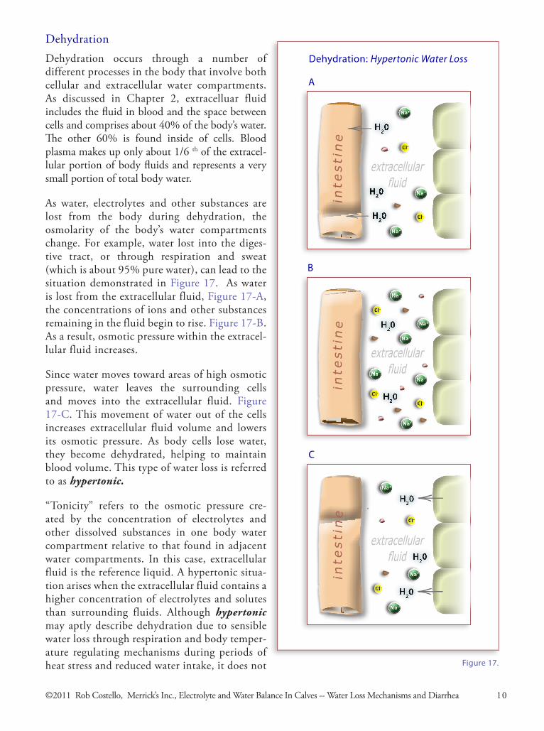
Figure17.
Dehydration:Hypertonic Water Loss
extracellular fluid
inte
sti
ne
Na+
Na+
Na+
A
extracellular fluid
inte
sti
ne
Na+
Na+
Na+
Na+
Na+
Na+
B
extracellular fluid
inte
sti
ne
Na+
Na+
Na+
C
Dehydration occurs through a number of different processes in the body that involve both cellular and extracellular water compartments. As discussed in Chapter 2, extracelluar fluid includes the fluid in blood and the space between cells and comprises about 40% of the body’s water. The other 60% is found inside of cells. Blood plasma makes up only about 1/6 th of the extracel-lular portion of body fluids and represents a very small portion of total body water.
As water, electrolytes and other substances are lost from the body during dehydration, the osmolarity of the body’s water compartments change. For example, water lost into the diges-tive tract, or through respiration and sweat (which is about 95% pure water), can lead to the situation demonstrated in Figure 17. As water is lost from the extracellular fluid, Figure 17-A, the concentrations of ions and other substances remaining in the fluid begin to rise. Figure 17-B. As a result, osmotic pressure within the extracel-lular fluid increases.
Since water moves toward areas of high osmotic pressure, water leaves the surrounding cells and moves into the extracellular fluid. Figure 17-C. This movement of water out of the cells increases extracellular fluid volume and lowers its osmotic pressure. As body cells lose water, they become dehydrated, helping to maintain blood volume. This type of water loss is referred to as hypertonic.
“Tonicity” refers to the osmotic pressure cre-ated by the concentration of electrolytes and other dissolved substances in one body water compartment relative to that found in adjacent water compartments. In this case, extracellular fluid is the reference liquid. A hypertonic situa-tion arises when the extracellular fluid contains a higher concentration of electrolytes and solutes than surrounding fluids. Although hypertonic may aptly describe dehydration due to sensible water loss through respiration and body temper-ature regulating mechanisms during periods of heat stress and reduced water intake, it does not
Dehydration
©2011 Rob Costello, Merrick’s Inc., Electrolyte and Water Balance In Calves -- Water Loss Mechanisms and Diarrhea 10
©2011 Rob Costello, Merrick’s Inc., Electrolyte and Water Balance In Calves -- Water Loss Mechanisms and Diarrhea 11
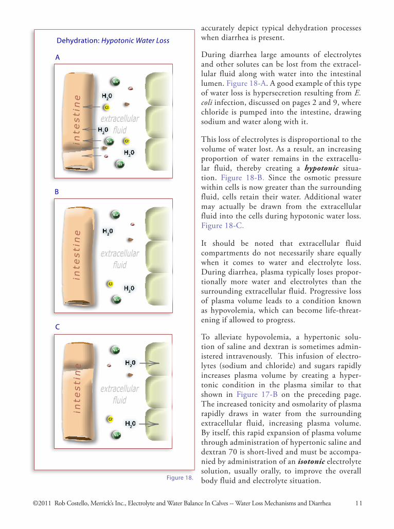
accurately depict typical dehydration processes when diarrhea is present.
During diarrhea large amounts of electrolytes and other solutes can be lost from the extracel-lular fluid along with water into the intestinal lumen. Figure 18-A. A good example of this type of water loss is hypersecretion resulting from E. coli infection, discussed on pages 2 and 9, where chloride is pumped into the intestine, drawing sodium and water along with it.
This loss of electrolytes is disproportional to the volume of water lost. As a result, an increasing proportion of water remains in the extracellu-lar fluid, thereby creating a hypotonic situa-tion. Figure 18-B. Since the osmotic pressure within cells is now greater than the surrounding fluid, cells retain their water. Additional water may actually be drawn from the extracellular fluid into the cells during hypotonic water loss. Figure 18-C.
It should be noted that extracellular fluid compartments do not necessarily share equally when it comes to water and electrolyte loss. During diarrhea, plasma typically loses propor-tionally more water and electrolytes than the surrounding extracellular fluid. Progressive loss of plasma volume leads to a condition known as hypovolemia, which can become life-threat-ening if allowed to progress.
To alleviate hypovolemia, a hypertonic solu-tion of saline and dextran is sometimes admin-istered intravenously. This infusion of electro-lytes (sodium and chloride) and sugars rapidly increases plasma volume by creating a hyper-tonic condition in the plasma similar to that shown in Figure 17-B on the preceding page. The increased tonicity and osmolarity of plasma rapidly draws in water from the surrounding extracellular fluid, increasing plasma volume. By itself, this rapid expansion of plasma volume through administration of hypertonic saline and dextran 70 is short-lived and must be accompa-nied by administration of an isotonic electrolyte solution, usually orally, to improve the overall body fluid and electrolyte situation. Figure18.
Dehydration:Hypotonic Water Loss
extracellular fluid
inte
sti
ne
Na+
Na+
B
extracellular fluid
inte
sti
ne
Na+
Na+
Na+
A
C
extracellular fluid
inte
st i
ne
Na+
Na+
An isotonic solution is one with an ionic and solute concentration similar to that of the surrounding fluids. Isotonic water loss occurs when the same relative proportions of water, electrolytes and other solutes are lost into the digestive tract as are normally found in the extracellular fluid. During isotonic water loss, no osmotic gradients are created and water does not preferentially flow from one body compart-ment in response to osmotic changes.
Many factors, such as the type of diarrhea (ie. hypersecretion, malabsorption, etc.), infective organisms, severity of infection and treatments administered, influence the processes of dehy-dration, including tonicity.
Continuing dehydration. As dehydration progresses, tissues tend to shrink, skin becomes dry and wrinkled, and eyes become shrunken and soft. Fever develops as dehydration worsens. If water loss continues and plasma volume falls, the kidneys reduce urine output in order to conserve water. As urine output decreases, waste products accumulate in the blood.
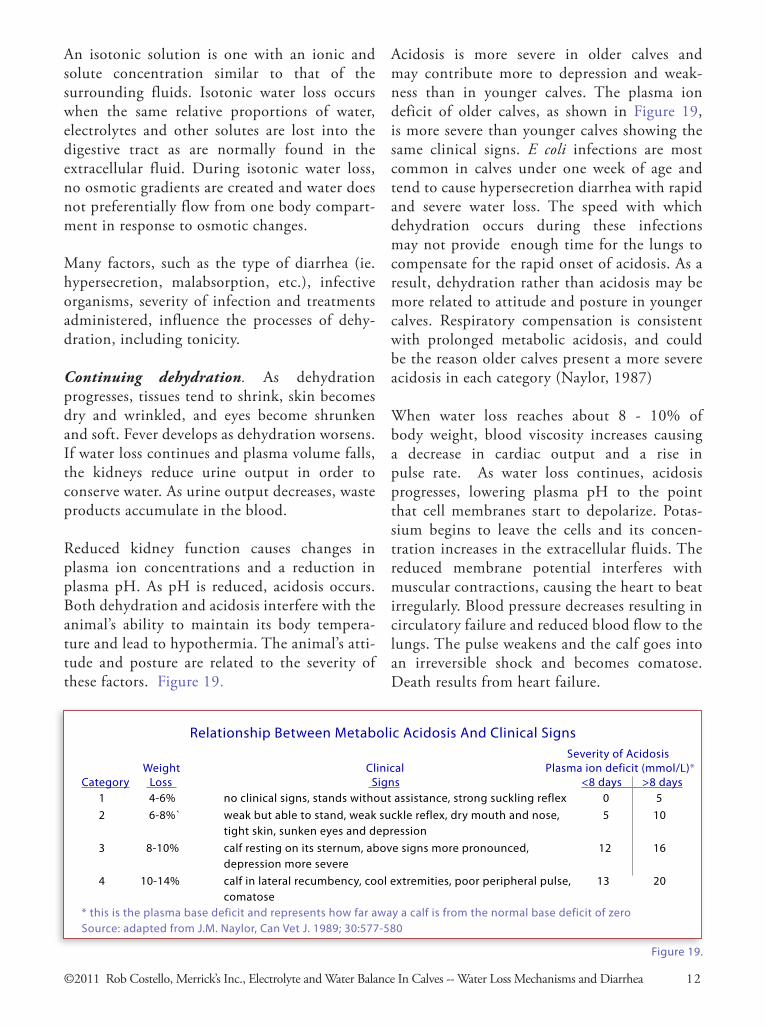
Reduced kidney function causes changes in plasma ion concentrations and a reduction in plasma pH. As pH is reduced, acidosis occurs. Both dehydration and acidosis interfere with the animal’s ability to maintain its body tempera-ture and lead to hypothermia. The animal’s atti-tude and posture are related to the severity of these factors. Figure 19.
Acidosis is more severe in older calves and may contribute more to depression and weak-ness than in younger calves. The plasma ion deficit of older calves, as shown in Figure 19, is more severe than younger calves showing the same clinical signs. E coli infections are most common in calves under one week of age and tend to cause hypersecretion diarrhea with rapid and severe water loss. The speed with which dehydration occurs during these infections may not provide enough time for the lungs to compensate for the rapid onset of acidosis. As a result, dehydration rather than acidosis may be more related to attitude and posture in younger calves. Respiratory compensation is consistent with prolonged metabolic acidosis, and could be the reason older calves present a more severe acidosis in each category (Naylor, 1987)
When water loss reaches about 8 - 10% of body weight, blood viscosity increases causing a decrease in cardiac output and a rise in pulse rate. As water loss continues, acidosis progresses, lowering plasma pH to the point that cell membranes start to depolarize. Potas-sium begins to leave the cells and its concen-tration increases in the extracellular fluids. The reduced membrane potential interferes with muscular contractions, causing the heart to beat irregularly. Blood pressure decreases resulting in circulatory failure and reduced blood flow to the lungs. The pulse weakens and the calf goes into an irreversible shock and becomes comatose. Death results from heart failure.
Figure19.
RelationshipBetweenMetabolicAcidosisAndClinicalSigns SeverityofAcidosis Weight Clinical Plasmaiondeficit(mmol/L)*CategoryLoss Signs <8days>8days1 4-6% noclinicalsigns,standswithoutassistance,strongsucklingreflex 0 52 6-8%̀ weakbutabletostand,weaksucklereflex,drymouthandnose, 5 10 tightskin,sunkeneyesanddepression3 8-10% calfrestingonitssternum,abovesignsmorepronounced, 12 16 depressionmoresevere4 10-14% calfinlateralrecumbency,coolextremities,poorperipheralpulse,13 20 comatose*thisistheplasmabasedeficitandrepresentshowfarawayacalfisfromthenormalbasedeficitofzeroSource:adaptedfromJ.M.Naylor,CanVetJ.1989;30:577-580
©2011 Rob Costello, Merrick’s Inc., Electrolyte and Water Balance In Calves -- Water Loss Mechanisms and Diarrhea 12
Electrolyte And Water Balance In Calves
Oral Rehydration Therapy4The amount and timing of electrolyte replacement therapy is critical for rapid recovery from dehydration. This section describes the relationship between the degree of water loss and the amount of electrolyte solution required to offset the loss. An overview of the effect of different pathogens on water loss and rehydration therapy, regulation of voluntary intake and effects of tube-feeding are presented. The importance of regular milk replacer feedings on the hydration status of the animal and in maintaining nutrient intake is also explored.
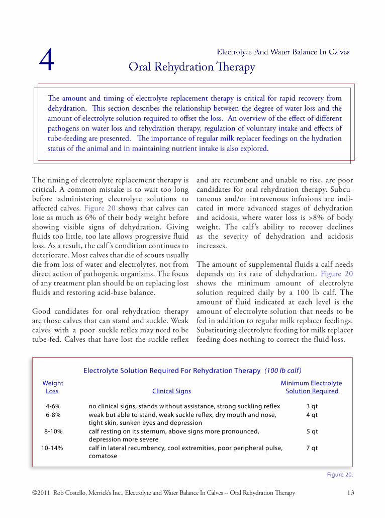
The timing of electrolyte replacement therapy is critical. A common mistake is to wait too long before administering electrolyte solutions to affected calves. Figure 20 shows that calves can lose as much as 6% of their body weight before showing visible signs of dehydration. Giving fluids too little, too late allows progressive fluid loss. As a result, the calf ’s condition continues to deteriorate. Most calves that die of scours usually die from loss of water and electrolytes, not from direct action of pathogenic organisms. The focus of any treatment plan should be on replacing lost fluids and restoring acid-base balance.
Good candidates for oral rehydration therapy are those calves that can stand and suckle. Weak calves with a poor suckle reflex may need to be tube-fed. Calves that have lost the suckle reflex
and are recumbent and unable to rise, are poor candidates for oral rehydration therapy. Subcu-taneous and/or intravenous infusions are indi-cated in more advanced stages of dehydration and acidosis, where water loss is >8% of body weight. The calf ’s ability to recover declines as the severity of dehydration and acidosis increases.
The amount of supplemental fluids a calf needs depends on its rate of dehydration. Figure 20 shows the minimum amount of electrolyte solution required daily by a 100 lb calf. The amount of fluid indicated at each level is the amount of electrolyte solution that needs to be fed in addition to regular milk replacer feedings. Substituting electrolyte feeding for milk replacer feeding does nothing to correct the fluid loss.
Figure20.
ElectrolyteSolutionRequiredForRehydrationTherapy(100 lb calf )
Weight MinimumElectrolyte Loss ClinicalSigns SolutionRequired
4-6% noclinicalsigns,standswithoutassistance,strongsucklingreflex 3qt6-8% weakbutabletostand,weaksucklereflex,drymouthandnose, 4qt tightskin,sunkeneyesanddepression8-10% calfrestingonitssternum,abovesignsmorepronounced, 5qt depressionmoresevere10-14% calfinlateralrecumbency,coolextremities,poorperipheralpulse, 7qt comatose
©2011 Rob Costello, Merrick’s Inc., Electrolyte and Water Balance In Calves -- Oral Rehydration Therapy 13
Consider a 100 lb calf that is scouring, but shows no other clinical signs. The calf is alert and attentive and has a strong suckle reflex. We can assume the calf has lost about 5% of its body weight due to diarrhea. For this calf, a 5% weight loss equals 5 pounds. If a gallon of water weighs 8 lb, each quart weighs 2 lb and each pint weighs 1 pound. Therefore, this calf has lost 5 pints or 2½ qt of water. Since absorption is not likely to be 100%, the calf should receive at least 3 qt of electrolyte solution daily while scouring.
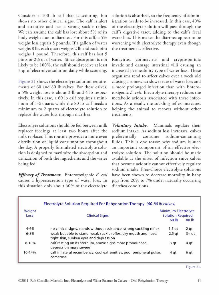
Figure 21 shows the electrolyte solution require-ments of 60 and 80 lb calves. For these calves, a 5% weight loss is about 3 lb and 4 lb respec-tively. In this case, a 60 lb calf requires a mini-mum of 1½ quarts while the 80 lb calf needs a minimum to 2 quarts of electrolyte solution to replace the water lost through diarrhea.
Electrolyte solutions should be fed between milk replacer feedings at least two hours after the milk replacer. This routine provides a more even distribution of liquid consumption throughout the day. A properly formulated electrolyte solu-tion is designed to maximize the absorption and utilization of both the ingredients and the water being fed.
Efficacy of Treatment. Enterotoxigenic E. coli causes a hypersecretion type of water loss. In this situation only about 60% of the electrolyte
solution is absorbed, so the frequency of admin-istration needs to be increased. In this case, 40% of the electrolyte solution will pass through the calf ’s digestive tract, adding to the calf ’s fecal water loss. This makes the diarrhea appear to be worsening with electrolyte therapy even though the treatment is effective.
Rotavirus, coronavirus and cryptosporidia invade and damage intestinal villi causing an increased permeability type of water loss. These organisms tend to affect calves over a week old causing a somewhat slower rate of water loss and a more prolonged infection than with Entero-toxigenic E. coli. Electrolyte therapy reduces the metabolic acidosis associated with these infec-tions. As a result, the suckling reflex increases, helping the animal to recover without other treatments.
Voluntary Intake. Mammals regulate their sodium intake. As sodium loss increases, calves preferentially consume sodium-containing fluids. This is one reason why sodium is such an important component of an effective elec-trolyte solution. The solution should be made available at the onset of infection since calves that become acidotic cannot effectively regulate sodium intake. Free-choice electrolyte solutions have been shown to decrease mortality in baby pigs from 20% to 7% under naturally occurring diarrhea conditions.
Figure21.
ElectrolyteSolutionRequiredForRehydrationTherapy(60-80 lb calves)
Weight MinimumElectrolyte Loss ClinicalSigns SolutionRequired 60lb 80lb
4-6% noclinicalsigns,standswithoutassistance,strongsucklingreflex1.5qt 2qt6-8% weakbutabletostand,weaksucklereflex,drymouthandnose,2.5qt 3+qt tightskin,sunkeneyesanddepression8-10% calfrestingonitssternum,abovesignsmorepronounced, 3qt 4qt depressionmoresevere10-14% calfinlateralrecumbency,coolextremities,poorperipheralpulse,4qt 6qt comatose
©2011 Rob Costello, Merrick’s Inc., Electrolyte and Water Balance In Calves -- Oral Rehydration Therapy 14
Tube feeding. It may be necessary to tube-feed calves with a weak suckle reflex. In this situa-tion, there is little or no stimulation for closure of the esophageal groove causing the electro-lyte solution to enter the rumen rather than the abomasum. Some solution overflows into the abomasum and is absorbed as efficiently as nursed solutions, but rumen bacteria may be washed away.
Although it is critical to rehydrate scouring calves, the possibility of negative effects of tube feeding on rumen microflora does exist. These effects depend on the calf ’s age and degree of rumen development. Most electrolyte solutions, for example, contain glucose. In the developing rumen glucose is fermented to volatile fatty acids and lactate causing a decrease in rumen pH. This lower pH can destroy certain rumen bacteria, slowing the calf ’s return to normal feed digestion and absorption.
Milk/Milk Replacer Feeding. Dehydration is reported as the primary reason that scouring calves die. A secondary reason is starvation. When normal digestive and absorptive func-tions of the intestine are impaired, calves cannot absorb adequate nutrients from the diet. Since young calves have precious little in the form of
stored nutrients to sustain them, digestive and absorptive problems can progressively lead to rapid weight loss, weakness and death. This situation is made worse when milk replacer is withheld during the treatment process.
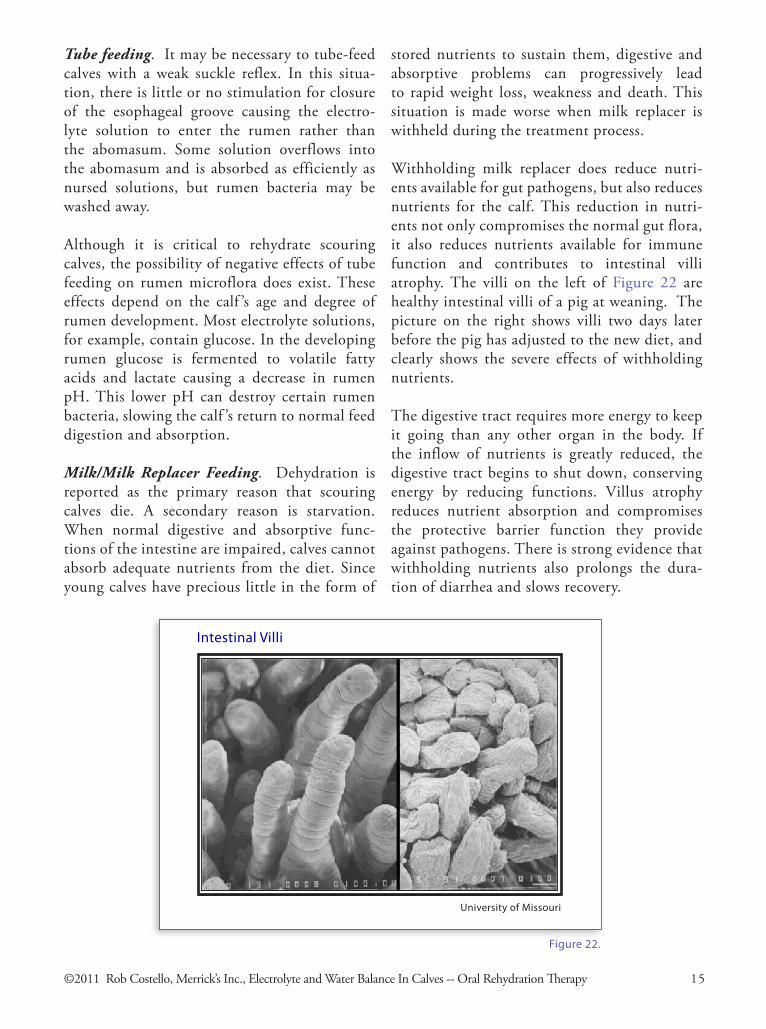
Withholding milk replacer does reduce nutri-ents available for gut pathogens, but also reduces nutrients for the calf. This reduction in nutri-ents not only compromises the normal gut flora, it also reduces nutrients available for immune function and contributes to intestinal villi atrophy. The villi on the left of Figure 22 are healthy intestinal villi of a pig at weaning. The picture on the right shows villi two days later before the pig has adjusted to the new diet, and clearly shows the severe effects of withholding nutrients.
The digestive tract requires more energy to keep it going than any other organ in the body. If the inflow of nutrients is greatly reduced, the digestive tract begins to shut down, conserving energy by reducing functions. Villus atrophy reduces nutrient absorption and compromises the protective barrier function they provide against pathogens. There is strong evidence that withholding nutrients also prolongs the dura-tion of diarrhea and slows recovery.
IntestinalVilli
UniversityofMissouri
Figure22.
©2011 Rob Costello, Merrick’s Inc., Electrolyte and Water Balance In Calves -- Oral Rehydration Therapy 15
Water is the most important nutrient for sustaining life. Water corrects dehydration and acts as a solvent for electrolytes.
Sodium (Na+) is the major extracellular ion in the body. Water follows the movement of sodium, making it the major electrolyte compo-nent as well. As sodium is absorbed from the digestive tract into the blood and into cells, it causes water to move along with it. Cells absorb sodium through simple diffusion. It is also co-transported into cells along with glucose and amino acids. Sodium has a critical role in acid-base balance and is used by the kidneys to adjust and maintain equilibrium.
Sodium is also involved in the voluntary uptake of electrolyte solutions. Since mammals regulate sodium intake, calves will preferentially drink sodium-containing solutions as sodium loss increases.
Glucose/Dextrose is a sugar, or carbohydrate, that facilitates sodium absorption. Glucose is co-transported with sodium from the digestive tract, enhancing sodium absorption and water uptake from the small intestine. Glucose also provides a minor energy source for the calf. However, an electrolyte solution should not be
Electrolyte And Water Balance In Calves
Components of Electrolyte Solutions5Electrolyte solutions should be formulated for their ability to enhance water absorption and water retention, to restore electrolyte levels and to reduce acidosis. Since diarrhea can have a serious effect on bacterial populations in the digestive tract, inclusion of specific direct-fed microbials favors conditions for growth of beneficial bacteria and reestablishment of a normal intestinal environment. Organisms that cause diarrhea often damage the intestinal lining and impair gut function. Electrolytes may also contain ingredients that can help repair damage and improve function. This chapter reviews the nature and function of the major ingredients found in electrolyte products.
©2011 Rob Costello, Merrick’s Inc., Electrolyte and Water Balance In Calves -- Components of Electrolyte Solutions 16
looked at as a replacement for energy provided by milk replacer.
High glucose electrolyte solutions are sometimes presented and used as a replacement for milk or milk replacer during diarrhea. Six quarts of such a solution provides about 75% of the daily energy needed by a baby calf for maintenance, while providing none of the protein required by the calf. Glucose, which is absorbed more quickly than lactose (milk sugar), causes a rapid increase in plasma glucose. Insulin is released into the calf ’s bloodstream to lower the elevated plasma glucose level. This insulin response is excessive in young calves. Within three hours after administration of the high glucose electro-lyte solution, plasma glucose is lower than the pretreatment level.
Glycine is an amino acid and is co-transported with sodium from the digestive tract. As with glucose, the inclusion of glycine facilitates sodium and water absorption. Glycine is the most easily synthesized amino acid and is most often included in electrolyte solutions.
Potassium (K+) is the major intracellular ion. Potassium helps maintain the integrity of the cell membrane and is involved in neural func-
tion and muscular contraction. Advanced dehy-dration leads to acidosis and severe electrolyte imbalance, causing a loss of cell membrane potential and cell death. A high supplemental level of potassium can be lethal.
Chloride (Cl-) is the major negative ion, or electrolyte in plasma and has a critical role in acid-base and water balance in the body. The kidneys use chloride to adjust and maintain equilibrium. Sodium chloride and potassium chloride are common chloride sources.
Ascorbic Acid (vitamin C) cannot be synthe-sized by calves until they are about 3 weeks old, and is therefore considered an essential nutrient for calves less than three weeks of age. Ascorbic acid is an antioxidant and is found in high concentrations in steroid secreting cells. The concentration of ascorbic acid in plasma is lower in stressed calves than non-stressed calves. Oral supplementation of ascorbic acid elevates the ascorbic acid level in plasma of preruminant calves.
Direct-Fed Microbials (DFMs) are specific, genetically superior species of bacteria that support conditions in the intestinal tract that are favorable to the growth of beneficial micro-organisms and are unfavorable for pathogens. DFMs help prevent intestinal colonization by pathogens through production of antimicro-bial compounds such as lactic acid, hydrogen peroxide, modified bile acids, and bacteriocins, which are effective bactericidal/bacteriostatic compounds. DFMs compete with pathogens for attachment sites for growth, compete for nutri-ents, neutralize toxins and stimulate the host immune system.
Bacillus bacteria are lumen organisms and do not attach to intestinal cells before providing their beneficial effects. This characteristic makes Bacillus an excellent microbial additive during diarrhea. Water moves rapidly into the digestive tract during diarrhea, making bacterial attachment and colonization more difficult.
Attachment is first required for most other lactic acid bacteria. Bacillus produce an abundant array of enzymes and antimicrobial compounds with activity against pathogens. Bacillus are very hardy organisms and survive under harsh condi-tions.
Lactobacillus bacteria rapidly colonize the newborn intestinal tract and are the predomi-nate microorganisms in the small intestine. They survive the digestive process and attach to the epithelial lining, making them an excellent compliment to Bacillus. They grow best at a pH of 5.5 and are very effective against E. coli.
Bifidobacteria are primary colonizers of the large intestine, growing best at a pH between 6.5 and 7.0. Bifidobacteria are antagonistic toward E. coli and Clostridium.
Fructo-oligosaccharides (FOS). FOS are natu-rally occurring plant sugars. When fed to animals, they travel intact to the large intestine where they provide a source of nutrients for beneficial bacteria such as Bifidobacteria . FOS have been shown to increase volatile fatty acid production in the large intestine and improve calcium and magnesium absorption. FOS cannot be digested by the animal or by pathogenic bacteria, and are an excellent complement to direct-fed micro-bials containing bifidobacteria.
Glutamine/Glutamate are amino acids that have been shown to improve villi height and overall intestinal morphology during periods of stress and following injury. Both glutamine and glutamate provide a local fuel source for entero-cytes, the absorptive cells of intestinal villi.
Alkalinizing Compounds are used as a formu-lation tool to help create a favorable ratio of positive to negative strong ions that improves plasma pH and helps alleviate symptoms of acidosis, a condition which often accompanies diarrhea and progressive dehydration. Alkali-nizing compounds are discussed in more detail in Chapter 6.
©2011 Rob Costello, Merrick’s Inc., Electrolyte and Water Balance In Calves -- Components of Electrolyte Solutions 17
When formulating or evaluating electrolytes we must work with the actual number of molecules of each ingredient used to make the product. For many of us who ventured into high school chemistry class, thinking in terms of molecules can lead to flashbacks of Avogadro’s number and the incomprehensible “mole”. But don’t panic, it won’t be that bad this time around.
The term or unit of measure most commonly used in electrolyte evaluations is millimole, ab-breviated mmol. Simply put, this is a measure of the concentration of a substance, such as so-dium, chloride, or glucose that is dissolved in a solution. The term osmolarity is typically used when talking about mmol concentrations.
There is another term, mEq (or milliequiv-alents), which is sometimes used instead of mmol. This term concerns itself with the number of available charges on each par-ticle of a dissolved substance. Since mmol refers specifically and only to the number
of dissolved particles, it is conceptually easier to understand, is perfect for the job at hand, and will be used throughout this discussion.
To evaluate an electrolyte product, we need to work with the solution the calf actually con-sumes, not just what’s in the package on the shelf. We start with the dry product, add wa-ter according to the label instructions and then evaluate. This is all figuratively speaking of course -- we do this on paper, not in an actual bucket.
The product label provides a list of ingredients and a guaranteed analysis. The Ingredient List is an accounting of the ingredients used to make the product, whereas the Guaranteed Analysis states the concentrations of various ingredients or specific nutrients provided in the dry prod-uct. Sometimes the details in the Guaranteed Analysis can be a bit skimpy, especially if a man-ufacturer is protecting a proprietary formula.
Electrolyte And Water Balance In Calves
Electrolyte Formulation and Function6This chapter draws upon the concepts and discussions presented in previous chapters, fusing them into a practical approach to evaluating electrolytes. Osmolarity, the relationship between strong ions and water, and their effects on acid-base chemistry are reviewed and applied to electrolyte formulation. The purpose is to enhance understanding of electrolyte formulation and its effects, not to provide instruction on how to formulate an electrolyte product. Acidosis, and the correction of this condition, is of special interest. A considerable portion of this chapter explores this metabolic condition, with special focus on the processes of alkalinization, alkalinizing compounds and their mode of action. Acid-base variables can be categorized as either independent, where changes in their concentrations affect acid-base balance, or they are categorized as dependent variables whose concentrations do not affect other variables nor do they affect acid-base mechanisms. This final discussion has profound implications.
Product Evaluation & Formulation
©2011 Rob Costello, Merrick’s Inc., Electrolyte and Water Balance In Calves -- Electrolyte Formulation & Function 18
That’s understandable since they may have a significant investment in the product and don’t want to give the formula away.
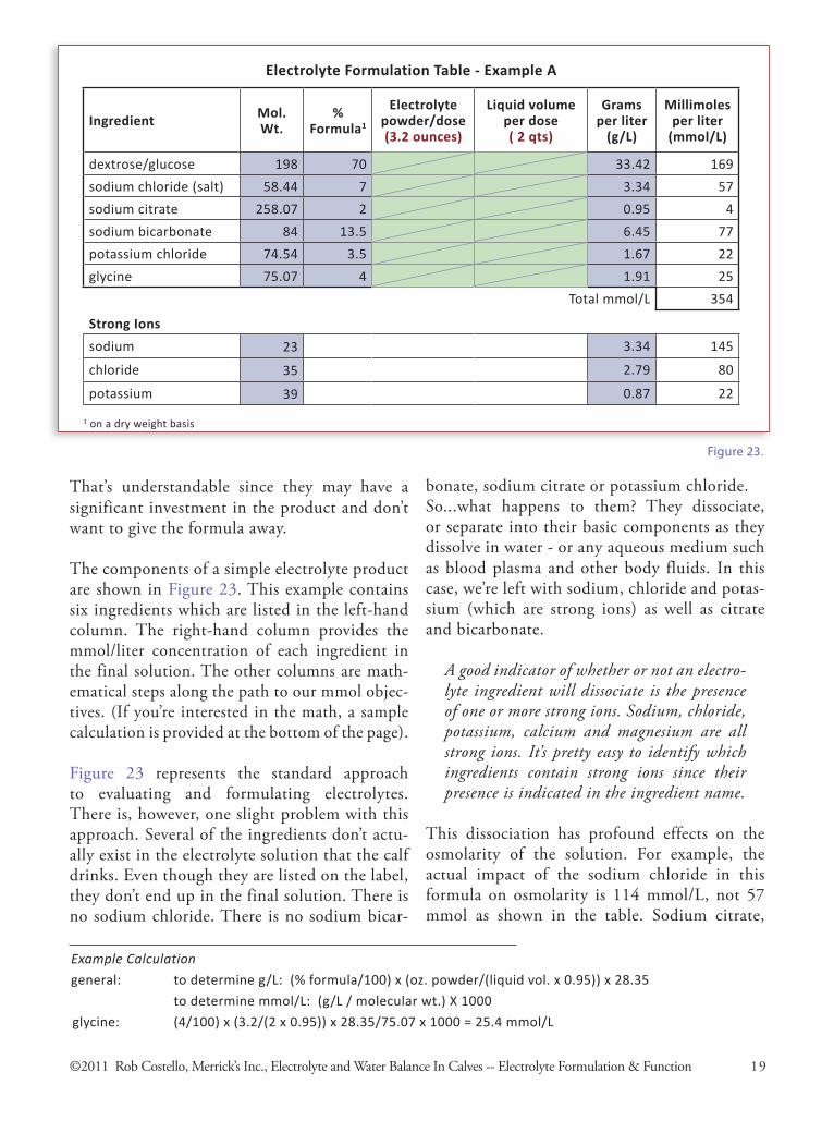
The components of a simple electrolyte product are shown in Figure 23. This example contains six ingredients which are listed in the left-hand column. The right-hand column provides the mmol/liter concentration of each ingredient in the final solution. The other columns are math-ematical steps along the path to our mmol objec-tives. (If you’re interested in the math, a sample calculation is provided at the bottom of the page).
Figure 23 represents the standard approach to evaluating and formulating electrolytes. There is, however, one slight problem with this approach. Several of the ingredients don’t actu-ally exist in the electrolyte solution that the calf drinks. Even though they are listed on the label, they don’t end up in the final solution. There is no sodium chloride. There is no sodium bicar-
bonate, sodium citrate or potassium chloride. So...what happens to them? They dissociate, or separate into their basic components as they dissolve in water - or any aqueous medium such as blood plasma and other body fluids. In this case, we’re left with sodium, chloride and potas-sium (which are strong ions) as well as citrate and bicarbonate.
A good indicator of whether or not an electro-lyte ingredient will dissociate is the presence of one or more strong ions. Sodium, chloride, potassium, calcium and magnesium are all strong ions. It’s pretty easy to identify which ingredients contain strong ions since their presence is indicated in the ingredient name.
This dissociation has profound effects on the osmolarity of the solution. For example, the actual impact of the sodium chloride in this formula on osmolarity is 114 mmol/L, not 57 mmol as shown in the table. Sodium citrate,
Electrolyte Formulation Table - Example A
Ingredient Mol.Wt.
%Formula1
Electrolyte powder/dose(3.2 ounces)
Liquid volume per dose( 2 qts)
Grams per liter
(g/L)
Millimolesper liter
(mmol/L)
dextrose/glucose 198 70 33.42 169
sodium chloride (salt) 58.44 7 3.34 57
sodium citrate 258.07 2 0.95 4
sodium bicarbonate 84 13.5 6.45 77
potassium chloride 74.54 3.5 1.67 22
glycine 75.07 4 1.91 25
Strong Ions
Total mmol/L 354
sodium 23 3.34 145
chloride 35 2.79 80
potassium 39 0.87 22
Figure23.
Example Calculation
general: to determine g/L: (% formula/100) x (oz. powder/(liquid vol. x 0.95)) x 28.35
to determine mmol/L: (g/L / molecular wt.) X 1000
glycine: (4/100) x (3.2/(2 x 0.95)) x 28.35/75.07 x 1000 = 25.4 mmol/L
©2011 Rob Costello, Merrick’s Inc., Electrolyte and Water Balance In Calves -- Electrolyte Formulation & Function 19
1 on a dry weight basis
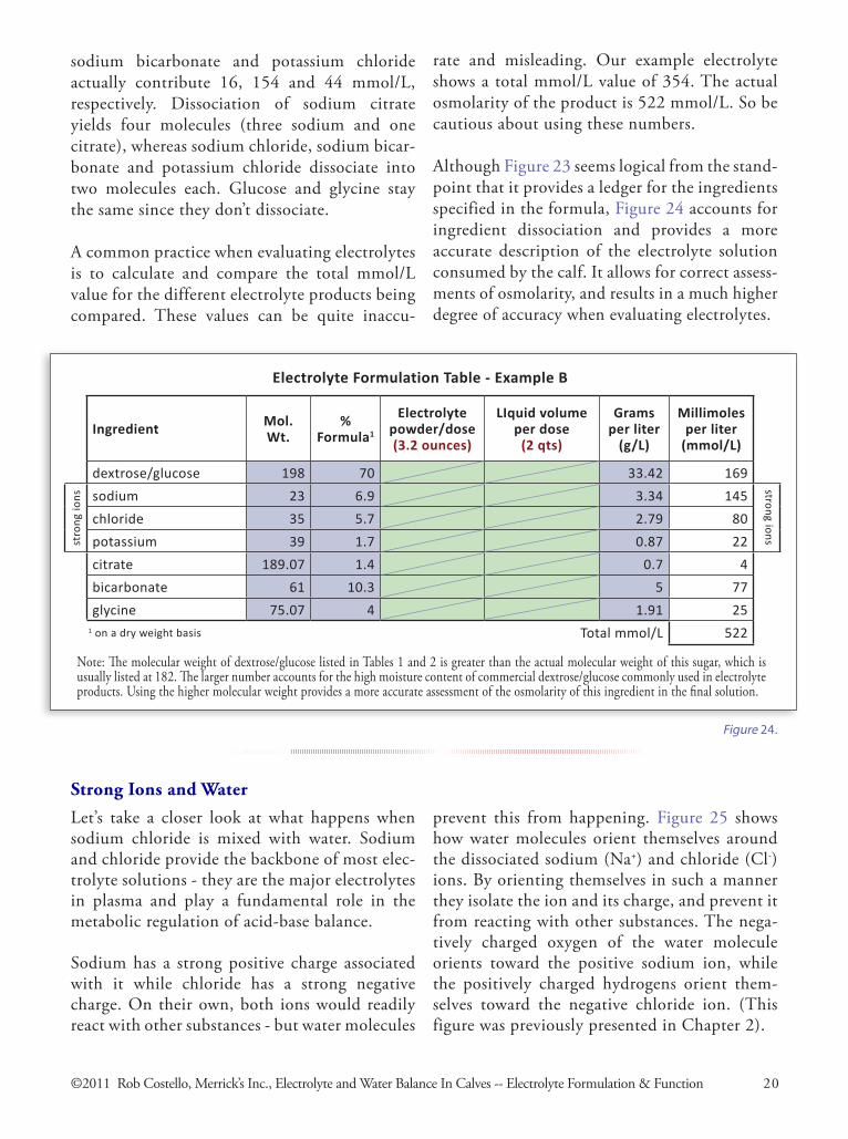
sodium bicarbonate and potassium chloride actually contribute 16, 154 and 44 mmol/L, respectively. Dissociation of sodium citrate yields four molecules (three sodium and one citrate), whereas sodium chloride, sodium bicar-bonate and potassium chloride dissociate into two molecules each. Glucose and glycine stay the same since they don’t dissociate.
A common practice when evaluating electrolytes is to calculate and compare the total mmol/L value for the different electrolyte products being compared. These values can be quite inaccu-
Electrolyte Formulation Table - Example B
Ingredient Mol.Wt.
%Formula1
Electrolyte powder/dose(3.2 ounces)
LIquid volume per dose
(2 qts)
Grams per liter
(g/L)
Millimolesper liter
(mmol/L)
dextrose/glucose 198 70 33.42 169
stro
ng io
ns sodium 23 6.9 3.34 145
strong ions
chloride 35 5.7 2.79 80
potassium 39 1.7 0.87 22
citrate 189.07 1.4 0.7 4
bicarbonate 61 10.3 5 77
glycine 75.07 4 1.91 25
Total mmol/L 522
Note: The molecular weight of dextrose/glucose listed in Tables 1 and 2 is greater than the actual molecular weight of this sugar, which is usually listed at 182. The larger number accounts for the high moisture content of commercial dextrose/glucose commonly used in electrolyte products. Using the higher molecular weight provides a more accurate assessment of the osmolarity of this ingredient in the final solution.
Let’s take a closer look at what happens when sodium chloride is mixed with water. Sodium and chloride provide the backbone of most elec-trolyte solutions - they are the major electrolytes in plasma and play a fundamental role in the metabolic regulation of acid-base balance.
Sodium has a strong positive charge associated with it while chloride has a strong negative charge. On their own, both ions would readily react with other substances - but water molecules
rate and misleading. Our example electrolyte shows a total mmol/L value of 354. The actual osmolarity of the product is 522 mmol/L. So be cautious about using these numbers.
Although Figure 23 seems logical from the stand-point that it provides a ledger for the ingredients specified in the formula, Figure 24 accounts for ingredient dissociation and provides a more accurate description of the electrolyte solution consumed by the calf. It allows for correct assess-ments of osmolarity, and results in a much higher degree of accuracy when evaluating electrolytes.
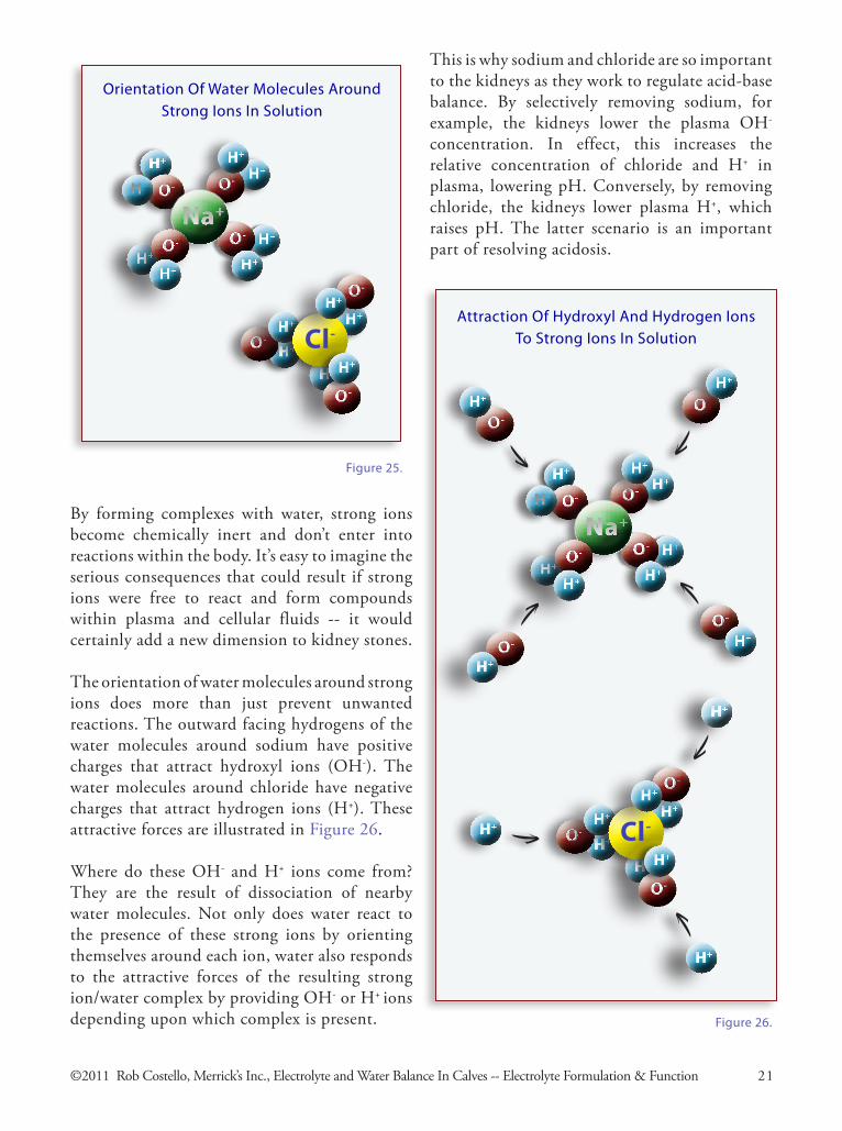
Strong Ions and Water
prevent this from happening. Figure 25 shows how water molecules orient themselves around the dissociated sodium (Na+) and chloride (Cl-) ions. By orienting themselves in such a manner they isolate the ion and its charge, and prevent it from reacting with other substances. The nega-tively charged oxygen of the water molecule orients toward the positive sodium ion, while the positively charged hydrogens orient them-selves toward the negative chloride ion. (This figure was previously presented in Chapter 2).
Figure24.
1 on a dry weight basis
©2011 Rob Costello, Merrick’s Inc., Electrolyte and Water Balance In Calves -- Electrolyte Formulation & Function 20
H+
O-H+H+
O-
H+
H+O-
H+
Na+
H+O-
H+
H+
O-H+
H+
H+
Cl-O-
H+
O-
H+
Figure25.
OrientationOfWaterMoleculesAroundStrongIonsInSolution
By forming complexes with water, strong ions become chemically inert and don’t enter into reactions within the body. It’s easy to imagine the serious consequences that could result if strong ions were free to react and form compounds within plasma and cellular fluids -- it would certainly add a new dimension to kidney stones.
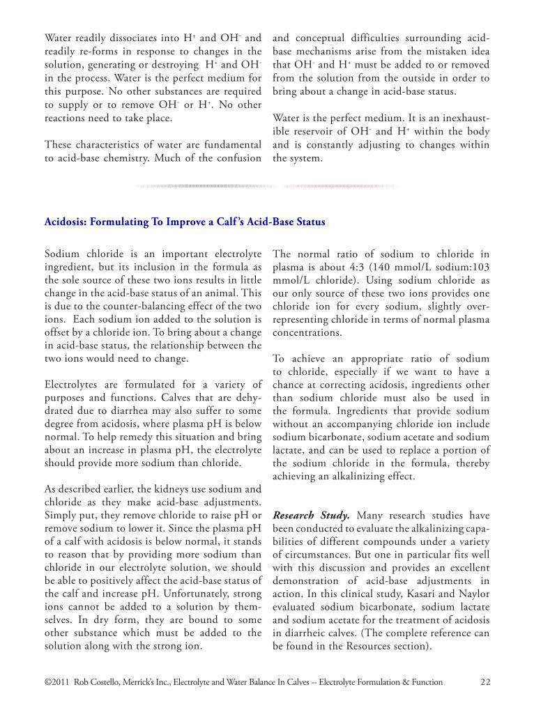
The orientation of water molecules around strong ions does more than just prevent unwanted reactions. The outward facing hydrogens of the water molecules around sodium have positive charges that attract hydroxyl ions (OH-). The water molecules around chloride have negative charges that attract hydrogen ions (H+). These attractive forces are illustrated in Figure 26.
Where do these OH- and H+ ions come from? They are the result of dissociation of nearby water molecules. Not only does water react to the presence of these strong ions by orienting themselves around each ion, water also responds to the attractive forces of the resulting strong ion/water complex by providing OH- or H+ ions depending upon which complex is present. Figure26.
H+
O-H+H+
O-
H+
H+O-
H+
Na+
H+O-
H+
O-
H+
O-
H+
O-
H+
O-
H+
H+
H+
H+
H+O-H+
H+
Cl-O-
H+
O-
H+
H+
AttractionOfHydroxylAndHydrogenIonsToStrongIonsInSolution
This is why sodium and chloride are so important to the kidneys as they work to regulate acid-base balance. By selectively removing sodium, for example, the kidneys lower the plasma OH- concentration. In effect, this increases the relative concentration of chloride and H+ in plasma, lowering pH. Conversely, by removing chloride, the kidneys lower plasma H+, which raises pH. The latter scenario is an important part of resolving acidosis.
©2011 Rob Costello, Merrick’s Inc., Electrolyte and Water Balance In Calves -- Electrolyte Formulation & Function 21
Water readily dissociates into H+ and OH- and readily re-forms in response to changes in the solution, generating or destroying H+ and OH-
in the process. Water is the perfect medium for this purpose. No other substances are required to supply or to remove OH- or H+. No other reactions need to take place.
These characteristics of water are fundamental to acid-base chemistry. Much of the confusion
Sodium chloride is an important electrolyte ingredient, but its inclusion in the formula as the sole source of these two ions results in little change in the acid-base status of an animal. This is due to the counter-balancing effect of the two ions. Each sodium ion added to the solution is offset by a chloride ion. To bring about a change in acid-base status, the relationship between the two ions would need to change.
Electrolytes are formulated for a variety of purposes and functions. Calves that are dehy-drated due to diarrhea may also suffer to some degree from acidosis, where plasma pH is below normal. To help remedy this situation and bring about an increase in plasma pH, the electrolyte should provide more sodium than chloride.
As described earlier, the kidneys use sodium and chloride as they make acid-base adjustments. Simply put, they remove chloride to raise pH or remove sodium to lower it. Since the plasma pH of a calf with acidosis is below normal, it stands to reason that by providing more sodium than chloride in our electrolyte solution, we should be able to positively affect the acid-base status of the calf and increase pH. Unfortunately, strong ions cannot be added to a solution by them-selves. In dry form, they are bound to some other substance which must be added to the solution along with the strong ion.
Acidosis: Formulating To Improve a Calf ’s Acid-Base Status
The normal ratio of sodium to chloride in plasma is about 4:3 (140 mmol/L sodium:103 mmol/L chloride). Using sodium chloride as our only source of these two ions provides one chloride ion for every sodium, slightly over-representing chloride in terms of normal plasma concentrations.
To achieve an appropriate ratio of sodium to chloride, especially if we want to have a chance at correcting acidosis, ingredients other than sodium chloride must also be used in the formula. Ingredients that provide sodium without an accompanying chloride ion include sodium bicarbonate, sodium acetate and sodium lactate, and can be used to replace a portion of the sodium chloride in the formula, thereby achieving an alkalinizing effect.
Research Study. Many research studies have been conducted to evaluate the alkalinizing capa-bilities of different compounds under a variety of circumstances. But one in particular fits well with this discussion and provides an excellent demonstration of acid-base adjustments in action. In this clinical study, Kasari and Naylor evaluated sodium bicarbonate, sodium lactate and sodium acetate for the treatment of acidosis in diarrheic calves. (The complete reference can be found in the Resources section).
and conceptual difficulties surrounding acid-base mechanisms arise from the mistaken idea that OH- and H+ must be added to or removed from the solution from the outside in order to bring about a change in acid-base status.
Water is the perfect medium. It is an inexhaust-ible reservoir of OH- and H+ within the body and is constantly adjusting to changes within the system.
©2011 Rob Costello, Merrick’s Inc., Electrolyte and Water Balance In Calves -- Electrolyte Formulation & Function 22
The criteria for calves in this study were:
• under 30 days of age• clinical signs of diarrhea• greater than 8% dehydration• venous blood pH less than 7.25
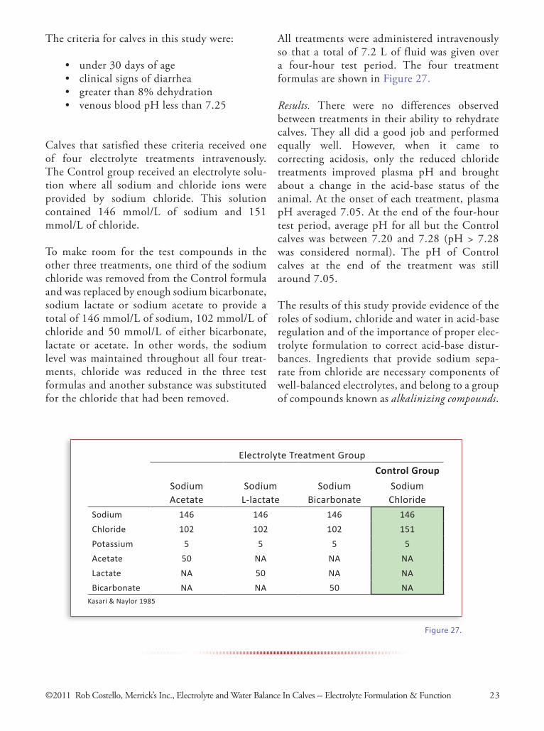
Calves that satisfied these criteria received one of four electrolyte treatments intravenously. The Control group received an electrolyte solu-tion where all sodium and chloride ions were provided by sodium chloride. This solution contained 146 mmol/L of sodium and 151 mmol/L of chloride.
To make room for the test compounds in the other three treatments, one third of the sodium chloride was removed from the Control formula and was replaced by enough sodium bicarbonate, sodium lactate or sodium acetate to provide a total of 146 mmol/L of sodium, 102 mmol/L of chloride and 50 mmol/L of either bicarbonate, lactate or acetate. In other words, the sodium level was maintained throughout all four treat-ments, chloride was reduced in the three test formulas and another substance was substituted for the chloride that had been removed.
©2011 Rob Costello, Merrick’s Inc., Electrolyte and Water Balance In Calves -- Electrolyte Formulation & Function 23
Electrolyte Treatment Group
Control Group
SodiumAcetate
SodiumL-lactate
SodiumBicarbonate
SodiumChloride
Sodium 146 146 146 146
Chloride 102 102 102 151
Potassium 5 5 5 5
Acetate 50 NA NA NA
Lactate NA 50 NA NA
Bicarbonate NA NA 50 NAKasari & Naylor 1985
Figure27.
All treatments were administered intravenously so that a total of 7.2 L of fluid was given over a four-hour test period. The four treatment formulas are shown in Figure 27.
Results. There were no differences observed between treatments in their ability to rehydrate calves. They all did a good job and performed equally well. However, when it came to correcting acidosis, only the reduced chloride treatments improved plasma pH and brought about a change in the acid-base status of the animal. At the onset of each treatment, plasma pH averaged 7.05. At the end of the four-hour test period, average pH for all but the Control calves was between 7.20 and 7.28 (pH > 7.28 was considered normal). The pH of Control calves at the end of the treatment was still around 7.05.
The results of this study provide evidence of the roles of sodium, chloride and water in acid-base regulation and of the importance of proper elec-trolyte formulation to correct acid-base distur-bances. Ingredients that provide sodium sepa-rate from chloride are necessary components of well-balanced electrolytes, and belong to a group of compounds known as alkalinizing compounds.
From a clinical evaluation standpoint, it’s fairly easy to gain insights into an animal’s acid-base status from a blood sample. A pH measurement gives an estimate of the hydrogen ion concen-tration, [H+], which can be used along with a CO2 measurement from a blood gas analyzer to calculate the concentration of bicarbonate, [HCO3
-], present in the animal’s bloodstream. Even better, these values can be used to develop an effective treatment protocol. It would there-fore seem logical that variables such as pH, [H+] and [HCO3
-] must be central to a problem and that they are determinant forces in acid-base physiology.
In reality, this assumption is at the core of much of the difficulty and confusion that so often accompanies a developing understanding of acid-base mechanisms. It leads, for example, to the common misinterpretation that substances such as bicarbonate, acetate and lactate are the alkali-nizing components of alkalinizing compounds. The thought is that metabolism and other reac-tions in the body involving these substances result in hydrogen ions being removed from the body, thereby bringing about alkalinization.
The first three sections in this chapter demon-strate that acid-base adjustments occur without regard for variables such as bicarbonate. This section is no different in that regard, and will explore how acid-base variables relate to each other and what that means to acid-base balance.
Dependent & independent variables. An impor-tant consideration when evaluating variables and their acid-base relevance is whether changes in their concentration occur independently or whether they depend on changes and interactions among other variables. The concentration of a dependent variable is governed by the concen-trations and changes of independent variables. In other words, dependent variables change only as a result of changes in independent ones.
Variables such as pH, hydrogen ion concen-tration [H+] and bicarbonate concentration [HCO3
-] are all dependent variables. As such, they cannot cause changes in each other or in independent variables. Their concentrations are simply the results of other factors. The idea that acid-base changes are brought about through direct manipulations of [H+] or [HCO3
-] is simply incorrect.
The other dependent variables are [OH-], carbonate [CO3
-2], individual weak acids [HA] and the dissociated form of individual weak acids [A-]. Weak acids are discussed in more detail under [ATOT] in Figure 28, which can be found on the next page.
On the other hand, independent variables are subject to independent variation. Their concen-trations are not under the control of other variables. As such, independent variables are amenable to change, and as a result, changes in their concentrations influence acid-base status. This has important implications when attempting to correct an acid-base disturbance.
There are three independent variables whose concentrations have major effects on acid-base balance. Two of these variables have already been introduced: the strong ion differ-ence, [SID], and the partial pressure of carbon dioxide, PCO2. The third independent variable is the total plasma concentration of weak acids, expressed as [ATOT].
Figure 28 provides a description of each of these independent variables. However, this discussion will focus on [SID] - we are already familiar with strong ions, and adjustments to [SID] are how electrolytes products affect acid-base status. [SID] is simply the sum of all positive strong ions and all negative strong ions. Normal plasma [SID] is around +40.
Dependent/Independent Variables & Their Effects on Acid Base Status
©2011 Rob Costello, Merrick’s Inc., Electrolyte and Water Balance In Calves -- Electrolyte Formulation & Function 24
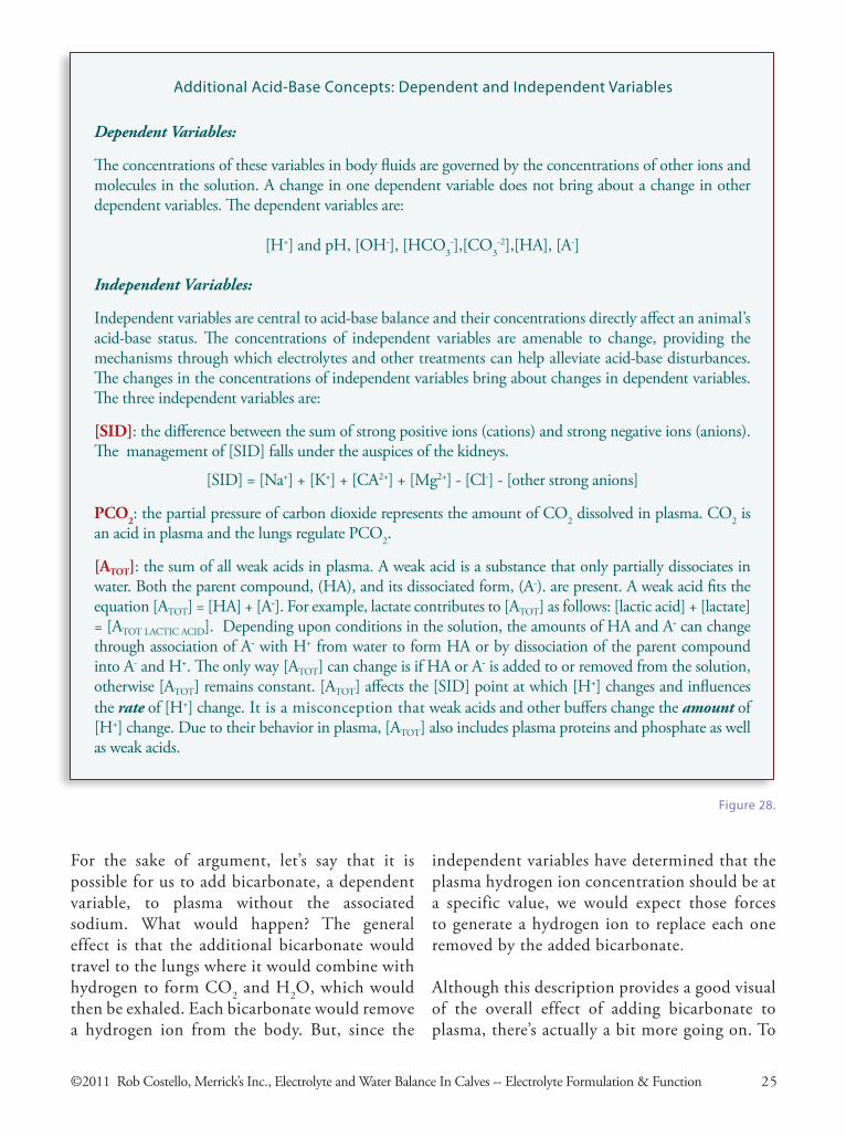
Dependent Variables:
The concentrations of these variables in body fluids are governed by the concentrations of other ions and molecules in the solution. A change in one dependent variable does not bring about a change in other dependent variables. The dependent variables are:
[H+] and pH, [OH-], [HCO3-],[CO3
-2],[HA], [A-]
Independent Variables:
Independent variables are central to acid-base balance and their concentrations directly affect an animal’s acid-base status. The concentrations of independent variables are amenable to change, providing the mechanisms through which electrolytes and other treatments can help alleviate acid-base disturbances. The changes in the concentrations of independent variables bring about changes in dependent variables. The three independent variables are:
[SID]: the difference between the sum of strong positive ions (cations) and strong negative ions (anions). The management of [SID] falls under the auspices of the kidneys.
[SID] = [Na+] + [K+] + [CA2+] + [Mg2+] - [Cl-] - [other strong anions]
PCO2: the partial pressure of carbon dioxide represents the amount of CO2 dissolved in plasma. CO2 is an acid in plasma and the lungs regulate PCO2.
[ATOT]: the sum of all weak acids in plasma. A weak acid is a substance that only partially dissociates in water. Both the parent compound, (HA), and its dissociated form, (A-). are present. A weak acid fits the equation [ATOT] = [HA] + [A-]. For example, lactate contributes to [ATOT] as follows: [lactic acid] + [lactate] = [ATOT LACTIC ACID]. Depending upon conditions in the solution, the amounts of HA and A- can change through association of A- with H+ from water to form HA or by dissociation of the parent compound into A- and H+. The only way [ATOT] can change is if HA or A- is added to or removed from the solution, otherwise [ATOT] remains constant. [ATOT] affects the [SID] point at which [H+] changes and influences the rate of [H+] change. It is a misconception that weak acids and other buffers change the amount of [H+] change. Due to their behavior in plasma, [ATOT] also includes plasma proteins and phosphate as well as weak acids.
Figure28.
AdditionalAcid-BaseConcepts:DependentandIndependentVariables
For the sake of argument, let’s say that it is possible for us to add bicarbonate, a dependent variable, to plasma without the associated sodium. What would happen? The general effect is that the additional bicarbonate would travel to the lungs where it would combine with hydrogen to form CO2 and H2O, which would then be exhaled. Each bicarbonate would remove a hydrogen ion from the body. But, since the
independent variables have determined that the plasma hydrogen ion concentration should be at a specific value, we would expect those forces to generate a hydrogen ion to replace each one removed by the added bicarbonate.
Although this description provides a good visual of the overall effect of adding bicarbonate to plasma, there’s actually a bit more going on. To
©2011 Rob Costello, Merrick’s Inc., Electrolyte and Water Balance In Calves -- Electrolyte Formulation & Function 25
begin with, we don’t add bicarbonate by itself, we also add sodium. When sodium and bicar-bonate are administered directly to plasma, they are added in a one-to-one ratio. But that’s not the case with sodium and bicarbonate in an oral electrolyte product.
Bicarbonate behaves differently in the digestive tract than it does in plasma. In the body, bicar-bonate only combines with hydrogen ions in specific lung tissue forming gas and water to be exhaled. Thankfully, that process does not occur in plasma. The controlling mechanisms that prevent this from happening, such as the partial pressure of carbon dioxide (PCO2), do not affect the gut. What’s inside the digestive tract is open to the outside environment.
Consequently, ingested bicarbonate can react with hydrogen ions in the stomach and intestine, generating gas and water and destroying bicar-bonate in the process. Also, there is no reason to expect that the remaining bicarbonate would be absorbed from the gut at the same rate or to the same extent as sodium. (The characteristics of sodium and its role in absorption are described in Chapter 1). As a result, there could be a large difference in the amounts of sodium and bicar-bonate that actually move from the digestive tract into plasma after oral administration of an electrolyte containing sodium bicarbonate.
Figure 28 shows that the total amount of weak acids in plasma equals the sum of all the weak acids plus their dissociated forms and fits the equation [ATOT] = [HA] + [A-]. Bicarbonate, acetate and lactate fit into this equation as:
[carbonic acid] + [bicarbonate][acetic acid] + [acetate][lactic acid] + [lactate]
Although bicarbonate, acetate and lactate are dependent variables, the equation shows that their addition to plasma increases total weak acids. Since the body works to maintain [ATOT] within narrow limits, the effect on [ATOT] would be temporary as the extra bicarbonate, acetate and lactate are metabolized -- bicarbonate in the lungs; acetate and lactate in the liver and other body tissues, generating energy.
The effect of a change in [ATOT] is to change the [SID] point at which the hydrogen ion concentra-tion changes. In other words, the amount of change in [SID] required to bring about a change in the hydrogen ion concentration increases or decreases in response to changes in [ATOT].
This effect is offset to some degree by the fact that we also add sodium every time we add bicarbonate. While bicarbonate is heading off to the lungs to be processed (or acetate and lactate to the liver and other body tissue) the added sodium is raising [SID] which lowers the hydrogen ion concentration and raises pH.
So, the notion that bicarbonate is an alkalinizing substance is obviously incorrect. The same can be said for literature that touts bicarbonate’s acid buffering benefits. Nonetheless, sodium bicarbonate is an alkalinizing compound and is frequently used as a low-cost formulation tool to generate an alkalinizing effect. Formulators often choose not to use sodium bicarbonate in oral electrolytes because of its potential for causing undesirable effects in the digestive tract. From a formulation standpoint, there is not a requirement that an alkalinizing compound must be used for an electrolyte to provide an alkalinizing effect. Having an alkalinizing compound on the label is no guarantee either. That’s up to the person doing the formulation.
©2011 Rob Costello, Merrick’s Inc., Electrolyte and Water Balance In Calves -- Electrolyte Formulation & Function 26
Angelos SA, Van Metre DC. Treatment of sodium balance disorders. Vet Clin North Am Food Anim Pract 1999; 15:587-607
Austgen, L; Bowen, R.A; Rouge, M. Pathophysiology of the digestive system. Colorado State University. 2001. http:/arbl.cvmbs.colostate.edu/hbooks.
Berchtold, J. Intravenous fluid therapy of calves. Vet Clin North Am Food Anim Pract 1999 15:505-531
Casiday R; Frey, R. Blood, sweat and buffers: pH regulation during exercise. Washington Univer-sity, Department of Chemistry. 2001.http://wunmr.wustl.edu/EduDev/LabTutorials/blood.htm.
Casiday R; Frey R. Maintaining the body’s chemistry: dialysis in the kidneys, Washington Univer-sity, Department of Chemistry. 2001.http://wunmr.wustl.edu/EduDev/LabTutorials/blood.htm.
Constable PD. Hypertonic saline.Vet Clin North Am Food Anim Pract 1999; 15:559-585
Dickman D.G, Hempson SJ, Anderson J, Lippe S, Zhao L, Burakoff R, Shaw, RD. Rotavirus alters paracellular permeability and energy metabolism in Caco-2 cells. Am J Physiol Gastrointest Liver Physiol 2000; 279 G757-G766
Dalton RG, Fisher EW, McIntyre WIM. Changes in blood chemistry, body weight and haematocrit of calves affected with neontatal diarrhoea. Br Vet J 1965; 121:34-41
Fayet J.C. Plasma and faecal osmolality, water kinetics and body fluid compartments in neonatal calves with diarrhoea. Br. Vet. J 1971; 127:37-43
Fisher E.W.; McEwan A.D. Death in neonatal calf diarrhoea. Pt. II: The role of oxygen and potas-sium. Br. Vet. J. 1967; 123:4-7
Fortran JS. Gas production after reaction of sodium bicarbonate and hydrochloric acid. Gastroen-terology 1984; 87(5):1014-1021
Grogono A.W. Acid-Base Balance. Tulane University School of Medicine, Department of An-esthesiology. 2001.www.acid-base.com
Groutides CP, Michell AR. Changes in plasma commposition in calves surviving or dying from diarrhoea. Br Vet J 1990; 146:205-210
Hunt E, Fu Q, Armstrong MU, Rennix DK, Webster DW, Galanko JA, Chen W, Weaver EM, Argenzio RA, Rhoads JM. Oral bovine serum concentrate improves cryptosporidial enteritis in calves. Pediatric Res 2002, 51, 3:370-376
Kasari TR. Metabolic acidosis in diarrheic calves: the importance of alkalinizing agents in therapy. Vet Clin North Am Food Anim Pract, 1990 6:29-43
Resources
©2011 Rob Costello, Merrick’s Inc., Electrolyte and Water Balance In Calves -- Resources 27
Kasari, TR; Naylor, JM: Clinical evaluation of sodium bicarbonate, sodium L-lactate, and sodium acetate for the treatment of acidosis in diarrheic calves. JAVMA, Vol 187, No. 4, August 15, 1985
Martin MJ, FitzSullivan E, Salim A, Berne TV, Towfigh S. Use of serum bicarbonate measure-ment in place of arterial base deficit in the surgical intensive care unit. Arch Surg 2005; 140:745-751
Naylor JM. A retrospective study of the relationship between clinical signs and severity of acido-sis in diarrheic calves. Can. Vet. J., 1989; 30:577-580.
Naylor JM. Oral electrolyhte therapy. Vet Clin North Am Food Anim Pract 1999; 15:487-504 1990; 6:51-67
Naylor JM. Oral fluid therapy in neonatal ruminants and swine. Vet Clin North Am Food Anim Pract
Naylor JM. Severity and nature of acidosis in diarrheic calves over and under one week of age. Can. Vet. J., 1987; 28:168-173
Naylor JM, Zello GA, Abeysekara S. Advances in oral and intravenous fluid therapy of calves with gastroeintestinal disease. Proc 24th World Buiatrics Congress 2006.
Phillips RW, Lewis LD, Knox KL. Alterations in body water turnover and distribution in neo-natal calves with acute diarrhea. Ann NY Acad Sci 1971; 176:231-243
Phillips RW. Fluid Therapy for diarrheic calves - what, how and how much. Vet Clin North Am Food Anim Pract 1985; 1:541-562
Phillips RW, Case GL. Altered metabolism, acute shock, and therapeutic response in a calf with severe coronavirus-induced diarrhea. Am J Vet Res 1980; 41:1039-1044
Stewart PA. How To Understand Acid-Base. A Quantitative Acid-Base Primer for Biology and Medicine. New York: Elsevier North Holland, Inc; 1981.
Tyler, Howard. Iowa State University, Ames Iowa. Personal communication
©2011 Rob Costello, Merrick’s Inc., Electrolyte and Water Balance In Calves -- Resources 28