eighth annual research symposium animal molecular...
TRANSCRIPT

EIGHTH ANNUAL RESEARCH SYMPOSIUM
ANIMAL MOLECULAR AND CELLULAR BIOLOGY GRADUATE PROGRAM
UNIVERSITY OF FLORIDA
Whitney Laboratory for Marine Bioscience St. Augustine, Florida
April 2‐3, 2010

1
2
Animal Molecular and Cellular Biology Graduate Program
Eighth Annual Research
Symposium
WELCOME
The AMCB symposium committee would like to welcome all faculty and students to this year’s Research Symposium which is held at Whitney Laboratory for Marine Bioscience. We wish all the best to Lilian Oliveira, Maria Padua, Sarah Reed, Luciano Silva, and Izabella Thompson, who completed their degrees in 2009. We welcome all new students and faculty to the premier scientific event in the AMCB calendar. As for the veteran students and faculty, we trust the Eigth Annual Symposium will be marked by good science, good fellowship, and good memories. Lokenga Badinga, AMCB Director Alan Ealy, AMCB Co‐Director
2
ACKNOWLEDGEMENTS
The faculty and students of the Animal Molecular and Cellular Biology Program
thank the following for support of the 8th Annual Research Symposium
Dr. Kirby Barrick, Dean for Academic Programs, IFAS, University of Florida
Dr. Mark McLellan, Dean of Research, IFAS, University of Florida
Dr. Winfred Phillips, Vice-President, Research and Graduate Programs, University of Florida
Appreciation is also expressed to those who have supported the AMCB
throughout the year
Dr. Kirby Barrick, Dean for Academic Programs, IFAS, University of Florida
Ms. Joann Fischer, Program Assistant, Dept. of Animal Sciences, University of Florida
Dr. Joel H. Brendemuhl, Professor, Dept. of Animal Sciences, University of Florida;
Graduate Coordinator, AMCB
Special thanks to Sarah Fields for help with the symposium

3
2010 Animal Molecular and Cellular Biology Distinguished Lecturer
Dr. Marc‐André Sirard
Marc-André Sirard DMV PhD started his carrier as a veterinarian in 1981. After a year in large animal practice, he begun graduate studies at University Laval on in vitro fertilization to generate the first clinical method to produce test-tubes calves in 1985. He then went for a post-doctoral training in the laboratory of Neal First in Wisconsin to study in vitro maturation of oocytes. He came back to Québec in 1987, and since has published over 180 scientific papers and has been invited to give over 28 invited lectures in international meetings. His current research activities are focus on oocyte quality and the link between the oocyte and the follicle in an animal model, the cow. He is associate editor of several journals as Reproduction, Reproduction Fertility and Development and Molecular Human Reproduction. He obtained an NSERC industrial Chair in 1990 and subsequently obtained the Synergy award with Semex Canada in 2000. He founded the CRBR (Centre de Recherche en Biologie de la Reproduction) in 1995 which has grown to include more than 100 people today. He obtained a senior Canadian Research Chair in 2000 on genomics applied to reproduction and is leading an international effort to define the normal genomic program in early mammalian embryos which has become an NSERC strategic network since 2008.
4
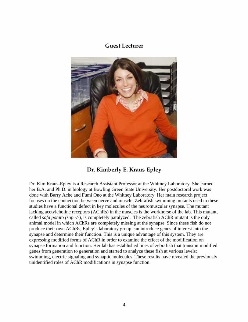
Guest Lecturer
Dr. Kimberly E. Kraus‐Epley
Dr. Kim Kraus-Epley is a Research Assistant Professor at the Whitney Laboratory. She earned her B.A. and Ph.D. in biology at Bowling Green State University. Her postdoctoral work was done with Barry Ache and Fumi Ono at the Whitney Laboratory. Her main research project focuses on the connection between nerve and muscle. Zebrafish swimming mutants used in these studies have a functional defect in key molecules of the neuromuscular synapse. The mutant lacking acetylcholine receptors (AChRs) in the muscles is the workhorse of the lab. This mutant, called sofa potato (sop -/-), is completely paralyzed. The zebrafish AChR mutant is the only animal model in which AChRs are completely missing at the synapse. Since these fish do not produce their own AChRs, Epley’s laboratory group can introduce genes of interest into the synapse and determine their function. This is a unique advantage of this system. They are expressing modified forms of AChR in order to examine the effect of the modification on synapse formation and function. Her lab has established lines of zebrafish that transmit modified genes from generation to generation and started to analyze these fish at various levels: swimming, electric signaling and synaptic molecules. These results have revealed the previously unidentified roles of AChR modifications in synapse function.
5
Committees of the AMCB, 2009‐2010 Director: Lokenga Badinga Co‐Director: Alan D. Ealy Graduate Coordinator: Joel H. Brendemuhl Program Assistant: Joann Fischer Academic / Assistantships Dr. Lokenga Badinga Dr. Sally Johnson Dr. David Julian Research / Symposium Dr. Lokenga Badinga Dr. Alan Ealy Dr. Peter Hansen Ms. Sarah Fields Mr. Kun Zhang Mr. Justin Fear Mr. Sha Tao Social Events Dr. Peter Hansen Dr. Alan Ealy Dr. Jose Santos Ms. Aline Bonilla Mr. Rafael Bisinotto Mr. Qien Yang Web Site Dr. Peter Hansen Dr. Alan Ealy
6
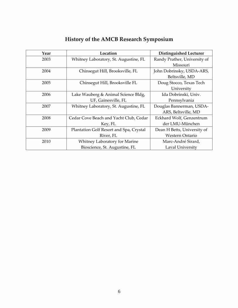
History of the AMCB Research Symposium
Year Location Distinguished Lecturer 2003 Whitney Laboratory, St. Augustine, FL Randy Prather, University of
Missouri 2004 Chinsegut Hill, Brooksville, FL John Dobrinsky, USDA‐ARS,
Beltsville, MD 2005 Chinsegut Hill, Brooksville FL Doug Stocco, Texas Tech
University 2006 Lake Wauberg & Animal Science Bldg,
UF, Gainesville, FL Ida Dobrinski, Univ.
Pennsylvania 2007 Whitney Laboratory, St. Augustine, FL Douglas Bannerman, USDA‐
ARS, Beltsville, MD 2008 Cedar Cove Beach and Yacht Club, Cedar
Key, FL Eckhard Wolf, Genzentrum
der LMU‐München 2009 Plantation Golf Resort and Spa, Crystal
River, FL Dean H Betts, University of
Western Ontario 2010 Whitney Laboratory for Marine
Bioscience, St. Augustine, FL Marc‐André Sirard, Laval University
7
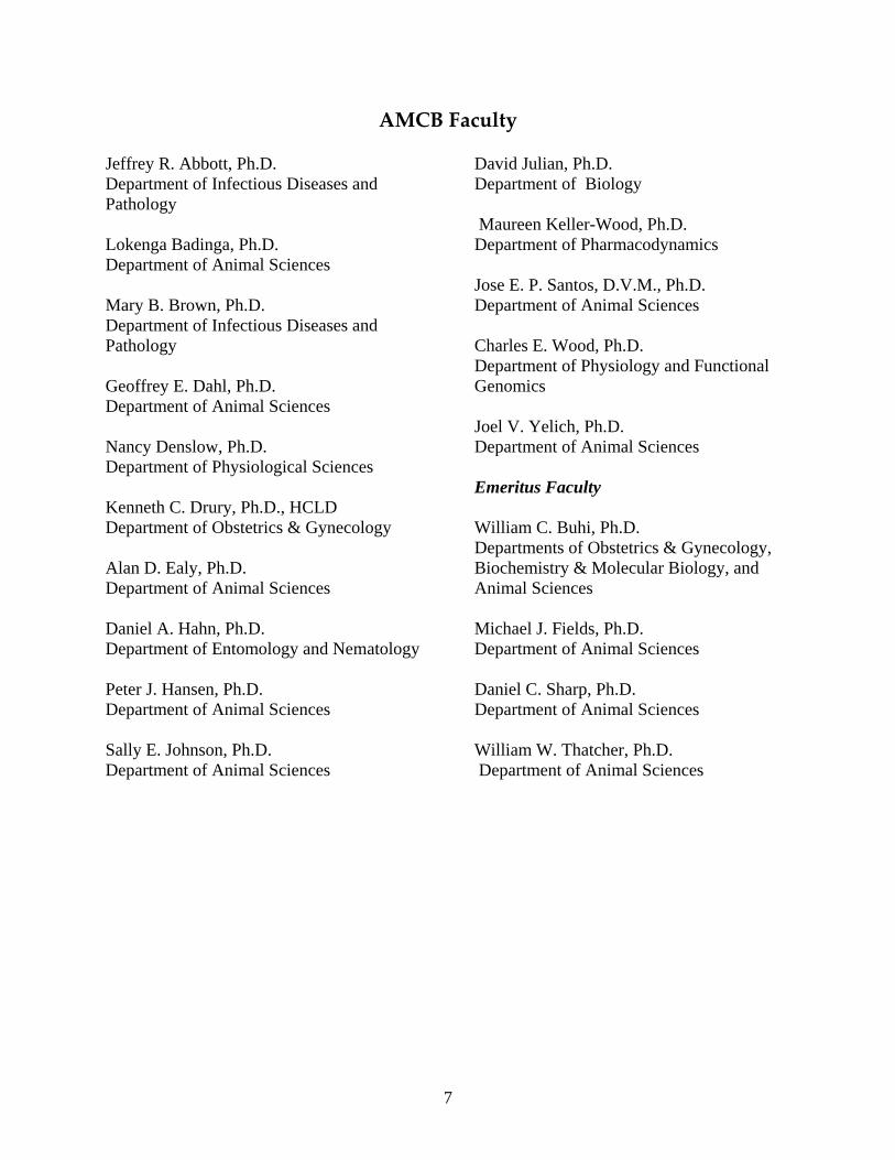
AMCB Faculty Jeffrey R. Abbott, Ph.D. Department of Infectious Diseases and Pathology Lokenga Badinga, Ph.D. Department of Animal Sciences Mary B. Brown, Ph.D. Department of Infectious Diseases and Pathology Geoffrey E. Dahl, Ph.D. Department of Animal Sciences Nancy Denslow, Ph.D. Department of Physiological Sciences Kenneth C. Drury, Ph.D., HCLD Department of Obstetrics & Gynecology Alan D. Ealy, Ph.D. Department of Animal Sciences Daniel A. Hahn, Ph.D. Department of Entomology and Nematology Peter J. Hansen, Ph.D. Department of Animal Sciences Sally E. Johnson, Ph.D. Department of Animal Sciences
David Julian, Ph.D. Department of Biology Maureen Keller-Wood, Ph.D. Department of Pharmacodynamics Jose E. P. Santos, D.V.M., Ph.D. Department of Animal Sciences Charles E. Wood, Ph.D. Department of Physiology and Functional Genomics Joel V. Yelich, Ph.D. Department of Animal Sciences Emeritus Faculty William C. Buhi, Ph.D. Departments of Obstetrics & Gynecology, Biochemistry & Molecular Biology, and Animal Sciences Michael J. Fields, Ph.D. Department of Animal Sciences Daniel C. Sharp, Ph.D. Department of Animal Sciences William W. Thatcher, Ph.D. Department of Animal Sciences

8
Current Students in the AMCB Ph.D. Students Aline Bonilla (Advisor: Pete. Hansen) Regina Esterman (Advisor: Joel Yelich) Sarah D. Fields (Advisor: Pete Hansen) Leandro Greco (Advisor: Jose Santos) Joe Kramer (Advisor: Ken Drury) Barbara Loureiro (Advisor: Pete Hansen) Kathleen Pennington (Advisor: Alan Ealy) Maria Belen Rabaglino (Advisor: Charles Wood) Sha Tao (Advisor: Geoff Dahl) Maria Christina Vasquez (Advisor: David Julian) Qien Yang (Advisor: Alan Ealy) Kun Zhang (Advisor: Alan Ealy) M.S. Students Rafael Bisinotto (Advisor: Jose Santos) Justin Fear (Advisor: Pete Hansen)
9
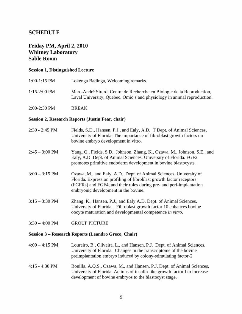
SCHEDULE Friday PM, April 2, 2010 Whitney Laboratory Sable Room Session 1, Distinguished Lecture 1:00-1:15 PM Lokenga Badinga, Welcoming remarks. 1:15-2:00 PM Marc-André Sirard, Centre de Recherche en Biologie de la Reproduction,
Laval University, Quebec. Omic’s and physiology in animal reproduction. 2:00-2:30 PM BREAK Session 2. Research Reports (Justin Fear, chair) 2:30 - 2:45 PM Fields, S.D., Hansen, P.J., and Ealy, A.D. T Dept. of Animal Sciences,
University of Florida. The importance of fibroblast growth factors on bovine embryo development in vitro.
2:45 – 3:00 PM Yang, Q., Fields, S.D., Johnson, Zhang, K., Ozawa, M., Johnson, S.E., and
Ealy, A.D. Dept. of Animal Sciences, University of Florida. FGF2 promotes primitive endoderm development in bovine blastocysts.
3:00 – 3:15 PM Ozawa, M., and Ealy, A.D. Dept. of Animal Sciences, University of
Florida. Expression profiling of fibroblast growth factor receptors (FGFRs) and FGF4, and their roles during pre- and peri-implantation embryonic development in the bovine.
3:15 – 3:30 PM Zhang, K., Hansen, P.J., and Ealy A.D. Dept. of Animal Sciences,
University of Florida. Fibroblast growth factor 10 enhances bovine oocyte maturation and developmental competence in vitro.
3:30 – 4:00 PM GROUP PICTURE Session 3 – Research Reports (Leandro Greco, Chair) 4:00 – 4:15 PM Loureiro, B., Oliveira, L., and Hansen, P.J. Dept. of Animal Sciences,
University of Florida. Changes in the transcriptome of the bovine preimplantation embryo induced by colony-stimulating factor-2
4:15 - 4:30 PM Bonilla, A.Q.S., Ozawa, M., and Hansen, P.J. Dept. of Animal Sciences, University of Florida. Actions of insulin-like growth factor I to increase development of bovine embryos to the blastocyst stage.
10
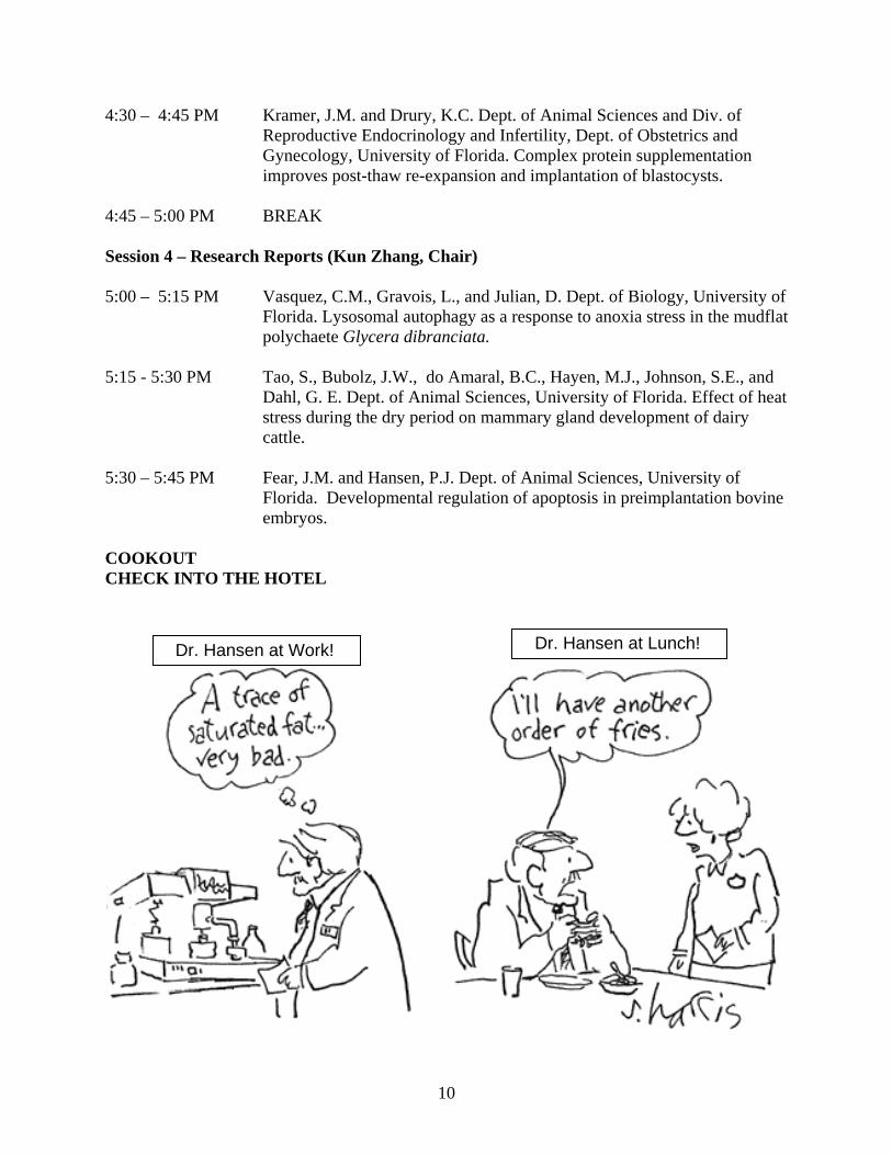
4:30 – 4:45 PM Kramer, J.M. and Drury, K.C. Dept. of Animal Sciences and Div. of Reproductive Endocrinology and Infertility, Dept. of Obstetrics and Gynecology, University of Florida. Complex protein supplementation improves post-thaw re-expansion and implantation of blastocysts.
4:45 – 5:00 PM BREAK Session 4 – Research Reports (Kun Zhang, Chair) 5:00 – 5:15 PM Vasquez, C.M., Gravois, L., and Julian, D. Dept. of Biology, University of
Florida. Lysosomal autophagy as a response to anoxia stress in the mudflat polychaete Glycera dibranciata.
5:15 - 5:30 PM Tao, S., Bubolz, J.W., do Amaral, B.C., Hayen, M.J., Johnson, S.E., and
Dahl, G. E. Dept. of Animal Sciences, University of Florida. Effect of heat stress during the dry period on mammary gland development of dairy cattle.
5:30 – 5:45 PM Fear, J.M. and Hansen, P.J. Dept. of Animal Sciences, University of
Florida. Developmental regulation of apoptosis in preimplantation bovine embryos.
COOKOUT CHECK INTO THE HOTEL
Dr. Hansen at Work! Dr. Hansen at Lunch!
11
Saturday AM, April 3, 2010 7:30 – 8:00 AM Breakfast Session 5 – Guest Lecture (Alan Ealy, Chair) 8:00 - 8:45 AM Kimberly E. Kraus-Epley, Whitney Laboratory for Marine Bioscience,
University of Florida. What can a 'lazy' fish teach us about synaptogenesis?
8:45 – 9:15 AM BREAK Session 6 – Research Reports (Belen Rabaglino, Chair) 9:15 – 9:30 AM Esterman, R.D., Austin, B.R., Woodall, S.A., McKinniss, E.M., and
Yelich, J.V. Dept. of Animal Sciences, University of Florida. Effect of day of the estrous cycle at the initiation of a CIDR protocol on ovulation rates and subsequent follicle developement in Bos taurus and Bos indicus x Bos taurus cows.
9:30 – 9:45 AM Araujo, D.B., Lopes, C.N., Vasconcelos, J.L.M., and Santos, J.E.P. Univ-
ersity of Florida, Gainesville, and São Paulo State University, Botucatu, Brazil. Effects of calcium salts of polyunsaturated fatty acids suppl-ementation on reproductive function of Nellore cows in Brazil.
9:45 – 10:00 AM Greco, L.F. Garcia, M., Favoreto, M.G. Marsola, R.S., Martins, L.T.,
Bisinotto, R.S., Ribeiro, E.S., Lima, F.S., Thatcher, W.W., Staples, C.R., and Santos, J.E.P. Dept. of Animal Sciences and Dept. of Large Animal Clinical Sciences, University of Florida. Fatty acid supplementation to periparturient dairy cows fed diets containing low amounts of fatty acids.
10:00 – 10:15 AM Bisinotto, R.S. Ribeiro, E.S., Greco, L.F., Lima, F.S., Favoreto, M.G.,
Ayres, H., Carvalho, M.R., Monteiro, A.P., Perdomo, M.C., Cerri, R.L.A., Risco, C.A., Thatcher, W.W., and Santos, J.E.P. Dept. of Animal Sciences and Dept. of Large Animal Clinical Sciences, University of Florida. Effect of follicular wave and concentration of progesterone during follicular growth on gene expression associated with conceptus development in dairy cows.
10:15 – 10:45 AM BREAK Session 7 – Research Reports (Christina Vasquez, Chair) 10:45 – 11:00 AM Cerri, R.L.A, Thompson, I.M., Kim, I.H., Ealy, A.D. Hansen, P.J., Staples,
C.R., Li, J.L.,and Thatcher, W.W. University of Florida, Gainesville and
12
Chungbuk National University, South Korea. Effects of lactation on endometrial gene expression in Holstein dairy cows.
11:00 – 11:15 AM Thompson, I.M., Bubolz, J., Ozawa, M., Yang, Q.E., and Dahl, D.E. Dept.
of Animal Sciences, University of Florida, Gainesville. Bovine luteal prolactin receptor expression: potential involvement in regulation of progesterone during the estrous cycle and pregnancy.
11:15 – 11:30 AM Pennington, K.A., Yang, Q.E., and Ealy, A.D. Dept. of Animal Sciences,
University of Florida. Candidate gene expression in bovine binucleated trophectoderm and changes in expression profiles after culture.
11:30 – 11:45 AM Rabaglino, M.B., Sumners, E., Denslow, N., and Wood, C.E.. Dept. of
Physiology and Functional Genomics and Dept. of Physiological Sciences, University of Florida. Microarray analysis of the genetic expression profile in ovine fetuses at the end of gestation.
ADJOURNMENT

13
ABSTRACTS
14
THE IMPORTANCE OF FIBROBLAST GROWTH FACTORS ON BOVINE EMBRYO DEVELOPMENT IN VITRO S. D. Fields, P. J. Hansen, and A. D. Ealy Department of Animal Sciences and D.H. Barron Reproductive and Perinatal Biology Research Program, University of Florida, Gainesville, Florida The objective was to test the hypothesis that fibroblast growth factors (FGF) regulate early embryonic development. The first two experiments were performed to determine if embryo-derived FGFs are required during development. In Exp 1, bovine embryos were produced in vitro and cultured in modified synthetic oviductal fluid (mSOF) containing either 20 µM SU5402, an FGF receptor inhibitor, or carrier (0.2% DMSO) at D 0 or 4. There was no effect of SU5402 on cleavage rate at Day 3. SU5402 reduced (P = 0.04) the percent of oocytes that were blastocysts on D 7 compared to controls when added at D 0 (5.9 ± 2.1 vs16.9 ± 2.4) but not when added at Day 4. Exp 2 tested if blocking FGF action at the blastocyst stage would affect subsequent cell number. D 8 blastocysts were placed into individual culture drops of mSOF containing 0.2% DMSO or 20 µM SU5402. SU5402 decreased (P = 0.04) the number of cells at D 11 (211.1 ± 27.5 vs 297.8 ± 25). Exp 3 was performed to test if supplemental FGF2 would enhance development to the blastocyst stage. Embryos were cultured in mSOF containing 0, 5, or 100 ng/mL FGF2. There was no effect of FGF2 on cleavage or percent of oocytes that were blastocysts at D 7 or 8. For Exp 4, 8-16 cell embryos were placed in fresh mSOF containing 0, 5, or 100 ng/mL FGF2 at D 5 after insemination. There was no effect of concentration of FGF2 on percent of oocytes that were blastocysts at D 7 or 8, number of trophectoderm cells or inner cell mass cells, or the ratio. Effects of higher concentrations of FGF2 were examined in Exp 5. Embryos received 0 ng/mL of FGF2 or 500 ng FGF2 at D 0, D 4, or D 0 and 4. There was no effect of FGF2 on cleavage. Addition of FGF2 at both D 0 and 4 (27.4 ± 1.3) increased (P ≤ 0.03) the percent of oocytes that became blastocysts on D 7 compared with control (19.7 ± 1.3) or FGF2 on D 4 (20.4 ± 1.3), but did not differ from FGF2 treatment on D 0 (23.2 ± 1.3). In summary, FGFs are important for normal blastocyst development. One of these, FGF2, increased the competence of embryos to become blastocysts at high concentrations. This work was supported by NRICGP Grant# 2008-35203-19106 and 2009-34135-20049 from the USDA-CSREES.
15
FGF2 PROMOTES PRIMITIVE ENDODERM DEVELOPMENT IN BOVINE BLASTOCYSTS Qi-En Yang, Sarah D Fields, Kun Zhang, Manabu Ozawa, Sally E Johnson and Alan D. Ealy Department of Animal Sciences, University of Florida, Gainesville, FL Cell fate specification begins in preimplantation embryo with the emergence of trophectoderm (TE). Primitive endoderm (PE) is the second extraembryonic lineage to form from cells within the inner cell mass (ICM) during embryogenesis. Ruminant conceptuses elongate dramatically prior to uterine attachment and extensive migration and proliferation of TE and PE is crucial for normal conceptus development and elongation. Work described here provides evidence that FGF2 and potentially other FGFs impact PE development during bovine embryonic development. Bovine blastocyst outgrowths were generated from in vitro-produced embryos using a feeder-layer free culture system containing a Matrigel-coated surface. Individually cultured bovine blastocysts were supplemented with FGF2 at the beginning of culture (day 8 post-IVF) and identification of PE-like cells was evaluated microscopically on days 13 and 15. In controls, only a single blastocyst outgrowth contained what appeared morphologically as PE cells (1.3% of all outgrowths). By contrast, 10.5±6.7%, 19.7±0.28% and 22.4±0.26% of outgrowths contained PE colonies on day 15 when embryos were exposed to 0.5, 5 and 50ng/ml FGF2, respectively. A subset of outgrowths (n=6 TE only and 6 PE-containing) were selected and cultured to day 20, then lineage-specific markers were evaluated using qRT-PCR. Transcripts for IFNT and CDX2 (TE markers) were present in TE outgrowths. Trace amounts of IFNT and CDX2 mRNA were detected in PE colonies (P<0.05) likely because residual TE cells still existed in some of these outgrowths. PE-containing outgrowths contained ample quantities of GATA4 and GATA6 mRNA (putative PE markers) whereas TE outgrowths contained no GATA4 and low amounts of GATA6 mRNA. None of the outgrowths contained NANOG mRNA whereas all TE and PE outgrowths contained OCT4 mRNA. Western blot analysis determined PE-containing outgrowths produced transferrin, a PE product. Moreover, immunoreactive GATA4 localized to nuclei of PE cells but was not detected in TE cells. By contrast, CDX2 localized to TE nuclei but was not detected in PE cells. Continued cultivation and selection for TE and PE yielded pure cell populations (n=6 of each). Differences in relative abundance of FGF receptors (FGFRs) and selective alternatively spliced receptor isoforms were evident between the cell types. Specifically, transcripts for FGFR2b and FGFR3 predominated in TE cells while FGFR1b represented the primary transcript in PE cells. TE and PE cells responded differently to FGF2 treatment. FGF2 supplementation did not affect the mitotic index of TE cells whereas the mitotic index of PE cells increased (P<0.05) following exposure to 5 or 50ng/ml FGF2 (35.1±0.09% and 34.7±0.11%, respectively versus 19.9±0.06% for controls). These observations indicate that the non-TE cells formed with blastocyst exposure to FGF2 are PE, and these cells respond differently to FGF2 than TE. Collectively, this work provides new insight into another vital activity for FGF2 and potentially other FGFs during early embryonic development in bovine. This project was supported by NRICGP number 2008-35203-19106 from the USDA-CSREES.
16
EXPRESSION PROFILING OF FIBROBLAST GROWTH FACTOR RECEPTORS (FGFRs) AND FGF4, AND THEIR ROLES DURING PRE- AND PERI-IMPLANTATION EMBRYONIC DEVELOPMENT IN THE BOVINE. Manabu Ozawa and Alan D. Ealy Department of Animal Sciences, University of Florida, Gainesville, FL The fact that preimplantation development and blastocyst formation occurs in the absence of uterine-derived factors indicates that regulatory molecules controlling embryogenesis are produced by the embryo itself. Several FGFs regulate embryogenesis in various ways in mice and other species, but their functions during embryonic development in cattle are not well described. Four genes encode FGFRs (termed FGFR1-4), and two predominant spliced variant forms (b or c isoforms) exist for FGFR1, 2, and 3 but not FGFR4. The overall aims of this work were to profile expression of FGFR and one embryonic FGF, FGF4, throughout early development and describe the impact of providing a chemical inhibitor to FGFR kinase activity (PD173074) on embryonic development in vitro. The relative transcript abundance of each major FGFR was determined at various stages of embryo development using qRT-PCR. FGFR1, 3 and 4 mRNA concentrations remained low from the zygote to 8-16-cell stage, then increased (P<0.05) at the morula and blastocyst stages. FGFR2 mRNA was evident in zygotes, progressively decreased (P<0.05) in 2-4 and 8-16 cell stage embryos, and then increased (P<0.05) at morula and blastocyst stages. The relative abundance of alternatively spliced forms of FGFR1 and 2 isoforms were also examined. At the morula and blastocyst stages, FGFR1b and FGFR2b mRNA were more abundant (P<0.05) than the c-isoforms for each receptor. Confocal immunofluorescence microscopy indicated that FGFR2 or FGFR4 were present exclusively in trophectoderm (TE) whereas FGFR1 and FGFR3 were found in both TE and ICM. FGF4 mRNA abundance was very low between the zygote and 8-16 cell stages and then increased (P<0.05) at morula and blastocyst stages. Interestingly, FGF4 mRNA abundance decreased (P<0.05) thereafter and was not detected in ovoid and elongating conceptuses collected at day 14-15 post-estrus from superovulated cows. The importance of FGF4 and potentially other embryo-derived FGFs during early embryo development was examined by treatment with 1 uM PD173074 beginning on day 5 post-fertilization (i.e. early morula stage). Exposure to this chemical prevented an increase of interferon-tau expression but did not affect the progression of embryos to blastocysts by day 8. However, the ability of blastocysts to form outgrowths when incubated on Matrigel-coated plates was compromised (P<0.05) by inhibitor treatment. On day 14 post-IVF, only 4.2±2.2% of inhibitor treated blastocysts formed outgrowths whereas 18.3±3.8% of the non-treated blastocysts formed outgrowths. These findings indicate that the expression of each FGFR and FGF4 occurs at a similar time after embryonic genome activation and that some FGFRs are expressed preferentially in TE whereas others are expressed throughout blastocysts. FGFR-mediated signaling does not appear to be essential for blastocyst formation but is important for subsequent outgrowth formation. This project was supported by NRICGP number 2008-35203-19106 from the USDA-CSREES.
17
FIBROBLAST GROWTH FACTOR 10 ENHANCES BOVINE OOCYTE MATURATION AND DEVELOPMENTAL COMPETENCE IN VITRO. Kun Zhang, Peter J. Hansen, and Alan D. Ealy. Dept. of Animal Sciences, University of Florida, Gainesville, Florida
An oocyte’s competency for generating viable offspring begins during folliculogenesis. Several endocrine and paracrine factors are suspected to play important roles in controlling oocyte competency. One candidate molecule is fibroblast growth factor 10 (FGF10). In cow, it is expressed by thecal cells and the oocyte, and its cognate receptor, FGFR2b, is expressed by granulosa cells. Moreover, FGF10 expression in theca cells is associated with follicle health in cattle. The aim of the present work was to describe how FGF10 impacts oocyte maturation and subsequent embryo development after in vitro fertilization (IVF). Cumulus-oocyte complexes (COCs) were collected and cultured with serum-free or serum-containing oocyte maturation medium supplemented with various concentrations of FGF10 and/or FGF10 neutralizing antibody. In vitro fertilization was performed by co-culture of matured COCs with a pool of frozen-thawed semen from three bulls. After 8-10 h, putative zygotes were cultured with modified synthetic oviductal fluid for 8 days. To evaluate whether FGF10 improves oocyte competence, FGF10 was added to maturation medium containing or lacking bovine steer serum. Subsequent fertilization rates were not affected by FGF10 treatment but increases (P<0.05) in the proportion of 8-16 cell embryos on d 3 and the blastocysts on d 7 and 8 post-IVF were evident when supplementing 50 ng/ml FGF10 in serum-containing medium and 0.5 ng/ml FGF10 in serum-free medium. In addition, FGF10 supplementation (50 ng/ml) in serum-containing medium increased the total number of cells in blastocysts (P=0.05) and number of inner cell mass cells (P=0.004). Addition of an antibody against FGF10 during oocyte maturation did not affect fertilization rates but prevented FGF10 from increasing the proportion of zygotes that formed blastocysts at d 7 (P<0.05). Moreover, the effect of anti-FGF10 could be reversed by providing a 3.9-fold molar excess of FGF10 (50 ng/ml). FGF10 supplementation acted in several capacities to impact oocyte maturation. Addition of FGF10 in serum-free maturation medium increased (P<0.05) first polar body extrusion rates, percentage of oocytes reaching telophase I and metaphase II (TI/MII), and cumulus expansion. Blocking endogenous FGF10 with anti-FGF10 decreased (P<0.05) the percentage of oocytes reaching MII and impeded cumulus expansion. Lastly, expression profiles of putative cumulus and oocyte competency markers were examined for their involvement in FGF10-mediated responses. FGF10 affected (P<0.05) the expression of several genes, including CTSB in cumulus cells and BMP15 and GDF9 in oocytes. In summary, FGF10 improved oocyte developmental competence and subsequent embryo development. This activity may be explained, at least in part, by benefiting meiotic and molecular maturation in COCs. This work provides new insights into how FGFs, and FGF10 in particular, function to control oocyte competency in cattle and potentially other animals. This project was supported by NRICGP number 2008-35203-19106 from the USDA CSREES.
18
CHANGES IN THE TRANSCRIPTOME OF THE BOVINE PREIMPLANTATION EMBRYO INDUCED BY COLONY-STIMULATING FACTOR-2 Barbara Loureiro, Lilian J. Oliveira and Peter J. Hansen Dept. of Animal Sciences and D.H. Barron Reproductive and Perinatal Biology Research Program, University of Florida Colony-stimulating factor-2 (CSF-2) is a cytokine expressed in bovine oviduct and endometrium that has been reported to improve the proportion of embryos that become blastocysts in vitro and survive after transfer to recipients. One effect of CSF-2 that may be related to increased embryonic survival is a preferential increase in the number of cells in the inner cell mass. The objective of the present study was to determine changes in the embryo transcriptome caused by CSF-2 that promotes blastocyst formation and establishment and maintenance of pregnancy after transfer. Bovine embryos were produced in vitro and cultured in KSOM-BE2 + 10 ng/ml recombinant BoCSF-2 added at day 5 after insemination. On day 6 (24 h after treatment), embryos at the morula and early blastocyst stage were harvested and stored in groups of 50 at -80°C. A total of 4 pools of GM-CSF treated blastocysts and 4 control blastocysts were subjected to transcriptional profiling using the Bos taurus Two Color Agilent chip (4 x 44K format). Prior to labeling, total RNA starting sample was spiked with control genes (artificial clones) of known concentration provided by Agilent. Labeling was done simultaneously with complimentary RNA (cRNA) amplification. Two rounds of linear RNA amplification were employed. Images were extracted using the Agilent Feature Extraction Software and normalized within arrays by the Lowess method. Statistical analysis was performed using JMP Genomics program (SAS Inst., Cary NC). The normalized data were transformed to log2 and the quantile normalization method was used to normalize data between arrays. Differences in gene expression were determined using PROC ANOVA (fixed false discovery rate = 0.01). Only genes with a 1.5-fold difference and P<0.05 were considered differentially expressed. A total of 216 genes were differentially expressed between CSF-2 and control embryos. Of these, 141 could be annotated (61 genes upregulated by CSF-2 and 80 genes downregulated). These included 13 genes involved in wnt pathways including 5 inhibitors of wnt signaling (FRP, MAB21L2, PCDH24, PDE7, PPPR23A) that were upregulated by CSF-2 and 5 genes involved in transmission of wnt signals (WNT16, ROR2, CSNK2B, CELSR2, DTX3) that were downregulated by CSF-2. Several other genes associated with differentiation were downregulated by CSF-2 including CXCL12, FEZF1, PLD2, and RGS12. Expression of one gene that inhibits apoptosis (PRKAR2B) was increased by CSF-2 while expression of 6 genes involved in apoptosis pathways (DAPK1, MADD, NOD2, PIK3IP1, RIPK3, RNF7) were downregulated. Results indicate that CSF-2 promotes pluripotency and decreases apoptosis in bovine preimplantation embryos. Research supported by USDA-AFRI. BL and LO were supported by a CAPES (Brazil)/Fulbright Fellowship.
19
ACTIONS OF INSULIN-LIKE GROWTH FACTOR I TO INCREASE DEVELOPMENT OF BOVINE EMBRYOS TO THE BLASTOCYST STAGE A.Q.S. Bonilla, M. Ozawa, and P.J. Hansen Department of Animal Sciences and D.H. Barron Reproductive and Perinatal Biology Research Program, University of Florida, Gainesville, Florida Insulin-like growth factor-I (IGF1) is a growth factor that increases the proportion of embryos becoming blastocysts in many species. The mechanism is unknown but could involve, among other possibilities, stimulation of blastomere proliferation, inhibition of apoptosis or cell-cycle arrest and stimulation of expression of genes involved in compaction and blastocyst formation. The overall objective of the present study was to determine the mechanism by which IGF1 increases the percent of oocytes becoming a blastocyst. Specifically, it was tested whether IGF1 acts before or after embryonic genome activation, stimulates development through the MAPK pathway, and increases transcript abundance for genes controlling compaction and blastocyst formation. Experiments were performed with embryos produced in vitro and cultured in a modified Synthetic Oviductal Fluid. In Exp. 1, it was demonstrated that recombinant human IGF1 could increase the proportion of oocytes becoming blastocysts and that concentrations of 10, 100 and 200 ng/mL were equally effective. In Exp. 2 it was tested whether IGF1 acts to increase blastocyst yield before or after day 4 of development. Embryos were cultured with the following treatments: 1) medium alone from day 0-8 post insemination (pi), 2) medium alone from day 0-4 and medium with 100 ng/mL IGF1 from day 4-8, 3) medium with 100 ng/mL IGF1 from day 0-4 and medium alone from day 4-8, or 4) medium plus 100 ng/mL IGF1 from day 0-8. As compared to controls (30.4% ± 1.89), IGF1 increased (P<0.01) the percent of oocytes that were blastocysts at day 8 pi when added from day 4-8 (42% ± 1.89) or day 0-8 (40% ± 1.89) but not from day 0-4 (33.6 ± 1.89). For Exp. 3, it was tested whether IGF1 increases blastocyst development by acting through MAPK signaling. At day 4, embryos were placed in fresh medium to achieve the following treatments: 1) medium + 0.1% DMSO (vehicle); 2) medium + 0.1% DMSO and 100 ng/mL IGF1; 3) medium + 0.1% DMSO (vehicle) + 100 µM PD 98059 (MAPK inhibitor); and 4) medium +DMSO + 100 ng/mL IGF1 + 100 µM PD 98059. In the absence of IGF1, PD 98059 did not decrease the percent of oocytes that became blastocysts at day 8 (21% vs 24% for control and PD 98059, pooled SEM=0.02). IGF1 increased blastocyst yield (P<0.001) and PD 98059 decreased this effect (37% vs 29% for IGF1 and IGF1 + PD 98059; P<0.05). In Exp. 4 quantitative real time PCR was used to analyze genes involved in compaction and blastocyst formation. At day 4, embryos received IGF1 (100 ng/mL) or vehicle. Embryos were harvested on day 5 (those ≥ 16 cells) or 6 (morulae and early blastocysts) and analyzed for expression of CDX2, CDH1, ATP1A1, OCLN and Hist2h2aa2. IGF1 tended to increase ATP1A1 at day 5 (1.3 fold; P=0.09) and increased the expression of ATP1A1 day 6 (1.68 fold; P<0.04), but there was no effect on IGF-1 on the other genes. In conclusion, IGF1 promotes development to the blastocyst stage by regulating MAPK-dependent events at day 4 or later. Among these events is upregulation of expression of ATP1A1, which is required for blastocoel formation (Support: USDA NRI Grants 2006-55203-17390 and 2007-35203-18070).
20
COMPLEX PROTEIN SUPPLEMENTATION IMPROVES POST-THAW RE-EXPANSION AND IMPLANTATION OF BLASTOCYSTS Joseph M. Kramer and Kenneth C. Drury
Department of Animal Sciences and Division of Reproductive Endocrinology and Infertility, Department of Obstetrics and Gynecology, University of Florida, Gainesville, FL While several studies have indicated supplementation of embryo culture medium with a globulin-rich protein source benefits embryo compaction and blastocyst formation, the affects of such complex proteins in cryopreservation and thaw solutions on blastocyst post-thaw survival and potential is unknown. The purpose of this study was to test whether the supplementation of freeze and thaw solutions with a commercial protein source containing globulins would improve post-thaw blastocyst developmental potential as measured by blastocyst re-expansion, total cell counts, pregnancy and implantation. Animal experimental studies compared thaw survival of control-rate cryopreserved murine blastocysts using the globulin-rich protein source, Serum Substitute Supplement (SSS, 10mg/ml HSA + 2mg/ml globulins; Irvine Scientific) or human serum albumin (HSA, 10mg/ml) alone in solutions for freeze and thaw, as well as the additional supplementation of post-thaw culture medium. Blastocysts with greater than 50% re-expansion were considered as survived. Re-expanded blastocysts were fixed on slides, stained with DAPI, and viewed using epifluorescent microscopy to determine total cell counts. The retrospective human study included analysis of 29 frozen embryo transfer (FET) cycles in which frozen blastocysts were thawed in solutions supplemented with HSA or SSS. Data were analyzed using fisher’s exact test, chi-square test, or student’s t-test where appropriate. A greater number of murine blastocysts re-expanded if frozen and thawed in solutions containing SSS compared to frozen and thawed in solutions containing only HSA (85.7% vs 66.1%; P<0.05). Total cell counts did not significantly differ between treatments, however, there was a trend supporting greater total cell numbers from re-expanded blastocysts that were frozen and thawed in solutions supplemented with SSS compared to frozen and thawed in solutions containing only HSA (80.0 vs 70.5; P=0.057). Retrospective analysis of human clinical FET cycles revealed higher rates of blastocyst survival (84.6% vs 43.9%), re-expansion (69.2% vs 15.8%), clinical pregnancy per thaw (60.0% vs 10.5%) and implantation (53.3% vs 13.3%) for blastocysts thawed in solutions supplemented with SSS compared to HSA (P<0.05). Our data demonstrate that the inclusion of a globulin-rich protein source, such as SSS, in cryopreservation and thaw solutions improves post-thaw survival and implantation.
21
LYSOSOMAL AUTOPHAGY AS A RESPONSE TO ANOXIA STRESS IN THE MUDFLAT POLYCHAETE GLYCERA DIBRANCIATA M. Christina Vasquez, Lauren Gravois and David Julian. Dept. of Biology, University of Florida, Gainesville Lysosomal autophagy functions to remove and recycle damaged and redundant cellular components that accumulate under stressful conditions. We hypothesize that this response allows marine organisms to survive variable abiotic stressors, such as oxidative stress. To investigate this, we studied lysosomal autophagy as a response to oxidative stress in erythrocytes of the mudflat polychaete Glycera dibranchiata, a species native to the anoxic mudflats of Maine. We quantified the average number of lysosomes present per cell during exposure to normoxia, following 24 h exposure to anoxia, and during a 24 h normoxia recovery period immediately following the anoxia exposure. Our preliminary data suggest that the number of lysosomes is reduced by 50% after 24 h in anoxia. After 1 hour in normoxia following anoxia, the average number of lysosomes doubles and then slowly decreases over an additional 24 h. During recovery from anoxia we observed the presence of putative autophagosomes, which are organelles that capture damaged cellular components to be delivered to the lysosome for degradation. These data suggest that lysosomal autophagy plays a role in survival of G. dibranchiata in its natural habitat by removing oxidatively damaged proteins and organelles, which could otherwise compromise the cell and ultimately lead to death.
22
EFFECT OF HEAT STRESS DURING THE DRY PERIOD ON MAMMARY GLAND DEVELOPMENT OF DAIRY CATTLE S. Tao, J. W. Bubolz, B. C. do Amaral, M. J. Hayen, S. E. Johnson and G. E. Dahl Dept. of Animal Sciences, University of Florida, Gainesville, FL Heat stress during the dry period affects immune status, alters hepatic metabolism and decreases milk production in the subsequent lactation. But, cellular mechanisms involved in the mammary response are unclear. Our objective was to evaluate the effects of heat stress during the dry period on mammary gland development of multiparous cows. Cows were dried off 46 d prior to expected calving and assigned to two treatments, heat stress (HT, n=15) or cooling (CL, n=14). Average THI during treatment was 76.6 for all cows, but CL cows had sprinklers and fans that came on when ambient temperatures reached 21.1 0C, whereas HT cows were in the same barn without fans or sprinklers. Rectal temperature (RT) was measured twice daily (0730 and 1430h) and respiration rates (RR) recorded at 1500h on a Mon-Wed-Fri schedule from dry-off to calving. After parturition, all cows were housed in a free-stall barn with sprinklers and fans. Milk yield was recorded daily to 147 DIM. Mammary biopsies were taken at dry-off, -20, +2 and +20 d relative to calving from a subset of cows (HT, n=7, CL, n=7) and infiltrated with paraffin. Numbers of Ki67 immunopositive epithelial and stromal cells were measured in 4 micron mammary tissue sections from each animal. Total cell numbers were measured following hematoxylin histology and percent proliferation was calculated as Ki67+/ total × 100. Compared with HT cows, CL cows had lower morning and afternoon RT (38.6 vs. 38.8 0C, 39.0 vs. 39.40C, P < 0.01, respectively) and lower RR (46 vs. 78 breaths/min, P < 0.01). Relative to HT cows, CL cows produced more milk (36.5 vs. 31.6 kg/d, P < 0.06). Compared with HT, CL cows had a higher percentage of proliferative epithelial cells at -20 d relative to calving (3.3 vs. 1.3%, P < 0.05), but there was no difference in labeled stromal cells (P > 0.1). We conclude that heat stress abatement during dry period improves milk production in the subsequent lactation possibly by increasing mammary epithelial cell proliferation during the dry period.

23
DEVELOPMENTAL REGULATION OF APOPTOSIS IN PREIMPLANTATION BOVINE EMBRYOS
Justin M. Fear and P.J. Hansen Department of Animal Sciences and D.H. Barron Reproductive and Perinatal Biology Research Program, University of Florida, Gainesville Florida
Preimplantation embryos undergo apoptosis in a stage-dependent manner. Apoptosis is blocked in 2-cell and 4-cell embryos and capacity for apoptosis is restored around the 8-16 cell stage (Paula-Lopes and Hansen, Biol Reprod 66:1169-1177, 2002). One of the developmental blocks is at the level of the mitochondrion (Brad et al., Reproduction 134: 789-797; de Castro et al., Mol. Reprod. Dev., 75:1063-1070, 2008). Whether depolarization of the mitochondrial membrane occurs in response to a pro-apoptotic signal depends upon the complex balance of anti- versus pro- apoptotic proteins. We hypothesized that 2-cell embryos have higher amounts of anti-apoptotic proteins and lower amounts of pro-apoptotic proteins when compared to embryos ≥16-cell embryos. Two-cell embryos were collected between 28 to 30 h post-insemination and embryos ≥16-cells were collected at Day 5 post-insemination. Experiment 1 measured steady state mRNA for Bcl2, Bcl2l1, Hspa1a, Bax, Bad, Dffb (caspase-activated DNase), Dffa (inhibitor of caspase-activated DNase), and Hist1h2ae using quantitative real-time RT-PCR comparing MII oocytes, 2-cell embryos, 2-cell embryos treated with a transcription inhibitor α-amanitin (50 µM), and ≥16-cell embryos. Cycle threshold (Ct) values were measured and analyzed using the General Linear Model procedure of Statistical Analysis System (SAS); only Dffb was not affected by developmental stage. ∆Ct was calculated using Dffb as the internal control and analyzed by least-squares analysis of variance using the General Linear Models procedure of SAS. The anti-apoptotic genes Bcl2 and Hspa1a were higher in oocytes, 2-cell embryos, and 2-cell embryos treated with α-amanitin compared to the ≥16-cell embryos ( P<.001) and the pro-apoptotic gene Bad was lower in oocytes, 2-cell embryos, and 2-cell embryos treated with α-amanitin compared to the ≥16-cell embryos (P<.0001). Steady state mRNA for Bcl2l1, Bax, Dffa, and Hist1h2ae was not affected by stage of development. Experiment 2 measured immunoreactive amounts of the anti-apoptotic proteins BCL2, BCL2L1 and HSPA1A and the pro-apoptotic proteins BAX and BAD by immunoflourescence microscopy. Mean pixel intensity was measured on a per embryo basis using ImageJ (NIH) and average mean pixel intensities for IgG control embryos were subtracted from the values of antibody-stained embryos to compensate for background. Amounts of immunoreactive BCL2 was greater at the 2-cell stage than for embryos ≥16-cells (327 ± 43.7 vs 101 ± 49.9; P=0.0010, n=111). In contrast, BAX (221 ± 25.0 vs 399 ± 20.1; P<.0001, n=142) was greater for embryos ≥16 cells. Amounts of immunoreactive BCL2L1, HSPA1A and BAD were not significantly affected by stage of development. To verify immunoflourescence results, Western blotting was performed for BCL2 and BAX. There was higher amounts of immunoreactive BCL2 in 2-cell embryos compared to ≥16-cell embryos. For BAX, multimers were identified in ≥16-cell embryos that were not present in 2-cell embryos. These data suggest that the developmental acquisition of the capacity for apoptosis is associated with the regulation of genes involved in apoptosis at the level of transcription and translation, and possibly post-translational modifications. Research supported by USDA NRICGP 2007-35203-18070.
24
EFFECT OF DAY OF THE ESTROUS CYCLE AT THE INITIATION OF A CIDR PROTOCOL ON OVULATION RATES AND SUBSEQUENT FOLLICLE DEVELOPEMENT IN BOS TAURUS AND BOS INDICUS × BOS TAURUS COWS
R.D. Esterman, B.R. Austin, S.A. Woodall, E.N. McKinniss, and J.V. Yelich Dept. of Animal Sciences, University of Florida, Gainesville.
Cattle of Bos indicus influence are commonly raised in subtropical regions like Florida
due to their superior heat and disease tolerance, and ability to utilize low quality forages compared to Bos taurus cattle. Subtle differences in the reproductive physiology of Bos indicus compared to Bos taurus influenced cattle, create challenges in developing effective synchronization protocols for Bos indicus influenced cattle. Stage of the estrous cycle when GnRH is administered effects ovulation rates in Bos taurus cattle but its effect is unclear in Bos indicus cattle. Cattle of Bos indicus breeding have a greater frequency of three and four follicle wave patterns compared to Bos taurus cattle suggesting that they have fewer opportunities for ovulation to GnRH, which could result in altered follicle development patterns during synchronization protocols in Bos indicus cattle.
Lactating, estrous cycling Angus (Bos taurus) and Brangus (Bos indicus × Bos taurus) cows at different stages of the estrous cycle (DOC) were utilized to evaluate the effectiveness of GnRH to initiate ovulation in cows receiving a 7-d CIDR synchronization protocol. Estrous cycling cows were pre-synchronized to start the synchronization protocol on either day 2, 6, 10, 14, or 18 of the estrous cycle (n = 5 Angus & n = 5 Brangus in each DOC group). Cows received GnRH (100 µg) at CIDR insertion followed by PGF2α (25 mg) 7-d later. Daily ultrasonography and blood sampling were conducted from CIDR insertion until3 d after PGF2α to evaluate follicle growth and progesterone concentrations. Cows were inseminated 8-10 h after exhibiting estrus and cows not exhibiting estrus were timed-AI + GnRH 73 to 80 h after PGF2α.
Ovulation rate to the initial GnRH and ovulatory follicle size were similar (P > 0.05) for Angus (56%; 13.9 ± 1.8 mm) and Brangus (52%; 14.1 ± 2.4 mm). However, ovulation rate to initial GnRH and ovulatory follicle size (mm) differed (P < 0.05) for cows on days 2 (0%; no ovulations), 6 (100%; 13.2 ± 2.1), 10 (30%; 15.7 ± 1.5), 14 (70%; 15.7 ± 1.5), and 18 (70%; 15.6 ± 1.5) of the estrous cycle. Brangus had a greater (P < 0.05) number of class I follicles (3-5 mm) compared to Angus. There tended (P = 0.10) to be a breed × day of synchronization effect on class II (6-9 mm) follicle number, suggesting a difference in follicle recruitment between breeds. There were DOC (P < 0.05) and DOC × day of synchronization (P < 0.05) effects on class III (≥ 10 mm) follicle numbers. Estrous response was greater (P < 0.05) for Brangus (48.0%) compared to Angus (28.0%), but conception, timed-AI pregnancy, and synchronized pregnancy rates were similar (P > 0.05) for Angus and Brangus. Estrous response, conception rate, timed-AI pregnancy, and synchronized pregnancy rates differed (P < 0.05) for cows on days 2 (0, 0, 10, 10%), 6 (10, 100, 11, 20%), 10 (30, 100, 57, 70%), 14 (60, 33, 100, 60%), and 18 (90, 44, 0, 40%), respectively. Further research will investigate effects of stage of follicular growth on oocyte competency in Angus and Brangus cows. Results generated from this experiment have led to a better understanding of follicle development in Bos indicus × Bos taurus cattle, which can be used to design more effective estrous synchronization protocols in these cattle. EFFECTS OF
25
CALCIUM SALTS OF POLYUNSATURATED FATTY ACIDS SUPPLEMENTATION ON REPRODUCTIVE FUNCTION OF NELLORE COWS IN BRAZIL D.B. Araujo1, C.N. Lopes2, J.L.M. Vasconcelos2 and J.E.P. Santos1 University of Florida, Gainesville, 2 São Paulo State University, Botucatu, Brazil Brazil is the world’s biggest beef exporter and it is ranked eighth in the gross domestic product (GDP) classification. All agribusiness operations together represent 25% of the total Brazilian GDP and beef cattle accounts for 27% of this amount. The maintenance of this framework depends on the profitability of the activity. Thus, management strategies that enhance reproductive performance of beef cows are beneficial to the productivity of cow-calf operations. Strategies that increase P4 concentrations in cattle before or after breeding have been positively associated with pregnancy rates and supplemental polyunsaturated fatty acids (PUFA) have been shown to increase P4 concentrations by several pathways. Four experiments were designed to evaluate the effects of calcium salts of fatty acids on reproductive performance of Nellore beef cows. In experiment 1, 51 ovulated non-lactating multiparous Nellore cows were used to evaluate if PUFA supplementation affects circulating P4 concentrations and timing of luteolysis. In experiment 2, 43 ovulated non-lactating multiparous Nellore cows were used to evaluate if PUFA supplementation alters the sensitivity of a 6-d corpus luteum to exogenous prostaglandin treatment. In experiment 3, 27 ovulated postpartum Nellore cows were used to evaluate if PUFA supplementation affects the incidence of premature luteolysis. Beginning at the d of estrus, the daily treatments in these 3 experiments were: Control (0.1 Kg Mineral + 0.1 Kg corn + 0.1 Kg kaolin); SFA (0.1 Kg Mineral + 0.1 Kg Megalac®-S + 0.1 Kg corn), this group was used just in experimental 1 and 2; PFA (0.1 Kg Mineral + 0.1 Kg Megalac®-E + 0.1 Kg corn). The SFA was not included as treatment during experiment 3. Treatments did not affect (P > 0.1) circulating concentrations of P4, timing of luteolysis, corpus luteum sensitivity to prostaglandin, and the incidence of premature luteolysis. Although when cows from experiment 1 and 2 were grouped, greater (P<0.05) concentration of P4 were observed in cows that were supplemented with PUFA. In experiment 4, we evaluated if the length of PUFA supplementation after timed-AI alters the pregnancy rate. Postpartum multiparous Nellore cows (n = 1457) received 0.1 Kg mineral + 0.1 Kg corn during the 28 d after timed-AI plus one of the following daily treatments: T1 (0.1 Kg kaolin during 28 d post-AI; n = 156); T2 (0.1 Kg Megalac® during the first 14 d post-AI + 0.1 Kg kaolin from d 15 to 28; n = 160); T3 (0.1 Kg Megalac®-E during the first 14 d post-AI + 0.1 Kg kaolin from d 15 to 28; n = 305); T4 (0.1 Kg Megalac®-S during the first 21 d post-AI + 0.1 Kg kaolin from d 22 to 28; n = 158); T5 (0.1 Kg Megalac®-E during the first 21 d post-AI + 0.1 Kg kaolin from d 22 to 28; n =252) T6 (0.1 Kg Megalac®-S during 28 d post-AI; n = 155); T7 (0.1 kg Megalac®-E during 28 d post-AI; n = 271). Cows supplemented with PUFA during 21 or 28 d post-AI had greater pregnancy rates (P< 0.05) than cows from other treatments. Additionally, there was no difference between T5 and T7 treatments. These experiments indicated that the possible mechanism for enhanced reproductive performance with supplemental PUFA post-AI may be due to effects on embryo development. Cows supplemented for 21 or 28 d post-AI had greater pregnancy rates with no changes in circulating concentrations of progesterone and corpus luteum maintenance.
26
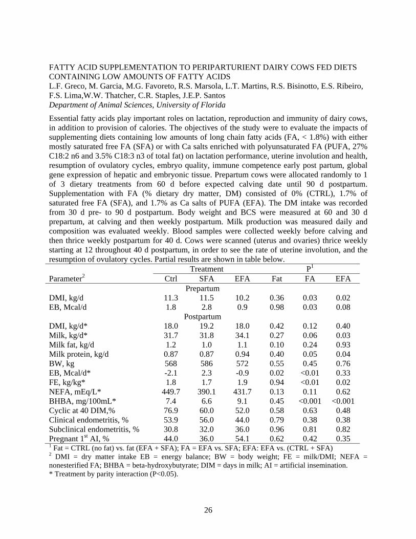
FATTY ACID SUPPLEMENTATION TO PERIPARTURIENT DAIRY COWS FED DIETS CONTAINING LOW AMOUNTS OF FATTY ACIDS L.F. Greco, M. Garcia, M.G. Favoreto, R.S. Marsola, L.T. Martins, R.S. Bisinotto, E.S. Ribeiro, F.S. Lima,W.W. Thatcher, C.R. Staples, J.E.P. Santos Department of Animal Sciences, University of Florida
Essential fatty acids play important roles on lactation, reproduction and immunity of dairy cows, in addition to provision of calories. The objectives of the study were to evaluate the impacts of supplementing diets containing low amounts of long chain fatty acids (FA, < 1.8%) with either mostly saturated free FA (SFA) or with Ca salts enriched with polyunsaturated FA (PUFA, 27% C18:2 n6 and 3.5% C18:3 n3 of total fat) on lactation performance, uterine involution and health, resumption of ovulatory cycles, embryo quality, immune competence early post partum, global gene expression of hepatic and embryonic tissue. Prepartum cows were allocated randomly to 1 of 3 dietary treatments from 60 d before expected calving date until 90 d postpartum. Supplementation with FA (% dietary dry matter, DM) consisted of 0% (CTRL), 1.7% of saturated free FA (SFA), and 1.7% as Ca salts of PUFA (EFA). The DM intake was recorded from 30 d pre- to 90 d postpartum. Body weight and BCS were measured at 60 and 30 d prepartum, at calving and then weekly postpartum. Milk production was measured daily and composition was evaluated weekly. Blood samples were collected weekly before calving and then thrice weekly postpartum for 40 d. Cows were scanned (uterus and ovaries) thrice weekly starting at 12 throughout 40 d postpartum, in order to see the rate of uterine involution, and the resumption of ovulatory cycles. Partial results are shown in table below. Treatment P1 Parameter2 Ctrl SFA EFA Fat FA EFA
Prepartum DMI, kg/d 11.3 11.5 10.2 0.36 0.03 0.02 EB, Mcal/d 1.8 2.8 0.9 0.98 0.03 0.08
Postpartum DMI, kg/d* 18.0 19.2 18.0 0.42 0.12 0.40 Milk, kg/d* 31.7 31.8 34.1 0.27 0.06 0.03 Milk fat, kg/d 1.2 1.0 1.1 0.10 0.24 0.93 Milk protein, kg/d 0.87 0.87 0.94 0.40 0.05 0.04 BW, kg 568 586 572 0.55 0.45 0.76 EB, Mcal/d* -2.1 2.3 -0.9 0.02 <0.01 0.33 FE, kg/kg* 1.8 1.7 1.9 0.94 <0.01 0.02 NEFA, mEq/L* 449.7 390.1 431.7 0.13 0.11 0.62 BHBA, mg/100mL* 7.4 6.6 9.1 0.45 <0.001 <0.001 Cyclic at 40 DIM,% 76.9 60.0 52.0 0.58 0.63 0.48 Clinical endometritis, % 53.9 56.0 44.0 0.79 0.38 0.38 Subclinical endometritis, % 30.8 32.0 36.0 0.96 0.81 0.82 Pregnant 1st AI, % 44.0 36.0 54.1 0.62 0.42 0.35 1 Fat = CTRL (no fat) vs. fat (EFA + SFA); FA = EFA vs. SFA; EFA: EFA vs. (CTRL + SFA) 2 DMI = dry matter intake EB = energy balance; BW = body weight; FE = milk/DMI; NEFA = nonesterified FA; BHBA = beta-hydroxybutyrate; DIM = days in milk; AI = artificial insemination. * Treatment by parity interaction (P<0.05).
27
EFFECT OF FOLLICULAR WAVE AND CONCENTRATION OF PROGESTERONE DURING FOLLICULAR GROWTH ON GENE EXPRESSION ASSOCIATED WITH CONCEPTUS DEVELOPMENT IN DAIRY COWS
R.S. Bisinotto*, E.S. Ribeiro, L.F. Greco, F.S. Lima, M.G. Favoreto, H. Ayres, M.R. Carvalho, A.P. Monteiro, M.C. Perdomo, R.L.A. Cerri, C.A. Risco, W.W. Thatcher, J.E.P. Santos.
Depts. of Animal Sciences and Large Animal Clinical Sciences, University of Florida
Anovular cows have reduced fertility because of lower pregnancy per insemination (P/AI) and increased pregnancy loss. One of the mechanisms for reduced fertility involve premature regression of the CL, but it is likely that this only explains a portion of the decrease in P/AI, and does not explain the increased pregnancy loss after d 28 of gestation. The poor fertility of anovular cows subjected to synchronization protocols has been linked to the ovulation of a first wave follicle, which grows under low and increasing concentrations of progesterone (P4). We speculate that P4 concentrations during the development of the ovulatory follicle impact endometrial and embryonic gene expression, which might be related to pregnancy responses of anovular cows. The hypothesis of the present study is that the ovulation of a first wave follicle reduces expression of genes related to histotroph secretion and conceptus elongation compared with the ovulation of a second wave follicle. This effect is mediated by the low concentrations of P4 during follicle development. Furthermore, we hypothesize that supplementation with exogenous P4 during the growth of the ovulatory follicle will attenuate the changes in gene expression observed with ovulation of the first wave dominant follicle. Non-lactating cyclic Holstein cows (n=36) had their estrous cycle pre-synchronized by an injection of GnRH and the insertion of a controlled internal drug release (CIDR) containing P4. The CIDR was removed 7 d later and 2 injections of PGF2α were administered 24 h apart. The Ovsynch protocol (d0 GnRH; d7 PGF2α; d8 PGF2α; d9.5 GnRH and AI; d10 AI) was initiated either 2 (FW and FWP4) or 8 d (SW) after the first PGF2α of pre-synchronization. Cows in the SW group received an injection of GnRH 2 d after the first PGF2α of pre-synchronization. Cows in the FWP4 group received 3 CIDR inserted 12, 24 and 48 h after the initial GnRH of the Ovsynch protocol. Ovaries were scanned at all treatments, 2 d after each GnRH injection, and 7 and 13 d after AI. Only cows that responded to all hormonal treatments were kept in the study. Blood was sampled daily starting at the initial GnRH of the Ovsynch protocol. Cows were slaughtered 17 d after AI. Data were analyzed using the GLIMMIX procedure of SAS. Orthogonal contrasts were used to determine effects of follicular wave (FW+FWP4 vs. SW) and P4 level (FW vs. FWP4). Partial results are presented. Twenty-eight cows have been slaughtered and 20 concepti were recovered (FW=7/13; FWP4=8/9; SW=5/8). The diameter of the ovulatory follicle at AI was larger (P<0.01) for first than second wave and P4 reduced (P<0.01) follicle diameter (FW=1.78±0.06, FWP4=1.50±0.07, and SW=1.39±0.07 cm). The area of the CL on d 7 (FW=2.87 ±0.28, FWP4=2.48±0.31, and SW=2.37±0.32 cm2) and d 13 after AI (FW=3.47±0.29, FWP4=3.32±0.30, and SW=3.45±0.31 cm2) was not affected (P>0.20) by neither follicular wave nor level of P4. Progesterone reduced (P=0.05) the volume of the CL on d 7 (FW=4.35±0.58, FWP4=3.00 ±0.62, and SW=2.94±0.64 cm3) but not on d 13 after AI (FW=5.31±0.78, FWP4=4.65±0.83, and SW=5.20±0.85 cm3; P=0.46). Progesterone tended to reduce (P=0.09) CL weight on the day of the slaughter (FW=5.47±0.66, FWP4=4.14±0.70, and SW=4.69±0.78 g). Follicular wave had no effect (P>0.25) on the volume of the CL on d 7 and 13 after AI, nor on CL weight at slaughter. The length of the concepti ranged from 3.5 to 24.0 cm and was not affected (P>0.90) either follicular wave or P4 (FW=9.94±5.10, FWP4=9.60±4.14, and SW=9.90±5.40 cm).

28
EFFECTS OF LACTATION ON ENDOMETRIAL GENE EXPRESSION IN HOLSTEIN DAIRY COWS R.L.A. Cerri1, I.M. Thompson1, I.H. Kim2, A.D. Ealy1, P.J. Hansen1, C.R. Staples1, J.L. Li1, and W.W. Thatcher1
1University of Florida, Gainesville, 2Chungbuk National University, South Korea. Objectives were to determine effects of lactation on endometrial gene expression on day 17 of the estrous cycle and pregnancy. Heifers (n=34) were assigned randomly after parturition to lactating (L, n=17) or non-lactating (NL, n=17) groups. Cows were subjected to an ovulation synchronization program for a timed artificial insemination (TAI); 10 cows in L and 12 in NL were inseminated. Slaughter occurred 17 days after the day equivalent to TAI, and conceptus and intercaruncular endometrial tissues collected. Only pregnant (L, n=8; NL, n=6) and non-inseminated cyclic (L, n=7; NL, n=4) cows were analysed. Microarray analysis used the bovine Affymetrix platform. Data were analyzed using Bioconductor GCRMA and Limma methods. Differentially expressed genes were selected with P-value < 0.01 and absolute expression > 40. Analyses detected 207 genes differentially expressed for lactation (134 down-regulated and 73 up-regulated). Gene ontology (GO) analyses of up-regulated genes during lactation revealed terms related to Immunoglobulin/major histocompatibility complex (InterPro:IPR003006), antigen binding (GO:0003823) and developmental process (GO:0032502). Genes down-regulated in lactating cows were associated with cytoskeleton (GO:0005856) and apoptosis (GO:0006915) among others. A number of up-regulated genes caused by lactation such as IGHG1, IGLL1, IGK, and TRDd were all related to immune function, particularly of B cells and γδ T cells. Developmental genes related to limb and neural development and glucose homeostasis (DKK1, FST, RELN, PDK4) were down-regulated by lactation. All the identified genes were validated by real time RT-PCR. Following steps to be pursued include the localization and quantification of the proteins. The stated genes associated with immune function and developmental genes expressed in the endometrium, that are impacted by lactational state, are possible candidate genes for interventions aiming to improve fertility of lactating dairy cows.
29
BOVINE LUTEAL PROLACTIN RECEPTOR EXPRESSION: POTENTIAL INVOLVEMENT IN REGULATION OF PROGESTERONE DURING THE ESTROUS CYCLE AND PREGNANCY I. M. Thompson, J. Bubolz, M. Ozawa, Q. E. Yang and G. E. Dahl. Dept. of Animal Sciences, University of Florida, Gainesville. Prolactin is a multifunctional hormone synthesized and secreted by the anterior pituitary gland and numerous other tissues including the ovary. In rodents, prolactin has long been identified as a luteotropic factor, characterized by enhanced progesterone secretion. A decrease in prolactin receptor expression in the corpus luteum (CL) of rodents leads to an increased expression of 20α-hydroxysteroid dehydrogenase (20α-HSD) and a subsequent decrease of serum progesterone concentrations. Thus, prolactin receptors are key components in regulation of progesterone secretion and maintenance of the CL. In the present study, we performed quantitative RT-PCR (qRT) to examine changes in gene expression of prolactin receptors (long form: l-PRLR; short form: s-PRLR) and 20α-HSD in bovine CL throughout the estrous cycle and during pregnancy. In addition, western blotting was used to determine protein abundance. Ovaries with CL were collected at a local abattoir and luteal stages were classified by macroscopic observation as early (days 1- 4 after ovulation; n = 6), mid- (days 5-10; n = 6), late (days 11-17; n = 6) and regressing (days 18-20; n = 6). Pregnant CL (n = 6) was determined by the presence of conceptus (d28-term). Quantitative RT-PCR revealed that the mRNAs for both forms of prolactin receptor were expressed at all the luteal stages. Expression of s-PRLR and l-PRLR mRNA was less during the early and regressing luteal stages compared with mid- and late stages. Pregnant CL expression of the s-PRLR was greater than early, mid- and regressing CL and did not differ from late luteal stage expression. Expression of l-PRLR did not differ among pregnant and mid and late luteal CL stages. However, a greater expression of l-PRLR was observed in pregnant versus early and regressing CL. In addition, qRT results showed the presence of 20α-HSD mRNA during all luteal stages of the estrous cycle, with the greatest expression observed during the regressing luteal stage. Interestingly, expression of 20α-HSD mRNA was significantly greater than either form of prolactin receptor during the regressing luteal stage. Moreover, relative to prolactin receptors, 20α-HSD mRNA expression was the lowest during pregnancy and the late luteal stage. Western blotting revealed transcripts of both prolactin receptors during all luteal stages and pregnancy, with a prevalence of the s-PRLR protein. Densitometry analysis indicated a significant decrease in both prolactin receptor’s protein level during the regressing luteal stage. Protein levels of s-PRLR were greater than l-PRLR during early, mid and late luteal stages and did not differ during the regressing luteal stage. In addition, 20α-HSD protein levels were lowest during early and greatest during regressing luteal stages. Moreover, protein levels of both prolactin receptors were lower than 20α-HSD during the regressing luteal stage. In conclusion, results of the current study suggest a possible involvement of prolactin receptors, especially s-PRLR, in regulation of progesterone levels during bovine estrous cycle and pregnancy.
30
CANDIDATE GENE EXPRESSION IN BOVINE BINUCLEATED TROPHECTODERM AND CHANGES IN EXPRESSION PROFILES AFTER CULTURE K.A. Pennington, Q.E. Yang and A.D. Ealy Department of Animal Sciences, University of Florida, Gainesville A giant, hyperploidic, fusogenic cell termed the binucleate cell (BNC) comprises 15 to 20% of the trophectoderm in ruminant placentae. This cell differentiates from mononucleated trophoblast cells (MNCs) by unidentified mechanisms. In the bovine, BNCs migrate and form a feto-maternal syncytium made up of trinucleated feto-maternal hybrid cells. This laboratory previously described a fluorescence-activated cell sorting (FACS) procedure for obtaining BNCs from bovine placental homogenates. The primary objectives of this work were to describe the expression of specific trophectoderm genes in BNCs and determine if BNCs isolated from mid-gestation bovine placentae could be maintained in culture. In one study, several trophectoderm specific genes were examined in enriched BNC and MNC samples using qRT-PCR. Transcript abundance for the trophectoderm lineage specifier, CDX2, was not different between MNCs and BNCs. The relative abundance of CSH1 and PAG1 mRNA was greater (P<0.05) in BNCs than MNCs (13.4±6.0 and 9.9±4.0 fold induction, respectively). A second a study was completed to describe differences between MNCs and BNCs in mRNA concentrations for putative placental differentiation factors. Primary transcripts of interest were HAND1 (heart and neural crest derivative 1), an endoreduplication factor, and GCM1 (glial cells missing homolog-1), a fusogenic factor. HAND1 mRNA concentrations tended to be greater (P=0.07) in BNCs than in MNCs (3.5±1.97 fold induction). GCM1 mRNA concentrations were greater (P<0.05) in BNCs than in MNCs (2.2±0.9 fold induction). The abundance of these transcripts was also evaluated in one bovine and one ovine trophectoderm cell line (CT1 and oTR). In both cells, HAND1 mRNA was barely detectable and GCM1 expression was absent. In a final study, the maintenance of BNCs in culture was tested. FACS-enriched BNC samples (n=4 isolations) were cultured in serum containing DMEM on Matrigel™ or no coating (plastic) for 3.5 days at 38.5ºC. A greater (P<0.01) number of BNCs and percentage of CSH1-immunoreactive cells were observed when plated on Matrigel (12.15±2.2 BNCs/field; 91±2.1% CSH1-positive) than when plated on plastic only (5.5±1.2 BNCs; 51±7.4% CSH1-positive). Transcripts for CDX2, CSH1, PAG1 and CYP19 were evident after culture, but the abundance of these transcripts decreased (P<0.05) when compared to BNCs examined immediately after collection. To summarize, FACS was effective at enriching BNCs from bovine placental homogenates, and these BNCs contained transcripts for several trophectoderm- and BNC-specific genes. Transcripts for two factors that may regulate BNC formation and function also were found in greater abundance in BNCs than MNCs, and these factors were absent in trophectoderm cell lines. FACS-enriched BNCs maintained their morphology after several days in culture. However, key BNC-specific genes were lost during culture, suggesting these cells rapidly lost their ability to produce trophectoderm- and BNC-specific factors. The loss of these BNC-specific features makes them a poor model for studying placental biology in cattle.
31
MICROARRAY ANALYSIS OF THE GENETIC EXPRESSION PROFILE IN OVINE FETUSES AT THE END OF GESTATION Maria Belen Rabaglino, Elaine Sumners, Nancy Denslow and Charles E. Wood Dept. of Physiology & Functional Genomics and Dept. of Physiol. Sciecnes, Univ. of Florida Introduction: One of the major applications of microarray technique is to measure global changes in gene expression. This application is particularly useful for studying physiological reproductive process such as parturition. At the beginning of 2010, Agilent Technologies has commercially validated a single-channel microarray designed for Ovis aries. Therefore, a number of experiments have been proposed in our lab for the study of gene expression related with parturition using Ovis aries as animal model. The first experiment is related with the fact that in ruminants, estrogen produced by placenta plays an essential role stimulating the hypothalamus-pituitary-adrenal axis in the fetal brain. In fetal circulation, estrogen is presented mostly as sulfoconjugated estrogen (estrone sulfate and estradiol sulfate) that needs to be deconjugated by the enzyme steroid sulfatase before it can bind to the estrogen receptor. Thus, the objective of this first experiment was to compare gene expression in the brain between twin ovine fetuses treated or not with estrone sulfate. Materials and methods: A total of 4 sets of chronically-catheterized ovine twin fetuses were studied with one infused with estradiol-3-sulfate intracerebroventricularly (1 mg/day) and the other remained untreated (control fetus). After euthanasia, mRNA was extracted from different brain regions. In the present experiment, the mRNA isolated from hypothalamus was analyzed. RNA purification was achieved according the RNA STAT-60 Protocol. Purified RNA samples were analyzed with the Agilent 2100 Bioanalyzer to determine RNA integrity. A total of 8 samples were obtained, corresponding half and half to treatment and control groups. Microarray was performed according Agilent protocol for 1-color 8x15 microarrays. The 8 samples were hybridized into one slide containing 8 arrays, one for each sample. Each array contained a total of 15744 probes. Fluorescence intensity was measured with the Agilent Scanner. Microarray data analysis was performed using the JMP Genomics 4.1 software. Results: A total of 2050 genes showed significant differential regulation among estradiol sulfate treated fetus and control fetus (FDR<0.05%). From them, 401 genes were up-regulated. The next step would be to perform an enrichment analysis by comparing significance columns with a column that defines a set of annotation categories, obtained from an Annotation Data Set that contains biological information about genes. This dataset has not been completed for Ovis aries genome so far. Therefore, the nucleotide sequences of the most significant differentiated genes (<0.01, 74 genes in total) were blasted to determine their function by homology with other species. Among them, the most interesting genes found were the genes for progesterone receptor, prolactin regulatory element binding, interleukin 6 signal (that initiate the synthesis of PGE2i), glucagon, and oxysterol-binding proteins (involved with sterol synthesis and/or its regulation). Discussion and future goals: For our particular interests, microarrays can be applied to increase the knowledge of the parturition process. The results could help partially in understanding the mechanism of parturition. But, to obtain a more reliable conclusion of the processes significantly affected for the treatment with sulfoconjugated estrogens, we need to generate an annotation dataset for the sheep genome. With this, and through a function enrichment process, we will end with the identification of ontological categories (biological process, biochemical pathways, etc.) that are significantly associated with changes in the experimental conditions.
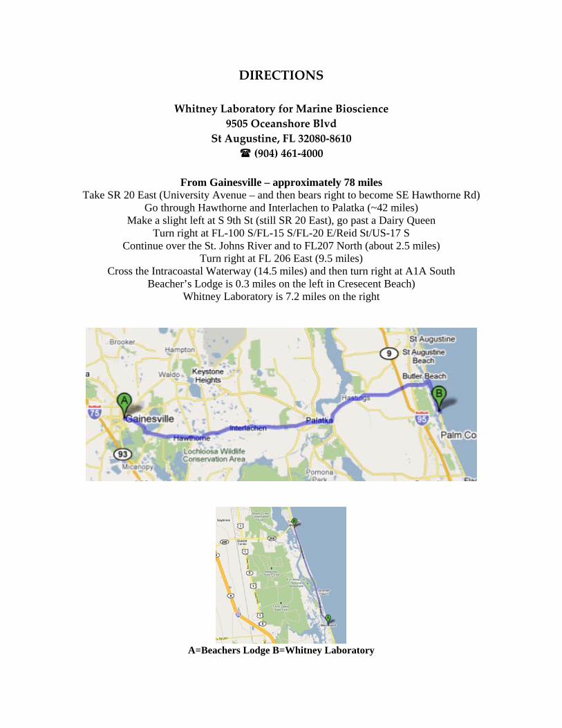
DIRECTIONS
Whitney Laboratory for Marine Bioscience 9505 Oceanshore Blvd
St Augustine, FL 32080‐8610 (904) 461‐4000
From Gainesville – approximately 78 miles
Take SR 20 East (University Avenue – and then bears right to become SE Hawthorne Rd) Go through Hawthorne and Interlachen to Palatka (~42 miles)
Make a slight left at S 9th St (still SR 20 East), go past a Dairy Queen Turn right at FL-100 S/FL-15 S/FL-20 E/Reid St/US-17 S
Continue over the St. Johns River and to FL207 North (about 2.5 miles) Turn right at FL 206 East (9.5 miles)
Cross the Intracoastal Waterway (14.5 miles) and then turn right at A1A South Beacher’s Lodge is 0.3 miles on the left in Cresecent Beach)
Whitney Laboratory is 7.2 miles on the right
A=Beachers Lodge B=Whitney Laboratory