egg inoculation new1
TRANSCRIPT

Virology
Laboratory Guide Line in Embryo inoculation
Technique

Contents
1. Sampling
2. virus isolation in embryonated eggs
2.1. Material
2.2. Inoculation site
2.3. Candling
2.4. Harvesting virus
2.5. Result

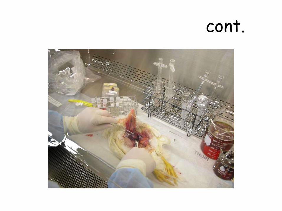
Sample Processing

What safe sampling is?

cont.

Cont.

cont.

cont.

cont.

cont.
cont.

cont.

Processing Tissues for Isolation of AIV and APMV
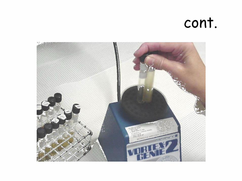
• Diluents Prepare 10% suspension in antibiotic
• Centrifuge at 1,500 x g – 20 minutes
• and place in sterile vial Remove supernatant with pipette
• Temperature Incubate 1 hour at room

cont.

cont.

cont.
cont.

Cont.

VIRUS CULTIVATION SYSTEMS
1. BIOLOGICAL SYSTEM
2. EMBRYONATED EGGS
3. TISSUE CULTURE SYSTEM

IN-VIVO / IN-VITRO?
1. Biological System
a) Natural host
b) Experimental animals In - vivo
c) Transgenic animals
2. Embryonated Eggs –
In - vivo & In - vitro
3. Tissue Culture System – In - Vitro

ANIMAL INOCULATION-DISADVANTAGES
• Cost• Maintenance• Interference of immune system• Individual variations• Difficulty in choosing of animals
for particular virus

EMBRYONATED EGGS
ADVANTAGES• Isolation and cultivation of many avian and few mammalian viruses
• Ideal receptacle for virus to grow
• Sterile & wide range of tissues and fluids

• Cost- much less
• Maintenance-easier
• Less labour
• Readily available

• Free from bacteria and manylatent viruses.
• Free from specific and nonspecific factors of defense.
• Sensitive to viruses which do not produce infection in adult birds.

virus isolation in embryonated eggs

Why Egg? The avian embryo, especially the chicken embryo, is a valuable and
widely used medium for the initial isolation and subsequentpassage of many viruses for stock cultures and the production ofvaccines. Chicken embryos are used almost exclusively because oftheir.
(1) Availability
(2) Economy
(3) Convenient size
(4) Relative freedom from latent infection and extraneous Contamination, and
(5) Lack of production of antibodies against the viral inoculums. Eggs only from healthy, disease-free flocks should be used. It is desirable to have one source of supply for reasons of uniformity of production and management of the breeder flock.

Material

EGG HOLDERS


Paraffin wax/Elmer’s Glue

Various routes of inoculation
a) Yolk sac
b) Allantoic sac
c) Chorioallantoic membrane
d) Amniotic cavity
e) Intravenous
f) embryo

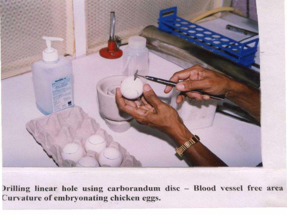
Inoculation site

Allantoic Cavity
1. Candle the egg and select an area of the chorioallantoicmembrane distant from the embryo and amnionic cavityand free of large blood vessels about 3 mm below thebase of the air cell. In this area make a pencil mark atthe point of inoculation.
2. Make a similar mark at the upper extremity of the shellover the air cell.
3. Drill a small hole through the shell at each mark but do notpierce the shell membrane.
4. Apply tincture of metaphen or another suitabledisinfectant to the holes and allow to dry

34
Cont.
• After all the eggs have been nicked, theyare inoculated with virus using a tuberculinsyringe – a 1 ml syringe fitted with a 1/2inch, 27 gauge needle.
• The needle passes through the hole in theshell, through the chorioallantoicmembrane. The hole in the shell is sealedwith melted paraffin, and the eggs areplaced at 37 °C for 48 hours

ALLANTOIC ROUTE – INOCULATION SITE DETERMINATION

ALLANTOIC ROUTE
• Most popular
• Most of avian viruses
• High titered virus
• Simple technique

AMNIOTIC ROUTE
• Primary isolation of influenza and mumps viruses
• Growth of virus detected by haemagglutination
Hemorrhagic lesions in the proventriculus, seen at necropsy in fowl with avian influenza
Influenza Virus
Mumps Virus
CHORIOALLANTOIC MEMBRANE
It is inoculation employs 10- to 12-day-oldembryos and inoculum of 0.1-0.5 cc. Thisroute is particularly effective for primaryisolation and cultivation ofthe viruses ofvaccinia, variola, fowl pox, laryngotracheitis of chickens, and pseudo rabieswhich produce easily visible foci or"pocks." The chorioallantoic membrane is asuitable site for study ofthe developmentof pathologic alterations and inclusionbodies, and titration of viruses by the pockcounting technic.
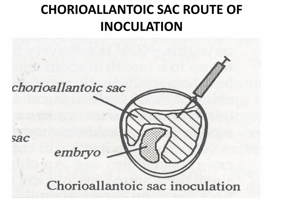
CHORIOALLANTOIC SAC ROUTE OF INOCULATION


CAM ROUTE OF INOCULATION

Tips
• Pox and Herpes viruses.
• ‘Pock Lesions’
• Suitable for plaque studies

YOLK SAC inoculation
YOLK SAC inoculation is performedwith 5- to 8-day-old embryos andinoculum of 0.2-1.0 cc. This route maybe used for initial isolation of mumpsvirus

YOLK SAC ROUTE

YOLK SAC ROUTE
Advantages
•Simplest method
•Mostly mammalian viruses
•Immune interference for most of avian viruses
Disadvantages
•Not suited for avian viruses

Intracerebral inoculationIntracerebral inoculation can be performed with8- to 14-day-old embryos and inoculum of 0.01-0.02 cc. This route may be employed in studies ofpathologic alterations of the brain followinginfection. The viruses of herpes simplex andrabies may be cultivated by this route.
Embryos are incubated after inoculation for aperiod appropriate for the virus employedandthey are examined at least once daily. Deathof the embryo within the first 24 hours afterinoculation is generally considered to be due tononspecific causes such as trauma. Some viruses.

Cont.kill all embryos and mortality is the criterion ofinfection. Newcastle disease virus is an example in whichembryos are killed in two to four days depending uponthe strain of the virus. With some viruses such asinfluenza virus the mortality rate varies on initialpassage but may increase with subsequent passage.
The criterion of infection with herpes and pox viruses isthe formation of pock lesions on the chorioallantoicmembrane. Other gross pathologic manifestations ofinfection of the embryo may be curling and dwarfing ofthe embryo, fibrosis of the amnionic membrane, edemaof the chorioallantoic membrane, and urates in thekidney and mesonephros such as produced by aviancoronaviruses on initial and low passage in the embryo.

Cont.
Various types of cytologic changes,including inclusion bodies with certainviruses, may be detected by microscopy.
The embryo should be examined soon afterdeath so that postmortem changes do notobscure any specific pathologic alterations.Chilling of the embryos for several hoursor for overnight before collection ofextraembryonic fluids is recommended toreduce hemorrhage into the fluids.

Intravenous inoculation
Intravenous inoculation does not have widepractical application for study ofexperimental infections of the avianembryo.The procedure is generallyemployed for hematologic studies. Embryosof 10- to 15-days incubation are mostsuitable for this route. The amountofinoculum may vary from 0.02 to 0.05 cc.

BLOOD VESSELS

INTRAVENOUS ROUTE
• Blue tongue virus• Cherry red embryo• Highly cumbersome• Most sophisticated procedure

Student Task 1. Why we select egg for cultivation of viral inoculums?
2. How can we detect the viability of embryo while candling?
3. What is the relationship b/n embryo inoculation site and viral inoculums?
4.The best selective site for pox viral inoculum is m/m how it could be?
5. White egg is selective for embryo inoculation technique than other colored egg, why this is so?

Student TaskQ) What is your base on site selection ?
Q) What is the effect of maternal immunity on injected inoculums?
Q) What types of viral samples selectively inoculated in yolk sac?
Q) If u encountered death of embryo within 24hr after inoculation of fertile egg ,it is indicative of ____?

How to select Needle ?
25 23 gauge 1
2222 gauge
25 gauge
22 gauge1½” needle

CandlingIntroduction
Candling is the process of holding a strong light above or below the egg toobserve the embryo. A candling lamp consists of a strong electric bulbcovered by a plastic or aluminum container that has a handle and anaperture. The egg is placed against this aperture and illuminated by thelight. If you do not have a candling lamp, improvise.

Equipment Needed for Egg Candling and Inoculation
• 4 eggs/student
• Egg flat
• Drill
• 70% Ethanol spray bottle
• Marker
• Glue
• 23 guage 1” needle
• Egg labels
• Sharps container

What is the role of candling?

CANDLING BOX

Cont.

Storage and cleaning of eggs
• Do not buy dirty eggs.
• Eggs that are stained can be disinfected by washing in awarm (37°C) solution of 0.1 percent Chloramin B (benzinesulfonamide sodium salt) or wiped with a 70 percentalcohol solution.
• Fertile eggs that have not been incubated can bepurchased. They can then be placed in an incubator whenthey are delivered. Alternatively, they can be stored forseveral days in cool conditions (16°C to 18°C) prior toincubation. This may reduce the number of viable embryos,as some embryos may not develop after storage.

Check for embryo location and mark the side opposite the embryo midway along the long
axis where the vein structure is well developed


Candle 10-11 day-old embryos and check for embryo vitality –
mark the air cell line

Marking the inoculation site
1. Hold the blunt end of the egg against theaperture of the candling lamp and note theposition of the head of the embryo.
2. Turn the egg a quarter turn away from the head.
3. Draw a line on the shell marking the edge of the airsac.
4. Draw an X approximately 2 mm above this line.
5. The X marks the inoculation site.

Cont.
Note:
In some eggs the air sac will have notdeveloped on the blunt end but half waydown the egg. These eggs are not suitablefor vaccine production. They can be usedfor inoculation during routine titrations toestablish infectivity titres.

cont.
Candle Embryos Prior to Inoculation Check embryo for:
• Proper fertility
• Proper growth of embryo
• Placement of air sac
• Development of chorio-allantoic membrane

cont.

Eggs should lie in a horizontal position with the inoculum and air
cell holes glued shut

Cleaning and decontamination of incubators
Keep surfaces clean by wiping out with awet cloth and disinfecting with 70percent alcohol solution or a non-corrosive disinfectant.

Incubation of eggs after inoculation
Inoculated eggs contain virus and shouldbe placed in a different incubator. Eggsinoculated with virulent strains ofNewcastle disease virus should not beincubated in the same incubator as usedfor eggs inoculated with the avirulent I-2strain of Newcastle disease virus.
Inoculated eggs are incubated under thesame conditions as uninoculated eggs butdo NOT turn the eggs.

Egg incubator 37-39 C relative humidity 60-70%

Tips
Humidity should be maintained at 60 to 65percent. A tray filled with water andplaced in the bottom of the incubator isusually sufficient to maintain this level ofhumidity.
Place the eggs in the incubator with the airsac on top.
Eggs should be turned three times a day.

Candle eggs dailyChecking for embryo
mortality
Determining the viability of the embryo
Under the candling lamp, the embryo appears as a darkshadow with the head as a dark spot. Healthy embryos willrespond to the light by moving Sometimes the movement isvery sluggish and it can take 30 to 40 seconds for theembryo to move when held under the candling lamp. Thisindicates the embryo is not healthy and the egg should bediscarded.
Look carefully at the blood vessels. They are well definedin a healthy embryo. After an embryo has died, the bloodvessels start to break down. They then appear as streaksunder the shell when viewed under the candling lamp.Candling will also reveal cracks in the eggshells. Eggs withcracked shells should be discarded.

TipsInfertile eggs: These are easy to detect, as the egg is clear. Discard
Deaths: The embryo has developed for several days and then died. Candling will reveal a small dark area and disrupted blood vessels. Often deteriorating blood vessels will appear as a dark ring around the egg. Discard.
Late Deaths: These are often difficult to tell apart from a viable embryo at the same stage of development. Look for the absence of movement and the breakdown of the blood vessels. Discard
Viable Embryos: These move in response to the light and have well defined blood vessels. Mark the air sac and the inoculation site and then return the eggs to the incubator ready for inoculation.

Live 12 day old chicken embryo prominent

Dead chicken embryo – no CAM

Refigerate embryo – embryos is deadand blood vessles are constricted

Harvesting

Equipment Needed for Harvesting
AAF from Dead Eggs
2 embryos/student
2 Forceps
Iodine box for forceps
5 ml pipettes and
pipette aid
Gloves
Blood Agar plate
Plastic loops

Cont.
• Snap cap tube with • labels• Ethanol spray bottle• Iodine bucket for • pipettes• Discard bucket with • bags for embryos• Plastic bags to discard • flats

Disinfection
cont.

Harvesting embryo

Harvesting AAF from Dead Embryos
1. Disinfect egg shell surface one time in the BSC
2. 2. Only open eggs from a single specimen at
3. Open egg from air cell end with forceps
4. Break allantoic sac with sterile forceps pipette tip with forceps
5. Hold membranes and embryo away from
6. Harvest AAF
7. Streak BA plate
8. Centrifuge 1,500 x g for 15 min.

Harvesting AAF for Live Embryosconstrict chorioallantoic vessels
1. Refrigerate embryo to kill embryo and
2. Disinfect shell
3. Drill small hole above air cell line
4. Aspirate AAF with 3cc 22 gauge 1½” syringe will bind with virus
5. Discard AAF with red blood cells – RBC

cont.


Harvesting and storage of allantoic fluid
Introduction
Allantoic fluid from inoculated eggs will be harvested for threereasons.
1. To prepare I-2 Newcastle disease working seed or vaccine.
2. To use as antigen in the haemagglutination inhibition test.
3. To be tested for the presence or absence of Newcastle diseasevirus by the haemagglutination test. These results are then usedto calculate the infectivity titre of a suspension of virus.
Note
Maternal antibody is confined to the yolk sac until about 14days of incubation and then enters the embryo. Virusharvested in allantoic fluid at 14 days will not have beenexposed to antibody.

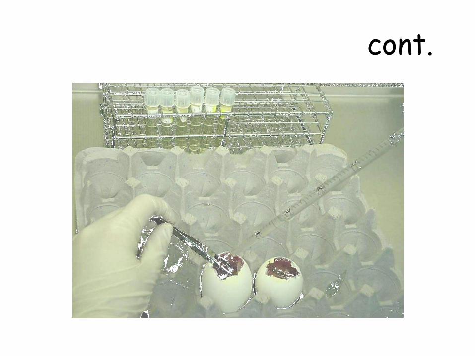
HARVEST OF ALLANTOIC FLUID

92
Cont.
• During the incubation period, the virus replicates inthe cells that make up the chorioallantoic membrane.As new virus particles are produced by budding, theyare released into the allantoic fluid. To harvest thevirus, the top of the egg shell – the part covering theair sac – is removed.
• The shell membrane and chorioallantoic membrane arepierced with a pipette which is then used to removethe allantoic fluid – about 10 ml per egg.

Harvesting allantoic fluid to prepare working seed, vaccine or antigen
MaterialsForceps or a small pair of scissorsAbsolute alcohol for flaming forcepsCotton wool70 percent alcohol solution in waterDiscard tray50 mL micropipette and tips or a wire loopSterile Pasteur pipettes with short blunt ends96 well microwell plate10 percent washed red blood cellsSterile containers for receiving the harvested
allantoic fluid.

Method1. Chill eggs at 4°C for at least two hours to kill the embryo and to reduce the
contamination of the allantoic fluid with blood during harvesting.
2. Remove stationery tape (if used to seal the eggs) and swab each egg with cotton wool soaked with 70 percent alcohol to disinfect and remove condensation from the shells.
3. Dip the forceps or scissors in disinfectant OR if using a Bunsen burner, dip the forceps or scissors in absolute alcohol and flame to sterilize. Remove the eggshell above the air space.
4. Discard embryos that are visibly contaminated.
5. Remove a sample of allantoic fluid from each egg. Use a micropipette and sterile tip, sterile glass pipette or a flamed loop. Test each sample for the presence of Newcastle disease virus by the haemagglutination (HA) test.
6. Discard embryos that do not test HA positive for Newcastle disease virus.
7. Use sterile glass Pasteur pipettes to harvest the allantoic fluid from the eggs. The pipettes can be either hand held or used unplugged and connected to a vacuum pump. Collect the fluid into sterile containers.

Preliminary Quality Control
This step involves the inoculation of a general purpose brothculture.
1. Test each container for bacterial contamination byinoculating tryptic soy broth with test samples andincubation at 37°C overnight.
2. Centrifuge the samples of allantoic fluid or standovernight at 4°C to allow particles including red blood cellsto settle. The allantoic fluid should appear clear aftercentrifugation or standing overnight.
3. After 24 hours, read the results of the tests for bacterialcontamination.
4. Use aseptic technique to transfer the clear allantoic fluidsupernatant from containers that showed no bacterialgrowth into a sterile container for storage. This step poolsthe fluid and ensures homogeneity.

Storage of allantoic fluid
The optimum temperature for storage of allantoic fluidcontaining live Newcastle disease virus is -70°C. Storageat -20°C is not as effective and the infectivity titre willslowly decrease. The action of the freezing and thawingalso decreases the infectivity titre of the virus. Allantoicfluid containing Newcastle disease virus has been storedfor up to 6 weeks at 4°C without significant loss of titre.Allantoic fluid will be stored for two purposes.
1. Antigen for use in the haemagglutination inhibition test.Prepare 1 mL aliquots of undiluted allantoic fluid in vials.
2. Preparation of vaccine.

Storage of allantoic fluid at 4°C for use as a wet vaccine
Diluents containing a stabilizing agent are used in thepreparation of wet Newcastle disease vaccine. Suitablestabilizing agents are gelatin and skim milk powder.
Diluents containing 2 percent gelatin solution or 8 percentskim milk powder in phosphate buffered saline aresterilized prior to use and mixed one part diluent with onepart allantoic fluid.
A further dilution step in two parts of PSG antibioticsolution will reduce the risk of growth of contaminatingbacteria during storage. Trials at John Francis VirologyLaboratory have shown that 1 percent gelatin is a superiorstorage agent to 4 percent skim milk.

Control allantoic fluid samples
• Negative and positive control samples are tested in boththe rapid and micro haemagglutination tests to ensure thevalidity of the test.
• Negative control allantoic fluid is harvested from 14-dayold embryonated eggs that have not been inoculated withNewcastle disease virus. It should always test negative forthe presence of haemagglutinins. There should not be anysign of haemagglutination.
• Positive control allantoic fluid is known to contain a highinfectivity titre of Newcastle disease virus. It shouldalways test positive for the presence of haemagglutinins.Haemagglutination should be visible.


100
An example: ILT

Result Interpretation

Newcastle Disease Virus

NDV – OCCIPITAL HAEMORRHAGE

Infectious Bronchitis Virus - CAM inoculation results in very stunted and tightly curled embryos

Multiple passages are required to produce typical lesions.
Urates in the kidney

Infectious LaryngotracheitisVirus
Inoculation by dropped CAM reveals formation of pox lesions

Infectious Bursal Disease


Avian Encephalomyelitis Virus –Muscle atrophy

BLUE TONGUE VIRUS – CHERRY RED EMBRYO

‘POCK’ LESIONS ON CAM