effects of ventromedial hypothalamic lesions on restricted feeding behavior in rats
TRANSCRIPT
Physiology and Behavior, Vol. 12. pp. 761-766. Brain Research Publications Inc.. 19"~a. Printed in the U.S.A.
Effects of Ventromedial Hypothalamic Lesions on Restricted Feeding Behavior in Rats
R O N A L D H. PETERS
Deparrrnent o f P~,chology. Iowa State University, Ames. Iowa 50010
(Received 27 N o v e m b e r 1973)
PETERS. R. H. Effects of ventromedial hypothaiamic lesions on restricted feeding behevior in rats. PHYSIOL. BEHAV. 12(5) 761-766, 1974. - Prevlous research has mown that VMH lesioned rats overeat when given free access to food yet undereat when daily access is restricted. In the present study VMH lesioned female rats in Experiment 1 and male rats in Experiment 2 consumed more food than controls during both free and restricted access feeding schedules. The factor that most likely accounts for the contrasUng outcomes is the extent of hyperpha~a displayed by the lesioned rats prior to restricted access feedin,2. The present data are consistent with the interpretation that VMH lesions increase hunger motivation.
VMH le¢ions Restricted access feeding
PANKSEPP [11] has recent ly p roposed a s imple neural model o f feeding behavior tha t subs tan t ia l ly modif ies cur ren t concep t ions of the regula tory func t ions of the ven t romed ia l h y p o t h a l a m u s (VMH). The hyperphag ia and subsequen t obesi ty tha t occur fol lowing VMH damage have general ly been a t t r i b u t e d to the d i s rup t ion of a neural sys tem tha t no rmal ly m o m t o r s pos t -prandia l sat ie ty st imuli [8 ] . Since a wide variety of exper imen ta l man ipu la t ions tha t decrease meal size in normal rats are also effect ive in VMH lesioned rats [3, 10, 15, 17] , Panksepp has suggested tha t the VMH mediates long- term ra the r than shor t - t e rm regula t ion of food intake. Tha t is, the VMH meters the nu t r i en t s tores of the organism in con t ras t to the immedia te consequences of food ingestion. Thus the VMH lesioned rat overeats and becomes obese because s tored nu t r i en t s are incapable of suppressing feeding behavior .
Suppor t for this i n t e rp re t a t i on of VMH func t ion was ob ta ined in several s tudies [ 11,12] designed to separate long- f rom shor t - t e rm cont ro l of feeding behavior . If VMH lesioned rats overeat on ad Lib diets because they are unable to a c c u r a t e l y m o n i t o r nu t r i en t stores, then , argued Panksepp, they may also be unable to increase the i r food in take appropr ia te ly when nu t r i en t s tores are deple ted . The data of several expe r imen t s suppor t ed this c o n t e n t i o n in tha t VMH lesioned rats did not respond to nu t r i en t deple- t ion as did normal ra:s, e.g., VMH lesioned rats consumed less food than normal rats when placed on restr ic ted feed- ing schedules.
Panksepp ' s neural model readily accounts for the VMH paradox, the decrease in hunger m o t i v a t i o n associated wi th hyperphagla in lesioned rats [9 ,18 ] . The model assumes tha t the VMH conta ins spon taneous ly active exc i t a to ry neurons tha t project to neurons in the lateral h y p o t h a l a m i c area (LHA). When these excitatory, neurons are des t royed , a cor responding decrease in hunger m o t i v a t i o n occurs. The hyperphagaa occurs because neurons located in the VMH
tha t me te r nu t r i en t stores are also des t royed. These la t ter neurons normal ly exer t i nh ib i to ry cont ro l over b o t h the exc i t a to ry neurons located in the VMH and neurons located in the LHA. The activity of these LHA neurons direct ly cont ro l s feeding responses. Post-prandial sat ie ty st imuli p resumably act d i rect ly on the LHA ra ther than the VMH.
Several studies IS, 13, 14] have recent ly demons t r a t ed , however , tha t the VMH paradox, in all l ikel ihood, does not exist. When be tween-group compar i sons were made at identical modera te depr iva t ion condi t ions , VMH lesioned rats pe r fo rmed at cons iderably higher levels than cont ro l rats on various hunger -mot iva ted and food-re inforced tasks. Ra the r than displaying mot iva t iona l deficits. VMH lesioned rats ran faster in a straight alley, pressed at higher rates on b o t h variable-interval (60 sec) and f ixed-rat io ( 6 4 : 1 ) r e i n - fo rcement schedules, and consumed more sucrose pellets in a cond i t ioned aversion paradigm. VMH lesioned rats also consumed more food than cont ro l rats dur ing the first 30 mm fol lowing p lacement of food in thei r home cages in a m o u n t s required to ma in ta in them at modera t e depriva- t ion levels [5 ] . This la t ter observa t ion cont ras t s with the data reported, by Panksepp [I 1 .12] . whose lesioned rats ate less t han con t ro l rats when pe rmi t t ed l imited daily access to food. Since the procedures of the exper imen t s yielding cont ras t ing ou t comes were qui te dissimilar, the present exper imen t s were run to examine the restr icted access food intake of VMH lesioned and con t ro l rats in this l abora to ry using procedures similar to those used by Panksepp.
EXPERIMENT l
Method
.qntrnats. The animals were 1S female hooded rats (Blue Spruce Farms, Inc.) 9 0 - 1 0 0 days old weighing 1 8 7 - 2 4 7 g at the beginning of the exper imen t . Ten rats received bi- lateral e lec t rolyt ic lesions aimed at the VMH. located by de
761
762 PETERS
Groot [2J coordinates (A = 5.8, H = -3.5, L = 0.7). The lesions were produced under sodium pentobarbital anes- thesia (40 mg/kg) by passing 2.0 mA anodal current for 20 sec between a 30 ga nichrome steel electrode insulated except at its cone-shaped tip and a rectal cathode, Eight rats served as unoperated controls.
Procedure. The rats were housed individually in a continuously lighted colony and were allowed free access to water throughout the expern'nent. The amount of food (Wayne Lab B l o x ) each rat consumed during 24-hr free access periods was measured for 3 days prior to surgery. Food in these and subsequent test periods was placed directly in the home cage. Food intake was measured to the nearest g and spillage beneath the cage was weighed. F o o d was not available for 24 hr after the day of surgery for both lesloned and control rats. Following free access to food for the next 3 days, food was removed for 23 hr and the amount consumed during daily I-hr access periods was measured for I0 days. Food intake was subsequently measured for I0 days at successive access periods of 1.5 and 2 hr. Body weights were obtained daily throughout the e x p e r i m e n t immediately prior to the placement of measured amounts of food in the home cage.
Following restricted access testing, the rats were glven food daily in amounts necessary to maintain them at their b o d y weights on the day of surgery. This procedure was used to establish comparable baseline body weights for the 2 groups prior to a return to free access testing. Since the body weights on the day of surgery were used to establish the common baseline, all rats were deprived with respect to normal growth. After all rats had been maintained at this baseline for 5 days, they were glven free access to food for 63 days, Daily measures of food intake and body weight
were recorded for the first I0 days and body weights were obtained after 63 days of free access to food. All rats in this experiment and Experiment 2 were deprived of food and water for 24 hr on 6 occasions during these 63 days as part of the procedures for unrelated research to be reported elsewhere.
The VMH lesioned rats were perfused with saline and 10% Formalin. Frozen sections were taken at 150 ~ in the coronal plane described by Korag and Klippel [7] . Photo= graphic enlargements of the unstained sections were used to assess the extent and location of tissue destruction.
Results and Discusszon
Three lesioned rats gained only 15, 23, and 41 g more than the largest weight gain for a control rat during 63 days of free access to food, and their data were not included in the analysis. Weight gains for the 7 remaining lesloned rats ranged between 171 -302 g whim control rats gained between 2 7 - 9 0 g. Mean weight gains for lesioned and control rats were 58 and 242 g, respectlvely.
The lesions in the 7 rats whose data were used in the analysis were typically large and symmetrical. Although the exact size and location of destruction varied considerably, the VMH was substantlaily damaged in each rat. The largest lesions also destroyed portions of the anterior, dorsomedial, and posterior hypothaiamic nuclei, and extended laterally to the plane of the fornix. The lesions for the 3 rats whose data were not analyzed were much smaller, and were either dorsal to the VMH or unilateral.
Mean daily food intake for 3 days unmediately pre- ceding and following the day of surgery, and for the subse- quent i0 days of I-hr access to food is presented in Fig. I.
A
0 v
L U
<
Z
0 0 u _
4 0
3 0
2 0
10
% %
O--o V M H F E M A L E S
4 1 HR A C C E S S ' - = ' *
C O N T R O L
j -
I I I I I I , I 1 I I
1 3 5 7 8 11 D A Y S
]4 17
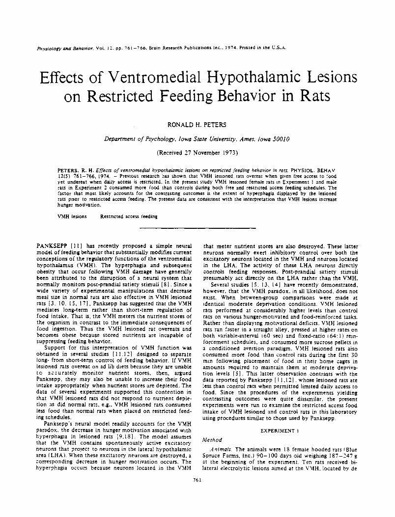
FIG. I. Daily t'ooc~ intakes ot lesioned and =ontroi :emaie rats during [-hr access to '3od.
VMH LESIONS AND RESTRICTED FEEDING BEHAVIOR 763
40 t,n
(3 30 144
~- 2 0 Z
O O LL-
10
FEMALES
. . e ' ' "
0 . - "
I I
I I
I I
P /
. . . . . . . . ~ C O N T R O L
I . I . I I
1.O 1.5 2.0 24.0 ACCESS iNTERVALS
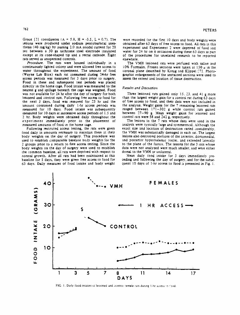
FIG. 2. Daffy food intakes of lesioned and control female rats at ea¢.h access interval
Al though the groups did not differ prior to surgery, the unrestricted intake of rats sustaining VMH destruct ion increased and was sigmficantiy higher than control intake during the 3 days fol lowing surgery (t = 7.26, d.f = 13, p<0.001) . Body weights for the lesioned rats increased correspondingly. Thus the rats with VMH lesions displayed considerable hyperphagJa at the outset of restricted access testing.
VMH lesioned rats consumed more food than control rats on each of the I0 days when access to food was limited to l hr each day. Whereas control rats gradually increased their mean food intake from the first (3.8 g) to the last day (9,5 g) of hourly access, lesioned rats consumed nearly as much on the first day ( I0 . I g) as on the last (12.6 g). The difference between groups was significant on both days (t = 5.95, d/c= 13, p<0 .001 , t = 3.16, d/c= 13, p<0 .01 , respec- tively).
Figure 2 presents the mean food intakes for lesioned and control rats over the 10-day test period for each access interval. Lesioned rats consumed more food than control rats at each interval (p<0.001 and d.t'= 13 for each t test). When access t ime was increased to 1.5 or 2 hr, control rats did not increase their intake appreciably from the amount consumed on the last day of hourly testing. Lesioned rats, however, increased their mean daily intake from 12.6 to l 5.7 g when shifted from I- to 1.5-hr access, and consumed 17.3 g on the first day of 2-hr access. Consumpt ion within both groups remained relatively constant across test days at the 1.5- and 2-hr access intervals. The body weights of the two groups reflected the differences in food consumpt ion. The mean body weight of the lesioned rats on the last day of 2-hr testing was 20 g higher than their weights on the
first day of l-hr testing, whereas control rats lost an average of 12 g dunng the 30 days of restricted access.
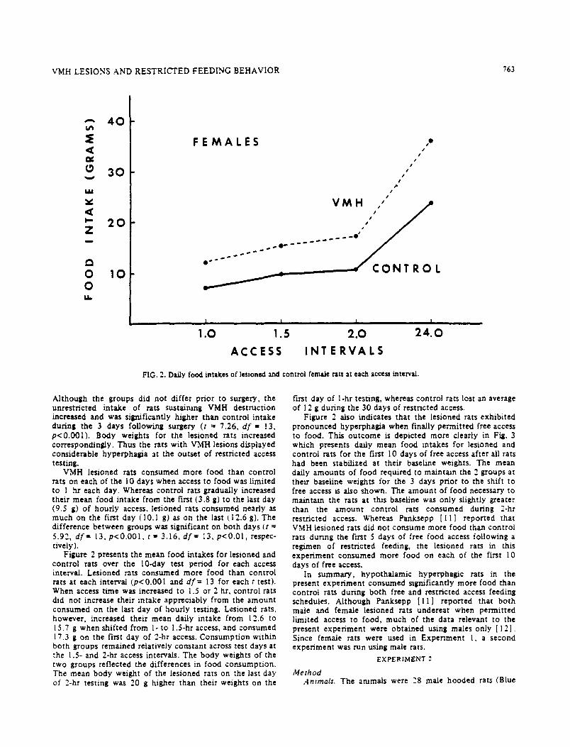
Figure 2 also indicates that the lesioned rats exhibi ted pronounced hyperphagia when finally permit ted free access to food. This ou tcome is depicted more clearly in Fig. 3 which presents daffy mean food intakes for lesioned and control rats for the first 10 days of free access after all rats had been stabilized at their baseline weights. The mean daffy amounts of food required to maintain the 2 groups at their baseline weights for the 3 days prior to the shift to free access ts also shown. The amount of food necessary to maintain the rats at this baseline was only slightly greater than the amount control rats consumed during 2-hr restricted access. Whereas Panksepp [ I | ] reported that VMH lesioned rats did not consume more food than control rats during the first 5 days of free food access following a regimen of restricted feeding, the lesioned rats in this exper iment consumed more food on each of the first 10 days of free access.
In summary, hypothalamic hyperphagic rats in the present exper iment consumed significantly more food than control rats during both free and restricted access feeding schedules. Al though Panksepp [ l I ] reported that both male and female lesioned rats undereat when permit ted limited access to food, much of the data relevant to the present exper iment were obtained using males only [12] . Since female rats were used in Exper iment I, a second exper /ment was run using male rats.
EXPERIMENT 2
Me th od .4nzmals. The animals were "8 male hooded rats (Blue
764 PETERS
A
,,,w,
0 V
t . U
, ( I , . -
Z m
r~
0 0 E L
4 0
:30
2 0
10
F E M A L E S I . - - 0
. i " V M H
% s l - . _ l s s
p s
% s S
, FREE ACCESS ,
I I I I I I I I I I
- 3 =I I 4 7 D A Y S
I I I
10
FIG. 3. Daily food intakes of lesioned and control female rats dunng free access to food.
Spruce Farms, Inc.) 8 0 - 9 0 days old weighing 2 6 5 - 3 1 5 g at the beginning of the exper iment . Lesions were made in 10 rats using the same stereotaxic coordinates and current parameters of Exper iment 1. Lesions were also made in 10 rats at a locus 1 mm anterior (AP = 6.8) to the placements for the other I0 lesioned rats since preliminary data indi- cated that the anterior lesions more effectively induced hyperphagla and obesity. Eight rats served as unopera ted controls.
Procedure. The procedures used in this exper iment were essentially identical to those of Exper iment I except for the inclusion of an additional 10 days of restricted access feeding when food was available daily dur.ng a .¢-hr access period.
R esuits
The data of -t !esioned rats were not included :n the analysis. These rats were not hyperphagic during free access testing and failed to become obese. Their ',esions were generally small and dorsal to the VMH.
The data for both groups of lesioned rats were 'ortually identical and were combined for simplicity of presentat ion. The lesions tn these rats were generally !arge, and similar in size and extent to those of Exper iment I. The anterior tesions often ex tended rostraily to the optic chlasm whereas the pos tenor lesions extended caudally to the mammillary region. The VMH was substantially des t royed at both e!ec- trode iocatlons.
Mean we:ghc gains "or Eesioned and controi rats after 63 days of free access ~o food were 244 and 143 g. respec- tiveiy. Weight gains for control rats ranged between
1 2 0 - 1 6 8 g while lesioned rats gamed between 1 4 2 - 3 9 0 g. Although 2 lesioned rats gained slightly less than the largest gain for a control rat, their data were included in the anal- ysis since they were hype rpha~c and could not be excluded on the basis of histological findings.
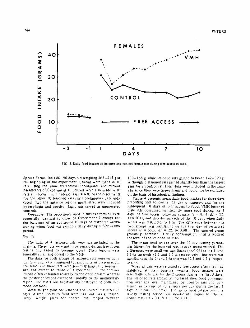
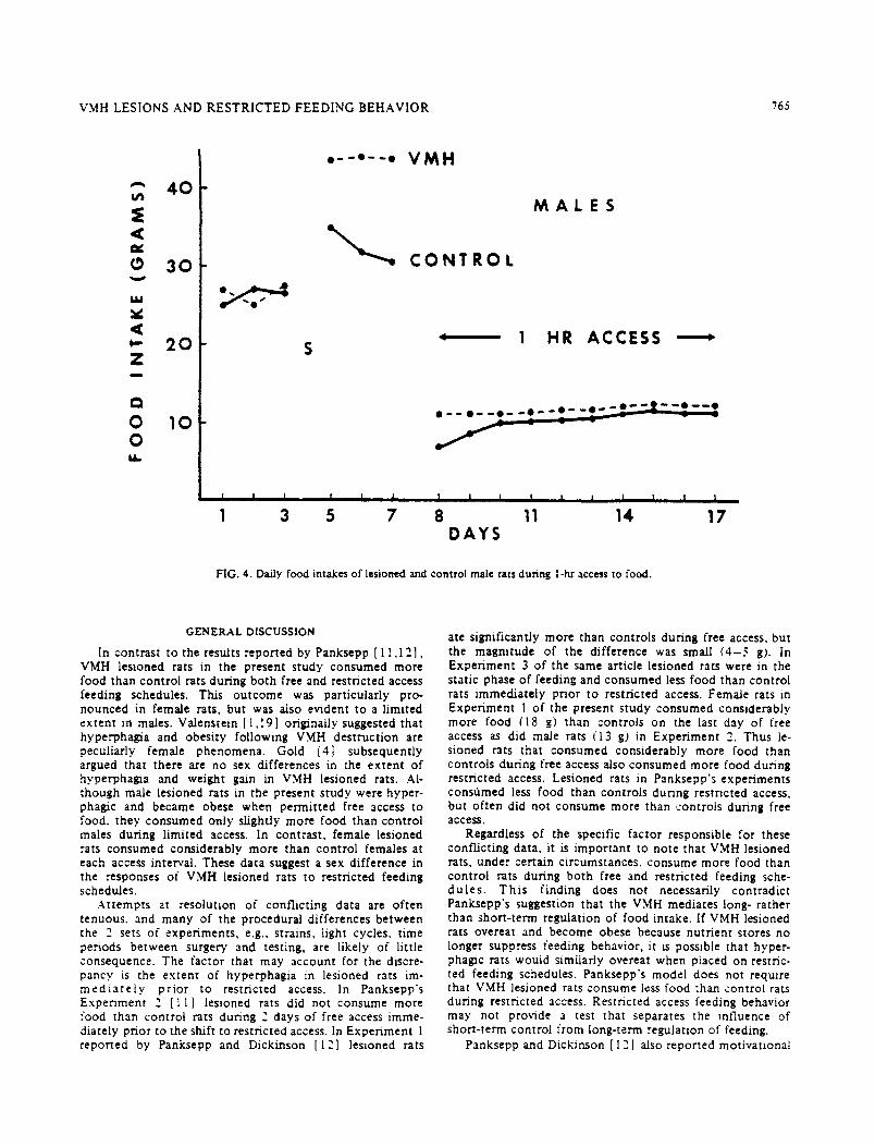
Figure 4 presents mean dally food intakes for three days preceding and following the day of surgery., and for the subsequent 10 clays of 1-hr access to food. VMH lesioned male rats consumed stgnit'icantly more food during the 3 days of free access following surgery , t = 8.14. d f = 2"2_. p<0.001) , and also during each of the I0 days when daily access was restricted to l hr. The difference between the two groups was sigmficant on the first day of r e smc ted access (: = 55.5. d.t = 22, ~<0.001) . The control grouo gradually increased its dady consumpt ion untia it reached the level of the !eszoned animals.
The mean food retake over :he 10-day testing periods was higher for the lesioned rats at each access tnterva!. The differences were small yet significant (p<0.05) at the I- and 1.5-hr intervals (1.3 and I ." g, respectively), but were not significant at the 2- and 5-hr ~ntervals (2.5 and 1.3 g. respec- tively ).
When all rats were returned to free access after they had stabilized at their baseline weights, food intakes were essentially identical for the 2 groups during ehe first 3 days. The lesioned rats gradually increased their food consump- tion over the level maintained by zontroi rats and con- sumed an average of ~3 g more per day dunng the ',ast 3 days of measured intake. The mean food ,ntake over :he 10-day testing period was stgmficantly hlgi~er rot ,he le- sioned rats (~" = -) 00. dt = 22. p<.9.001 ,.
VMH LESIONS AND R E S T R I C T E D F E E D I N G B E H A V I O R "165
A 4 0
,¢
3 0
L U
, - " 2 0 Z
c~
O 10 O u _
O - --ql)-- - - O
S
V M H
M A L E S
C O N T R O L
' 1 HR ACCESS '
• - - O - - @ - - o ' - O - - o - - ' O - - ! - 2 - - - Oi - - - - @
I i I ! I I l I I i i | I I I l
1 3 S 7 8 11 14 17 DAYS
FIG. 4. Daily food intakes of lesioned ~ d control male rats dunns l-ha" access to food.
GENERAL DISCUSSION
In cont ras t to the results repor ted by Panksepp [ I 1 ,12] , VMH lesioned rats in the present s tudy consumed more food than cont ro l rats during b o t h free and restr ic ted access feeding schedules. This ou t com e was par t icular ly pro- nounced in female rats, bu t was also evident to a l imited ex t en t in males. Valens te in [ l , 1 9 l o r i~na i ly suggested that hyperphas~a and obesi ty fol lowing VMH des t ruc t ion are peculiarly female p h e n o m e n a . Gold [4] subsequen t ly argued tha t there are no sex di f ferences in ihe e x t e n t of hyperphas~a and weight gain in VMH lesioned rats. Al- though male lesioned rats in the present s tudy were hyper- phagic and became obese when pe rmi t t ed free access to food. they consumed only slightly more food than con t ro l males dur ing l imited access. In cont ras t , female lesioned rats consumed cons iderably more than con t ro l females at each access interval. These data suggest a sex di f ference in the responses of VMH lesioned rats to res t r ic ted feeding schedules.
A t t e m p t s at resolut ion of conf l ic t ing data are o f t en tenuous , and many of the p rocedura l d i f ferences be tween the 2 sets of exper iments , e.g., strains, light cycles, t ime periods be tween surgery and test ing, are likely of lit t le consequence . The fac tor tha t may accoun t for the discre- pancy is the ex t en t of hyperphagia in lesioned rats im- m e d i a t e l y p r i o r to restr icted access. In Panksepp ' s Exper imen t 2 [ i I ] lesioned rats did not consume more food than cont ro l rats dur ing 2 days of free access imme- diately prior to the shift to res t r ic ted access. In Exper imen t I repor ted by Panksepp and Dickinson [12] lesioned rats
ate s ignif icant ly more than cont ro ls d u n n g free access, but the magni tude of the dif ference was small (4- ,~ g). In Expe r imen t 3 of the same article lesioned rats were in the static phase of feeding and consumed less food than cont ro l rats immedia te ly pr ior to restr ic ted access. Female rats in Exper imen t I of the present s tudy consumed cons iderably more food (18 g) than cont ro ls on the last day of free access as did male rats ('13 g) in Exper imen t 2. Thus le- s ioned rats tha t consumed cons iderably more food than cont ro ls during free access also consumed more food dur ing restr ic ted access. Lesioned rats in Panksepp ' s exper imen t s consumed less food than cont ro l s dur ing restr icted access, bu t of ten did not consume more than cont ro ls during free access.
Regardless of the specific fac tor responsible for these conf l ic t ing data , it is i m p o r t a n t to note tha t VMH lesioned rats, under cer tain c i rcumstances , consume more food than cont ro l rats during b o t h free and restr ic ted feeding sche- d u l e s . T h i s f ind ing does not necessarily con t rad ic t Panksepp ' s suggestion tha t the VMH media tes long- ra ther than shor t - t e rm regula t ion of food intake. If VMH lesioned rats overeat and become obese because nu t r i en t stores no longer suppress feeding behavior , it is possible tha t hyper- p h a ~ c rats would similarly overeat when placed on restric- ted feeding schedules. Panksepp ' s model does not require that VMH lesioned rats consume less food than cont ro l rats during restr ic ted access. Rest r ic ted access feeding behavior may not provide a test tha t separates the inf luence of shor t - te rm cont ro l f rom long-term regulat ion of feeding.
Panksepp and Dickinson [I 2] also repor ted mot iva t lona l
766 PETERS
deficits in VMH lesioned rats. The outcomes of compari- sons between VMH lesioned and control rats on hunger- motivated tasks interact, however, with many procedural variables [5, 6, 16]. The present data are consistent with the recent in terpreta t ion that VMH lesions increase rather than decrease hunger motivat ion [5, 13, 14, 201. Not only do les~oned rats overeat during free access to food because of increased hunger, they similarly overeat when access is restricted. Panksepp's model could be modif ied to accom-
modate tl'us in terpreta t ion of motivational increments following VMH destruct ion. The model could simply drop the requirement that the VMH contains spontaneously active exci ta tory .neurons and instead state that motiva- tional increments occur when neural tissue that normally responds to nutr ient repletion is destroyed. The model does not have to account for the VMH paradox if. in fact, the paradox does not exist.
REFERENCES
1. Cox, V. C., J, W. Kakolewsld and E. S. Valemtein. Sex differ- ences in hyperphqia and obe.~ty. /. comp. physioL Prychol. 67: 320-326, 1969.
2. de Groot, J. The rat forebram in stereotaxic coordinates. Trans. Roy. :Verh. Acad. ScL 52: 1.-40. t959.
3. Epstein, A. N. Suppresston of eating and dnnkang by ampheta- mine and other drugs in normal and hyperpha~ic rats. I. comp. physiol, thT. chol. 52: 37-45, 1959.
4. Gold. R. M. Hypothalamic hyperpha~a: males get just as fat as females./, comp. physiC. Plychol. 71: 347-356, 1.970.
5. Kent, M. A. and R. H. Peters. Effects of ventromedial hypo- thalamic lesions on hunger-motivated behavior m rats. J. comp. physiol. P~ychoL 83: 92-97, 1973.
6. King, B. M. and M. G. Gaston. The effects of pretraanmg on the bar-pr~,sing performance of VMH-tesioned rats. Physiol. Behav. 11: t61-1.66, 1973.
7. Konig, J. F. R. and R. A. Klippel. The Rat Brain. Baltimore: Williams and Wilkins, 1963.
8. Mayer, J. and D. W. Thomas. R~ulatlon of food intake and obesity. Science 156: 328-337. 1967.
9 Miller. N. E., C. J. Bailey and J. A. F. Stevenson. Decreased hunger but increased food intake resulting from hypothalamic lesions. Science 112: 256-259, 1950.
10. Panksepp. J. Is "satiety" mediated by rhe VMH? PhysZol. Behav. 7: 381-384. 1.971.
11. Panlc~pp, J. A re.~xamination of the role of the ventromedial hypo tha l amus in feeding behavior. Physiol. Behav 7: 385-394, 1971..
12. Panlcsepp, J. and A. Dickinson. On the motivational deficits a f ter medial hypothalamic lesions. Physiol. Behav. 9: 609-614, 1972.
13. Peters, R. H. and M. J. Reich. Effects of ventromedial hypo- thaJamic lesions on conditloned sucrose aversions in rats. /. cornp, phy~ol. P~chol. 84: 502-506. 1973.
14. Peters, R. H., L. D. Sensentg and M. J. Reich. Fixed-ratzo performance following ventromediai hypothaJamtc lesions in rats. Physiol. th'ychol. 1: 1.36-138, 1973.
15. Reynolds, R. W. and J. Kimm. Effect of glucose on food retake in hypothalamic hyperpha~c rats. J. comp. physiol. P~chol. 60: 438-440, 1965.
16. Singh, D. Effect of preoperative training on food-mouvated behavior of hypothalamic hyperphaglc rats. J. comp. physiol. th'ychoL 84: 47-52. 1973.
17. Srruth, .M.H., R. Salisbury and H. Wemberg. The reacnon of hypothalamic-hyperpha~ic rats to stomach preloads. Z comp. physioL P~ychoL 54: 660-664, 1961.
18. Teidebaum, P. Random and food-directed acuvity in hyper- phaglc and normal rats. J. comp. physiol. I~vchoL 50: 486-490, 1.957.
19. Valenstein, E. $., V C. Cox and J. W. Kakolewski. Sex differ- ences in hyperpha.~a and body weight following hypothalamlc damage. Ann.V. Y. Acad. Scl. 157: 1030-1046. 1969.
20. Wampler, R. S. Increased mouvauon tn rats with ventromediai hypothalamtc lesions. J. comp. phy;iol. P~.chol. 84: 275-285. 1973.