effects of maternal stress during different gestational periods on the serotonergic system in adult...
TRANSCRIPT
Pharmacology Biochemistry & Behavior, Vol. 31, pp. 83%843. © Pergamon Press plc, 1989. Printed in the U.S.A. 0091-3057/88 $3.00 + .00
Effects of Maternal Stress During Different Gestational Periods on
the Serotonergic System in Adult Rat Offspring
D A V I D A. V. P E T E R S
Depar tment o f Pharmacology, Faculty o f Health Sciences, University o f Ottawa 451 Smyth Road, Ottawa, Ontario, Canada K 1 H 8M5
Rece ived 16 Oc tober 1987
PETERS, D. A. V. EfJects of maternal stress during diJJbrent gestational periods on the serotonergic system in adult rat offspring. PHARMACOL BIOCHEM BEHAV 31(4) 83%843, 1988.--Pregnant Sprague-Dawley rats were exposed to mild stress treatments during different gestational periods and the offspring were investigated at 60 days of age. In the first study, stress from embryonic day (ED) 11 to ED 20 produced effects similar to those reported following stress throughout pregnancy; increased numbers of 5-HT2 binding sites in cerebral cortex and a reduced intensity of the behavioral syndrome produced by injections of the 5-HT agonist 5-methoxy-N,N-dimethyitryptamine (5-MeODMT). In the second study, stress from ED 3 to ED 14 had no significant effect on the intensity of the 5-MeODMT-elicited 5-HT syndrome while stress from ED 15 to ED 20 had a similar effect as stress throughout pregnancy. These data provide evidence that the critical period for prenatal stress-induced changes in brain 5-HT neurons is between ED 15 and birth. This suggests that the mechanism involves an interaction with developmental events occurring within this time span such as the growth of nerve axons and the formation of synaptic contacts. Our findings also provide further evidence that stress during the final trimester of pregnancy may have serious adverse effects on fetal brain development.
Prenatal Stress Brain development 5-Methoxy-N,N-dimethyltryptamine
5-Hydroxytryptamine 5-HT syndrome 5-HT receptors
SEVERAL studies have shown that adult behavior in ro- dents can be significantly influenced by prenatal events such as maternal stress (1, 2, 4, 8). However, there is little evi- dence at present to suggest a possible mechanism by which rodent behavior may be modified by prenatal stress and even the stage of development during which the effects originate is not known.
We have previously reported that maternal stress affects several biochemical measures associated with central 5-HT neurons in the adult offspring, suggesting the possibility of permanent structural and functional changes. For example, prenatal stress increased 5-HT2 receptor binding in several brain regions at 60 days of age while 5-HT1 receptor binding was increased in cortex, decreased in hippocampus and un- changed in hindbrain (15). The increased 5-HT2 receptor binding was present as early as 21 days of age (15) and per- sisted until at least 100 days, the latest age studied (unpub- lished data). Prenatal stress also reduced the intensity of the behavioral syndrome produced by injection of the 5-HT agonist 5-methoxy-N,N-dimethyltryptamine (5-MeODMT) (17) suggesting that the biochemical changes may be func- tionally significant.
The effect of maternal stress on the intensity of the 5-HT syndrome produced by 5-MeODMT injections appears to be
drastically affected by the postnatal environment. For example, when the litters were left with the previously stressed mother until weaning, the intensity of the 5-HT syn- drome was consistently increased instead of decreased (16). Moreover, a similar increase could be demonstrated in con- trol offspring that had been reared by a previously stressed mother (16). Maternal stress can therefore affect the intensity of the 5-HT behavioral syndrome both prenatally and during early postnatal life, suggesting that the critical period for stress-induced changes in the development of central serotonergic neurons spans the prenatal and neonatal periods.
In an attempt to define the prenatal critical period more precisely we administered stress treatments at different ges- tational periods. We now report that both the increased 5-HT2 receptor binding and the reduced 5-HT syndrome could be demonstrated in adult offspring following stress ex- posure during the final 10 days of pregnancy alone. A further study showed that the critical period could be narrowed to a stage starting after the peak of differentiation for 5-HT neurons of the raphe nuclei but including the formation of synaptic contacts. These data suggest that the mechanism associated with the stress-induced changes operates in the final few days of gestation. We suggest that a factor released during the stress response interferes with the process of for-
839
84O PETERS
mation and/or verification of synaptic contacts in the serotonergic system, resulting in permanent changes in the functioning of central serotonergic neurons.
METHOD
Pairs of adult male and female Sprague-Dawley rats were placed together overnight between 17:00 hr and 07:00 hr. The morning on which sperm-positive smears were obtained was noted as embryonic day (ED) 0. The females were kept in pairs until the appropriate day of gestation when they were randomly assigned to either the control or stress groups. Stress treatments consisted of crowding (5 females in a 20× 40 cm breeding cage) combined with once daily saline injections (0.01 ml saline, intramuscular) as previously described (16). Control females were kept in pairs in identical cages and were undisturbed except for routine cage changing.
For the first study the stress treatment was given from ED 11 to ED 20. At the end of this period the rats were trans- ferred to individual breeding cages and no further treatments were given. Within 6 hours of birth all litters were reduced to 10 pups. At the same time the prenatal stress litters were transferred to control mothers that had given birth within the identical period and the control litters were discarded. A control group of offspring was obtained by cross-fostering litters between control mothers.
For the second study the stress treatment was given either from ED 3 to ED 14 (early stress group) or from ED 15 to ED 20 (late stress group). Rats in the early stress group were exposed to the stress treatment up to the end of ED 14 and were then placed under control conditions (2 rats/cage, no further injections). Pregnant females assigned to the late stress group were kept in pairs until ED 15 and were then transferred to crowding/injections stress conditions until the end of ED 20. The litters were cross-fostered within 13 hr of birth as in the first study.
The litters were weaned at 22 days of age and maintained in groups of 4 separated by treatment and sex until testing commenced at day 60. After weaning the animals were kept under a reverse light cycle with lights on at 22:00 hr and offat 10:00 hr. Behavioral testing was carried out between 10:30 hr and 12:00 hr.
5-HT Syndrome
Injection of a 5-HT agonist such as 5-MeODMT produces a group of behaviors usually referred to as the 5-HT syn- drome. These include head weaving, forepaw treading, hindlimb abduction, Straub taft, tremor and piloerection (7). We selected three behaviors which we had previously found to occur in almost all 5-MeODMT-treated rats and which could be easily quantified. The types of receptor involved in the different components of the 5-HT syndrome have not all been firmly established. Two of the 5-HT-related behaviors that we examined (forepaw treading and head weaving) are believed to be mediated by 5-HTla receptors (20) but a different population appears to mediate the hindlimb abduction (20).
Sixty-day-old offspring were given single 4.0 mg/kg intra- peritoneal injections of 5-MeODMT in random order and placed individually in 20x40 cm polycarbonate cages. The cages were immediately transferred to a small sound proofed, temperature and humidity controlled cubicle and were viewed through one-way glass. An observer, unaware of the group identity of each rat, rated the animals over a 10 min period for the presence of head weaving, forepaw tread-
ing and hindlimb abduction on a scale of 0 (absent), 1 (pres- ent), 2 (moderate) and 3 (severe). Four animals were rated simultaneously. The 4 mg/kg dose of 5-MeODMT has been routinely used in our studies because at this dose the differ- ent behaviors can be clearly differentiated. At higher dose levels the control animals become completely prostrate and the individual components are not easily distinguished.
[H ~] Spiperone Binding
[H 3] Spiperone was used to label 5-HT2 receptors in cere- bral cortex (6). Sixty day male offspring were killed by de- capitation and the brains removed and dissected. The cere- bral cortex from each rat was quickly weighed and homogenized in 40 volumes of ice-cold 50 mM pH 7.4 tris- HC1 buffer using a Brinkman Polytron. The homogenates were centrifuged at 35000 x g for 20 min and the pellet washed twice more in the same volume of buffer. The final pellet was suspended in 80 volumes of 50 mM pH 7.4 tris- HC1 buffer containing ascorbic acid (5.7 raM) and CaCI2 (4 mM). [H :~] Spiperone binding to the suspended membranes was measured by the method of Creese and Snyder (6). Bound ligand was isolated by rapid filtration through What- man GF/B glass fiber filters followed by two washes of 5 ml ice-cold 50 mM tris-HCl, pH 7.4. The filters were dried, placed in scintillation vials containing PCS (Amersham) and counted in a Beckman LS 8100 liquid scintillation counter. Ten concentrations of the tritiated ligand in the concentra- tion range of 0.02-5 nM were used to determine the number of binding sites (Bmax) and the apparent dissociation constant (K~) by the Scatchard method (18). Cinanserine (1 tzM) was used to define nonspecific binding. The protein content of the membrane fraction was assayed by the method of Lowry et al. (13).
RESULTS
None of the prenatal stress treatments significantly af- fected the gestation period, the litter size or the birth weight. Similarly, there were no significant weight differences be- tween treatment groups at weaning or at 40 or 60 days of age.
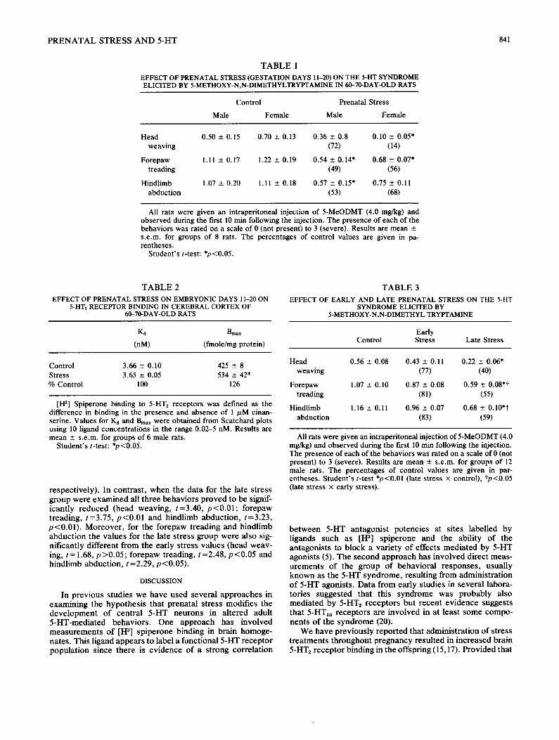
In the first study involving stress on ED 11-20, 8 control and 8 prenatal stress litters were available. This treatment resulted in a significantly reduced intensity of the behavioral syndrome produced by a 4 mg/kg injection of 5-MeODMT (Table 1). A 2-way ANOVA (treatment x sex) showed signif- icant treatment effects for head weaving, F(1,28)=12.74, p <0.005, forepaw treading, F(1,28)= 14.47, p <0.001, and the hindlimb abduction, F(1,28)=6.99, p <0.025. There were no significant sex effects or treatment x sex interactions. Scatchard plots of the binding of [H 3] spiperone to cortical membranes showed that stress on ED 11-20 resulted in a significant increase in the Bmax (Student's t-test; t=2.55, p<0.05) without affecting the I~ (Table 2).
In the second study, stress treatments were given either on ED 3-14 (early stress) or ED15-20 (late stress). Twelve litters in each of the three groups were available. Because the previous study showed no significant sex differences or treatment x sex interactions only male rats were used in this experiment. The early stress appeared to slightly reduce the intensity of the behavioral syndrome elicited by a 4 mg/kg injection of 5-MeODMT (Table 3) but the group means proved not to be statistically different when the data were analysed by Student's t-tests (t values: 0.96, 1.56 and 1.53 for the head weaving, forepaw treading and hindlimb abduction
P R E N A T A L S T R E S S A N D 5-HT 841
T A B L E 1
EFFECT OF PRENATAL STRESS (GESTATION DAYS 11-20) ON THE 5-HT SYNDROME ELICITED BY 5-METHOXY-N,N-DIMETHYLTRYPTAMINE IN 60-70-DAY-OLD RATS
Control Prenatal Stress
Male Female Male Female
Head 0.50 ± 0.15 0.70 ± 0.13 0.36 ± 0.8 0.10 --- 0.05* weaving (72) (14)
Forepaw 1.11 ± 0.17 1.22 ± 0.19 0.54 ± 0.14" 0.68 ± 0.07* treading (49) (56)
Hindlimb 1.07 ± 0.20 1.11 +- 0.18 0.57 +- 0.15" 0.75 ± 0.11 abduction (53) (68)
All rats were given an intraperitoneal injection of 5-MeODMT (4.0 mg/kg) and observed during the first 10 min following the injection. The presence of each of the behaviors was rated on a scale of 0 (not present) to 3 (severe). Results are mean ± s.e.m, for groups of 8 rats. The percentages of control values are given in pa- rentheses.
Student's t-test: *p<0.05.
T A B L E 2
EFFECT OF PRENATAL STRESS ON EMBRYONIC DAYS 11-20 ON 5-HT2 RECEPTOR BINDING IN CEREBRAL CORTEX OF
60-70-DAY-OLD RATS
Kd Bmax
(nM) (fmole/mg protein)
Control 3.66 - 0.10 425 _+ 8 Stress 3.65 ± 0.05 534 ± 42* % Control 100 126
[1-13] Spiperone binding to 5-HTz receptors was defined as the difference in binding in the presence and absence of 1 jzM cinan- serine. Values for Kd and Bmax were obtained from Scatchard plots using 10 ligand concentrations in the range 0.02-5 nM. Results are mean ± s.e.m, for groups of 6 male rats.
Student's t-test: *p<0.05.
respect ively) . In con t ras t , w h e n the da ta for the late s t ress g roup were e x a m i n e d all t h ree behav io r s p r o v e d to be signif- icant ly r educed (head weaving , t =3 .40 , p < 0 . 0 1 ; fo repaw t read ing , t=3 .75 , p < 0 . 0 1 and h ind l imb abduc t ion , t=3 .23 , p < 0 . 0 1 ) . Moreove r , for the fo repaw t read ing and h ind l imb a b d u c t i o n the va lues for the late s t ress g roup were also sig- ni f icant ly di f ferent f rom the early s t ress va lues (head weav- ing, t=1 .68 , p > 0 . 0 5 ; fo repaw t reading, t =2 .48 , p < 0 . 0 5 and h ind l imb abduc t ion , t =2.29, p < 0 . 0 5 ) .
DISCUSSION
In p rev ious s tudies we have used severa l app roaches in examin ing the hypo thes i s tha t p rena ta l s t ress modif ies the d e v e l o p m e n t o f cen t ra l 5 -HT neu r ons in a l t e red adul t 5 -HT-med ia t ed behav io r s . One a p p r o a c h has invo lved m e a s u r e m e n t s o f [1-13] sp ipe rone b inding in b ra in homoge- na tes . This l igand appea r s to label a func t iona l 5 -HT recep to r popu la t ion s ince the re is ev idence of a s t rong cor re la t ion
T A B L E 3
EFFECT OF EARLY AND LATE PRENATAL STRESS ON THE 5-HT SYNDROME ELICITED BY
5-METHOXY-N,N-DIMETHYL TRYPTAMINE
Early Control Stress Late Stress
Head 0.56 ± 0.08 0.43 ± 0.11 0.22 --- 0.06* weaving (77) (40)
Forepaw 1.07 -+ 0.10 0.87 - 0.08 0.59 ± 0.08*t treading (81) (55)
Hindlimb 1.16 - 0.11 0.96 --- 0.07 0.68 _+ 0.10*t abduction (83) (59)
All rats were given an intraperitoneal injection of 5-MeODMT (4.0 mg/kg) and observed during the first 10 rain following the injection. The presence of each of the behaviors was rated on a scale of 0 (not present) to 3 (severe). Results are mean -+ s . e . m , for groups of 12 male rats. The percentages of control values are given in par- entheses. Student's t-test *p<0.01 (late stress × control), tp<0.05 (late stress x early stress).
b e t w e e n 5-HT an tagon i s t po tenc ies at s i tes label led by l igands such as [I-I 3] sp ipe rone and the abil i ty of the an tagon is t s to b lock a var ie ty of effects med ia ted by 5-HT agonis ts (5). The second a p p r o a c h has invo lved direct meas- u r e m e n t s o f the g roup o f behav io ra l r e sponses , usual ly k n o w n as the 5-HT s y n d r o m e , resul t ing f rom admin i s t r a t ion o f 5 -HT agonis ts . Da t a f rom ear ly s tudies in severa l labora- tor ies sugges ted tha t this s y n d r o m e was p robab ly also med ia t ed by 5-HT2 recep to r s bu t r ecen t ev idence suggests tha t 5-HT, a r ecep to r s are invo lved in at leas t some compo- nen t s of the s y n d r o m e (20).
We have p rev ious ly r epo r t ed tha t admin i s t r a t ion o f s t ress t r e a t m e n t s t h r o u g h o u t p r egnancy resu l ted in inc reased b ra in 5-HT~ r ecep to r b inding in the offspr ing (15,17). P rov ided tha t
842 PETERS
the offspring were reared by control females from birth to eliminate postnatal effects were were also able to demon- strate decreased behavioral responses to 5-MeODMT. It was of interest to determine the critical period of these prenatal stress-induced changes to provide evidence that would be useful in formulating hypotheses on possible mechanisms.
Serotonergic neurons make their appearance early in brain development (14). Recent immunohistochemical studies (11, 12, 21) have generally confirmed the timetable for 5-HT neuron development established earlier from fluorescence histochemical observations (14,19). Thus, the earliest appearance of5-HT perikarya occurs on ED 12-13 in the mesencephalon and 1-2 days later in the medulla. Growth of axons proceeds rapidly after the first detection of 5-HT immunoreactivity and by ED 17 5-HT axons reach the basal forebrain. [H 3] Spiperone binding to 5-HT2 sites be- comes measurable after ED 15 (3). By ED 19 the 5-HT path- ways to all major regions of the forebrain have been estab- lished (11) but innervation of the cerebral cortex is not com- plete until the end of the third postnatal week (11). From ED 19 until the end of the first postnatal week there is a rapid growth of 5-HT dendrites (12).
For our first study we chose ED 11-20 as the stress period. This period extends from immediately before the first appearance of 5-HT-containing cells until shortly before birth, at which time pathways to all major areas of the fore- brain have been established and the extended period of de- velopment of terminal fields just begun (11). Stress exposure during this time produced similar changes to those reported for stress treatments throughout gestation, namely, in- creased 5-HT2 receptor binding and a reduced 5-HT syn- drome (17), confirming that the critical period was between ED 11 and birth.
The second study attempted to discriminate between ef- fects on early events such as 5-HT cell proliferation and growth of axons, and later stages such as the period of de- velopment of terminal fields and synapse formation. Three groups of pregnant rats were studied, unstressed controls, an early stress group treated on ED 3 to ED 14, and a late stress group treated on ED 15 to ED 20. There is considerable overlap between the stages of 5-HT neuron development and it is impossible to completely separate the stages. We selected the beginning of ED 15 as the break between the early and late stress period because both 5-HT cell subdivi- sion and the period of initial axon elongation are nearly complete at this time, while formation of the terminal fields has only just begun. ED 15 is also about the time that fore- brain 5-HTz receptors become detectable (3) suggesting that some synapse formation may have occurred.
Delaying the start of stress treatments until ED 15 again resulted in a reduction in the intensity of the 5-HT syndrome. In contrast, stress treatments starting on ED 3 and ending on ED 14, a period which included the peak period of raphe cell differentiation, did not significantly affect the intensity of the 5-HT syndrome. Thus, the critical period for changes in the central serotonergic system appears to start after the peak of 5-HT raph6 cell differentiation and corresponds to a period of rapid formation of synaptic contacts. Since the process of synapse formation in the rat is incomplete at birth, these data are consistent with our previous finding that the same biochemical and behavioral parameters can also be influ- enced during the early postnatal period.
Several authors have concluded that monoamines may play an important trophic role in the early development of brain cells before synaptic transmission is established [see discussion in (10)]. There is evidence that 5-HT may influ- ence the development of cells which will later receive 5-HT synaptic contacts. For example, the 5-HT-depleting drug p-chlorophenylalanine (PCPA) was found to retard the onset of neuronal differentiation in regions known to contain 5-HT terminals or to have a high 5-HT content in the adult while the stress of daily vehicle injections had the opposite effect (9). It is of interest that there is evidence that prenatal stress elevates fetal brain levels of tryptophan and 5-HT (D.A.V.P., unpublished observation). At least some of the effects of maternal stress on fetal development may there- fore be mediated by a stress-induced increase in fetal brain 5-HT synthesis. If such an increase occurred during the period starting about ED 15, when the peak of differentiation of cells in 5-HT terminal fields takes place (10), then the development of neurons which receive a serotonergic input may be affected.
In summary, we report evidence that the critical period of prenatal stress-induced changes in the intensity of at least some 5-HT mediated behaviors is during the final 7 days of pregnancy. Since this period commences toward the end of the period of differentiation of 5-HT raph6 neurons and after the time that these cells synthesise 5-HT, it is suggested that prenatal stress affects the later stage of neuron development such as the formation of synaptic contacts. Moreover, if 5-HT acts as a differentiation signal then stress-induced in- creases in fetal brain 5-HT may also affect the development of nonserotonergic neurons.
ACKNOWLEDGEMENTS
The author thanks Mr. Peter Borodchak for expert technical assistance and the Ontario Mental Health Foundation for financial support.
REFERENCES
1. Ader, R.; Conklin, P. M. Handling of pregnant rats: effects on emotionality of their offspring. Science 143:411-412; 1963.
2. Ader, R. ; Plaut, S. M. Effects of prenatal handling and differen- tial housing on offspring emotionality, plasma corticosterone levels and susceptibility to gastric erosion. Psychosom. Med. 30:277-286; 1968.
3. Bruinink, A.; Lichtensteiger, W.; Schlumpf, M. Pre- and postnatal ontogeny and characterisation of dopaminergic I)2, serotonergic S~ and spirodecanone binding sites in rat forebrain. J. Neurochem. 40:1227-1236; 1983.
4. Chapman, R.; Masterpasqua, F.; Lore, R. The effects of crowd- ing during pregnancy on offspring emotional and sexual behav- ior in rats. Bull. Psychon. Soc. 7:475-477; 1976.
5. Conn, P. J.; Sanders-Bush, E. Central serotonin receptors: ef- fector systems, physiological roles and regulation. Psycho- pharmacology (Berlin) 92:267-277; 1987.
6. Creese, I.; Snyder, S. H. [all] spiroperidol labels serotonin re- ceptors in rat cerebral cortex and hippocampus. Eur. J. Phar- macol. 49:201-202; 1978.
7. Green, A. R. 5-HT-Mediated behavior. Animal studies. Neuropharmacology 23:1521-1528; 1984.
8. Keeley, K. Prenatal influence on behavior of offspring of crowded mice. Science 135:44-45; 1962.
9. Lauder, J. M.; Krebs, H. Serotonin as a differentiation signal in early neurogenesis. Dev. Neurosci. 1:i5-30; 1978.

P R E N A T A L STRESS A N D 5-HT 843
10. Lander, J. M.; Wallace, J. A.; Krebs, H. Roles for serotonin in neuroembryogenesis. Adv. Exp. Med. Biol. 133:477-506; 1981.
l l. Lidov, H. G. W.; Molliver, M. E. An immunohistochemical study of serotonin neuron development in the rat. Ascending pathways and terminal fields. Brain Res. Bull. 8:389-430; 1982.
12. Lidov, H. G. W.; Molliver, M. E. Immunohistochemical study of the development of serotonergic neurons in the rat CNS. Brain Res. Bull. 9:559-694; 1982.
13. Lowry, O. J.; Rosenbrough, M. J.; Farr, A. L.; Randall, R. J. Protein measurement with the Folin phenol reagent. J. Biol. Chem. 193:265-275; 1951.
14. Olsen, L.; Seiger, A. Early prenatal ontogeny of central monoamine neurons in the rat: fluorescence histochemical ob- servations. Z. Anat. Entwickl.-Gesch. 137:301-316; 1972.
15. Peters, D. A. V. Prenatal stress: Effect on development of rat brain serotonergic neurons. Pharmacol. Biochem. Behav. 24:1377-1382; 1986.
16. Peters, D. A. V. Prenatal stress increases the behavioral re- sponse to serotonin agonist and alters open field behavior in the rat. Pharmacol. Biochem. Behav. 25:873-877; 1986.
17. Peters, D. A. V. Both prenatal and postnatal factors contribute to the effects of maternal stress on offspring behavior and cen- tral 5-hydroxytryptamine receptors in the rat. Pharmacol. Biochem. Behav. 30:669-673; 1988.
18. Scatchard, G. The attractions of proteins for small molecules and ions. Ann. NY Acad. Sci. 51:660-672; 1949.
19. Seiger, A.; Olson, L. Late prenatal ontogeny of central monoamine neurons in the rat: fluorescence histochemical ob- servations. Z. Anat. Entwickl.-Gesch. 140:281-318; 1973.
20. Smith, L. M.; Peroutka, S. J. Differential effects o1 5-hydroxytryptamine selective drugs on the 5-HT behavioral syndrome. Pharmacol. Biochem. Behav. 24:1513-1519; 1986.
21. Wallace, J. A. ; Lauder, J. M. Development of serotonergic sys- tem in the rat embryo: An immunocytochemical study. Brain Res. Bull. 10:459-470; 1983.