effects of hypothalamic and thalamic lesions on prolactin secretion in goldfish (carassius auratus)
TRANSCRIPT

Al’ CT
HOC NAH
NAPv NAT NTIL
NDLI NDNI
NDTL ND; NC: NT1
NLG NLTa ?JLTi NLTl
NLTp
‘Supported by Grant A-6371 to R. E. P. and Grant, A-6978 to 1%. A. IVI. from the National Re- search Council of Canada.
Sl’( :I NP( ;m
Kl’O NPl’
NT’Pv N PT
Xl: x1:1, N 1:P NS\’ KTI’
iKVS1 OT
()Tev I’ T
To date tllcm> is rclatirclv little known about t,lic cmt,rol of In-olnc& secretion in telcosts (for rc+v: Ball, 1969; Ball and Balxr, 1969; IS:111 et trl., 1972; Peter, 1973). Tndircct c~rirltmcc~ suggests that tjhe liypo- tmllnlamus norinnll~ inhibits the tendency for

(!OKTHOL OF PROLA(‘TlS IS GOLDFISH 439


CONTltOL OP PROLACTIN IS GOLDFISH 111
ISsperimental gro,Ip 2:
P:xperiment~nl group 1 :
Experimental grorrp 2:
Experiment 4
Normal control groly: Sham-operated grollp:
E:xperiment,al group 1:
Experimental group 3:
target : nllcleu:, prcopli- (‘,lS
wordinates: + I .O, III, 1X2.2
c,rwrent : 1 m-4 for 20 sec. 20 animals target : nllvlerw lateral
trlberis pars inferioris coot,dillat,es: +0.2, Izl,
I)“.6 cr1rrent: I mA for l‘l se,:
13 animal,, 12 animals target: rnlclelw lateral
trtberis pars lateralis cwordillittes: fO.7, IJ).a,
1 x2.0, the11 +O.i, RK-1, I):{.0
20 animals Inrget : nllclekw lateral
tuberis pars posterioris wordinates: +0.6, 11,
1)S.L’ crlrrent : 1 111A for I2 dec 20 animals target : n\lvlerls lateral
tuberis pars lateralis coordinates: +0.7. L0.4,
1)3.0, then +0.7, 110.4, 1>:1.0
ctwent: 1 m.4 for 12 SC< each placement
12 animals 12 animals target,: nucleus lateral
tuberis pars anterioris coordinates: + 0.0, RI,
112.9 20 animals target : nucleus lateral
tuberis pars anterioris coordinates: +0.9, RI,
1X2.9 current: I mh for 12 set 20 animals target: nucleus lateral
tuberis pars lateralis coordinates: f0.8, L0.3,
D3.0, t,hen +O.S, R0.3, D3.0
current: 1 mA for 16 WC
each placement
fl.rperimen t 1
The lesions in groul) 1 of ISspt I \Vc’I‘C
The lesions in groul) 2 w(‘re directccl at the nucl~s anterior tuhcris (SBT) in the dorsal liyl~otl~al:tn~us. Figurcb 2 is a sum- mary plot8 of t’he Icsiona that. clestroyc~cl :i major 1)ortion of tile nucl(W. =Zs shown in Tahlr 1, lcsioning of the NAT had no sig- nificant effect. on serum or l)itllit:try l’ro- lnctin levels.
The lesions in group I were directctl at the nucleus preopticus (NJ?<)). Figure 3 is a summary plot of the lesions that, were correctly plncc~l. In six of the lesionc~cl animals includctl in the group there were no stainahlc iieurosccrctory cells of the NPO evident. In five animals included in the group there were up to 25 stainable cells (nv = 17.2) of the pars magnoccl- lulnris remaining. Since the total number of st,ainahlc cells in the SPO is approximately TiOOO, of which the pars magnocellularis constitutes about 40% (Peter, 1970)) it w-as felt that including the five lesioncd animals each with not more than 25 stainable cells remaining would not invalidate the results. In addition to the destruction of the NPO, the lesions did substantial damage to the nllclcus prcopticus lkvcntricularis (NPP) and the nucleus anterioris periventricularis (KAPvi. Thcrc wer(l no significant effects on serum or pituitary prolactin levels due to lcsioning the NH), NPP and NAPv (Table 2 1.
Figure 4 is a summary plot of lesions from group 2 of Expt 2. The t,arget for the

442
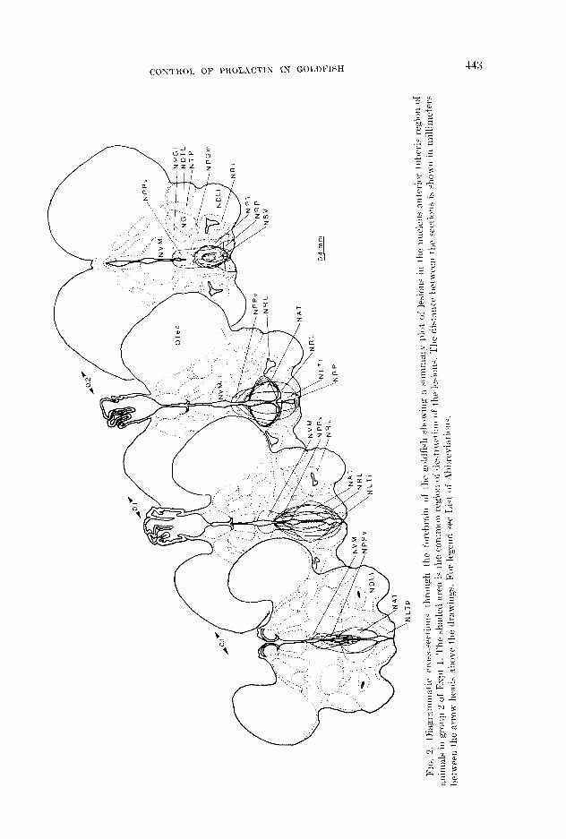
FIG. 1. l>iagrammati~~ cross;-sections through the forebrain of the goldfish showing a summary plot of lesions in the nucleus lateral tllheris pars :tntjerioris regiol~ of animals in group 1 of Espt I. The shaded area is t.he common region of destruction of the lesions. The distance between the sections is shown in millimet,ers between the arrow heads above the drawings. For legend see Lisl of Abbreviations.
lesions was the nucleus latrral t~uhcris pars Imkrioris ( NIJ’I‘IJ.). L4 SUlrnll~~ry plot (Jf the
inferioris (NLTi) Lesionin g of the KI,Ti lesions is shown in Fig. 5. AR shown in Table had no significant8 effcrt on serum or pitu- 3, lesioning the !SI,Tp had no significant cf- itary prolnctin lcvcls (Tnblc 2). fccts on wrum or pituitary prolartin lwels.
The Iwion:: Ezperimenf 3
in group 2 of Expt 3 were clirc~ctc~cl at) tliv nuc*lcus lntcrnl tubcris pars
The lesions in groul) 1 of Esl)t 3 new lntcmlis (W,Tl) A swnnlnry plot of the directed at the nucleus lateral tuberis pars lesions t,hnt ccm~~l~~tc~ly hilntcrxlly destroyccl
Tr\HI,b: I I':FFICCTS ok‘ I,~cslo.us 121 ml,: Nr-c.1.1,:r-s I,.\TW.\L TL:lcb:Irrs I'.\IlS .4sTl:Irlonls (NLTfl), .,sn
IS TIT, , : NVCI>I:UH ih-T~:IiIOll T~-I~I,:I<Is (SAT) I:I.:GIONS ON S~:~tcal
\S,) I’ITI-I’I’.\It’- 1’1101. \(“I’IS I,I~:Tl~:I,S IS ‘I’H,: (:OI,l)b’lSH
Gronp
04
mm

444 PETER AND BICKEOWN
(;roup
Normal control (n = 12) Sham control (n = 12) NPO lesioned (n = I1 1 NLTi lesioned (IL = S)
* NS = not significant compared with normal or sham conlrol groups.
the NLTl is shown in Fig. 6. As shown in differ significantly from levels found in the Table 3, lesioning the NLTl caused a sig- control groups. n&ant increase in serum prolactin lwels Two animals in group 2 in which lesion- compared to both the normal and sham ing of the NI,Tl was attempted had only control groups. Pituitary prolactin lewls of unilateral destruction of the nucleus. Strum the NLTl lcsioncd group, however, did not prolactin levels of these two animals (121.24
FIG. 3. 1 )iagrammat ic truss-sect ions through the forebrnill of the goldfish showing a summary plot of lesions in the nllclells preopiiuls region of animals in gro~~p 1 of F:xpt 2. The shaded area is the common region of destruction of the lesiotls. The distance between the sectioll:: is shown in millimeters between the arrow heads above the drawings. For legend see List of Ahtxevi:ttions.

CONTROL OF PROLACTIIS IX GOLDFISH 445
FIG. 1. I )i:tgrmmmtic muss-sections t hrollgh the forebrain of 1 he goldfish hhowing a summ:try plot rrf lesilms ill the nllclerw lateral tuberis pars inferioris region of animals ill grunp 2 of I5xpt 2. The shaded area is the common region of destruc*tion of the lesions. The distance betwee the swtions is shown in millimeters between the arrow heads nbcwe the drnwiugs. For legend we I.ist of Abbrevintic~tls.
units/ml, and 122.75 units/ml) ww wry aimilnr to values for the cLoutro1 groups (WC Table 3).

446
FIG. 5. IXagrammatic cross-sec+iorls throrgh the forebrain of the goldfish showing it sltmmary plot, of lesions in the nllclerw lateral trlberis pars posterioris region of animals in grc,,q, 1 of Pkpt :3. The shaded area 1s the common region of destrllrtion of the lesiolls. The tlistancae het,ween the sectsions is shown ill mllhmeters between the arrow heads above the ~I.BWIII~H. For Legend see IA of Abbreviations.
The rcnmintlw of the animnl~ in group 2 of Expt 4 (77 = 12‘1 hot1 sniallcr lesions in various lowtiolls of the AH-?\IT rc$on ~sulmnary plot of IcAons not, showni, The smun prolnctin ltwl for tllrsc nnim:~ls was 159.92 f 6.77 mitsjnil and the pitllitary prolnctin lrwl ‘rvw 116.77 + 3.50 ullits,/mg. Scithm of tliwc~ ‘\xIurs cliffrr significantly from tlir wren or I)ituitnry prolnctin 1~~~1s.

CONTROL OF PHOL.4C’TIK IN GOLDFISH
FIG. 6. 1)iagrammstic cross-sections through the forebrain of IShe goldfish showing a s\lrnmary plot of lesions in the nllcleus lateral trtheris pars lateralis region of animals in group 2 of Kspt 3. The shaded area is the common region of destruction of t,he lesions. The distance between t,he sections is shown in millimeters between the arrow heads above the drawings. For legend see I,ist of Abbreviations.
Expt 4; see t)clow for discussion of this r(‘- sult) . View-cd together the lesions intlicatc~ that the NLTl done has :m inhibitory cf- feet on prolwctin secretion. This finchg sNl)- l)orts the idea tliat~ the liyt~otli:~laillus nor- mally inhibits l)rolactin sccrct~ion. Surll all inliihitorp t4fcbct is l)resurnably iwcli:itc~tl
hy tllc ncurohornwnc) PIF suggested to Ix, present in the liypotlinlnmw of tel(wt:: (sv(~ Introcluction).

448

CONTROL OF PKOLACTIN IN GOLDFISH
.NPGm
-NRL
FIG. 8. lktgrammatic cross-sections through the forebrain of the goldfish showing R summary plot of lesions in anterior hypothalamic-medial thalamic region of animals in grollp 2 of Expt 4. The shaded area is the common region of destruction of the lesions. The distance between the sections is shown in millimet,ers bet ween the arrow heads above the sections. For legend see List of Abbreviations.
volvecl in wgulation of polactin secretion in the goldfisli and in mammals.
Iii the rat, lcsioning of an area including the parnoeutricular nucleus (PVN), or
dorsolatcral to the PTW, also induces pro- lnct~in secretion (Cook, 19.59; Flnmcnt- Durand and nw:cnlin, 1964; DcVoc, Rnmirez, anti IllcClnriii, 1966). III the 1)rcscnt study lcsioning thcl NPO, the l~on~ologue of the PVN and suprnoptic nucleus, htl no effect
on prol!lctin wcretion. Elcrtricxl stimula- tion s:tl;diw in mnmmals have sl~on~i~ that wrernl hyl)oth:~lamic regions in addition to the l)asnl tulwnl and PVN regions are iii- volv~~l in stimulation of prolactin secretion (e.g., E:\,erctt8 and Quinn, 1966; Quinn and
Everett. 1967; Tidal and Knaggs, 1972). In view of the osmoregrilntory function of prolartin in tclcosts, it would he of great interest, to study the cffccts of rlertricnl
stimulation of various l~ypotlial:~mic regions on prolactin secretion in teleosts.
The tlrcrcascd serum 1)rolactin levels found in animals with large lcsiuns in the AH-MT (group 2, Espt 4) cannot, he fully csplainctl. Simll lwions in the smic region had no rfl’ccks. The dcrreasc in prolact,in secretion as a result of the lnrgc lesions (yin hc intcrprcted in scvcrd ways. It could h:tvc resultc~cl from the destruction of n center that is the origin of n factor that stimulates the secretion of prolwtin. Evidence from l~pl~otlinl:tmic extract stritlics indicatw that a prolwtin rolrnsing factor (PR.F) is pres- ent in birds (Tisicr-Vidnl and Gourclji, 1972). In mnmmnls PRF activity has xlso been reportd in hy1~otlialan~ic extrxcts (for review: Blnckwcll and CTuillemin, 1973; McCann and Porter, 1969 ; Schally, Arimur:\ and Kastin, 1973). In t,elco3ts there is only


COIVTNOL OF PROLACTIN 1X GOLDFISH 451
