effects of β-estradiol on germination and growth …effects of β-estradiol on germination and...
TRANSCRIPT

EFFECTS OF β-ESTRADIOL ON
GERMINATION AND GROWTH IN ZEA MAYS L.
A THESIS PRESENTED TO THE DEPARTMENT OF NATURAL SCIENCES
IN CANDIDACY FOR THE DEGREE OF MASTER OF SCIENCE
BY KELSEY MARIE BOWLIN
NORTHWEST MISSOURI STATE UNIVERSITY MARYVILLE, MISSOURI 2014

Running Head: EFFECTS OF β-ESTRADIOL ON GERMINATION AND GROWTH
EFFECTS OF β-ESTRADIOL
ON THE GERMINATION AND GROWTH
IN ZEA MAYS L.
Kelsey Marie Bowlin
Northwest Missouri State University
Thesis Approved
Thesis Advisor Date
Dean of Graduate School Date
iii
Abstract
Water is one of the most important resources for an ecosystem. Pollution of
major water sources has become a serious problem across much of the world. One
example of water pollution caused by humans is the dumping of waste products as
effluent into major rivers and waterways. While documentation for the effects of water
contaminants on a variety of animals has been widely researched and documented,
studies on the effects of these same contaminants on plants are relatively new. Current
research has focused on the effects of these contaminants either on plant germination
or on vegetative plant growth. The purpose of this research was to investigate the
effects of a major pollutant, β-estradiol, on the germination and vegetative growth of
corn (Zea mays L.). The concentrations used in these experiments were 50 μg/L, 0.1
mg/L, 1.0 mg/L and 10 mg/L. In the germination experiment, total percent germination,
mean hour of germination, primary root length, coleoptile length, and number of
adventitious roots was investigated. The parameters used in the growth experiment
were overall root length, overall shoot length, number of leaves, and chlorophyll
content. Corn kernel germination and corn seedling growth were consistently inhibited
by the 10 mg concentration. The 0.1 mg treatment augmented germination and
seedling growth. Future experiments could be carried out to follow the development of
the corn seedlings through maturation and the production of fruit to determine if the
high doses of β-estradiol affect the later stages of development as well.
iv
TABLE OF CONTENTS
Page
ABSTRACT……………………………………………………………………………………………. iii
LIST OF FIGURES…………………………………………………………………………………… v
CHAPTER 1 – INTRODUCTION………………………………………………………………. 1
CHAPTER 2 – THE EFFECT OF β-ESTRADIOL ON GERMINATION OF CORN
(ZEA MAYS L.)………………………………………………………………………………………. 12
CHAPTER 3 – THE EFFECT OF β-ESTRADIOL ON GROWTH OF CORN
(ZEA MAYS L.)............................................................................................. 39
CHAPTER 4 – THE EFFECT OF β-ESTRADIOL ON CHLOROPHYLL
CONCENTRATION OF CORN (ZEA MAYS L.)……………………………………………. 57
CHAPTER 5 – CONCLUSION……………………………………………………………………. 64
LITERATURE CITED………………………………………………………………………………… 79
ACKNOWLEDGEMENTS…………………………………………………………………………. 81
v
LIST OF FIGURES
Page
FIGURE 1.1 The molecular structure of the mammalian sex hormone 17β-estradiol………………………………………………. 5
FIGURE 1.2. Value of corn production in the United States for the
years 1952 to 2012 according to the United States Department of Agriculture (University of Missouri Extension, 2012)…………………………………………………………. 12
FIGURE 2.1. Maure corn (Zea mays L.) kernel…………………………………. 15
FIGURE 2.2. Kernels used in this experiment were obtained from Croplan Genetics Research Plot Seed L78X3068 MF 113, Land O’Lakes Ag Services, Fort Dodge, IA, USA…….. 17
FIGURE 2.3. Ten corn (Zea mays L.) kernels were placed into petri dishes lined with filter paper. The dishes were then
randomly assigned a treatment group of various β-estradiol concentrations (10 mg/L, 1.0 mg/L, 0.1 mg/L or 50 μg/L) or control……………………………………….. 18
FIGURE 2.4. Petri dishes containing 10 corn (Zea mays L.) kernels and various concentrations of β-estradiol were placed into a Thermo Scientific incubator (model 818) at 26⁰C without light……………………………………………………………… 19
FIGURE 2.5. Fifteen mL test tubes in which each contained a germinated corn (Zea mays L.) kernel. Each kernel rests upon a piece of filter paper soaked in 100 μL of β-estradiol at the assigned concentration………………….. 20
FIGURE 2.6. Diagram illustrating the parts of a corn (Zea mays L.) kernel before (left) and after (right) germination. Germination was determined to be when the primary root began to emerge from the coleorhiza. After five days of growth both the coleoptile and the primary root were removed and measured (mm). The number of adventitious roots were also counted and recorded…… 21
vi
LIST OF FIGURES CONT.
Page
FIGURE 2.7. Corn (Zea mays L.) seedling after five days of growth post germination. The seedlings were grown in 15 mL test tubes in a Thermo Scientific incubator (model 818) without light at 26 ⁰C…………………………………………………. 23
FIGURE 2.8. The coleoptile from a corn (Zea mays L.) seedling grown in the presence of 17β-estradiol for five days was removed and measured for length (mm)……………. 24
FIGURE 2.9. The mean number of hours for 444 corn kernels (Zea mays L.) subjected to different β-estradiol concentrations to germinate. Bars denote a 97.5% confidence interval……………………………………………………. 26
FIGURE 2.10. Corn (Zea mays L.) kernels from the 10 mg β-estradiol treatment group after five days of growth with noticeable lack of primary root growth while exhibiting coleoptile (→) growth (A, B, C). A kernel from the 10 mg treatment group that germinated then proceeded to degenerate and die during the five days of growth in the 15 mL test tube (D)…………………… 28
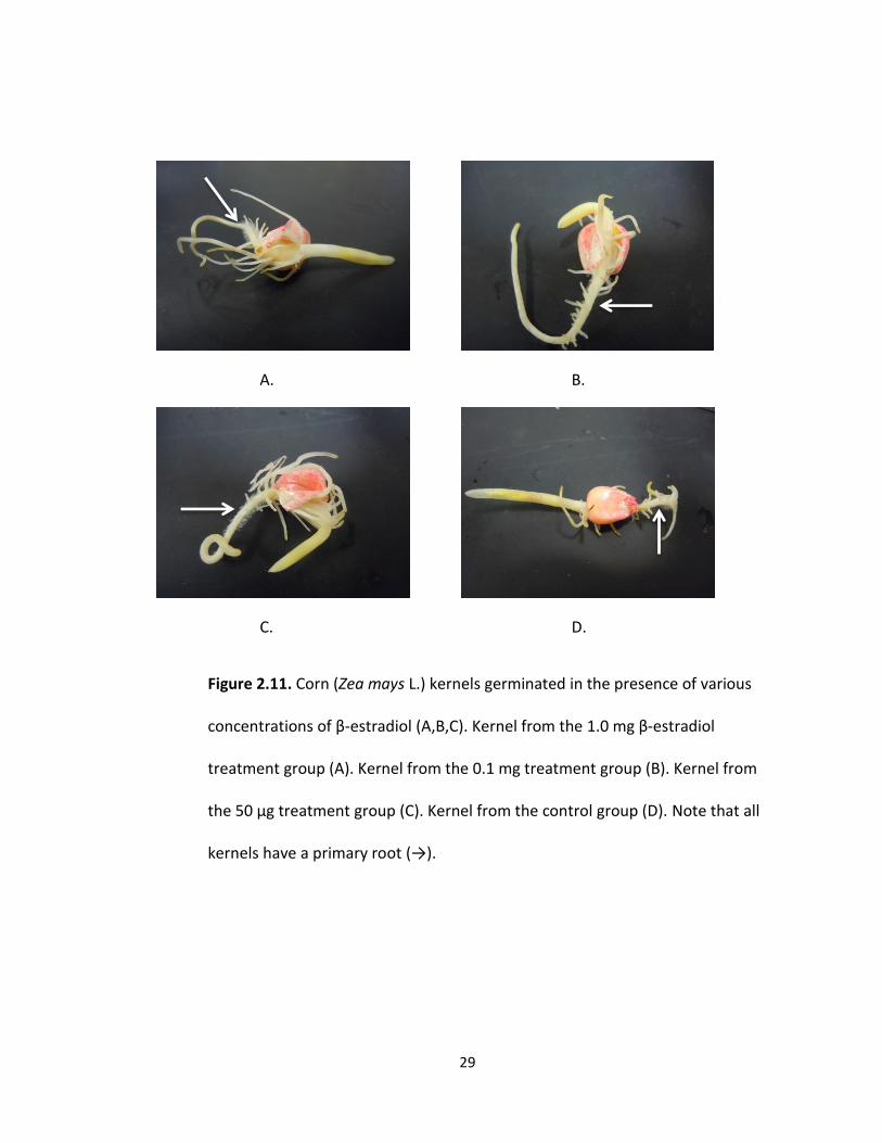
FIGURE 2.11. Corn (Zea mays L.) kernels germinated in the presence of various concentrations of β-estradiol (A,B,C). Kernel from the 1.0 mg β-estradiol treatment group (A). Kernel from the 0.1 mg treatment group (B). Kernel from the 50 μg treatment group (C). Kernel from the control group (D). Note that all kernels have a primary root (→). 29
FIGURE 2.12. The mean primary root length (mm) of 5 day old corn (Zea mays L.) seedlings germinated and grown in different concentrations of β-estradiol. Vertical bars are based on a 97.5% confidence interval……………………. 30
FIGURE 2.13. The mean length (mm) of the coleoptile of a 5 day old corn (Zea mays L.) seedlings germinated and grown in different concentrations of β-estradiol. Vertical bars are based on a 97.5% confidence interval……………………. 32
vii
LIST OF FIGURES CONT.
Page
FIGURE 2.14. The mean number of adventitious roots of 5 day old corn (Zea mays L.) seedlings germinated and grown in different concentrations of β-estradiol. Vertical bars are based on a 97.5% confidence interval……………………. 34
FIGURE 2.15. The overall percentage of germination after 96 hours in various concentrations of β-estradiol. A corn (Zea mays L.) kernel that germinated was assigned a 1 and a kernel that did not germinate was assigned a 0. Vertical bars are based on a 97.5% confidence interval………………………………………. 36
FIGURE 3.1. Fifty individual growth tubes (Blowmolded cells D16 L 2”x7”, Stuewe & Sons Inc.) were lined with triple ply cheese cloth, completely filled with vermiculite and randomly assigned to one of four concentrations of β-estradiol or a control. Tubes were then placed into a holding tray. One germinated kernel was placed into each of the 50 tubes and covered with additional vermiculite……….. 43
FIGURE 3.2. Corn (Zea mays L.) kernels were placed into a Thermo Scientific incubator (model 818) at 26⁰ C with a photoperiod set at 12 hours for 20 days. On days 4, 10, and 16 the seedlings received 20 mL of nutrient water. On days 7, 13, and 19 the seedlings received treatments of 50 mL of the assigned β-estradiol concentrations or sterile water for the control tubes.. 44
FIGURE 3.3. The corn (Zea mays L.) kernels were removed from the incubator after 20 days of growth in various concentrations of β-estradiol. Measurements (mm) were taken of both the length of the entire shoot system as well as the entire root system………………….. 45
viii
LIST OF FIGURES CONT.
Page
FIGURE 3.4. Corn (Zea mays L.) plants after 20 days of growth in 50 μg/mL, 0.1 mg/mL, 1.0 mg/mL and 10 mg/mL concentrations of β-estradiol (B,C,D,E respectively) or in a control solution (A). Note the shallow root system (white arrow) that was typical for the 10 mg/mL treatment group and that the leaves on the shoot system (black arrow) were starting to die (E)…… 48
FIGURE 3.5. The mean shoot system height of corn (Zea mays L.) seedlings after 20 days of growth in various concentrations of β-estradiol. There are 30 plants in each treatment group for a total of 150 plants. The vertical bars are based on a 97.5% confidence interval……………………………………………………………………. 49
FIGURE 3.6. The mean length of corn (Zea mays L.) root systems
of plants after 20 days of growth in various concentrations of β-estradiol. Thirty plants were in each treatment group. The vertical bars are based on a 97.5% confidence interval………………………………… 52
FIGURE 3.7. The mean number of corn (Zea mays L.) leaves of plants after 20 days of growth in various concentrations of β-estradiol. Thirty plants were in each treatment group. The vertical bars are based on a 97.5% confidence interval……………………………………… 53
FIGURE 4.1. The mean amount of chlorophyll in leaves from corn seedlings (Zea mays L.) after 20 days of growth in various concentrations of β-estradiol. There were 30 plants per treatment. The vertical bars are based on a 97.5% confidence interval……………………………………… 62
1
Chapter 1: Introduction
Ecosystems consist of complex and intricate relationships between organisms
and their biotic and abiotic surroundings. Each species in an ecosystem occupies a niche,
and often their role in the community is vital to the functionality of it. When there is a
change to any part of the biotic or abiotic resources in an ecosystem, it can be
drastically altered (Islam and Tanaka 2004). One of the most important resources for an
ecosystem is water. As a key reactant in photosynthesis, water helps to form the base of
any ecosystem by providing the necessary resources for photosynthetic organisms,
which then in turn provide sustenance for heterotrophic organisms as well as oxygen for
aerobic organisms (Campbell et al. 2011). The sum of all of these organisms and the flow
of energy from one trophic level to the next creates an intricate and balanced chain or
web. If disturbed or altered, the flow of energy can become highly disrupted causing
collapse of the entire ecosystem (Campbell et al. 2011). As water helps form the base of
these intricate relationships, any alteration of the water supplied to an ecosystem is
likely to have drastic effects on the organisms that live there.
Properties of Water
Water contains unique properties that allow it to sustain life on this planet. The
molecule itself, made of two hydrogen atoms bonded to one oxygen atom, is held
together by covalent bonds. These bonds are considered to be polar due to the high
electronegativity, or stronger pull on the shared electrons, of the oxygen atom in
comparison to the hydrogen (Campbell et al. 2011). The polar covalent bonds between

2
the atoms create a molecule that has a partial negative and a partial positive pole due to
the negatively charged electrons spending the majority of their time around the oxygen
atom. When the molecules are next to each other they align themselves so that the
negative pole of one water molecule is associated with the positive pole of another.
These create hydrogen bonds between water molecules. While one hydrogen bond by
itself is rather weak, the accumulation of many hydrogen bonds creates an extremely
strong collective force that ends up giving water its unique properties that allow it to
sustain life as we know it (Campbell et al. 2011).
One of these properties is the ability of water to hydrogen bond with itself and
create a high surface tension. This property is known as cohesion and is the result of
many hydrogen bonds working together. Cohesion becomes an important part of the
process by which plants will move water into the root tissue and up through the
vascular tissue. The process by which water is transported through a plant’s tissues is
known as adhesion-cohesion theory of water movement (Taiz and Zieger 2006). The
driving force behind this is transpiration. This involves the evaporation of water through
openings in the leaves, known as stomata. The hydrogen bonds between the molecules
create a chain of water that gets pulled through the xylem tissue of the root, up the
stem and ultimately evaporates into the atmosphere through the stomata (Campbell et
al. 2011). Adhesion, or the ability of water molecules to cling to other surfaces, also
plays a role as it allows the water molecules to adhere to the cell walls of xylem tissue
and help counter the effects of gravity (Campbell et al. 2011).
3
Another life supporting property of water is its ability to be a universal solvent
for almost any other polar molecule. This property allows water to dissolve many
different substances, and then transport and cycle them through organisms’ tissues as
well as through the environment (Campbell et al. 2011). There can be quite severe
consequences to organisms because of the solvent properties of water, as it is not a
selective process. Several toxic and synthetic substances that are polar could be
dissolved in water and consequently taken up into the organism’s tissues. Potentially,
these substances can then accumulate and cause damage or harm to the organism
(D’Abrosca et al. 2008).
Water pollution
One example of water pollution caused by humans is dumping waste products as
effluent into major rivers and waterways. Although this practice has occurred for
centuries, the damaging environmental effects have only recently been brought to light
by the research that is being conducted by scientists worldwide on various organisms
across most kingdoms and phyla (Islam and Tanaka 2004). As rivers take their course,
ecosystems downstream are affected. As these waterways flow into the oceans through
estuaries many coastal and marine ecosystems are now being impacted as well. To date,
the greatest amount of waste that ends up in marine ecosystems is sewage (Islam and
Tanaka 2004). Sewage effluent can include industrial waste, municipal waste, animal
remains, slaughterhouse wastes, water and wastes from domestic baths, kitchen
4
wastes, fecal matter and numerous other things (Islam and Tanaka 2004). For a
population of 800,000 people it has been estimated that approximately 3600 tons of
organic matter are dumped annually (Islam and Tanaka 2004). With worldwide
populations now approaching 8 billion people, the amount of organic waste ending up
in aquatic ecosystems is astronomical.
Endocrine Disruptors
Sometimes naturally occurring chemicals, such as hormones, can also build up in
these aquatic environments due to waste effluent. One specific class of these is called
reproductive endocrine disruptors, or REDs. Several studies have been conducted that
show where there are high levels of REDs in habitats there tends to be disruptions in the
morphology and physiology of the organisms that reside there (Islam and Tanaka 2004).
Examples of synthetic and naturally occurring chemicals that act as REDs include
estrogenic and anti-androgenic substances. There are also chemicals that are not
hormonal in nature but can have estrogenic properties, such as alkyphenols, industrial
pesticides, and chlorinated hydrocarbons (Islam and Tanaka 2004). One of the most
common estrogenic sources in waste water effluent comes from the urine of women
who take birth control pills. These hormone treatments contain the synthetic
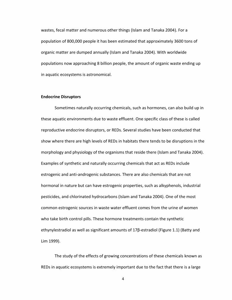
ethynylestradiol as well as significant amounts of 17β-estradiol (Figure 1.1) (Batty and
Lim 1999).
The study of the effects of growing concentrations of these chemicals known as
REDs in aquatic ecosystems is extremely important due to the fact that there is a large
5
Figure 1.1. The molecular structure of the mammalian sex hormone 17β-
estradiol.
6
number of organisms that show hormone-dependent sexual dimorphism, or hormonal
differences that are present in the development of males versus females. Reproductive
disorders in wildlife can include anything from altered fertility and reduced offspring
viability to impaired hormone functionality and modified reproductive anatomy
(Guillette et al. 2000). The most common disruption caused from REDs is due to
estrogen mimicry. These contaminants have the ability to induce cellular proliferation of
estrogen sensitive cells (such as breast and ovarian tissue) as well as bind to estrogen
receptors (Guillette et al. 2000).
A study was conducted on the mosquitofish, Gambusia affinis holbrooki, to
determine if there was an effect on the organisms from polluted aquatic environments
(Batty and Lim 1999). In this study fish were collected from multiple sections of the
same river. One of the sites was located upstream of a sewage/waste water treatment
facility and another was located downstream. This mosquitofish is one example of an
organism that exhibits hormone-dependent sexual dimorphism. The males have a
modified anal fin called the gonopodium that is used to aid in sperm packet transfer to
females (Batty and Lim 1999). The elongation and development of the gonopodium is
under androgenic control. Thus, when the researchers found that gonopodium length
and overall number of male fish present was significantly reduced in populations caught
downstream of the sewage effluent, there was evidence to support the hypothesis that
REDs are having an impact on wildlife (Batty and Lim 1999). These fish were originally
introduced to help keep mosquito populations at bay. They have since become an
integral part of the ecosystem by also providing food for other species of aquatic
7
organisms. By reducing their reproductive success, their numbers are starting to
dwindle downstream of the sewage effluent which in turn will start to impact other
organisms dependent on them (if they themselves are not already directly impacted by
the effluent).
Another organism that is exhibiting the effects of REDs is Alligator
mississippiensis, or the American alligator. Studies of populations in polluted Florida
lakes have shown that juvenile alligators have different plasma concentrations of
hormones along with altered reproductive tract anatomy and hepatic functioning
compared to juvenile alligators from non-polluted lakes nearby (Guillette et al. 2000). It
was also noted that clutch viability, or the survival rate of the eggs, was greatly reduced
in the polluted environments. Several alterations in the reproductive tract were noted in
hatchlings and persisted throughout the life of an alligator. More specifically, it was
noted that males had reduced phallus size as well as lower plasma levels of testosterone
than normal. This would indicate that the REDs are affecting the development of the
alligators starting when they are still an embryo (Guillette et al. 2000). This poses a
serious problem for appropriate alligator growth as the developing embryo shows
extreme sensitivity to chemical signals.
While documentation for the effects of water contaminants on a variety of
animals has been widely researched and documented, studies on the effects of these
same contaminants on plants are relatively new. Mammalian sex hormones were
originally found in plants and documented in 1926 by Dohrn et al. (Erdal et al. 2010).
Between the original discovery and up until the 1990’s the presence of the hormones
8
was determined in 128 plant species from 50 families but the effects of the hormones
were not studied in great detail (Janeczko and Skoczowski 2005). With the large
environmental movement that ushered in the millennium, scientists started to take
another look at the role that contaminants play in the environment and especially
started to look at their effects on plants. Current research has focused on the effects of
these contaminants either on plant germination or on vegetative plant growth
(D’Abrosca et al. 2008).
Estradiol
A potent mammalian sex hormone produced primarily in the ovaries that is
derived from cholesterol is 17β-Estradiol (Carreau et al. 2002). The chemical formula of
17β-Estradiol is C18H24O2 and it has a molecular weight of 272.38 grams per mol (Figure
1.1). The steroid can freely enter an animal cell and when bound to a ligand has the
ability to enter the nucleus; here it is involved with regulating gene transcription. In
female mammals this steroid acts as a growth hormone for specific tissues in
reproductive organs such as breast tissue, and is also responsible for maintaining the
oocytes in the ovaries (Carreau et al. 2002). Estradiol also exerts wide and varying
effects on other tissues in the body such as the ability to change the shape of bone and
joints, as well as affect fat deposition and skin composition.
To treat cases of hypoestrogenism, or low estrogen levels, medication containing
17β-Estradiol is often given to women. It is also one of the main chemical compounds
(or derivatives of 17β-Estradiol) in most oral birth control pills (Carreau et al. 2002).
9
Mammals naturally excrete 17β-Estradiol through sweat glands and in their urine, but
increased consumption through synthetic methods can lead to increased excretion.
These excretions then end up in the effluent of our waste water and are carried to
waste water treatment facilities. The environmental protection agency (EPA) has listed
17β-Estradiol as an unregulated drinking water contaminate on their Contaminate
Candidate List 3 (CCL3). Contaminates that are found on CCL3 are not subject to any
national primary drinking water regulations (United States Environmental Protection
Agency, 2012). The Safe Drinking Water Act (SDWA) of the United States originally
passed by Congress in 1974 (amended in 1986 and 1996) ensures that Americans have
access to quality drinking water and that actions are taken to protect drinking water and
its sources. The contaminants listed on the CCL3 are those that are toxic enough that,
under the SWDA, may require regulations in the future (United States Environmental
Protection Agency, 2012).
Seed Germination
Seed germination is a complex physiological process that includes the action of
multiple processes. Plant hormones, water relations, light responses, temperature, and
stimulation of the expression of genes all have a part in regulating germination. Once all
of the conditions have been met a seed will begin to germinate, or leave the dormant
stage and continue growth (Taiz and Zeiger, 2006). The entire process of germination
will transform the embryonic plant into a seedling. In order for growth of the embryo to
continue the cells will require energy in the form of ATP provided by the process of
10
cellular respiration. Respiration requires glucose as well as oxygen. In order to obtain
high oxygen levels the seed coat must be penetrated (Taiz and Zeiger, 2006). In most
plants this occurs when the seed imbibes, or uptakes, large quantities of water. This
causes the tissues to swell, cracking the seed coat in the process.
Ions and molecules in the water are taken up into the tissues of the seed as well.
Compounds contained in the water could have an effect on the germination of the seed.
Studies across both Europe and North America have shown that surface water, ground
water, and drinking water systems contain contaminants (D’Abrosca et al. 2008). Studies
conducted on varying species of plants demonstrated that β-estradiol decreased the
germination percentage of lettuce, carrot, and tomato seeds (Lactuca sativa L., Daucus
carota L., and Lycopersicon esculentum Mill. respectively). Germination was reduced by
57% in L. sativa, 6% in D. carota, and 18% in L. esculentum when compared to the
controls (D’Abrosca et al. 2008). In the same study the germinated seedlings were
collected and the root length was measured. Seedlings of two species exhibited marked
reduction in root length when compared to the control. There was a 34% reduction in L.
sativa and 22% reduction in L. esculentum. However, D. carota actually exhibited an 11%
increase in overall root length (D’Abrosca et al. 2008).
Another study performed on chickpea seeds (Cicer arietinum L.) resulted in a
dramatically different set of results. Both β-estradiol and progesterone increased seed
germination. After 48 hours seed germination increased from 85% in the control to
100% in the β-estradiol treated group (Erdal and Dumlupinar, 2010). Subsequently,
11
these treated seedlings also demonstrated a significant amplification to both the root
length and the shoot length. Yet another study performed on wheat (Triticum aestivum
L.) showed that there was no significant effect on seed germination (Nirmala et al.
2008).
Importance of Grain Crops
Farmland across the Midwest portion of the United States is typically dominated
by soybeans (Glycine max (L.) Merr.) or field corn (Zea mays L.). Corn (or maize) is
primary grown for livestock feed and silage, although a significant percentage is now
being used in the production of ethanol. One bushel of corn will provide 10.6 L of
ethanol, 7.7 kg of dried distillers grain, 25.4 kg of grain and 14.5 kg of starch or 14.9 kg
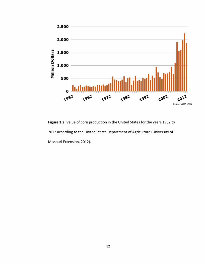
of sweetener or .73 kg of corn oil (University of Missouri Extension, 2012). The economic
value of corn in the U.S. (Fig 1.2) is at an all-time high and was recorded to be around
two billion dollars in 2010 (University of Missouri Extension, 2012). Understanding the
possible effects of environmental pollutants on corn production and yield is therefore
one of great consequence to farmers and the American economy. The purpose of the
experiments in subsequent chapters was to determine if a contaminate selected from
the CCL3 list would have an effect on the germination or growth of corn. The
contaminant chosen was 17β-Estradiol. One of the reasons for this decision was the
close association between farmland and large livestock operations. 17β-Estradiol can be
found in significant quantities in both the excrement and urine of the livestock in these
operations, and often works its way into the water systems and also onto fields (Caron
et al. 2012).
12
Figure 1.2. Value of corn production in the United States for the years 1952 to
2012 according to the United States Department of Agriculture (University of
Missouri Extension, 2012).

13
Chapter 2: The effect of β-estradiol on germination of corn (Zea mays L.)
As previously stated in the introductory chapter, hormones such as β-estradiol
can significantly affect the growth and physiology of both animals and plants.
Investigations previously conducted on plants have looked at the effect of β-estradiol on
the seed germination in a wide variety of different plant species. β-estradiol decreased
the germination percentage of lettuce, carrot, and tomato seeds (Lactuca sativa L.,
Daucus carota L., and Lycopersicon esculentum Mill. respectively), while in chickpea
seeds (Cicer arietinum L.) the germination percentage was increased. The variations in
the results of studies suggests that the effect of the hormone will depend on the species
of plant exposed to the hormone.
Corn kernels are actually fruits. Each kernel is technically classified as a caryopsis,
as the simple seed inside is surrounded by the pericarp, the mature ovary wall. Fruits
classified as caryopsis are also known as grains. In these fruits the seed coat is fused to
the pericarp (Evert and Eichhorn 2013). The corn embryo contains a well-established
embryonic axis. The radicle (root) is surrounded by the coleohriza (Fig 2.1) and the
epicotyl (shoot) is enclosed by the coleoptile. The axis is attached to a single cotyledon,
also known as the scutellum. Surrounding the cotyledon is the endosperm, which serves
as food and energy storage that will be utilized in germination (Evert and Eichhorn
2013).
In order to germinate the kernel must break dormancy. This often requires a
number of different things including proper temperature, plant hormones, water
14
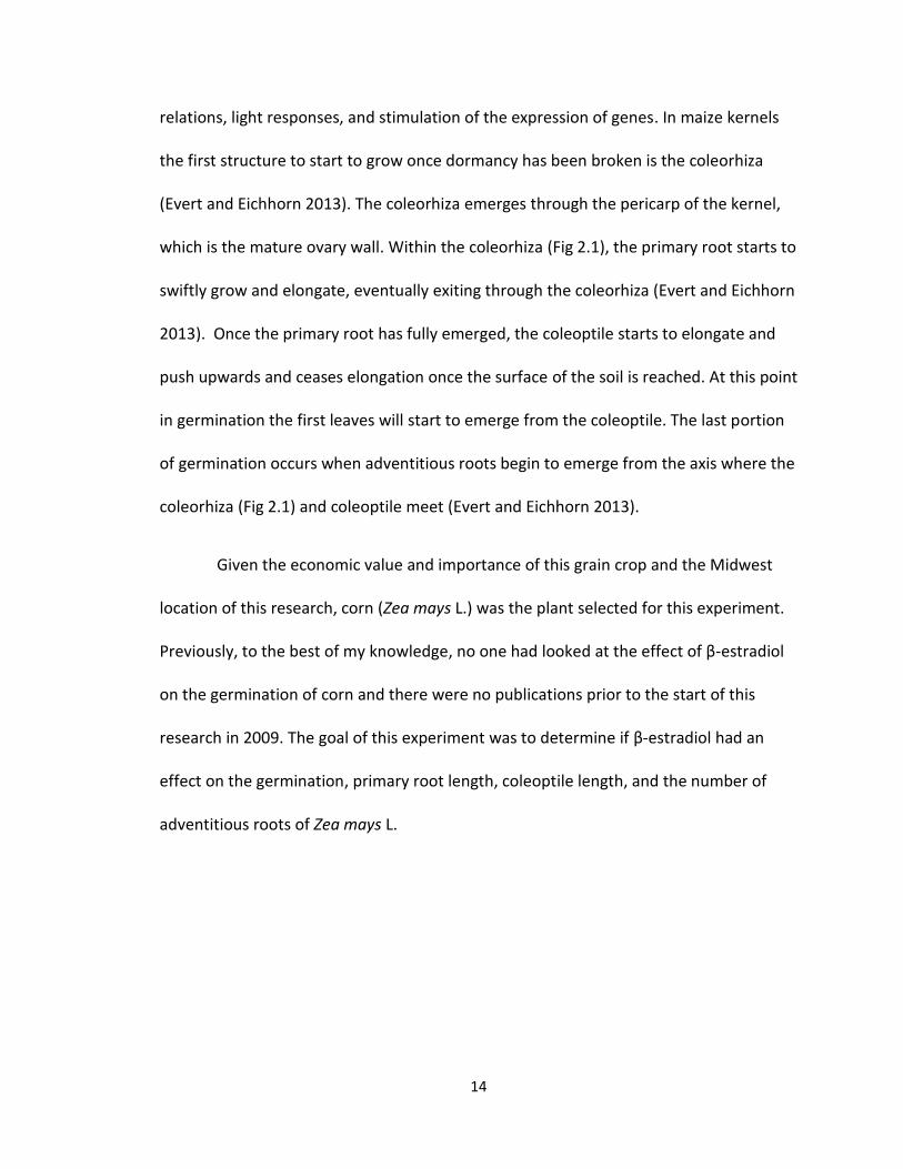
relations, light responses, and stimulation of the expression of genes. In maize kernels
the first structure to start to grow once dormancy has been broken is the coleorhiza
(Evert and Eichhorn 2013). The coleorhiza emerges through the pericarp of the kernel,
which is the mature ovary wall. Within the coleorhiza (Fig 2.1), the primary root starts to
swiftly grow and elongate, eventually exiting through the coleorhiza (Evert and Eichhorn
2013). Once the primary root has fully emerged, the coleoptile starts to elongate and
push upwards and ceases elongation once the surface of the soil is reached. At this point
in germination the first leaves will start to emerge from the coleoptile. The last portion
of germination occurs when adventitious roots begin to emerge from the axis where the
coleorhiza (Fig 2.1) and coleoptile meet (Evert and Eichhorn 2013).
Given the economic value and importance of this grain crop and the Midwest
location of this research, corn (Zea mays L.) was the plant selected for this experiment.
Previously, to the best of my knowledge, no one had looked at the effect of β-estradiol
on the germination of corn and there were no publications prior to the start of this
research in 2009. The goal of this experiment was to determine if β-estradiol had an
effect on the germination, primary root length, coleoptile length, and the number of
adventitious roots of Zea mays L.
15
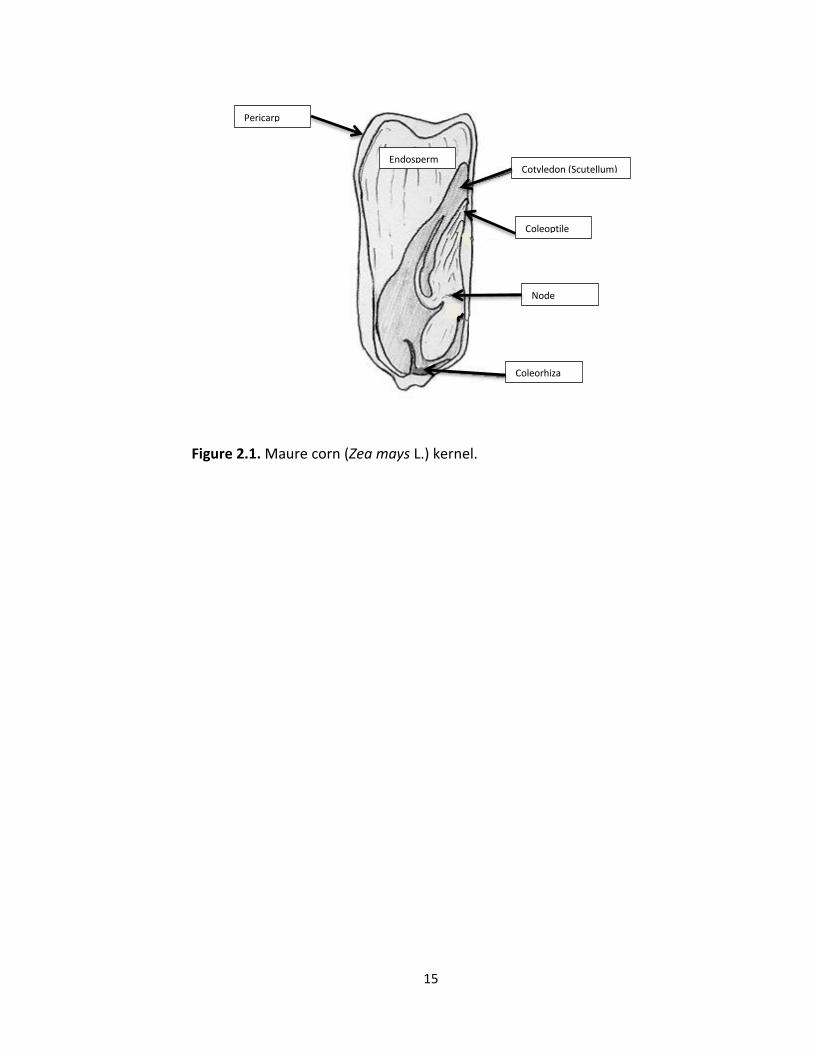
Figure 2.1. Maure corn (Zea mays L.) kernel.
Endosperm Cotyledon (Scutellum)
Endosperm
Coleoptile
Node
Pericarp
Coleorhiza
16
Materials and Methods
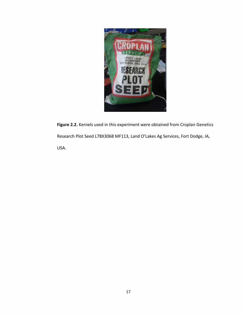
Corn kernels (Zea mays L.)(Croplan Genetics Research Plot Seed
L78X3068 MF113, Land O’Lakes Ag Services, Fort Dodge, IA, USA) (Fig 2.2) were surface
sterilized in a 10% household bleach solution (Clorox Bleach) for 20 minutes at room
temperature, and then rinsed several times with sterile deionized water. Kernels were
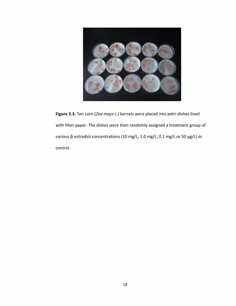
then placed randomly into groups of ten and using a random number generator
assigned to one of 25 petri dishes (Fig 2.3) lined with filter paper. Ten mg of β-estradiol
(Sigma-Aldrich Co., St. Louis, MO, USA) was dissolved in 1 mL of 95% ethanol and diluted
with sterile water to create concentrations of 10 mg/L, 1.0 mg/L, 0.1 mg/L and 50 μg/L.
Each of the 25 petri dishes was then randomly assigned to one of these four
concentrations or a control (sterile water). Fifteen mL of the assigned concentration was
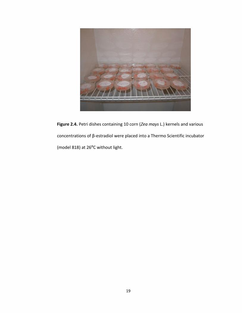
added to each dish. Petri dishes were then placed into an incubator (Thermo Scientific
model 818) without light and incubated at 26 ⁰C (Fig 2.4). After 24 hours, the dishes
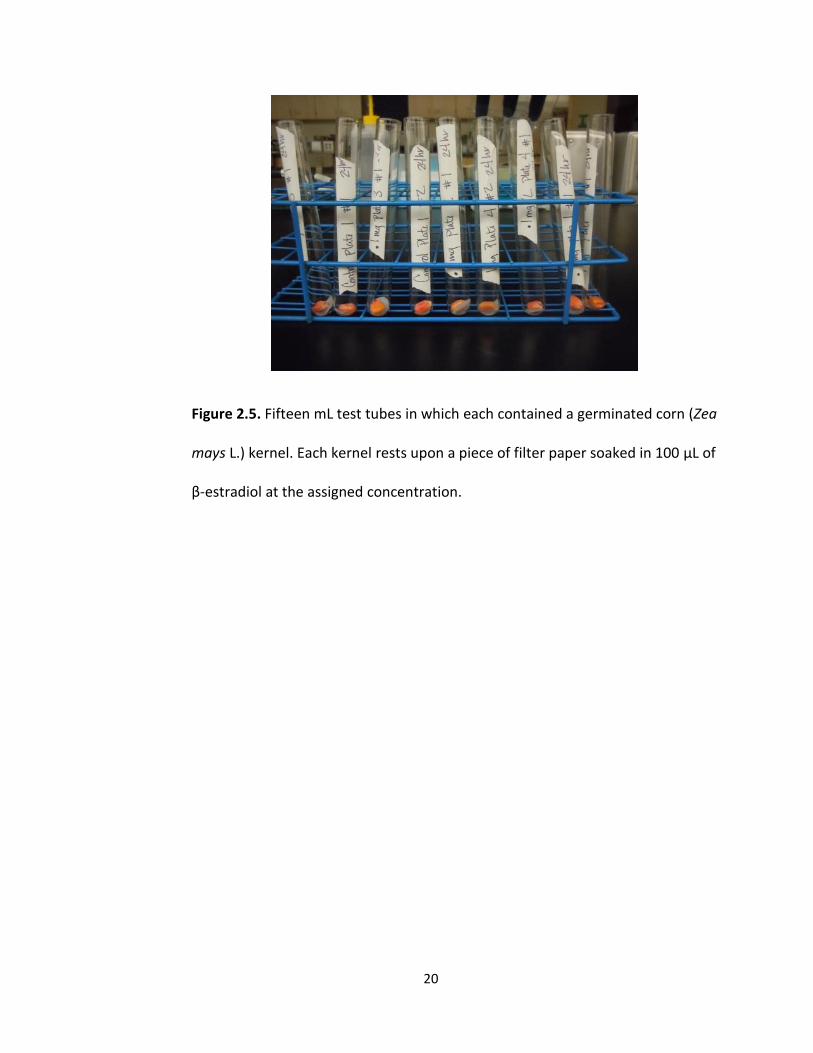
were surveyed for kernels that had germinated. All germinated kernels were removed
from petri dishes and placed into individual 15 ml test tubes in which a small circular
piece of filter paper had been placed at the bottom along with 100 μL of β-estradiol at
the assigned concentration (Fig 2.5). Germination was determined to be when the
primary root began exiting the coleorhiza (Fig 2.6). Test tubes were then placed back
into the incubator and kernels were allowed to grow for an additional five days. Petri
dishes were also checked at 48, 72, and 96 hours and each time any kernels germinated
they were put into test tubes as described above and placed back into the incubator for
an additional five days.
17
Figure 2.2. Kernels used in this experiment were obtained from Croplan Genetics
Research Plot Seed L78X3068 MF113, Land O’Lakes Ag Services, Fort Dodge, IA,
USA.
18
Figure 2.3. Ten corn (Zea mays L.) kernels were placed into petri dishes lined
with filter paper. The dishes were then randomly assigned a treatment group of
various β-estradiol concentrations (10 mg/L, 1.0 mg/L, 0.1 mg/L or 50 μg/L) or
control.
19
Figure 2.4. Petri dishes containing 10 corn (Zea mays L.) kernels and various
concentrations of β-estradiol were placed into a Thermo Scientific incubator
(model 818) at 26⁰C without light.
20
Figure 2.5. Fifteen mL test tubes in which each contained a germinated corn (Zea
mays L.) kernel. Each kernel rests upon a piece of filter paper soaked in 100 μL of
β-estradiol at the assigned concentration.
21
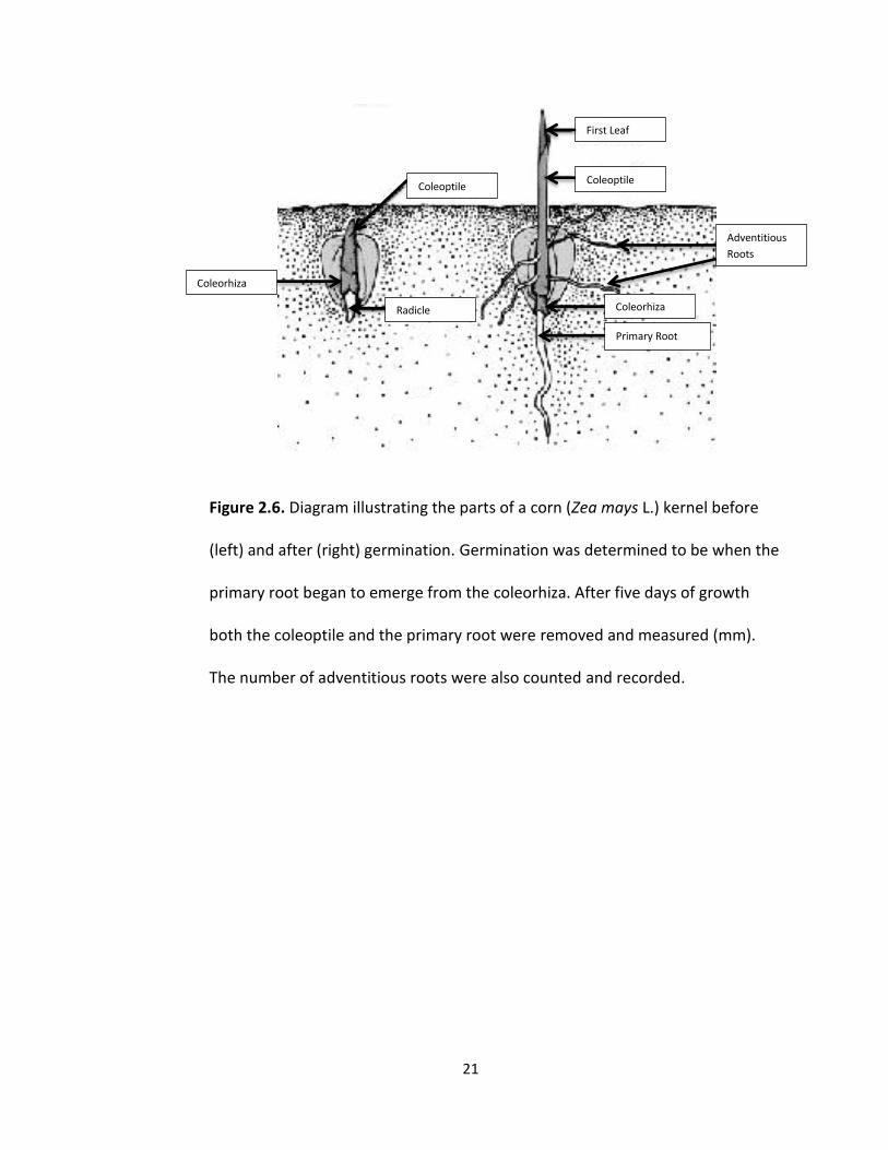
Figure 2.6. Diagram illustrating the parts of a corn (Zea mays L.) kernel before
(left) and after (right) germination. Germination was determined to be when the
primary root began to emerge from the coleorhiza. After five days of growth
both the coleoptile and the primary root were removed and measured (mm).
The number of adventitious roots were also counted and recorded.
Coleoptile
Radicle
Coleoptile
Adventitious
Roots
Primary Root
Coleorhiza
First Leaf
Coleorhiza
22
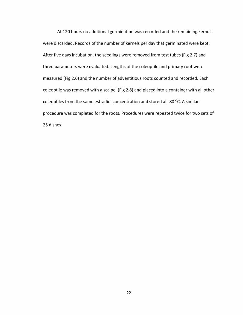
At 120 hours no additional germination was recorded and the remaining kernels
were discarded. Records of the number of kernels per day that germinated were kept.
After five days incubation, the seedlings were removed from test tubes (Fig 2.7) and
three parameters were evaluated. Lengths of the coleoptile and primary root were
measured (Fig 2.6) and the number of adventitious roots counted and recorded. Each
coleoptile was removed with a scalpel (Fig 2.8) and placed into a container with all other
coleoptiles from the same estradiol concentration and stored at -80 ⁰C. A similar
procedure was completed for the roots. Procedures were repeated twice for two sets of
25 dishes.
23
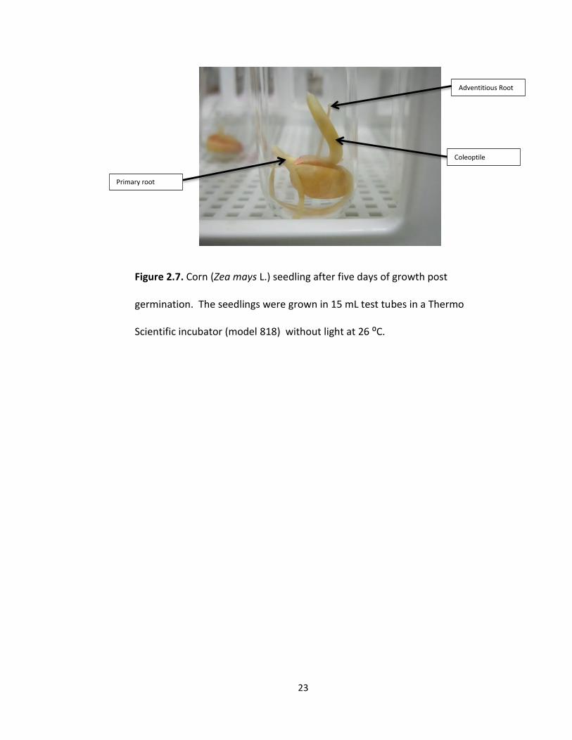
Figure 2.7. Corn (Zea mays L.) seedling after five days of growth post
germination. The seedlings were grown in 15 mL test tubes in a Thermo
Scientific incubator (model 818) without light at 26 ⁰C.
Coleoptile
Adventitious Root
Primary root
24
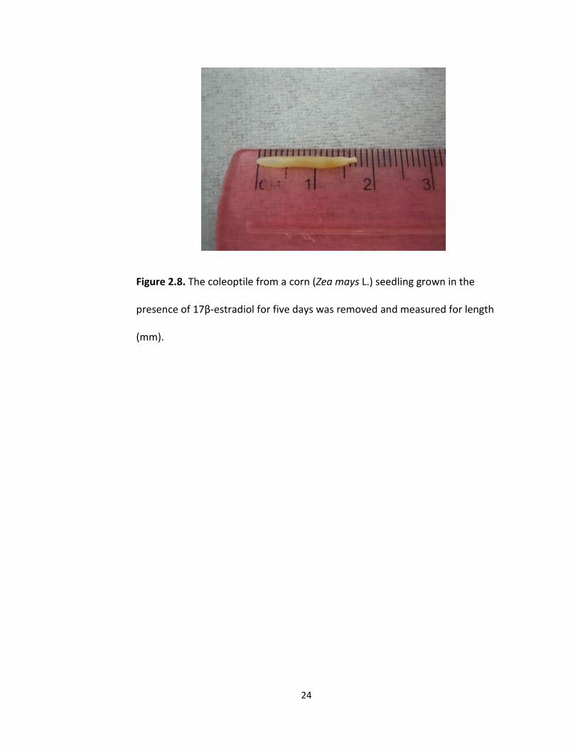
Figure 2.8. The coleoptile from a corn (Zea mays L.) seedling grown in the
presence of 17β-estradiol for five days was removed and measured for length
(mm).
25
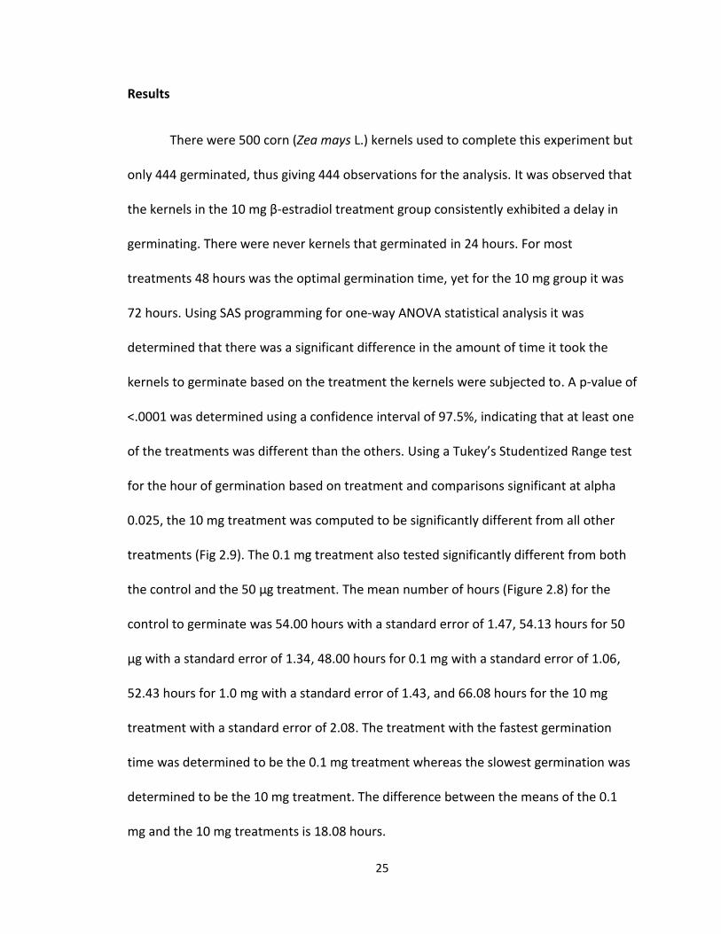
Results
There were 500 corn (Zea mays L.) kernels used to complete this experiment but
only 444 germinated, thus giving 444 observations for the analysis. It was observed that
the kernels in the 10 mg β-estradiol treatment group consistently exhibited a delay in
germinating. There were never kernels that germinated in 24 hours. For most
treatments 48 hours was the optimal germination time, yet for the 10 mg group it was
72 hours. Using SAS programming for one-way ANOVA statistical analysis it was
determined that there was a significant difference in the amount of time it took the
kernels to germinate based on the treatment the kernels were subjected to. A p-value of
<.0001 was determined using a confidence interval of 97.5%, indicating that at least one
of the treatments was different than the others. Using a Tukey’s Studentized Range test
for the hour of germination based on treatment and comparisons significant at alpha
0.025, the 10 mg treatment was computed to be significantly different from all other
treatments (Fig 2.9). The 0.1 mg treatment also tested significantly different from both
the control and the 50 μg treatment. The mean number of hours (Figure 2.8) for the
control to germinate was 54.00 hours with a standard error of 1.47, 54.13 hours for 50
μg with a standard error of 1.34, 48.00 hours for 0.1 mg with a standard error of 1.06,
52.43 hours for 1.0 mg with a standard error of 1.43, and 66.08 hours for the 10 mg
treatment with a standard error of 2.08. The treatment with the fastest germination
time was determined to be the 0.1 mg treatment whereas the slowest germination was
determined to be the 10 mg treatment. The difference between the means of the 0.1
mg and the 10 mg treatments is 18.08 hours.
26
Figure 2.9. The mean number of hours for 444 corn kernels (Zea mays L.) subjected to
different β-estradiol concentrations to germinate. Bars denote a 97.5% confidence
interval.
0
10
20
30
40
50
60
70
80
Control 50 μg 0.1 mg 1 mg 10 mg
Me
an N
um
be
r o
f H
ou
rs t
o G
erm
inat
e
Estradiol Concentration
Mean Number of Hours for Germination of Corn Kernels Based
on Treatment with Estradiol
27
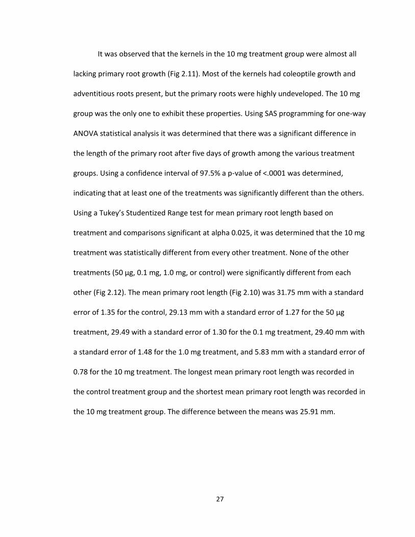
It was observed that the kernels in the 10 mg treatment group were almost all
lacking primary root growth (Fig 2.11). Most of the kernels had coleoptile growth and
adventitious roots present, but the primary roots were highly undeveloped. The 10 mg
group was the only one to exhibit these properties. Using SAS programming for one-way
ANOVA statistical analysis it was determined that there was a significant difference in
the length of the primary root after five days of growth among the various treatment
groups. Using a confidence interval of 97.5% a p-value of <.0001 was determined,
indicating that at least one of the treatments was significantly different than the others.
Using a Tukey’s Studentized Range test for mean primary root length based on
treatment and comparisons significant at alpha 0.025, it was determined that the 10 mg
treatment was statistically different from every other treatment. None of the other
treatments (50 μg, 0.1 mg, 1.0 mg, or control) were significantly different from each
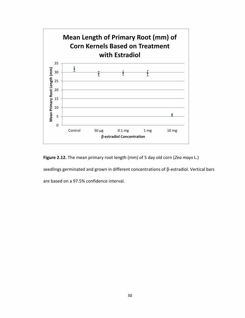
other (Fig 2.12). The mean primary root length (Fig 2.10) was 31.75 mm with a standard
error of 1.35 for the control, 29.13 mm with a standard error of 1.27 for the 50 μg
treatment, 29.49 with a standard error of 1.30 for the 0.1 mg treatment, 29.40 mm with
a standard error of 1.48 for the 1.0 mg treatment, and 5.83 mm with a standard error of
0.78 for the 10 mg treatment. The longest mean primary root length was recorded in
the control treatment group and the shortest mean primary root length was recorded in
the 10 mg treatment group. The difference between the means was 25.91 mm.
28
A. B.
C. D.
Figure 2.10. Corn (Zea mays L.) kernels from the 10 mg β-estradiol treatment group
after five days of growth with noticeable lack of primary root growth while exhibiting
coleoptile (→) growth (A, B, C). A kernel from the 10 mg treatment group that
germinated then proceeded to degenerate and die during the five days of growth in the
15 mL test tube (D).
29
A. B.
C. D.
Figure 2.11. Corn (Zea mays L.) kernels germinated in the presence of various
concentrations of β-estradiol (A,B,C). Kernel from the 1.0 mg β-estradiol
treatment group (A). Kernel from the 0.1 mg treatment group (B). Kernel from
the 50 μg treatment group (C). Kernel from the control group (D). Note that all
kernels have a primary root (→).
30
Figure 2.12. The mean primary root length (mm) of 5 day old corn (Zea mays L.)
seedlings germinated and grown in different concentrations of β-estradiol. Vertical bars
are based on a 97.5% confidence interval.
0
5
10
15
20
25
30
35
Control 50 μg 0.1 mg 1 mg 10 mg
Me
an P
rim
ary
Ro
ot
Len
gth
(m
m)
β-estradiol Concentration
Mean Length of Primary Root (mm) of Corn Kernels Based on Treatment
with Estradiol
31
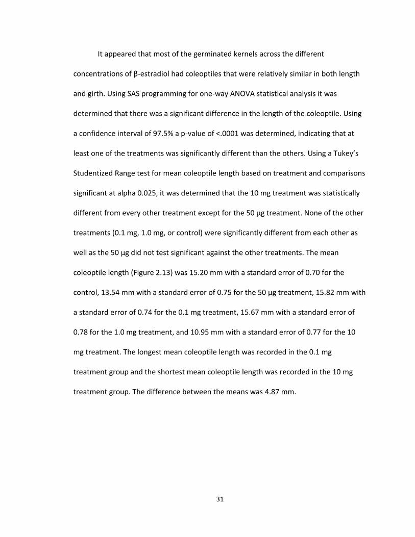
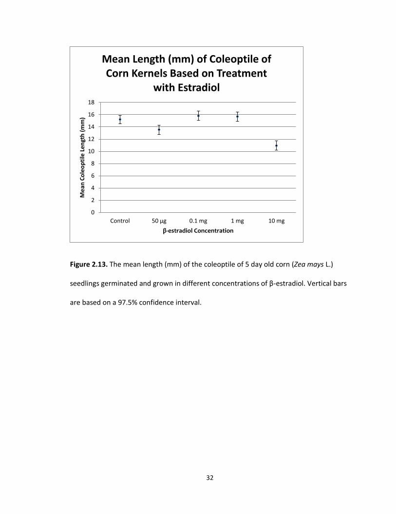
It appeared that most of the germinated kernels across the different
concentrations of β-estradiol had coleoptiles that were relatively similar in both length
and girth. Using SAS programming for one-way ANOVA statistical analysis it was
determined that there was a significant difference in the length of the coleoptile. Using
a confidence interval of 97.5% a p-value of <.0001 was determined, indicating that at
least one of the treatments was significantly different than the others. Using a Tukey’s
Studentized Range test for mean coleoptile length based on treatment and comparisons
significant at alpha 0.025, it was determined that the 10 mg treatment was statistically
different from every other treatment except for the 50 μg treatment. None of the other
treatments (0.1 mg, 1.0 mg, or control) were significantly different from each other as
well as the 50 μg did not test significant against the other treatments. The mean
coleoptile length (Figure 2.13) was 15.20 mm with a standard error of 0.70 for the
control, 13.54 mm with a standard error of 0.75 for the 50 μg treatment, 15.82 mm with
a standard error of 0.74 for the 0.1 mg treatment, 15.67 mm with a standard error of
0.78 for the 1.0 mg treatment, and 10.95 mm with a standard error of 0.77 for the 10
mg treatment. The longest mean coleoptile length was recorded in the 0.1 mg
treatment group and the shortest mean coleoptile length was recorded in the 10 mg
treatment group. The difference between the means was 4.87 mm.
32
Figure 2.13. The mean length (mm) of the coleoptile of 5 day old corn (Zea mays L.)
seedlings germinated and grown in different concentrations of β-estradiol. Vertical bars
are based on a 97.5% confidence interval.
0
2
4
6
8
10
12
14
16
18
Control 50 μg 0.1 mg 1 mg 10 mg
Me
an C
ole
op
tile
Le
ngt
h (
mm
)
β-estradiol Concentration
Mean Length (mm) of Coleoptile of Corn Kernels Based on Treatment
with Estradiol
33
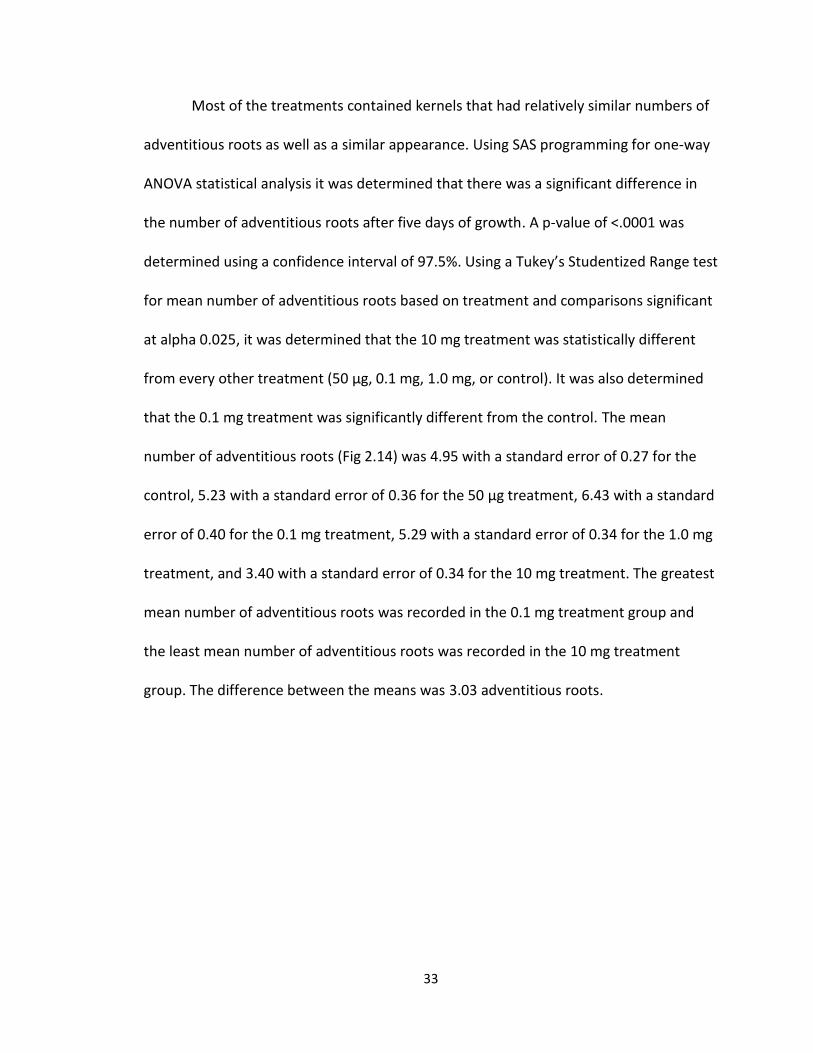
Most of the treatments contained kernels that had relatively similar numbers of
adventitious roots as well as a similar appearance. Using SAS programming for one-way
ANOVA statistical analysis it was determined that there was a significant difference in
the number of adventitious roots after five days of growth. A p-value of <.0001 was
determined using a confidence interval of 97.5%. Using a Tukey’s Studentized Range test
for mean number of adventitious roots based on treatment and comparisons significant
at alpha 0.025, it was determined that the 10 mg treatment was statistically different
from every other treatment (50 μg, 0.1 mg, 1.0 mg, or control). It was also determined
that the 0.1 mg treatment was significantly different from the control. The mean
number of adventitious roots (Fig 2.14) was 4.95 with a standard error of 0.27 for the
control, 5.23 with a standard error of 0.36 for the 50 μg treatment, 6.43 with a standard
error of 0.40 for the 0.1 mg treatment, 5.29 with a standard error of 0.34 for the 1.0 mg
treatment, and 3.40 with a standard error of 0.34 for the 10 mg treatment. The greatest
mean number of adventitious roots was recorded in the 0.1 mg treatment group and
the least mean number of adventitious roots was recorded in the 10 mg treatment
group. The difference between the means was 3.03 adventitious roots.
34
Figure 2.14. The mean number of adventitious roots of 5 day old corn (Zea mays L.)
seedlings germinated and grown in different concentrations of β-estradiol. Vertical bars
are based on a 97.5% confidence interval.
0
1
2
3
4
5
6
7
8
Control 50 μg 0.1 mg 1 mg 10 mg
Me
an N
um
be
r o
f A
dve
nti
tio
us
Ro
ots
β-estradiol Concentration
Mean Number of Adventitious Roots of Corn Kernels Based on Treatment
with Estradiol
35
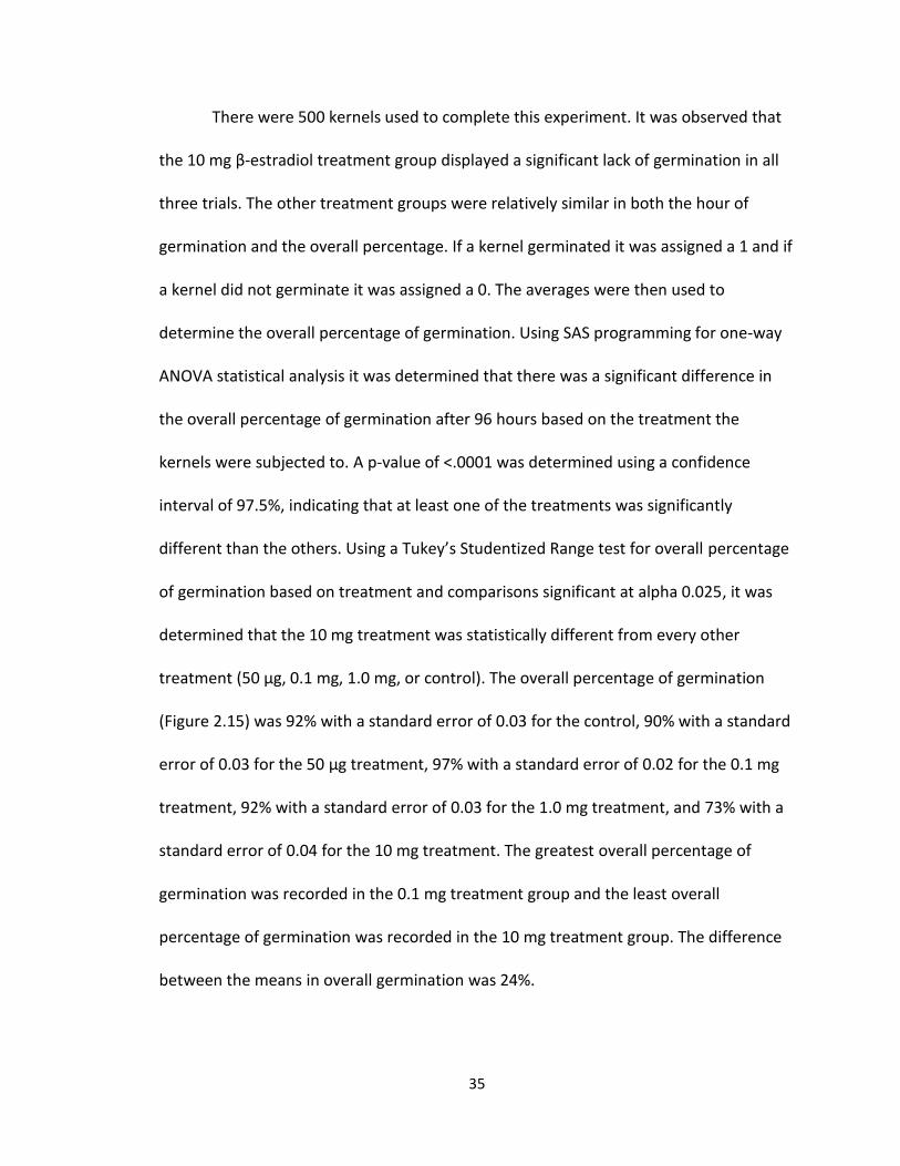
There were 500 kernels used to complete this experiment. It was observed that
the 10 mg β-estradiol treatment group displayed a significant lack of germination in all
three trials. The other treatment groups were relatively similar in both the hour of
germination and the overall percentage. If a kernel germinated it was assigned a 1 and if
a kernel did not germinate it was assigned a 0. The averages were then used to
determine the overall percentage of germination. Using SAS programming for one-way
ANOVA statistical analysis it was determined that there was a significant difference in
the overall percentage of germination after 96 hours based on the treatment the
kernels were subjected to. A p-value of <.0001 was determined using a confidence
interval of 97.5%, indicating that at least one of the treatments was significantly
different than the others. Using a Tukey’s Studentized Range test for overall percentage
of germination based on treatment and comparisons significant at alpha 0.025, it was
determined that the 10 mg treatment was statistically different from every other
treatment (50 μg, 0.1 mg, 1.0 mg, or control). The overall percentage of germination
(Figure 2.15) was 92% with a standard error of 0.03 for the control, 90% with a standard
error of 0.03 for the 50 μg treatment, 97% with a standard error of 0.02 for the 0.1 mg
treatment, 92% with a standard error of 0.03 for the 1.0 mg treatment, and 73% with a
standard error of 0.04 for the 10 mg treatment. The greatest overall percentage of
germination was recorded in the 0.1 mg treatment group and the least overall
percentage of germination was recorded in the 10 mg treatment group. The difference
between the means in overall germination was 24%.
36
Figure 2.15. The overall percentage of germination after 96 hours in various
concentrations of β-estradiol. A corn (Zea mays L.) kernel that germinated was assigned
a 1 and a kernel that did not germinate was assigned a 0. Vertical bars are based on a
97.5% confidence interval.
70
75
80
85
90
95
100
Control 50 μg 0.1 mg 1 mg 10 mg
Me
an P
erc
en
t G
erm
inat
ion
β-estradiol Concentration
Percent of Germination of Corn Kernels Based on Treatment with
Estradiol
37
Discussion
According to the results, β-estradiol has a significant effect on the germination of
Zea mays L. kernels. At the highest concentration of the hormone, 10 mg/mL, the
overall germination was reduced to 73%. This indicates that at some level β-estradiol is
toxic to the kernels at high concentrations. At smaller concentrations it appears that β-
estradiol causes a slight escalation in overall germination, 97% in 0.1 mg when
compared to the control at 92%. Germination is a key phase in the initiation of plant
growth. If germination is being inhibited by high concentrations of the hormone, this
could indicate that kernels sown in fields that are exposed to contaminated water or
soil, may have reduced germination rates and therefore reduced overall yield. It can also
be established that high levels of β-estradiol appears to cause a significant delay in the
germination process. This can be seen in the results above which shows the 10 mg
treatment group taking on average 66 hours to germinate, while kernels that were
exposed to the 0.1 mg treatment group took on average 48 hours to germinate
compared to the control that averaged 54 hours.
These results seem to fall in line with the plant species Lactuca sativa L. (lettuce)
and Lycopersicon esculentum Mill. (tomato) which also exhibited a reduction in the
overall rate of germination when exposed to β-estradiol (D’Abrosca et al 2008).
However, the results are at odds with the outcomes of experiments previously
performed on both Cicer arietinum L. (chickpea) and Daucus carota L. (carrot) which
both displayed an increase in the overall rates of germination (Erdal and Dumlupinar
38
2010). Both L. sativa and L. esculentum are dicots, which demonstrated the same results
as Z. mays which is a monocot. Therefore it cannot be assumed that the reason that
there was a difference was due to a difference in monocots versus dicots (D’Abrosca et
al 2008). Legumes such as C. arietinum are known to have high levels of endogenous
compounds known as phytoestrogens (Erdal and Dumlupinar 2010). These compounds
are extremely close in chemical structure and nature to β-estradiol. The influence on
germination may be positive due to that close connection and increased binding of
phytoestrogen receptors with β-estradiol. The negative effect of β-estradiol on the corn
kernels observed in this experiment would indicate that the hormone is inhibiting the
mechanisms of germination either by binding to receptors or in some other way down-
regulating the gene expression for cellular components involved in the process.
Additionally, it has been also been suggested that these steroids might be involved in a
disturbance of the Na/K balance of plant cells (Agarwal 1993).
It was also observed in the results that β-estradiol had a significant effect on the
primary root development. The average length of the primary root after 5 days of
growth after germination in the 10 mg treatment group was only 5.83 mm compared to
31.75 mm in the control group. This same phenomenon was observed in both H. annuus
and S. lycopersicum (Janeczko and Skoczowski 2005). This may illustrate that exposure
to high concentrations of β-estradiol could inhibit the functioning of the primary root
apical meristem. The process of cell elongation within the root could also be affected,
which is a primary means for increasing length (Taiz and Zieger 2006). Adventitious
roots begin to grow after the primary, so perhaps by this point in time the β-estradiol
39
had denatured to a point where it was no longer causing inhibition of root growth.
Several seedlings exposed to the highest concentration of β-estradiol from this portion
of the experiment would exhibit regular coleoptile growth, but would have severely
limited or no root growth. This indicates that the coleoptile growth is not hindered quite
like the primary root. This could perhaps come from the fact that the primary root is the
first to emerge followed by the coleoptile, about one day later (Nielson 2010). With the
initial imbibition of water containing β-estradiol directed towards root development,
the coleoptile may not be as directly affected. The results of this experiment also
showed that there was a significant reduction in the coleoptile length as well as in the
number of adventitious roots of the 10 mg treatment goup. While statistically
significant, from a purely observational standpoint these did not appear as starkly
contrasting when compared to the other treatment groups as the primary root
measurements did.
It should also be noted that in the dishes containing the highest concentrations
of β-estradiol there was occasional mold growth even though the procedures were
carried out under as sterile conditions as possible. This may be indicative of a
relationship between fungi and β-estradiol. Previous reports have shown that in some
instances fungal growth can be stimulated by the hormone (Stoka 1999). The growth of
mold was not observed in any other dishes.
40
Chapter 3: The effect of β-estradiol on growth of corn (Zea mays L.)
In mammals, β-estradiol plays a key role in controlling the processes revolving
around development and reproduction as well as being involved in the control of both
mineral and protein metabolism (Carreau et al. 2002). Knowing that this hormone is a
contaminant in several key water sources for plants, the question arises whether or not
there is also an effect upon plant growth and development (Islam and Tanaka 2004).
Several studies have been carried out on different plant species in order to determine
this.
In sunflower seedlings (Helianthus annuus L.), β-estradiol concentrations of 1 μg
per plant increased overall shoot growth, but was shown to inhibit overall root growth
(Janeczko and Skoczowski 2005). In tomato (Lycopersicon esculentum Mill.) seedlings
the hormone was shown to reduce overall root growth as well as the overall number of
roots present in nutrient solutions with a 1 μM concentration of β-estradiol (Janeczko
and Skoczowski 2005). In alfalfa plants (Medicago sativa L.) the lower concentrations of
β-estradiol used (0.005-0.5 μg.dm-3) in the experiment favored increased growth, while
the higher concentrations used (50-500 μg.dm-3) inhibited growth of both roots and
shoots (Janeczko and Skoczowski 2005). In chickpeas (Cicer arietinum L.), the hormone
significantly enhanced the root and shoot growth of the seedlings at concentrations of
10-4, 10-9, 10-12, and 10-15 M (Erdal and Dumlupinar 2010).
Thus, there appears to be a wide variation in the effects of β-estradiol on the
growth of different plant species. This suggests that how a plant responds depends on

41
the species of plant and the concentration of β-estradiol that the plant is exposed to.
The goal of this experiment was to determine if β-estradiol had an effect on the growth
of corn (Zea mays L.) seedlings.
42
Materials and Methods
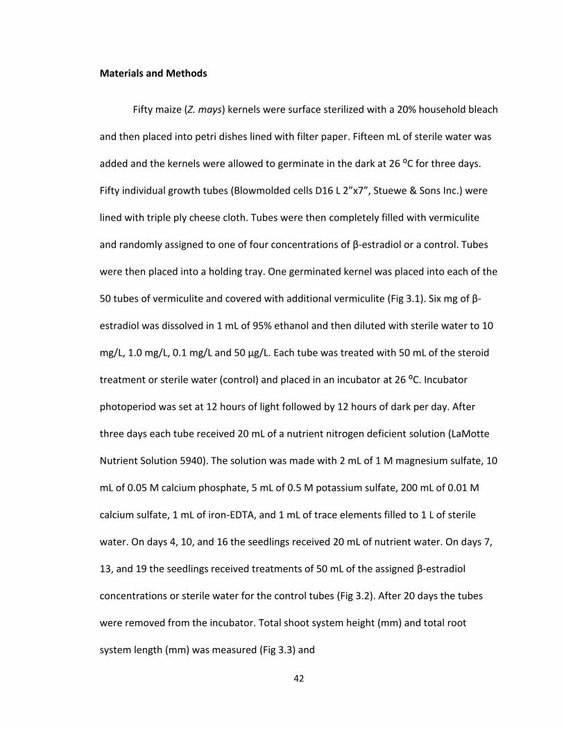
Fifty maize (Z. mays) kernels were surface sterilized with a 20% household bleach
and then placed into petri dishes lined with filter paper. Fifteen mL of sterile water was
added and the kernels were allowed to germinate in the dark at 26 ⁰C for three days.
Fifty individual growth tubes (Blowmolded cells D16 L 2”x7”, Stuewe & Sons Inc.) were
lined with triple ply cheese cloth. Tubes were then completely filled with vermiculite
and randomly assigned to one of four concentrations of β-estradiol or a control. Tubes
were then placed into a holding tray. One germinated kernel was placed into each of the
50 tubes of vermiculite and covered with additional vermiculite (Fig 3.1). Six mg of β-
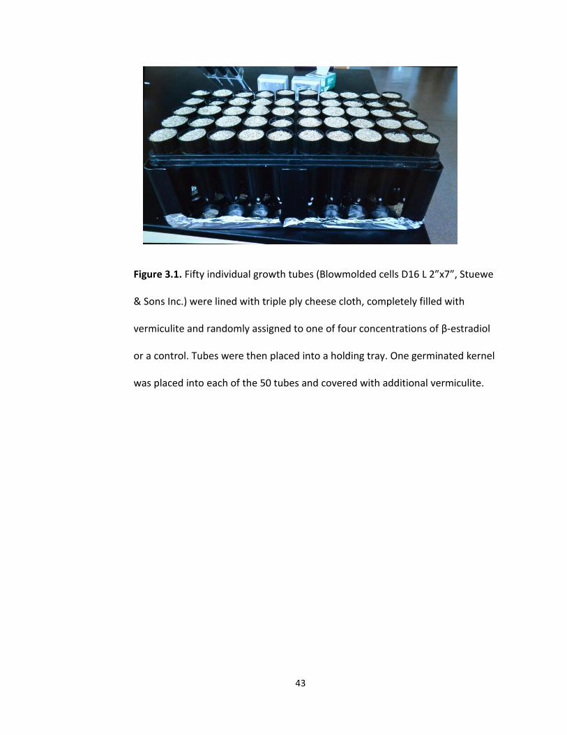
estradiol was dissolved in 1 mL of 95% ethanol and then diluted with sterile water to 10
mg/L, 1.0 mg/L, 0.1 mg/L and 50 μg/L. Each tube was treated with 50 mL of the steroid
treatment or sterile water (control) and placed in an incubator at 26 ⁰C. Incubator
photoperiod was set at 12 hours of light followed by 12 hours of dark per day. After
three days each tube received 20 mL of a nutrient nitrogen deficient solution (LaMotte
Nutrient Solution 5940). The solution was made with 2 mL of 1 M magnesium sulfate, 10
mL of 0.05 M calcium phosphate, 5 mL of 0.5 M potassium sulfate, 200 mL of 0.01 M
calcium sulfate, 1 mL of iron-EDTA, and 1 mL of trace elements filled to 1 L of sterile
water. On days 4, 10, and 16 the seedlings received 20 mL of nutrient water. On days 7,
13, and 19 the seedlings received treatments of 50 mL of the assigned β-estradiol
concentrations or sterile water for the control tubes (Fig 3.2). After 20 days the tubes
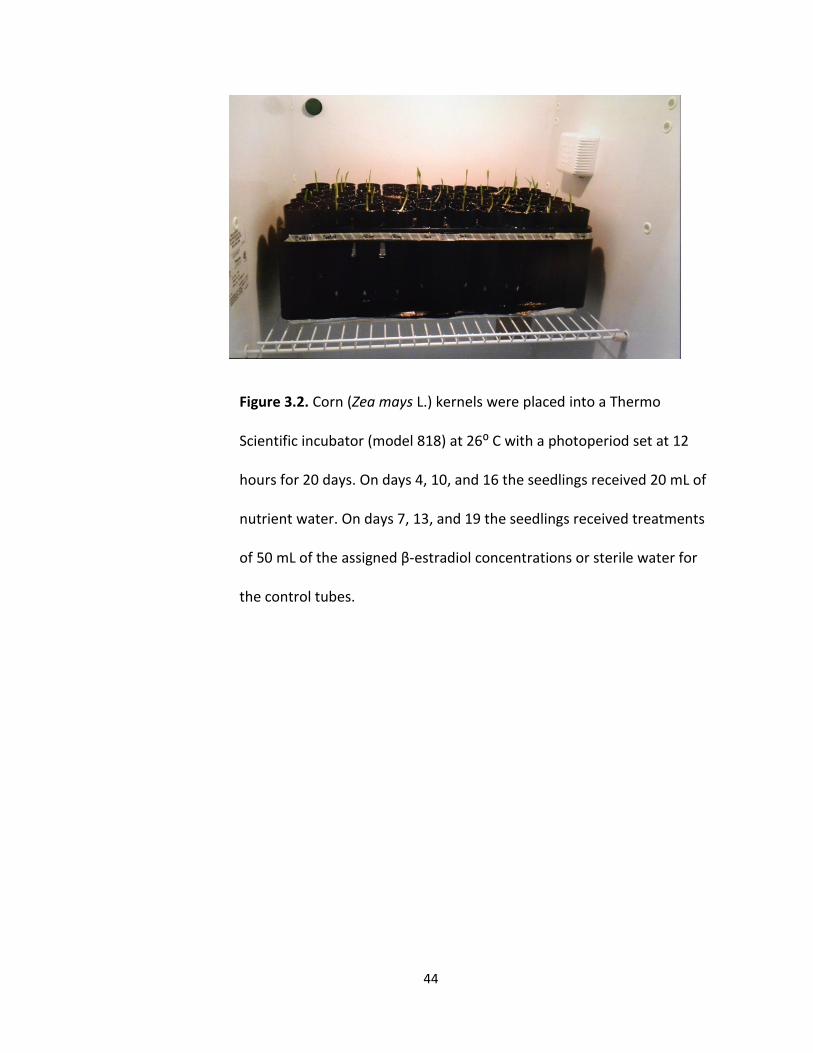
were removed from the incubator. Total shoot system height (mm) and total root
system length (mm) was measured (Fig 3.3) and
43
Figure 3.1. Fifty individual growth tubes (Blowmolded cells D16 L 2”x7”, Stuewe
& Sons Inc.) were lined with triple ply cheese cloth, completely filled with
vermiculite and randomly assigned to one of four concentrations of β-estradiol
or a control. Tubes were then placed into a holding tray. One germinated kernel
was placed into each of the 50 tubes and covered with additional vermiculite.
44
Figure 3.2. Corn (Zea mays L.) kernels were placed into a Thermo
Scientific incubator (model 818) at 26⁰ C with a photoperiod set at 12
hours for 20 days. On days 4, 10, and 16 the seedlings received 20 mL of
nutrient water. On days 7, 13, and 19 the seedlings received treatments
of 50 mL of the assigned β-estradiol concentrations or sterile water for
the control tubes.
45
Figure 3.3. The corn (Zea mays L.) kernels were removed from the incubator
after 20 days of growth in various concentrations of β-estradiol. Measurements
(mm) were taken of both the length of the entire shoot system as well as the
entire root system.
Adventitious (Prop Roots)
Primary Root
Shoot System
Root System
(including
lateral roots)

46
recorded. The total number of leaves was also recorded for each plant. The entire shoot
system of each plant was removed with a scalpel and placed into a container with all
other shoot systems from the same β-estradiol concentration and stored at -80 ⁰C.
Similar procedures were completed for roots. This entire procedure was repeated three
times for each concentration, resulting in 150 plants.
47
Results
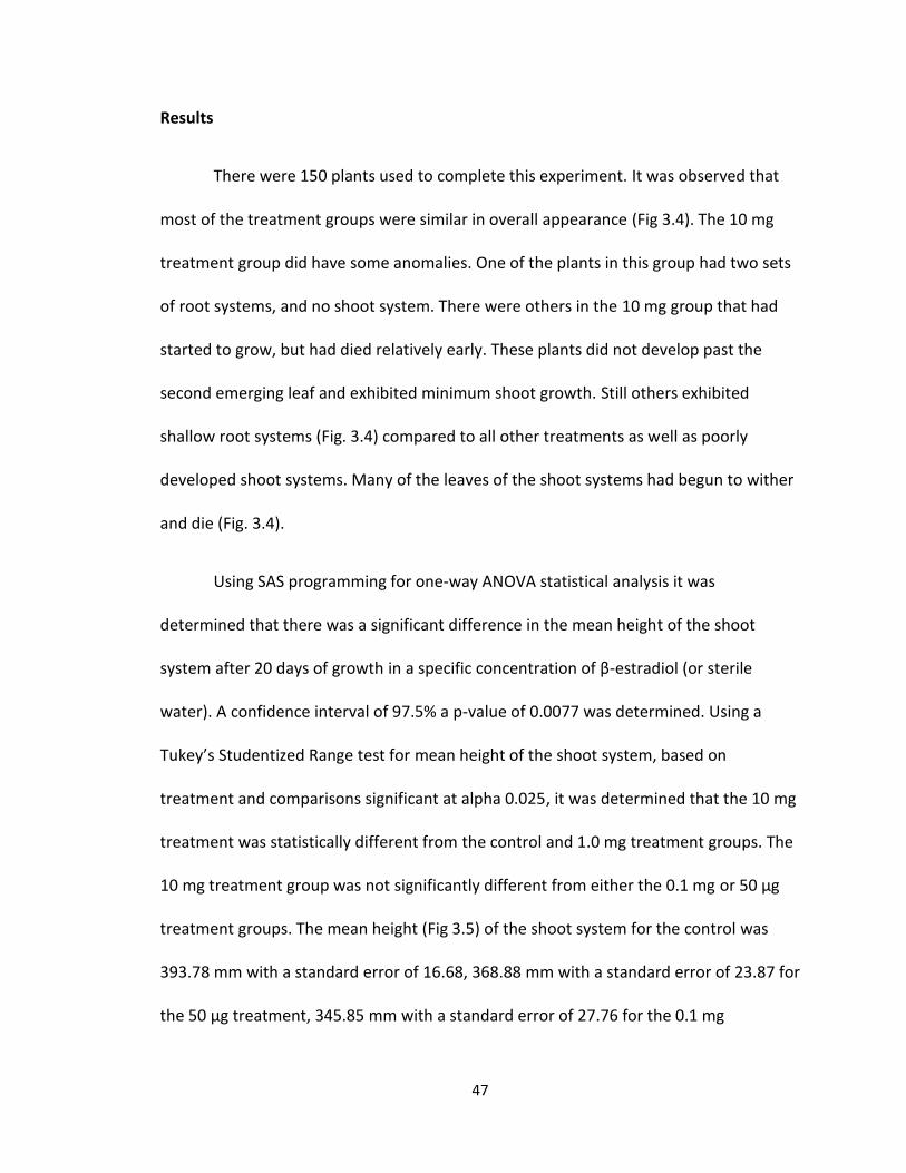
There were 150 plants used to complete this experiment. It was observed that
most of the treatment groups were similar in overall appearance (Fig 3.4). The 10 mg
treatment group did have some anomalies. One of the plants in this group had two sets
of root systems, and no shoot system. There were others in the 10 mg group that had
started to grow, but had died relatively early. These plants did not develop past the
second emerging leaf and exhibited minimum shoot growth. Still others exhibited
shallow root systems (Fig. 3.4) compared to all other treatments as well as poorly
developed shoot systems. Many of the leaves of the shoot systems had begun to wither
and die (Fig. 3.4).
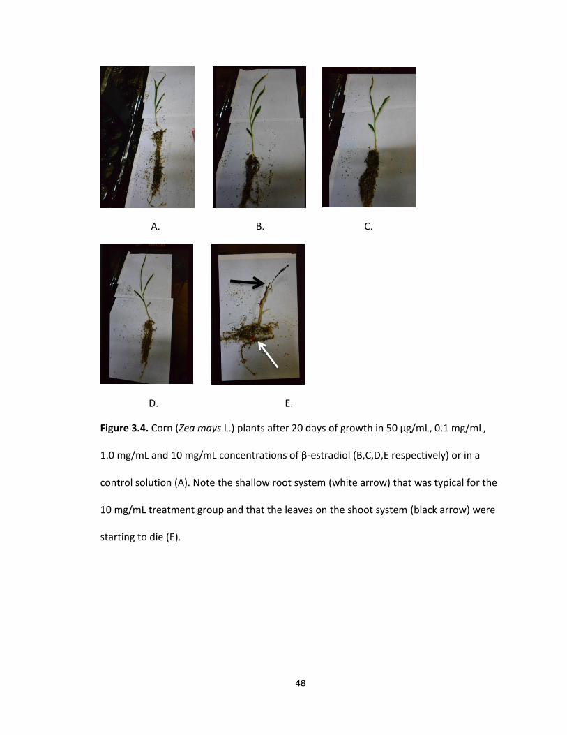
Using SAS programming for one-way ANOVA statistical analysis it was
determined that there was a significant difference in the mean height of the shoot
system after 20 days of growth in a specific concentration of β-estradiol (or sterile
water). A confidence interval of 97.5% a p-value of 0.0077 was determined. Using a
Tukey’s Studentized Range test for mean height of the shoot system, based on
treatment and comparisons significant at alpha 0.025, it was determined that the 10 mg
treatment was statistically different from the control and 1.0 mg treatment groups. The
10 mg treatment group was not significantly different from either the 0.1 mg or 50 μg
treatment groups. The mean height (Fig 3.5) of the shoot system for the control was
393.78 mm with a standard error of 16.68, 368.88 mm with a standard error of 23.87 for
the 50 μg treatment, 345.85 mm with a standard error of 27.76 for the 0.1 mg
48
A. B. C.
D. E.
Figure 3.4. Corn (Zea mays L.) plants after 20 days of growth in 50 μg/mL, 0.1 mg/mL,
1.0 mg/mL and 10 mg/mL concentrations of β-estradiol (B,C,D,E respectively) or in a
control solution (A). Note the shallow root system (white arrow) that was typical for the
10 mg/mL treatment group and that the leaves on the shoot system (black arrow) were
starting to die (E).
49
Figure 3.5. The mean shoot system height of corn (Zea mays L.) seedlings after 20 days
of growth in various concentrations of β-estradiol. There are 30 plants in each treatment
group for a total of 150 plants. The vertical bars are based on a 97.5% confidence
interval.
0
50
100
150
200
250
300
350
400
450
Control 50 μg 0.1 mg 1 mg 10 mg
Me
an P
lan
t H
eig
ht
(mm
)
β-estradiol Concentration
Mean Height of Plants Based on Treatment with Estradiol
50
treatment, 400.2 mm with a standard error of 9.18 for the 1.0 mg treatment, and 296.9
mm with a standard error of 25.83 for the 10 mg treatment. The greatest mean height
(mm) was recorded in the 1.0 mg treatment group and the least mean height (mm) was
recorded in the 10 mg treatment group. The greatest difference between the means in
plant height was 103.3 mm.
It was observed that the majority of plants had very similar root systems both in
overall structure and size. The only group that stood out visually from the rest and
exhibited a few irregularities was the 10 mg β-estradiol treatment group (Fig 3.4). The
roots of the 10 mg group were usually shorter in overall length and overall girth when
compared to the rest of the treatment groups. There was one seedling in the 10 mg
group that actually had two root systems and no shoot system. There were roots
coming out of both ends of the kernel. Using SAS programming for one-way ANOVA
statistical analysis it was determined that there was a significant difference in the mean
length of the entire root system after 20 days of growth in assigned β-estradiol
concentrations. Using a confidence interval of 97.5% a p-value of 0.0174 was
determined. Using a Tukey’s Studentized Range test for mean length of the entire root
system based on treatment and comparisons significant at alpha 0.025, it was
determined that the 10 mg treatment was statistically different from the 0.1 mg
treatment group. The 10 mg treatment group was not significantly different from any
other treatment groups. The 0.1 mg group was not significantly different from any other
treatment group. The mean length (Fig 3.6) of the entire root system (mm) was 151.7
mm with a standard error of 3.94 for the control, 156.7 mm with a standard error of
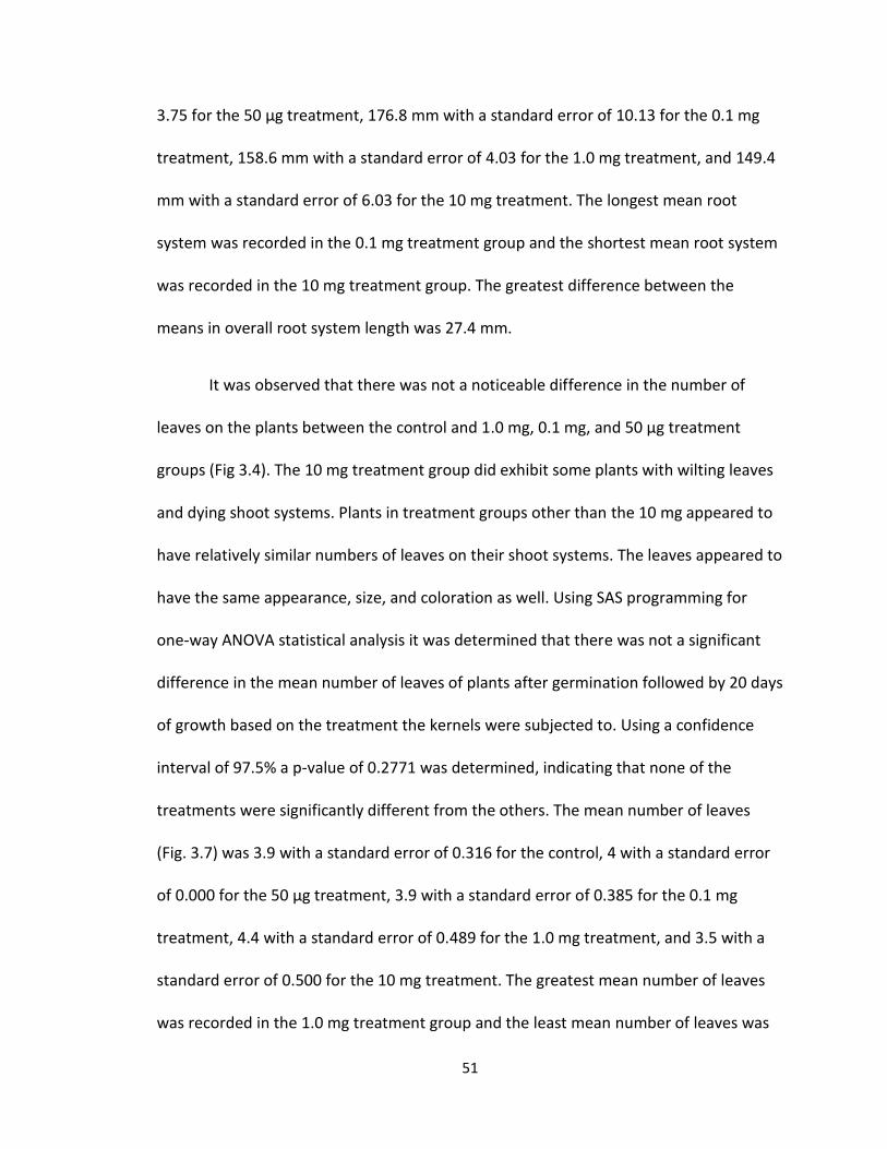
51
3.75 for the 50 μg treatment, 176.8 mm with a standard error of 10.13 for the 0.1 mg
treatment, 158.6 mm with a standard error of 4.03 for the 1.0 mg treatment, and 149.4
mm with a standard error of 6.03 for the 10 mg treatment. The longest mean root
system was recorded in the 0.1 mg treatment group and the shortest mean root system
was recorded in the 10 mg treatment group. The greatest difference between the
means in overall root system length was 27.4 mm.
It was observed that there was not a noticeable difference in the number of
leaves on the plants between the control and 1.0 mg, 0.1 mg, and 50 μg treatment
groups (Fig 3.4). The 10 mg treatment group did exhibit some plants with wilting leaves
and dying shoot systems. Plants in treatment groups other than the 10 mg appeared to
have relatively similar numbers of leaves on their shoot systems. The leaves appeared to
have the same appearance, size, and coloration as well. Using SAS programming for
one-way ANOVA statistical analysis it was determined that there was not a significant
difference in the mean number of leaves of plants after germination followed by 20 days
of growth based on the treatment the kernels were subjected to. Using a confidence
interval of 97.5% a p-value of 0.2771 was determined, indicating that none of the
treatments were significantly different from the others. The mean number of leaves
(Fig. 3.7) was 3.9 with a standard error of 0.316 for the control, 4 with a standard error
of 0.000 for the 50 μg treatment, 3.9 with a standard error of 0.385 for the 0.1 mg
treatment, 4.4 with a standard error of 0.489 for the 1.0 mg treatment, and 3.5 with a
standard error of 0.500 for the 10 mg treatment. The greatest mean number of leaves
was recorded in the 1.0 mg treatment group and the least mean number of leaves was
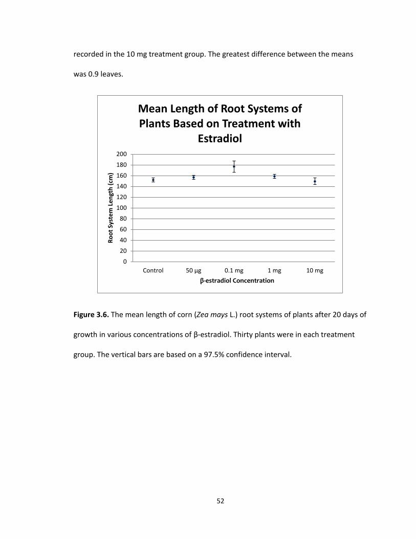
52
recorded in the 10 mg treatment group. The greatest difference between the means
was 0.9 leaves.
Figure 3.6. The mean length of corn (Zea mays L.) root systems of plants after 20 days of
growth in various concentrations of β-estradiol. Thirty plants were in each treatment
group. The vertical bars are based on a 97.5% confidence interval.
0
20
40
60
80
100
120
140
160
180
200
Control 50 μg 0.1 mg 1 mg 10 mg
Ro
ot
Syst
em
Le
ngt
h (
cm)
β-estradiol Concentration
Mean Length of Root Systems of Plants Based on Treatment with
Estradiol
53
Fig 3.7. The mean number of corn (Zea mays L.) leaves of plants after 20 days of growth
in various concentrations of β-estradiol. Thirty plants were in each treatment group. The
vertical bars are based on a 97.5% confidence interval.
0
1
2
3
4
5
6
Control 50 μg 0.1 mg 1 mg 10 mg
Me
an N
um
be
r o
f Le
ave
s
β-estradiol Concentration
Mean Number of Leaves of Plants Based on Treatment with Estradiol
54
Discussion
According to the results there was an effect on both the height of the shoot
system (mm) and the length of the root system (mm) of Zea mays L. due to exposure to
β-estradiol. The highest β-estradiol concentration used caused a significant decrease in
both the overall shoot height as well as the overall root system length. The 10 mg β-
estradiol treatment group had a mean overall shoot length of 296.9 mm which was
significantly lower than the control group at 393.78 mm. The 10 mg treatment group
had a mean overall root system length of 149.4 mm which was lower than all of the
other groups, and significantly lower than the 0.1 mg treatment group at 176.8 mm. This
appears to follow the same results that were found in studies in some other species of
plants. For example, in tomato seedlings (L. esculentum) β-estradiol concentrations of 1
μM was shown to reduce overall root growth as well as the overall number of roots
present (Janeczko and Skoczowski 2005). In alfalfa (M. sativa) plants, the lower
concentrations of β-estradiol used (0.005-0.5 μg.dm-3) in the experiment favored
increased growth, while the higher concentrations used (50-500 μg.dm-3) inhibited
growth of both roots and shoots (Janeczko and Skoczowski 2005).
The negative effect of β-estradiol on the corn plants observed in this experiment
was predominantly found within the 10 mg/mL concentration group and would indicate
that the hormone (at high concentrations) may be inhibiting the mechanisms of cell
elongation or cellular division either by binding to receptors or in some other way down-
regulating the gene expression for cellular components involved in the processes.
55
Previous studies have shown that phenolic compounds (such as β-estradiol) are well
established regulators of gene expression (Shore et. al 1992). The exact mechanism(s)
by which expression is regulated by β-estradiol is still under investigation. There are
over 8,000 known phenolic compounds in plants and their functions range from cell wall
structure, plant defense, color of woods and barks, flower color, and flavors within plant
tissues. Examples of some phenolic compounds commonly found in vascular plants
include flavonols, anthocyanins, tannins, lignins, and salicylic acid.
While the highest concentration of β-estradiol used (10 mg/mL) consistently had
negative impacts on various aspects of Zea mays L. seedling growth, the lower
concentrations (1.0 mg/mL, 0.1 mg/mL, and 50 μg/mL) were not statistically significant
from the control group. In fact, the longest mean root system length was recorded in
the 0.1 mg/mL treatment group, while the greatest mean shoot system height was
recorded in the 1.0 mg/mL treatment group. Although, there have been relatively few
studies conducted on the growth of plants in the presence of β-estradiol, it should be
noted that the plants used in previous studies were dicotyledonous species (H. annuus,
L. esculentum, M. sativa, C. arietinum). Zea mays L. is a monocotyledonous species.
There may be a difference in the physiology and pathways determining the growth of
tissues between dicotyledonous and monocotyledonous species, although this has not
yet been investigated.
It should also be noted that in the blow-molded cells containing the highest
concentrations (10 mg) of β-estradiol there was occasional mold growth even though
56
the procedures were carried out under as sterile conditions as possible. Three tubes in
this treatment group exhibited the mold growth. This is indicative of a relationship
between fungi and β-estradiol. Previous reports have shown that in some instances
fungal growth can be stimulated by the hormone (Stoka 1999). The growth of mold was
observed in one blow-molded cell in the 1.0 mg concentration as well, however, it was
never found in any of the other treatment groups.
57
Chapter 4: The effect of β-estradiol on chlorophyll concentration of corn (Zea mays L.)
The presence of chlorophyll in the chloroplasts of plants is what allows them to
be autotrophic , in that they are able to absorb light energy from the sun and convert it
to chemical energy (Campbell et al. 2011). The amount of chlorophyll present in plant
tissues can be a good indicator of how well the plant will be able to photosynthesize and
produce sugars as well as other carbohydrates, and therefore indicates how well a plant
may be able to grow. An increase or decrease in the amount of chlorophyll present
would have a direct influence on the amount of resources available for a plant to use for
growth. Producing new tissues will allow the plant to increase the height of the shoot
system, length of the root system, and eventually lead to flowering and seed production
(D’Abrosca et al. 2008).
Previous studies have been conducted to determine whether or not β-estradiol
had an effect on the amount of chlorophyll present in the tissues of various species of
plants. In Daucus carota L. the presence of β-estradiol at concentrations of 3-12 mg.dm-3
was shown to favor chlorophyll synthesis in leaves (Janeczko and Skoczowski 2005). The
same phenomenon of an increased production of chlorophyll was observed using β-
estradiol concentrations of 10-12 to 10-7 M in the algal cells of Chlorella vulgaris M.
Beijerinck (a type of green alga) that resulted in increased sugar and protein content in
these cells as well (Janeczko and Skoczowski 2005). In another study the presence of β-
estradiol at concentrations of 1 μM and 1 nM resulted in a 30-50% reduction of
chlorophylls and carotenoids in Lactuca sativa L. leaves which then led to a 50%
58
reduction in the sugar content (D’Abrosca et al. 2008). The results of these studies
indicate that the effects of β-estradiol on the amount of chlorophyll in plants and other
photosynthetic organisms depends on the specific species as well as the β-estradiol
concentration. The goal of this experiment was to determine what effect different
concentrations of β-estradiol would have on the amount of chlorophyll produced in Zea
mays L. leaves.
59
Materials and Methods
Samples for this experiment were taken from the leaves of Zea mays L. that had
been collected and frozen at -80 C⁰ after the experiment on the effects of β-estradiol on
growth (Chapter 3). The shoot systems were thawed and leaves from each individual
plant were collected until a 0.1 g sample was obtained. Chlorophyll extraction was
carried out according to the process outlined in Investigating Plant Physiology by
Camellia Okpudu (2001). Each 0.1 g tissue sample was placed into an individual 1.5 ml
Eppendorf tube along with 1 mL of anhydrous methanol (Sigma-Aldrich Co., St. Louis,
MO, USA). Once filled with anhydrous methanol, tubes were vortexed, allowed to sit for
30 minutes, and then vortexed again. Samples were then centrifuged at 10,000G for 10
minutes. Half of a mL of supernatant from each tube was collected and added to a new
Eppendorf tube and the volume was brought to 1 mL with distilled water. A
spectrophotometer (Biorad Smartspec Plus) was used to read the absorbance of diluted
samples at three separate wavelengths: 420 nm, 645 nm, and 663 nm. The absorbance
at 420 nm was used to determine the optimum dilution of samples. Samples that are
too concentrated would not provide accurate readings from the spectrophotometer.
Mass of chlorophyll/mL for each sample tube was calculated using the following
equation from Okpodu (2001):
micrograms chlorophyll/mL= (20.2 × 𝐴645) + (8.02 × 𝐴663),
where 𝐴645 is the optical density measured from absorbance at 645 nm, and 𝐴663 is the optical density measured from absorbance at 663 nm
60
.
Dilution factors were then determined by taking total sample tube volumes and
dividing these by the volume of original extract used. Total micrograms of chlorophyll
were determined by taking the dilution factor and multiplying it by the mass of
chlorophyll/mL. There was a 0.1 g sample taken from each plant used during the growth
experiment, leading to a total of 150 samples.
61
Results
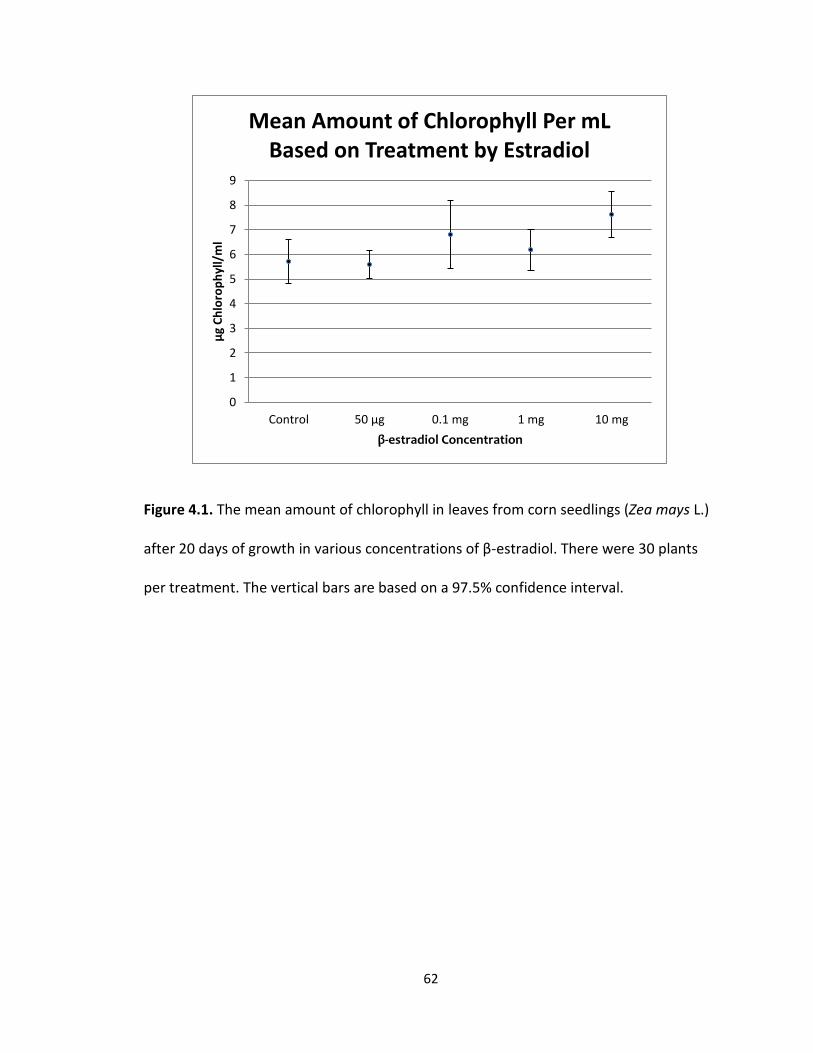
There were 150 plant samples used to complete this experiment. It was
observed that there was quite a bit of variation in the depth of color of the samples
used in the spectrophotometer. There was variation in the visual appearance not only
between the treatment groups, but within them as well. Overall the darkest (most
saturated appearing) samples seemed to come from the 10 mg samples. Using SAS
programming for one-way ANOVA statistical analysis it was determined that there was
not a significant difference in the mean amount of chlorophyll in μg/mL after 20 days of
growth in assigned β-estradiol concentrations. Using a confidence interval of 97.5% a p-
value of 0.5381 was determined, indicating that none of the treatments were
significantly different from the others. The mean amount of chlorophyll in μg/mL (Figure
4.1) was 5.72 μg with a standard error of 0.895 for the control, 5.60 μg with a standard
error of 0.560 for the 50 μg treatment, 6.81 μg with a standard error of 1.396 for the 0.1
mg treatment, 6.18 μg with a standard error of 0.828 for the 1.0 mg treatment, and 7.63
μg with a standard error of 0.934 for the 10 mg treatment. The greatest mean amount
of chlorophyll in μg/mL was recorded in the 10 mg treatment group and the least mean
amount of chlorophyll in μg/mL was recorded in the 50 μg treatment group. The
greatest difference between the mean amount of chlorophyll in μg/mL was 2.03 μg.
62
Figure 4.1. The mean amount of chlorophyll in leaves from corn seedlings (Zea mays L.)
after 20 days of growth in various concentrations of β-estradiol. There were 30 plants
per treatment. The vertical bars are based on a 97.5% confidence interval.
0
1
2
3
4
5
6
7
8
9
Control 50 μg 0.1 mg 1 mg 10 mg
μg
Ch
loro
ph
yll/
ml
β-estradiol Concentration
Mean Amount of Chlorophyll Per mL Based on Treatment by Estradiol
63
Discussion
According to the results there was not a significant effect of β-estradiol on the
amount of chlorophyll in the leaves of corn plants grown in various concentrations of
the hormone for 20 days. There was a wide variation in the amount of chlorophyll
amongst the samples within each concentration group. The variations were found in all
of the treatment levels as well as the control group. The high level of variation in
chlorophyll levels within each of the treatment groups could indicate that there is no
correlation between the β-estradiol and chlorophyll production in Zea mays L. It could
also be indicative of experimental error and follow up studies could be conducted to
corroborate the results. It should be noted that the 10 mg group did have the highest
mean amount of chlorophyll. Even though the chlorophyll levels exhibited in the 10 mg
group were not significant from the other concentrations, the increase in chlorophyll
content is consistent with the experiments carried out on D. carota and C. vulgaris
(Janeczko and Skoczowski 2005). These species also exhibited an increase in the amount
of chlorophyll with the presence of the hormone.
64
Chapter 5: Conclusion
Recap water pollution and effects of β-estradiol
Ecosystems consist of complex and intricate relationships between organisms
and their biotic and abiotic surroundings. When there is a change to any part of the
biotic or abiotic resources in an ecosystem, it can be drastically altered (Islam and
Tanaka 2004). One of the most important resources for an ecosystem is water. As a key
reactant in photosynthesis, water helps to form the base of any ecosystem by providing
the necessary resources for photosynthetic organisms, which then in turn provide
sustenance for heterotrophic organisms as well as oxygen for aerobic organisms
(Campbell et al. 2011).
The pollution of water sources and the damaging results have been well
documented over the past few years. To date, the greatest amount of waste that ends
up in aquatic ecosystems is sewage (Islam and Tanaka 2004). Sewage effluent can
include waste from industry, municipalities, animal remains, slaughterhouses, domestic
baths and kitchens, fecal matter and effluent from numerous other sources (Islam and
Tanaka 2004). Included among the sewage categories is a class of compounds known as
Reproductive Endocrine Disruptors, or REDs. Several studies have been conducted that
show where there are high levels of REDs in habitats there tends to be disruptions in the
morphology and physiology of the organisms that reside there (Islam and Tanaka 2004).
Examples of synthetic and naturally occurring chemicals that act as REDs include
estrogenic and anti-androgenic substances. There are also chemicals that are not
65
hormonal in nature but can have estrogenic properties, such as alkyphenols, industrial
pesticides, and chlorinated hydrocarbons (Islam and Tanaka 2004). One of the most
common estrogenic sources in waste water effluent comes from the urine of women
who take birth control pills. These hormone treatments contain the synthetic
ethynylestradiol as well as significant amounts of 17β-estradiol (Batty and Lim 1999).
Recap of previous experiments, and their results.
In mammals, β-estradiol plays a key role in controlling the processes revolving
around development and reproduction as well as being involved in the control of both
mineral and protein metabolism (Carreau et al. 2002). Knowing that this hormone is a
contaminant in several key water sources for plants, the question arises whether or not
there is also an effect upon plant growth and development (Islam and Tanaka 2004).
Several studies have been carried out on different plant species in order to determine
this.
Previous investigations have examined the effect that β-estradiol has on seed
germination in a wide variety of different plant species. β-estradiol decreased the
germination percentage of lettuce, carrot, and tomato seeds (Lactuca sativa L., Daucus
carota L., and Lycopersicon esculentum Mill. respectively), while in chickpea seeds (Cicer
arietinum L.) the germination percentage increased. In sunflower seedlings (Helianthus
annuus L.), β-estradiol concentrations of 1 μg per plant increased overall shoot growth,
but was shown to inhibit overall root growth (Janeczko and Skoczowski 2005). In tomato
66
(Lycopersicon esculentum Mill.) seedlings the hormone was shown to reduce overall
root growth as well as the overall number of roots present in nutrient solutions
containing β-estradiol (Janeczko and Skoczowski 2005). In alfalfa plants (Medicago
sativa L.), lower concentrations of β-estradiol favored increased root and shoot growth,
while the higher concentrations used inhibited growth (Janeczko and Skoczowski 2005).
In chickpeas (C. arietinum), the hormone significantly enhanced the root and shoot
growth of the seedlings (Erdal and Dumlupinar 2010). Previous studies have also been
conducted to determine whether or not β-estradiol had an effect on the amount of
chlorophyll present in the tissues of various species of plants. In D. carota the presence
of β-estradiol was shown to favor chlorophyll synthesis in leaves (Janeczko and
Skoczowski 2005). The same phenomenon of an increased production of chlorophyll
was observed in algal cells of Chlorella vulgaris M. Beijerinck (a type of green alga)
(Janeczko and Skoczowski 2005). In another study the presence of β-estradiol exposure
resulted in a 30-50% reduction of chlorophylls and carotenoids in L. sativa leaves
(D’Abrosca et al. 2008).
Thus, there appears to be a wide variation in the effects of β-estradiol on the
germination, growth, and chlorophyll content of different plant species. This suggests
that the effect on the growth of plants will depend on the species of plant and the
concentration of β-estradiol that the plant is exposed to. Although, there have been
relatively few studies conducted on the growth and chlorophyll content of plants in the
presence of β-estradiol, it should be noted that the plants used in previous studies were
dicotyledonous species (H. annuus, L. esculentum, M. sativa, C. arietinum). Zea mays L.
67
is a monocotyledonous species. There may be a difference in the physiological and
biochemical response to β-estradiol between dicotyledonous and monocotyledonous
species, although this has not yet been investigated.
Recap of current germination experiment on corn (Zea mays L.).
Previously, no one had looked at the effect of β-estradiol on the germination,
growth, and chlorophyll content of corn (Zea mays L.) and there were no publications
prior to the start of this research in 2009. The goal of this experiment was to determine
if β-estradiol had an effect on the germination of corn kernels and the growth of corn
seedlings. Parameters investigated during germination were primary root length,
coleoptile length, the number of adventitious roots, and the overall percentage of
germination. The parameters examined during corn seedling growth were overall shoot
system height, root system length, the number of leaves, and chlorophyll content.
For the experiment on the germination of Z. mays kernels in various
concentrations of β-estradiol, it was determined that the 10 mg β-estradiol treatment
group consistently exhibited a delay in germinating. For most other concentrations 48
hours was the optimal germination time, yet for the 10 mg group it was 72 hours. The
10 mg treatment was statistically significant from all other treatments (Fig 2.9). The 0.1
mg treatment group, which exhibited the fastest germination time, also tested
significantly different from both the control and the 50 μg treatment.
68
It was observed that the kernels in the 10 mg treatment group were almost all
lacking primary root growth (Fig 2.11). Most of the kernels had coleoptile growth and
adventitious roots present, but the primary roots were highly undeveloped. The 10 mg
group was the only one to exhibit these properties. It was determined that the 10 mg
treatment was statistically different from all other treatments. Primary root length of
the other treatments (50 μg, 0.1 mg, 1.0 mg, or control) were not significantly different
from each other (Fig 2.12). The longest mean primary root length was recorded in the
control treatment group while the shortest mean primary root length was recorded in
the 10 mg treatment group.
It appeared that most of the germinated kernels across the different
concentrations of β-estradiol had coleoptiles that were relatively similar in both length
and girth. It was determined that the coleoptile length in the 10 mg treatment was
statistically different from every other treatment except for the 50 μg treatment. None
of the other treatments (0.1 mg, 1.0 mg, or control) were significantly different from
each other as well as the 50 μg did not test significant against the other treatments. The
longest mean coleoptile length was recorded in the 0.1 mg treatment group and the
shortest mean coleoptile length was recorded in the 10 mg treatment group.
Most of the treatments contained kernels that had relatively similar numbers of
adventitious roots as well as a similar appearance. The difference between the means of
the 10 mg and 0.1 mg treatments was 3.03 adventitious roots. The 10 mg group had the
least mean number of adventitious roots and was statistically different from every other
69
treatment (50 μg, 0.1 mg, 1.0 mg, or control). It was also determined that the 0.1 mg
treatment was significantly different from the control in having the greatest mean
number of adventitious.
Recap of current growth experiment on corn (Zea mays L.).
For the growth portion of the experiments, it was observed that most of the
treatment groups were similar in overall appearance in reference to their shoot systems
(Fig 3.4). The 10 mg treatment group did have some anomalies. A few started to
develop a shoot system, but died relatively early. These plants did not develop past the
second emerging leaf and exhibited minimum shoot growth. Still others exhibited poorly
developed shoot systems. Many of the leaves of the shoot systems had begun to wither
and die (Fig. 3.4). It was determined that the shoot length of 10 mg treatment was
statistically different from the control and 1.0 mg treatment groups. The 10 mg
treatment group was not significantly different from either the 0.1 mg or 50 μg
treatment groups. The greatest mean shoot height (mm) was recorded in the 1.0 mg
treatment group and the least mean height (mm) was recorded in the 10 mg treatment
group.
It was observed that the majority of plants had very similar root systems both in
overall structure and size. The only group that stood out visually from the rest and
exhibited a few irregularities was the 10 mg β-estradiol treatment group (Fig 3.4). The
roots of the 10 mg group were usually shorter in overall length and overall girth when
compared to the rest of the treatment groups. There was one seedling in the 10 mg
70
group that actually had two root systems and no shoot system. This particular seedling
had roots coming out of both ends of the kernel. It was determined that the 10 mg
treatment was statistically different from the 0.1 mg treatment group. The 10 mg
treatment group was not significantly different from any other treatment groups. The
0.1 mg group was not significantly different from any other treatment group.
It was observed that there was not a noticeable difference in the number of
leaves on the plants (around 4 leaves) between the control and 1.0 mg, 0.1 mg, and 50 μ
treatment groups (Fig 3.4). The 10 mg treatment group did exhibit some plants with
wilting leaves and dying shoot systems. Plants in treatment groups other than the 10 mg
appeared to have relatively similar numbers of leaves on their shoot systems. The leaves
exhibited the same appearance, size, and coloration as well. It was determined that
none of the treatments were significantly different from the others.
When visually observing the chlorophyll extract of Z. mays there was quite a bit
of variation in the depth of color of the samples used in the spectrophotometer to
determine chlorophyll content. There was variation not only between the treatment
groups, but within them as well. Overall the darkest (most saturated appearing) samples
seemed to come from the 10 mg samples. It was determined that none of the
treatments were statistically significantly different from the others.
Summary of germination and growth experiments on corn (Zea mays)
Overall, the experimental results seem to indicate that higher concentrations of
β-estradiol (10 mg/L) had a negative impact on aspects of both the germination and
71
seedling growth of Z. mays. The germination of kernels was significantly altered by the
highest dose of β-estradiol. The mean number of hours before germination occurred for
the 10 mg treatment group was almost a full day later than the lower concentrations.
The overall percentage of germination was also drastically lower than the other
treatment groups. The 10 mg treatment group averaged 72 percent and all the others
averaged between 90 and 100 percent germination. This may be indicative of the
hormone inhibiting the mechanisms of germination either by binding to receptors or in
some other way down-regulating the gene expression for cellular components involved
in the process. Another consistent abnormality observed in the 10 mg treatment group
was either underdeveloped growth or the absence of primary root growth. This may
illustrate that exposure to high concentrations of β-estradiol could inhibit the
functioning of the primary root apical meristem. The process of cell elongation within
the root could also be affected, which is a primary means for increasing length (Taiz and
Zieger 2006).
The same trends of stunted root development were observed in the growth of Z.
mays seedlings exposed to the highest concentration of β-estradiol. Statistically, the 10
mg treatment group had a much smaller overall root system length than the other
treatment groups. This again suggests the hormone may be affecting some aspect of the
root apical meristem and cell elongation through inhibition of the genes, or other
aspects of cell regulation. Surprisingly, the hormone β-estradiol did not have a
statistically significant effect on either the number of leaves or the chlorophyll content
of the seedlings. With the significant alterations in the root systems, it was expected
72
that portions of the shoot system would also be affected. While, the total shoot system
height was reduced in the 10mg treatment group, the other variables measured
(number of leaves and chlorophyll content) did not test statistically significant from the
other treatment groups.
It should be noted that while the 10 mg treatment group showed consistent
inhibition among most of the parameters looked at in both the corn kernel germination
and corn seedling growth experiments, the 0.1 mg treatment group almost always
exhibited the highest measurements in all of the parameters observed including: mean
hour of germination, coleoptile length, number of adventitious roots, overall percent
germination, and overall root length. While not always statistically significant from the
control group, this may give credence to previous research (Janeczko and Skoczowski
2005) conducted which demonstrates that most species have augmented reactions to
low concentrations of β-estradiol, while high concentrations cause inhibitory effects.
Different study performed on Zea mays L. with conflicting data.
When the above set of experiments was started there had been no other
previous study that investigated the effects of β-estradiol on Z. mays. It should be noted
that during the course of experimentation and data collection a study was published
that investigated the effect of β-estradiol on the germination of corn kernels (Erdal et al.
2011). The experimental procedures as well as concentration of β-estradiol used in this
study were vastly different than those reported in this thesis study, which might explain
73
the variance in results between the two. In the study by Erdal et al. (2011) the Z. mays
kernels were only exposed to the hormone for six hours in concentrations of 10-4, 10-6,
10-9, 10-12, and 10-15 M before they were placed into a sterile environment with 10 ml of
distilled water. The steroid used in both studies did come from the same manufacturer.
In the study by Erdal et al. (2011) they found that all concentrations of β-
estradiol significantly increased the kernel germination rate when compared to the
control. However, by the end of the 5th day all treatment groups as well as the control
group had reached 100% germination. In their study they also found that the root and
coleoptile elongation were significantly stimulated in the presence of the hormone, with
the optimal treatment groups reported at 10-9 and 10-12 M. Their final conclusions were
that the hormone β-estradiol (at the concentrations used) augmented germination
velocity, as well as the length of the root and shoot. Erdal et al. (2011) do note that in
studies on other plant species it has been reported that low doses of the hormone may
enhance germination, while often times higher doses reduced or prevented it. This may
be part of the difference between the results presented in this thesis and the studies by
Erdal et al. (2011). Not only were the concentrations of β-estradiol used in the current
study vastly different, but the length of time the kernels were exposed to the hormone
in this thesis was 5 days, not six hours.
74
Implications of this current experiment
Mammalian sex hormones are constantly excreted into the environment through
the urine and feces of mammals. A woman naturally can excrete around 10 μmol per
day (Shore et al. 1993). The amount excreted in the urine increases with the intake of
birth control pills and during ovulation. Chicken manure can contain over 1 μmol of
estrogen in every gram (Shore et al. 1993). Larger mammals such as cows, horses, and
swine can naturally excrete much larger amounts every day. When exposed to the
environment either directly or through washing techniques and manure spreading, the
estrogen in the excrement gets transferred into the soil as well as gets carried into
various water sources. Several studies by independent researchers as well as the
Environmental Protection Agency (EPA) have shown that estrogen is a persistent
chemical in the environment and various water sources (Shore et al. 1993; United States
Environmental Protection Agency, 2012). Mammalian sex hormones have been found in
both lake sources as well as sewage water sources, some of which are used for
irrigation. Both sources contain hormone concentrations that have been shown to affect
plant growth and development (Shore et al. 1993).
It should be noted that there have been studies conducted on the ability of
specialized sewage sludge bacteria to degrade β-estradiol and its metabolites into a
non-estrogenic form. Through a proposed series of chemical reactions estradiol is
degraded by bacteria into tricarboxylic acid (Lee and Liu 2001). In one study it was
shown that aerobic degradation by activated sludge (sewage that is aerated and
75
contains aerobic bacteria) was able to remove about 88% of the estradiol. However, the
anaerobic sludge bacteria were only able to degrade 50% of the hormone (Lee and Liu
2001). It was also noted in this same study that the type of sewage treatment had an
effect on the ability of the bacteria to degrade the hormone. It was shown that
activated sludge was much more efficient at degrading β-estradiol than a trickling filter
system (Lee and Liu 2001). The conclusion was drawn that the vigorous mixing and
aeration, as well as the high levels of microbial activity found in the activated sludge
techniques, was the reason for the higher efficiency of degrading natural estrogens (Lee
and Liu 2001).
How estrogens in water gets into field crops
Not all estrogen contaminated water undergoes treatment in waste water
facilities. Much of the estrogen contamination occurs when runoff from farms and
livestock facilities goes into natural water sources such as rivers and streams. These
water sources can then be applied directly to fields through irrigation techniques or
even periodic flooding. Once the β-estradiol is in soil, the crops can readily take up the
hormone along with other nutrients and ions when water is taken into the root tissue
through the root hairs. The water and dissolved nutrients and ions then traverse the
dermal and ground tissue finally entering the xylem tissue where they become xylem
sap. The process by which water is transported through a plant’s tissues is known as
cohesion-adhesion-tension theory of water movement (Taiz and Zieger 2006). Once
76
within the cells of xylem tissue (tracheids and vessel elements), the water molecules
experience a pull upward through the tissue, the driving force being transpiration or loss
of water vapor from the stoma of leaves. The water molecules each exhibit an unequal
sharing of electrons causing a partial positive and partial negative charge across each
molecule. These partial charges then form hydrogen bonds with neighboring water
molecules in a property known as cohesion. The molecules also adhere to the surface of
the xylem tissue through these partial charges and this is known as adhesion. The
cohesion-adhesion-tension theory of water along with the process transpiration is
considered to be the primary mechanism by which the xylem sap moves upward against
gravity and into the shoot system of plants. Another property of water is its ability to be
a universal solvent. This property allows water to dissolve many different substances,
and then transport and cycle them through organisms’ tissues as well as through the
environment (Campbell et al. 2011).
Possible effects on staple grain crops and consequences
Farmland across the Midwest portion of the United States is typically dominated
by soybeans (Glycine max (L.) Merr.) or field corn (Zea mays L.). A large portion of
farmland is also dedicated to wheat (Triticum aestivum L.) and sorghum (Sorghum
bicolor (L.) Moench) production. Corn, wheat, and sorghum are all staple grain crops in
the United States. Their production has an enormous economic impact. In the U.S. the
economic value of corn is at an all-time high and was recorded to be around two billion
77
dollars in 2010 (University of Missouri Extension, 2012). If the germination, growth, or
overall yield of these staple crops are being affected by β-estradiol contamination of
water sources this could have serious consequences on a global economic scale. In this
study it was concluded that high doses of the hormone had a negative impact on Zea
mays L. kernels and seedlings in several of the parameters tested including germination
percentages and root development.
Further tests should be conducted to follow the development of the seedlings
through maturation and the production of fruit to determine if the high doses of β-
estradiol affect the later stages of development as well. A field study might also be
beneficial for this area of research. So far, previous studies as well as this current study
have all looked at the effects of β-estradiol on germination and growth in sterile
laboratory conditions. However, as stated above there are bacteria that can degrade the
hormone into different conjugates and non-estrogenic forms (Lee and Liu 2001).
The basic methodology described in this thesis study also needs to be continued
on with field studies. The mass production of corn, sorghum, and wheat does not occur
in a laboratory, but out in a field exposed to its biotic and abiotic surroundings. These
grain crops are harvested for livestock feed and silage as well as human consumption
and it would be important to understand the effects β-estradiol might have on overall
production. Researching growth in the field would also take into account the presence
of soil bacteria and natural climate conditions as well. Therefore that is one direction

78
future studies should take in order to determine how much of an effect β-estradiol has
on corn where the actual growth of these plants takes place.
79
Literature Cited
Agarwal M. 1993. Receptors for mammalian steroid hormones in microbes and plants. Federation of European Biochemical Societies 322(3): 207-210. Batty J and Lim R. 1999. Morphological and reproductive characteristics of male
mosquitofish (Gambusia affinis holbrooki) inhabiting sewage-contaminated waters in New South Wales, Australia. Archives of Environmental Contamination and Toxicology 36: 301-307.
Campbell N, Reece J, Taylor M, Simon E, and Dickey J. 2012. Campbell Biology Concepts
& Connections. San Fransico: Pearson Benjamin Cummings. D’Abrosca B, Fiorentino A, Izzo A, Cefarelli G, Pascarella MT, Uzzo P, and Monaco P.
2008. Phytotoxicity evaluation of five pharmaceutical pollutants detected in surface water on germination and growth of cultivated and spontaneous plants. Journal of Environmental Science and Health Part A 43: 285-294.
Erdal S and Dumlupinar R. 2010. Progesterone and β-estradiol stimulate seed germination in chickpea by causing important changes in biochemical parameters. Zeitschrift Naturforsch 65: 239-244.
Erdal S and Dumlupinar R. 2011. Exogenously treated mammalian sex hormones affect
inorganic constituents of plants. Biol Trace Elem Res 143: 500-506. Erdal S and Dumlupinar R. 2011. Mammalian sex hormones stimulate antioxidant
system and enhance growth of chickpea plants. Acta Physiol Plant 33: 1011-1017.
Guillette L, Crain A, Gunderson M, Kools S, Milnes M, Orlando E, Rooney A, and Woodward A. 2000. Alligators and endocrine disrupting contaminants: a current perspective. American Zoology 40: 438-452.
Islam S and Tanaka M. 2004. Impacts of pollution on coastal and marine ecosystems
including coastal and marine fisheries and approach for management: a review and synthesis. Marine Pollution Bulletin 48: 624-649.
Janeczko A and Skoczowski A. 2005. Mammalian sex hormones in plants. Folia
Histochemica Et Cytobiologica 43(2): 71-79. Koes R, Quattrocchhio F, and Mol J. 1994. The flavonoid biosynthetic pathway in plants:
function and evolution. Bioessays 16(2): 123-132.
80
Lee H and Liu D. 2002. Degredation of 17β-estradiol and its metabolites by sewage bacteria. Water, Air, and Soil Pollution 134: 353-368.
Nimala G, Veena T, Jyothi M, and Suchitra B. 2008. Effect of estrogen and progesterone on seed germination. Veterinary World 1(8): 241-242. Raven P, Evert R, and Eichhorn S. 2005. Biology of Plants. 7th ed. New York: W.H.
Freeman and Company. Shore L, Gurevitz M, Shemesh M. 1993. Estrogen as an environmental pollutant.
Environmental Contamination and Toxicology. 51: 361-366. Shore L, Kapulnik Y, Ben-Dor B, Fridman Y, Wininger S, and Shemesh M. 2006. Effects of
estrone and β-estradiol on vegetative growth of Medicago sativa. Physiologia Plantarum 84(2): 217-222.
Stoka A. 1999. Phylogeny and evolution of chemical communication: an endocrine approach. Journal of Molecular Endocrinology 22: 207-225.

81
Acknowledgements
I would like to first and foremost thank my thesis committee for their long hours
and multiple revisions of this body of work. Dr. Karen Schaffer, I thank you for coming
up to me at the vending machines in Garrett-Strong and asking if I had any research
projects in mind. If it hadn’t been for you, the ball never would have started rolling on
this project. Dr. Jeff Thornsberry, I thank you for your knowledge of corn and your help
with my experimental design. I also thank you for always answering my questions
regarding all things maize. Dr. Ahmed Malkawi, I thank you for your knowledge of
chemistry and the inner workings of molecules and their interactions, especially when it
came to dissolving estradiol.
I gratefully acknowledge Dean Gregory Haddock and the Northwest Missouri
State University Graduate School, Dean Charles McAdam and the College of Arts and
Sciences and the Department of Natural Sciences for the funding of my research as well
as the lab space and use of equipment. I would also like to thank Dr. Mark Corson for
allowing me to work with Northwest Missouri State University while completing this
body of work. Your continual support and commitment to my completion has truly been
a gift.
For the rigorous and challenging comprehensive exam questions that allowed
me to broaden my knowledge and really think about the writing process, I must thank
Dr. Peter Adam. Even though I hated those five behemoths you called short essays, they
were absolutely essential in this process.

82
Dr. Gregg Dieringer and David Vlieger, I thank you for your involvement and for
teaching me the innerworkings of SAS statistical programming. Your teachings made the
analysis of my results easy to work with and present to the public.
The following people must be commended for their continuous encouragement
and support of this ongoing and strenuous project: Lisa Crater, Alex Bolick, Samantha
Barton, and Shannel Phillips. Your ability to discourage my procrastination habits was
truly a gift and I thank you every day for your involvement as well as for keeping me
sane. To the countless other people involved in this journey, I must also give thanks.
Your involvement, no matter how minor, will never be forgotten.
The biggest thanks of all must go to my parents David and Cynthia Bowlin. Your
countless hours on the phone, encouragement, support, and questions have all led to
this completed body of work. This is the culmination of not only my hard work, but
yours as well. For you are the ones who have always pushed me to do my best and to
always strive for success in all that I do. You both mean the absolute world to me and I
cannot thank you enough. I know that you will smile as I do when I say, “I finished!”