effect of osteopontin (opn) on in vitro embryo development in cattle
TRANSCRIPT

Effect of osteopontin (OPN) on in vitro embryo
development in cattle
E. Monaco a,*, B. Gasparrini b, L. Boccia b, A. De Rosa b,L. Attanasio b, L. Zicarelli b, G. Killian a
a John O. Almquist Research Center, Department of Dairy and Animal Science, Pennsylvania State University, University Park, PA, USAb Department of Scienze Zootecniche ed Ispezione degli Alimenti, Faculty of Veterinary Medicine, ‘‘Federico II’’ University,
Via F. Delpino 1, 80137 Naples, Italy
Received 17 November 2007; received in revised form 31 July 2008; accepted 6 August 2008
Abstract
Fertility-related phosphoprotein osteopontin (OPN) is present in the bovine oviduct epithelium and fluid. The objectives were to
determine the effects of OPN on percentages of cleavage and embryo development in vitro in cattle, and to assess the ability of OPN
to induce in vitro capacitation of bovine sperm. In vitro-matured bovine oocytes were fertilized in the presence of 0, 10, 20, or 40 mg/
mL OPN. There were greater percentages (P < 0.01) of cleavage and compact morulae-blastocysts (79.7 and 43.3%, respectively)
with 10 mg/mL OPN than in the control group (without OPN; 71.2 and 32.1%, respectively). Furthermore, percentages of advanced
blastocysts were greater in the group receiving 40 mg/mL OPN versus control (56.4% vs. 42.0%, P < 0.05).
Capacitation was assessed by the ability of sperm to undergo the acrosome reaction after incubation with lysophosphatidylcho-
line. Semen from three bulls was incubated for 2 h in either TALP medium alone (control) or with TALP medium containing
0.01 mM heparin, or with TALP medium containing 10 or 20 mg/mL OPN. Incubation with 10 and 20 mg/mL OPN produced more
(P < 0.01) capacitated sperm (14.4 and 13.6%, respectively) than the untreated control group (8.3%), but both untreated sperm and
those treated with OPN had significantly fewer capacitated sperm than those treated with 0.01 mM of heparin (30.5%). In
conclusion, OPN improved the efficiency of bovine in vitro embryo production and influenced sperm capacitation.
# 2009 Elsevier Inc. All rights reserved.
Keywords: Osteopontin; Embryo development; IVF; Sperm capacitation; Cattle
www.theriojournal.com
Available online at www.sciencedirect.com
Theriogenology 71 (2009) 450–457
1. Introduction
Since 1981, when the first in vitro produced (IVP)
calf was born, remarkable discoveries have been made
to define the metabolic needs of the embryo. There
are differences between in vivo and in vitro produced
* Corresponding author. Present address: Department of Animal
Sciences, University of Illinois at Urbana-Champaign, 1207 West
Gregory Drive, Urbana, IL 61801, USA. Tel.: +1 217 2443150;
fax: +1 217 3338286.
E-mail address: [email protected] (E. Monaco).
0093-691X/$ – see front matter # 2009 Elsevier Inc. All rights reserved.
doi:10.1016/j.theriogenology.2008.08.012
embryos in metabolic and morphologic profiles at the
cellular level [1–3], as well as in gene expression [4].
The oviduct provides an optimal environment for
sperm capacitation, the acrosome reaction, fertilization,
and early embryo development; oviduct fluid (ODF)
facilitated sperm capacitation, fertilization, and early
embryo development in vitro (reviewed in [5]). More-
over, ODF proteins associated with bovine sperm [6],
ova [7], and embryos [8]. Therefore, formulation of
defined media for in vitro fertilization (IVF) and in vitro
culture (IVC) that mimic the oviductal environment
may improve in vitro embryo production (IVEP) [9–11].
E. Monaco et al. / Theriogenology 71 (2009) 450–457 451
One of the proteins present in bovine oviduct fluid
is osteopontin [12]. Osteopontin was first described
in bone matrix, but has also been detected in epithelial
cells and in secretions of the gastrointestinal tract,
kidneys, thyroid, mammary gland, oviduct, uterus,
placenta, ovary, and testes [13–18]. Structurally, OPN
is an acid single chain phosphorylated glycoprotein that
ranges in length from 264 to 301 amino acids and
undergoes extensive post-translational modifications
that result in molecular weight variations ranging
from 25 to 75 kDa [19]. The resulting multiple
functional protein contains a Gly-Arg-Gly-Asp-Ser
(GRGDS) sequence that binds to cell surface integrins
to promote cell–cell adhesion, cell-extracellular matrix
(ECM) communication, migration of osteocytes and
immune cells [20,21]. It also stimulates immunoglo-
bulin production by B cells, alters intracellular calcium
concentrations, promotes calcium phosphate deposi-
tion in bone, and affects the mineralization of the
tissues (reviewed in [19]).
Osteopontin also appears to be important in
reproduction. Killian et al. [22] and Cancel et al.
[23] reported that more OPN was expressed in the
seminal plasma of high-fertility compared to low-
fertility bulls. Proteomic analyses demonstrated that
high-fertility Holstein bulls had more OPN in their
accessory sex glands fluid than low-fertility bulls
[24]. There are three molecular weight forms (55, 48,
and 25 kDa) of OPN in bovine ODF during the
estrous cycle [12]. These forms are present in both
the ampullary and isthmic regions, with the greatest
amount of OPN found in ampullary non-luteal
oviductal fluid (ANL-ODF) when serum progesterone
concentrations were �1.5 ng/mL [12]. Osteopontin
has been detected on the zona pellucida (ZP) of both
immature and mature bovine oocytes [25]. Goncalves
et al. [26] have recently showed that addition of rabbit
polyclonal IgG antibody against purified bovine milk
OPN to bovine sperm, oocytes, or both, decreased
IVF compared to untreated controls. In swine, Hao
et al. investigated the role of OPN on IVF [27]. They
demonstrated that fertilization rates were improved
when OPN was included in the IVF medium, and the
percentage of acrosome-reacted sperm bound on
the oocytes ZP was increased after 4 h incubation
with 1.0 mg/mL OPN. Based on these observations,
the objectives of our study were to evaluate the effect
of supplementing IVF medium with OPN on bovine
cleavage and post-fertilization embryo development,
and to evaluate the ability of various concentrations
of OPN to induce in vitro capacitation of bovine
sperm.
2. Materials and methods
Unless otherwise stated, all chemicals used were
purchased from Sigma Chemical Co. (St. Louis, MO,
USA). The OPN used in this study was purified (in
our laboratory) from bovine skim milk, as previously
described in Ref. [28].
2.1. Oocyte collection and maturation
Ovaries were harvested at a local abattoir from a
variety of cattle breeds and transported to the laboratory
in saline solution (0.9% NaCl) at 30–35 8C. Immature
cumulus–oocyte complexes (COCs) were aspirated
from follicles of 1 to 6 mm in diameter using an 18-
gauge needle (B-D1, Rutherford, NJ, USA) attached to
a 10 mL disposable syringe (B-D1). The aspirate was
distributed among sterile petri dishes. Oocytes with
uniform cytoplasm and multilayered cumulus cells
were selected, washed twice in TCM aspiration
(Medium 199 supplemented with 25 mM of Hepes,
2 mM sodium bicarbonate, 2 mM sodium pyruvate,
1 mM L-glutamine, 10 mL/mL amphotericin B, and
540 mg/mL heparin) and once in TCM-IVM (Medium
199 supplemented with 15% bovine serum (BS),
0.5 mg/mL FSH, 5 mg/mL LH (Sioux Biochemical1,
Sioux Center, IA, USA), 0.8 mM L-glutamine, and
50 mg/mL gentamicin). The COCs were then placed to
mature, 25/well (NunclonTM, Nunc, Roskilde, Den-
mark), in 400 mLTCM-IVM covered with mineral oil in
the incubator for 22–24 h at 39 8C and 5% CO2 in air.
2.2. Sperm preparation and in vitro fertilization
Mature COCs were washed in Tyrode’s modified
medium [29] without glucose and bovine serum
albumin (BSA), supplemented with 30 mg/mL heparin,
30 mM penicillamine, 15 mM hypotaurine, 0.15 mM
epinephrine, and 1% BS (IVF-TALP). They were
transferred, 25/well, into 300 mL IVF-TALP covered
with mineral oil.
Frozen sperm from one Limousine bull, previously
screened to perform satisfactorily in IVF (percentages
of fertilized oocytes were typically 70% and percen-
tages of expanded blastocysts usually 30%), were
thawed at 37 8C for 40 s and layered carefully into a
15 mL conical tube containing 2 mL 80% Percoll
solution in the bottom and 2 mL 45% Percoll solution
on top of it. Percoll solutions were made in Tyrode’s
modified medium [29] without glucose and BSA
(Sperm-TALP). Semen was centrifuged for 25 min at
300 � g. After centrifugation, the pellet was recon-

E. Monaco et al. / Theriogenology 71 (2009) 450–457452
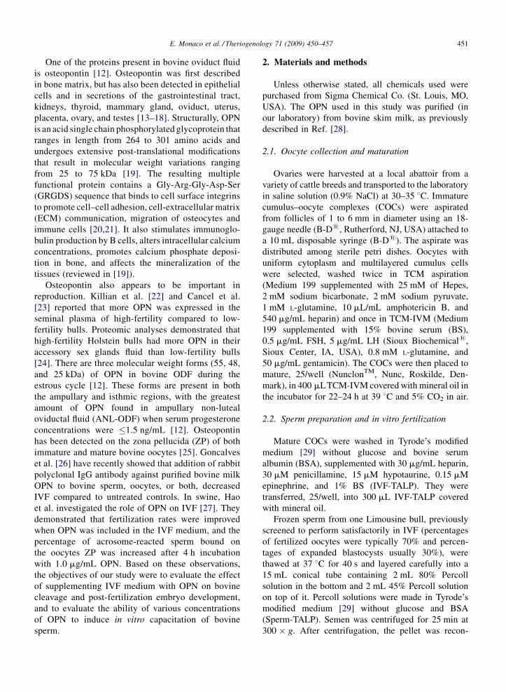
Fig. 1. Two viable bovine sperm (absence of trypan blue staining); the
sperm on the left has an intact acrosome (intense pink), whereas the
one on the right is acrosome reacted (light pink). (For interpretation of
the references to color in this figure legend, the reader is referred to the
web version of the article.)
stituted into 2 mL of Sperm-TALP and centrifuged
twice (160 and 108 � g). The pellet was diluted with
modified IVF-TALP and added in the fertilization wells
(concentration, 1 � 106 sperm/mL). Gametes were co-
incubated for 20–22 h at 39 8C in 5% CO2 in air.
2.3. In vitro embryo culture
After 20–22 h of co-incubation, presumptive
zygotes were vortexed for 2 min to remove cumulus
cells in Medium 199 supplemented with 25 mM Hepes,
2 mM sodium bicarbonate, 2 mM sodium pyruvate,
1 mM L-glutamine, and 5% BSA (TCM-Hepes).
Presumptive zygotes were then washed twice in the
same medium and transferred, 30–50/well, into 400 mL
synthetic oviduct fluid (SOF) modified medium [30],
supplemented with 30 mL/mL essential amino acids,
10 mL/mL non-essential amino acids, 0.34 mM tri-
sodium citrate, 2.77 mM myo-inositol, and 5% BS.
Zygotes were incubated in a humidified mixture of
5% CO2, 7% O2, and 88% N2 in air at 39 8C. The
percentages of cleaved embryos and oocytes reaching
blastocyst (BL) and advanced blastocyst (expanding
and expanded blastocyst, hatching and hatched
blastocyst, and expanded hatched blastocyst) were
determined at Day 7 of the culture (Day 0 = fertiliza-
tion day).
2.4. Sperm capacitation assay
Frozen sperm, from three Limousine bulls, which
were previously tested to perform well in IVF,
were thawed at 37 8C for 40 s and separated through
Percoll gradients, as described above. Sperm were
then exposed to lysophosphatidylcholine (LPC) as
described by Parrish et al. [31]. After 15 min of
incubation at 38 8C in 5% CO2 in air, sperm were fixed
and stained with Trypan blue-Giemsa [32]. Slides were
then rinsed and left to dry at air covered with a glass
cover slip and sealed with Entellan. Sperm were
counted under differential interference light micro-
scopy and the number of live with acrosome intact,
dead with acrosome intact, live with acrosome reacted,
or dead with acrosome reacted sperm was evaluated
(Fig. 1).
2.5. Experimental design
2.5.1. OPN and IVEP
A total of 1486 COCs were selected and matured in
vitro. At Day 0 of culture, they were randomly assigned
to four experimental groups:
(1) 3
78 oocytes were fertilized in IVF-TALP withoutOPN (control group);
(2) 3
72 oocytes were fertilized in IVF-TALP with10 mg/mL OPN;
(3) 3
72 oocytes were fertilized in IVF-TALP with20 mg/mL OPN;
(4) 3
64 oocytes were fertilized in IVF-TALP with40 mg/mL OPN.
At Day 7 of culture, the percentages of cleavage and
oocytes reaching blastocyst were evaluated. The
experiment was repeated six times (range, 25–99
oocytes/replicate/treatment).
2.5.2. OPN and semen capacitation
The ability of various concentrations of OPN (0, 10,
and 20 mg/mL) to induce in vitro capacitation of bovine
sperm was investigated. Capacitation was assessed
indirectly by estimating the percentage of acrosome
reacted sperm after incubation with lysophosphatidyl-
choline (LPC).
For each treatment, one straw of semen was thawed
per bull and two slides were prepared. An aliquot of
semen was taken immediately after thawing, and two
slides were prepared to estimate the percentage of
sperm without acrosomes prior to treatment. A second
aliquot was also prepared for counting after sperm were
incubated for 2 h in either TALP medium alone (control
group) or with TALP medium containing 0.01 mM
heparin or with TALP medium containing 10 or 20 mg/
mL of OPN. The percentage of live and dead sperm

E. Monaco et al. / Theriogenology 71 (2009) 450–457 453
Table 1
Effects of various doses of osteopontin (OPN) on development (%) of IVP bovine embryos to cleavage, compact morulae–blastocysts (CM–BL), and
advanced blastocyst (BL) stages
Embryo stage OPN (mg) SEM P value
0 10 20 40
Cleavage 71.2a 79.7b 77.5b 73.1ab 1.6 <0.01
CM–BL 32.1a 43.3b 36.8ab 29.0a 2.6 <0.01
Advanced BL 42.0ab 52.9bc 40.0a 56.4c 3.8 0.02
a–cWithin a row, percentages without a common superscript differed (P < 0.05).
for each treatment was determined by counting 160–
240 sperm/slide.
2.6. Statistical analyses
Data from both experiments were analysed using
PROC GLM of SAS (Release 8.0; SAS Institute, Cary,
NC, USA) with a repeated statement. The model
included the fixed effect of treatment (three treatments
plus control in both experiments), replicates (n = 6) for
the experiment OPN and IVEP, or bull (n = 3) for the
experiment OPN and semen capacitation, and treat-
ment � replicate or bull interaction. The test for
normality was run using PROC UNIVARIATE NOR-
MAL, and a Shapiro–Wilkins normality test >0.9 was
used as criterion to determine normal distribution of
data. All data were normally distributed; therefore, no
transformation was required prior to MANOVA
analysis. Significant was set at P � 0.05.
3. Results
3.1. Effect of OPN on embryo development
Greater percentages (P < 0.01) of cleaved zygotes
were observed in groups cultured with 10 and 20 mg/mL
of OPN than in the control group (Table 1). Furthermore,
significantly greater percentages of compact morulae–
blastocysts (CM–BL) were obtained after incubation
with 10 mg/mL OPN compared to the control group
Table 2
Effects of various doses of osteopontin (OPN), or 0.01 mM of heparin (EPA
acrosomes relative to total live, and sperm without acrosomes relative to to
Sperm OPN (mg)
0 10
Total live 49.6 54.4
Without acrosome/total live 8.3a 14.5b
Without acrosome/total 16.7a 26.9b
a–cWithin a row, percentages without a common superscript differed (P <
(Table 1). Greater percentages (P < 0.05) of advanced
blastocysts were present in the 40 mg/mL OPN group
versus the control group (Table 1).
3.2. Effect of OPN on sperm capacitation
At time = 0, none of the sperm randomly counted
for each slide showed acrosome lost just after thawing.
The percentages of capacitated sperm were greater
(P < 0.01) in treatments receiving 10 and 20 mg/mL of
OPN than the control group, but both untreated sperm
and those treated with OPN had fewer capacitated
sperm (P < 0.01) than those treated with 0.01 mM of
heparin (Table 2). There were no significant differences
among groups in the percentages of viable sperm.
4. Discussion
In the present study, the addition of OPN signifi-
cantly improved IVEP. Given that the percentages of
cleaved zygotes and transferable embryos produced in
vitro were greatest at the lower concentration of OPN,
we infer that there was a biphasic response pattern to
incremental doses of OPN. However, the percentages of
advanced blastocysts were greatest in the 40 mg/mL
group. Perhaps 10 mg/mL OPN is optimal for the
development of oocytes to CM-BL stage, but a higher
concentration is needed for advanced blastocyst
formation. In vivo, an accumulation of OPN on the
embryo is possible from both ODF [33] and the
), on percentages of live sperm relative to total sperm, sperm without
tal sperm
EPA SEM P value
20
53.4 65.4 5.2 0.26
13.7b 30.5c 1.4 <0.01
27.3b 47.4c 2.5 <0.01
0.05).

E. Monaco et al. / Theriogenology 71 (2009) 450–457454
endometrium [34], as it moves from the oviduct to the
uterus. Uterine OPN expression has been detected
during peri-implantation in humans [35,36], pigs [34],
and rabbits [37]. Perhaps with 40 mg/mL OPN there is a
carry-over effect of the protein that is not evident until
the latest stage of embryo development in vitro.
Our results were not consistent with those of Hao
et al. [27] for swine, where an overall increase in the
fertilization efficiency was obtained with 0.01 and
0.1 mg/mL OPN. In preliminary studies, we tested 0.1
and 1 mg/mL of OPN, but these concentrations were
ineffective. The different responses between porcine
and bovine to similar doses of OPN may be due to
species differences, or to the source of the OPN. We
used OPN that was from purified skim bovine milk,
whereas Hao et al. [27] used recombinant rat OPN.
Furthermore, in our study, the end point measured for
the effect of OPN on IVF was the percentage of
cleavage, whereas in the previous study [27], penetra-
tion rates, polyspermic fertilization rates, male pro-
nuclear formation rate, normal fertilization efficiency
(number of oocytes with one male and one female
pronucleus/total number of oocytes inseminated), and
number of sperm penetrated per oocyte were deter-
mined.
The ability of osteopontin to positively affect IVEP
may be due to direct interactions with both sperm and
oocyte. In a previous study, we detected OPN on the
zona pellucida (ZP) and oolemma of both immature and
mature bovine oocytes [25,33]. We also detected OPN
on the post-equatorial segment, acrosomal cap, and
midpiece of ejaculated bull sperm [33], and its distri-
bution on sperm changed after incubation in bovine
oviduct fluid [33].
The interaction of OPN with sperm or ova may occur
through its GRGDS sequence and CD44 [26,38]. The
GRGDS peptide is known to interact with integrin cell
receptors [15,39], which have been identified on the
surface of several cells. Different integrins have been
described on the oolemma of bovine fertilized oocytes
[40] and human sperm [33,41]. Integrins are known to
promote cell–cell adhesion and may facilitate fertiliza-
tion in mammalian species by promoting oocyte–sperm
binding [42]. Goncalves et al. [26] reported that
addition of OPN antibodies to fertilization media
reduced both the number of sperm bound to oocytes and
the percentage of fertilized oocytes in cattle. Further-
more, treatment of sperm or oocytes with an RGD
peptide, or with a5 and av integrin antibodies, reduced
the number of sperm bound to the ZP and fertiliza-
tion rates similar to what was found using antibodies
against OPN [26]. Perhaps OPN participates in sperm–
oocyte interaction (with potential involvement of
integrins).
Gabler et al. [12] detected three distinct molecular
weight forms of OPN protein in the bovine ODF from
both the ampullary and isthmic regions during the
estrous cycle. The greatest amount of OPN was in
ampullary non-luteal oviductal fluid, whereas the
minimum amount was present in both isthmus and
ampullary luteal fluid. Coincidently, the expression of
av e b1 integrin subunits in the oviduct during the luteal
phase was the lowest among the estrous cycle phases.
Moreover, Gabler et al. [12] observed that the quantity
of oviductal cells mRNA for the integrin subunit b3
significantly increased, starting from the latest luteal
phase until just before ovulation. Integrin beta (3)
mRNA content increased significantly from the lowest
level during the late luteal phase to the highest level
before ovulation. Taken together, the Gabler et al. [12]
work suggests the potential role of an osteopontin–
integrin interaction during normal oviduct physiology,
including early embryo development.
Previously, we suggested that the interaction of OPN
with sperm and oocyte may occur in two ways [33]. One
possibility is that OPN binds to sperm and the resulting
sperm–OPN complex adheres to the ZP of the oocyte.
Alternatively, the sperm–OPN complex may adhere to
the ovary/oviduct derived OPN, previously bound to the
ZP, since OPN forms bonds with other OPN molecules
[43,44]. However, in either case, once sperm entered the
perivitelline space, OPN may also mediate the
interaction of sperm with the oolemma, through
integrins and/or CD44. Numerous intracellular signals
are activated within the egg after the fusion with the
sperm, which are believed to have important roles in the
resumption of meiosis and consequent development of
the zygote [45,46]. We suggest that OPN, adhering to
the oolemma, triggers intracellular signals via interac-
tion with integrins and/or CD44. It is also likely that
OPN activates zygote genes involved in promoting/
accelerating development. This would explain not only
the improved percentages of IVEP after treatment with
OPN, but also the highest percentage of advanced
blastocysts obtained in this study. Perhaps the IVEP
effect of OPN may be derived from sperm which were
capacitated in the presence of OPN and heparin.
Although our results confirmed that heparin was the
best capacitating agent in vitro, they also showed that
OPN promoted in vitro sperm capacitation without
the negative effect on sperm viability associated with
heparin.
Sperm capacitation and the acrosome reaction are
dependent on increases in sperm intracellular calcium.

E. Monaco et al. / Theriogenology 71 (2009) 450–457 455
Because osteopontin influences intracellular calcium
concentrations [39], perhaps the positive effect of OPN
on bovine sperm capacitation is through its ability to
influence calcium transients.
Mammalian sperm capacitation is a maturation step
that enables sperm to achieve an acrosome reaction and
ova penetration. For capacitation and an acrosome
reaction to occur, calcium is necessary in the extra-
cellular environment. During sperm capacitation and
acrosome reaction, there is an increase in intracellular
Ca2+ concentration ([Ca2+]i) that finally leads to the
exocytose of the acrosomal vesicle and to the fusion of
the two gametes. Evidence suggests that ligand binding
to various integrins triggers an increase in [Ca2+]i
[47,48]. Rat osteoclasts or mouse osteoclasts-like cells
had a transient [Ca2+]i increase when perfused with
osteopontin and other integrin ligands [49]. This rise in
[Ca2+]i came from intracellular stores, although extra-
cellular Ca2+ was necessary to produce the [Ca2+]i
changes. During sperm capacitation and acrosome
reaction, the presence of extracellular Ca2+ is necessary
for an increase in [Ca2+]i and believed to be due to the
known Ca2+ requirement for ligand binding. However,
the mechanism by which OPN triggers an increase in
intracellular calcium is unknown.
Zimolo et al. [49] reported that binding of avb3-
ligands to the osteoclasts triggers cellular changes that
have second messenger characteristics. Both a- and b-
chains have relatively short intracellular domains that
lack kinase activity or typical domains known to
interact with G proteins. Nevertheless, both intracellular
domains contained tyrosine residues that, if phosphory-
lated, may be the regions for Src homology (SH) region
2 binding shown to modulate interactions in signal
transduction pathway [50,51].
Heparin is routinely used to induce sperm capacita-
tion in vitro; the heparin-induced capacitation in cattle
is mediated by heparin binding to sperm membrane
[31], with consequent Ca2+ uptake and an increase in
intracellular free calcium [52]. Perhaps heparin inter-
action with the sperm membrane occurs through OPN.
In that regard, OPN contains a calcium binding site and
two heparin binding domains. In a recent study, it was
highlighted that the phosphorylation of many bull sperm
proteins was important for the heparin induction of
sperm capacitation [53]. Remarkably, we showed OPN
to be present on the bovine sperm membrane [33] and it
is known that OPN undergoes extensive post-transla-
tional modifications, including phosphorylation [19].
Our hypothesis is that OPN present on the sperm surface
at ejaculation binds to heparin through its heparin
domain and that this complex OPN-heparin guides
calcium transients and sperm capacitation. In this
regard, it would be interesting to test the combined
effect of OPN and heparin on sperm capacitation in
vitro, since in vivo, the OPN present on the sperm at
ejaculation is supplemented with OPN from the ODF.
In conclusion, fertilization medium containing
10 mg/mL OPN improved in vitro embryo production
and OPN positively influenced sperm capacitation in
vitro. On a practical basis, addition of OPN or other
oviductal proteins to IVF systems may significantly
improve the percentage of live embryos produced in
vitro. However, the specific functions of OPN, as well
as, the molecular mechanisms underlying its action,
remain to be investigated.
Acknowledgements
The assistance of the staff at the Department of
Scienze Zootecniche ed Ispezione degli Alimenti
‘‘Federico II’’ University, Naples, Italy is greatly
appreciated.
References
[1] Boni R, Tosti E, Roviello S, Dale B. Intercellular communication
in in vivo- and in vitro-produced bovine embryos. Biol Reprod
1999;61:1050–5.
[2] Khurana NK, Niemann H. Energy metabolism in preimplanta-
tion bovine embryos derived in vitro or in vivo. Biol Reprod
2000;62:847–56.
[3] Thompson JG. In vitro culture and embryo metabolism of cattle
and sheep embryos—a decade of achievement. Anim Reprod Sci
2000;60/61:263–75.
[4] Miles JR, Blomberg le A, Krisher RL, Everts RE, Sonstegard TS,
Van Tassell CP, et al. Comparative transcriptome analysis of in
vivo- and in vitro-produced porcine blastocysts by small ampli-
fied RNA-serial analysis of gene expression (SAR-SAGE). Mol
Reprod Dev 2008;75:976–88.
[5] Killian GJ. Evidence for the role of oviduct secretions in sperm
function, fertilization and embryo development. Anim Reprod
Sci 2004;82/83:141–53.
[6] McNutt T, Rogowski L, Vasilatos-Younken R, Killian G.
Adsorption of oviductal fluid proteins by the bovine sperm
membrane during in vitro capacitation. Mol Reprod Dev
1992;33:313–23.
[7] Wegner CC, Killian GJ. In vitro and in vivo association of an
oviduct estrus-associated protein with bovine zona pellucida.
Mol Reprod Dev 1991;29:77–84.
[8] Boice ML, Mavrogianis PA, Murphy CN, Prather RS, Day BN.
Immunocytochemical analysis of the association of bovine
oviduct-specific glycoproteins with early embryos. J Exp Zool
1992;263:225–9.
[9] Keskintepe L, Brackett BG. In vitro developmental competence
of in vitro-matured bovine oocytes fertilized and cultured in
completely defined media. Biol Reprod 1996;55:333–9.

E. Monaco et al. / Theriogenology 71 (2009) 450–457456
[10] McNutt TL, Killian GJ. Influence of bovine follicular and
oviduct fluids on sperm capacitation in vitro. J Androl
1991;12:244–52.
[11] Way AL, Schuler AM, Killian GJ. Influence of bovine ampullary
and isthmic oviductal fluid on sperm-egg binding and fertiliza-
tion in vitro. J Reprod Fertil 1997;109:95–101.
[12] Gabler C, Chapman DA, Killian GJ. Expression and presence of
osteopontin and integrins in the bovine oviduct during the
oestrous cycle. Reproduction 2003;126:721–9.
[13] Johnson GA, Burghardt RC, Spencer TE, Newton GR, Ott TL,
Bazer FW. Ovine osteopontin: II. Osteopontin and alpha(v)-
beta(3) integrin expression in the uterus and conceptus during the
periimplantation period. Biol Reprod 1999;61:892–9.
[14] Senger DR, Perruzzi CA, Papadopoulos A, Tenen DG. Purifica-
tion of a human milk protein closely similar to tumor-secreted
phosphoproteins and osteopontin. Biochim Biophys Acta
1989;996:43–8.
[15] Brown LF, Berse B, Van de Water L, Papadopoulos-Sergiou A,
Perruzzi CA, Manseau EJ, et al. Expression and distribution of
osteopontin in human tissues: widespread association with
luminal epithelial surfaces. Mol Biol Cell 1992;3:1169–80.
[16] Kohri K, Nomura S, Kitamura Y, Nagata T, Yoshioka K, Iguchi
M, et al. Structure and expression of the mRNA encoding
urinary stone protein (osteopontin). J Biol Chem 1993;268:
15180–4.
[17] Daiter E, Omigbodun A, Wang S, Walinsky D, Strauss III JF,
Hoyer JR, et al. Cell differentiation and endogenous cyclic
adenosine 30,50-monophosphate regulate osteopontin expression
in human trophoblasts. Endocrinology 1996;137:1785–90.
[18] Luedtke CC, McKee MD, Cyr DG, Gregory M, Kaartinen MT,
Mui J, et al. Osteopontin expression and regulation in the
testis, efferent ducts, and epididymis of rats during postnatal
development through to adulthood. Biol Reprod 2002;66:1437–
48.
[19] Johnson GA, Burghardt RC, Bazer FW, Spencer TE. Osteopon-
tin: roles in implantation and placentation. Biol Reprod 2003;69:
1458–71.
[20] Oldberg A, Franzen A, Heinegard D. Cloning and sequence
analysis of rat bone sialoprotein (osteopontin) cDNA reveals an
Arg-Gly-Asp cell-binding sequence. Proc Natl Acad Sci USA
1986;83:8819–23.
[21] Somerman MJ, Prince CW, Sauk JJ, Foster RA, Butler WT.
Mechanism of fibroblast attachment to bone extracellular matrix:
role of a 44 kilodalton bone phosphoprotein. J Bone Miner Res
1987;2:259–65.
[22] Killian GJ, Chapman DA, Rogowski LA. Fertility-associated
proteins in Holstein bull seminal plasma. Biol Reprod 1993;49:
1202–7.
[23] Cancel AM, Chapman DA, Killian GJ. Osteopontin is the 55-
kilodalton fertility-associated protein in Holstein bull seminal
plasma. Biol Reprod 1997;57:1293–301.
[24] Moura AA, Chapman DA, Killian GJ. Proteins of the accessory
sex glands associated with the oocyte-penetrating capacity of
cauda epididymal sperm from holstein bulls of documented
fertility. Mol Reprod Dev 2007;74:214–22.
[25] Monaco E, Gasparrini B, Boccia L, De Rosa A, Attanasio L,
Campanile G, et al. Association of oviductal fluid (ODF) pro-
teins with the bovine zona pellucida. J Anim Sci 2007;85:529.
[26] Goncalves RF, Wolinetz CD, Killian GJ. Influence of arginine–
glycine–aspartic acid (RGD), integrins (alphaV and alpha5) and
osteopontin on bovine sperm-egg binding, and fertilization in
vitro. Theriogenology 2007;67:468–74.
[27] Hao Y, Mathialagan N, Walters E, Mao J, Lai L, Becker D, et al.
Osteopontin reduces polyspermy during in vitro fertilization of
porcine oocytes. Biol Reprod 2006;75:726–33.
[28] Bayless KJ, Davis GE, Meininger GA. Isolation and biological
properties of osteopontin from bovine milk. Protein Expr Purif
1997;9:309–14.
[29] Parrish JJ, Susko-Parrish JL, Leibfried-Rutledge ML, Critser ES,
Eyestone WH, First NL. Bovine in vitro fertilization with
frozen–thawed semen. Theriogenology 1986;25:591–600.
[30] Tervit HR, Whittingham DG, Rowson LE. Successful culture in
vitro of sheep and cattle ova. J Reprod Fertil 1972;30:493–7.
[31] Parrish JJ, Susko-Parrish J, Winer MA, First NL. Capacitation of
bovine sperm by heparin. Biol Reprod 1988;38:1171–80.
[32] Kovacs A, Foote RH. Viability and acrosome staining of bull, boar
and rabbit spermatozoa. Biotech Histochem 1992;67:119–24.
[33] Souza CE, Moura AA, Monaco E, Killian GJ. Binding patterns
of bovine seminal plasma proteins A1/A2, 30 kDa and osteo-
pontin on ejaculated sperm before and after incubation with
isthmic and ampullary oviductal fluid. Anim Reprod Sci
2008;105:72–89.
[34] Garlow JE, Ka H, Johnson GA, Burghardt RC, Jaeger LA, Bazer
FW. Analysis of osteopontin at the maternal–placental interface
in pigs. Biol Reprod 2002;66:718–25.
[35] Apparao KB, Murray MJ, Fritz MA, Meyer WR, Chambers AF,
Truong PR, et al. Osteopontin and its receptor alphavbeta(3)
integrin are coexpressed in the human endometrium during the
menstrual cycle but regulated differentially. J Clin Endocrinol
Metab 2001;86:4991–5000.
[36] von Wolff M, Strowitzki T, Becker V, Zepf C, Tabibzadeh S,
Thaler CJ. Endometrial osteopontin, a ligand of beta3-integrin, is
maximally expressed around the time of the ‘‘implantation
window’’. Fertil Steril 2001;76:775–81.
[37] Apparao KB, Illera MJ, Beyler SA, Olson GE, Osteen KG,
Corjay MH, et al. Regulated expression of osteopontin in the
peri-implantation rabbit uterus. Biol Reprod 2003;68:1484–90.
[38] Mazzali M, Kipari T, Ophascharoensuk V, Wesson JA, Johnson
R, Hughes J. Osteopontin—a molecule for all seasons. QJM
2002;95:3–13.
[39] Denhardt DT, Guo X. Osteopontin: a protein with diverse
functions. FASEB J 1993;7:1475–82.
[40] Fenichel P, Durand-Clement M. Role of integrins during ferti-
lization in mammals. Hum Reprod 1998;13(Suppl. 4):31–46.
[41] Reddy VR, Rajeev SK, Gupta V. Alpha 6 beta 1 integrin is a
potential clinical marker for evaluating sperm quality in men.
Fertil Steril 2003;79(Suppl. 3):1590–6.
[42] Almeida EA, Huovila AP, Sutherland AE, Stephens LE, Calarco
PG, Shaw LM, et al. Mouse egg integrin alpha 6 beta 1 functions
as a sperm receptor. Cell 1995;81:1095–104.
[43] Kaartinen MT, Pirhonen A, Linnala-Kankkunen A, Maenpaa
PH. Cross-linking of osteopontin by tissue transglutaminase
increases its collagen binding properties. J Biol Chem
1999;274:1729–35.
[44] Goldsmith HL, Labrosse JM, McIntosh FA, Maenpaa PH,
Kaartinen MT, McKee MD. Homotypic interactions of soluble
and immobilized osteopontin. Ann Biomed Eng 2002;30:840–
50.
[45] McGinnis LK, Albertini DF, Kinsey WH. Localized activation of
Src-family protein kinases in the mouse egg. Dev Biol 2007;306:
241–54.
[46] Talmor-Cohen A, Tomashov-Matar R, Eliyahu E, Shapiro R,
Shalgi R. Are Src family kinases involved in cell cycle resump-
tion in rat eggs? Reproduction 2004;127:455–63.

E. Monaco et al. / Theriogenology 71 (2009) 450–457 457
[47] Jaconi ME, Theler JM, Schlegel W, Appel RD, Wright SD, Lew
PD. Multiple elevations of cytosolic-free Ca2+ in human neu-
trophils: initiation by adherence receptors of the integrin family.
J Cell Biol 1991;112:1249–57.
[48] Schwartz MA. Spreading of human endothelial cells on fibro-
nectin or vitronectin triggers elevation of intracellular free
calcium. J Cell Biol 1993;120:1003–10.
[49] Zimolo Z, Wesolowski G, Tanaka H, Hyman JL, Hoyer JR,
Rodan GA. Soluble alpha v beta 3-integrin ligands raise [Ca2+]i
in rat osteoclasts and mouse-derived osteoclast-like cells. Am J
Physiol 1994;266:C376–81.
[50] Egan SE, Giddings BW, Brooks MW, Buday L, Sizeland AM,
Weinberg RA. Association of Sos Ras exchange protein with
Grb2 is implicated in tyrosine kinase signal transduction and
transformation. Nature 1993;363:45–51.
[51] Koch CA, Anderson D, Moran MF, Ellis C, Pawson T. SH2 and
SH3 domains: elements that control interactions of cytoplasmic
signaling proteins. Science 1991;252:668–74.
[52] Handrow RR, First NL, Parrish JJ. Calcium requirement and
increased association with bovine sperm during capacitation by
heparin. J Exp Zool 1989;252:174–82.
[53] Galantino-Homer HL, Visconti PE, Kopf GS. Regulation of
protein tyrosine phosphorylation during bovine sperm capacita-
tion by a cyclic adenosine 3050-monophosphate-dependent path-
way. Biol Reprod 1997;56:707–19.